Derivation and Characterization of Induced Pluripotent Stem … · 2013-01-07 · ORIGINAL RESEARCH...
11
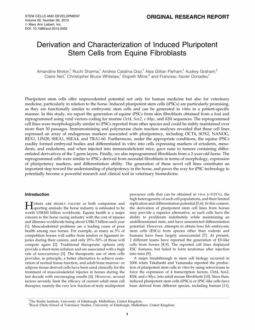
ORIGINAL RESEARCH REPORT Derivation and Characterization of Induced Pluripotent Stem Cells from Equine Fibroblasts Amandine Breton, 1 Ruchi Sharma, 1 Andrea Catalina Diaz, 1 Alea Gillian Parham, 1 Audrey Graham, 2 Claire Neil, 1 Christopher Bruce Whitelaw, 1 Elspeth Milne, 2 and Francesc Xavier Donadeu 1 Pluripotent stem cells offer unprecedented potential not only for human medicine but also for veterinary medicine, particularly in relation to the horse. Induced pluripotent stem cells (iPSCs) are particularly promising, as they are functionally similar to embryonic stem cells and can be generated in vitro in a patient-specific manner. In this study, we report the generation of equine iPSCs from skin fibroblasts obtained from a foal and reprogrammed using viral vectors coding for murine Oct4, Sox2, c-Myc, and Klf4 sequences. The reprogrammed cell lines were morphologically similar to iPSCs reported from other species and could be stably maintained over more than 30 passages. Immunostaining and polymerase chain reaction analyses revealed that these cell lines expressed an array of endogenous markers associated with pluripotency, including OCT4, SOX2, NANOG, REX1, LIN28, SSEA1, SSEA4, and TRA1-60. Furthermore, under the appropriate conditions, the equine iPSCs readily formed embryoid bodies and differentiated in vitro into cells expressing markers of ectoderm, meso- derm, and endoderm, and when injected into immunodeficient mice, gave raise to tumors containing differ- entiated derivatives of the 3 germ layers. Finally, we also reprogrammed fibroblasts from a 2-year-old horse. The reprogrammed cells were similar to iPSCs derived from neonatal fibroblasts in terms of morphology, expression of pluripotency markers, and differentiation ability. The generation of these novel cell lines constitutes an important step toward the understanding of pluripotency in the horse, and paves the way for iPSC technology to potentially become a powerful research and clinical tool in veterinary biomedicine. Introduction H orses are highly valued as both companion and sporting animals; the horse industry is estimated to be worth US$300 billion worldwide. Equine health is a major concern to the horse racing industry with the cost of injuries and illnesses worldwide being about US$6.5 billion each year [1]. Musculoskeletal problems are a leading cause of poor health among race horses. For example, as many as 5% of competition horses will suffer from tendon or ligament in- juries during their careers, and only 25%–50% of those will compete again [2]. Traditional therapeutic options only provide a short-term solution and are associated with a high rate of reoccurrence [3]. The therapeutic use of stem cells provides, in principle, a better alternative to achieve resto- ration of normal tissue function, and adult bone marrow- or adipose tissue-derived cells have been used clinically for the treatment of musculoskeletal injuries in horses during the last decade with encouraging results [4]. However, several factors severely limit the efficacy of current adult stem cell therapies, namely the very low fraction of truly multipotent precursor cells that can be obtained in vivo ( < 0.01%), the high heterogeneity of such cell populations, and their limited replication and differentiation potential [5,6]. In this context, the derivation of pluripotent stem cell lines from horses may provide a superior alternative, as such cells have the ability to proliferate indefinitely while maintaining an undifferentiated state, and have unrestricted differentiation potential. However, attempts to obtain bona fide embryonic stem cells (ESCs) from species other than rodents and humans have been largely unsuccessful [7]. At present, 2 different teams have reported the generation of ES-like cells from horses [8,9]. The reported cell lines displayed ESC features, but failed to form teratomas after injection into mice [9]. A major breakthrough in stem cell biology occurred in 2006 when Takahashi and Yamanaka reported the produc- tion of pluripotent stem cells in vitro by using retroviruses to force the expression of 4 transcription factors, Oct4, Sox2, Klf4, and c-Myc, into adult mouse fibroblasts [10]. Since then, induced pluripotent stem cells (iPSCs) or iPSC-like cells have been derived from different species, including human [11], 1 The Roslin Institute, University of Edinburgh, Midlothian, United Kingdom. 2 Royal (Dick) School of Veterinary Studies, University of Edinburgh, Midlothian, United Kingdom. STEM CELLS AND DEVELOPMENT Volume 00, Number 00, 2012 ȑ Mary Ann Liebert, Inc. DOI: 10.1089/scd.2012.0052 1
Transcript of Derivation and Characterization of Induced Pluripotent Stem … · 2013-01-07 · ORIGINAL RESEARCH...
ORIGINAL RESEARCH REPORT
Derivation and Characterization of Induced PluripotentStem Cells from Equine Fibroblasts
Amandine Breton,1 Ruchi Sharma,1 Andrea Catalina Diaz,1 Alea Gillian Parham,1 Audrey Graham,2
Claire Neil,1 Christopher Bruce Whitelaw,1 Elspeth Milne,2 and Francesc Xavier Donadeu1
Pluripotent stem cells offer unprecedented potential not only for human medicine but also for veterinarymedicine, particularly in relation to the horse. Induced pluripotent stem cells (iPSCs) are particularly promising,as they are functionally similar to embryonic stem cells and can be generated in vitro in a patient-specificmanner. In this study, we report the generation of equine iPSCs from skin fibroblasts obtained from a foal andreprogrammed using viral vectors coding for murine Oct4, Sox2, c-Myc, and Klf4 sequences. The reprogrammedcell lines were morphologically similar to iPSCs reported from other species and could be stably maintained overmore than 30 passages. Immunostaining and polymerase chain reaction analyses revealed that these cell linesexpressed an array of endogenous markers associated with pluripotency, including OCT4, SOX2, NANOG,REX1, LIN28, SSEA1, SSEA4, and TRA1-60. Furthermore, under the appropriate conditions, the equine iPSCsreadily formed embryoid bodies and differentiated in vitro into cells expressing markers of ectoderm, meso-derm, and endoderm, and when injected into immunodeficient mice, gave raise to tumors containing differ-entiated derivatives of the 3 germ layers. Finally, we also reprogrammed fibroblasts from a 2-year-old horse. Thereprogrammed cells were similar to iPSCs derived from neonatal fibroblasts in terms of morphology, expressionof pluripotency markers, and differentiation ability. The generation of these novel cell lines constitutes animportant step toward the understanding of pluripotency in the horse, and paves the way for iPSC technology topotentially become a powerful research and clinical tool in veterinary biomedicine.
Introduction
Horses are highly valued as both companion andsporting animals; the horse industry is estimated to be
worth US$300 billion worldwide. Equine health is a majorconcern to the horse racing industry with the cost of injuriesand illnesses worldwide being about US$6.5 billion each year[1]. Musculoskeletal problems are a leading cause of poorhealth among race horses. For example, as many as 5% ofcompetition horses will suffer from tendon or ligament in-juries during their careers, and only 25%–50% of those willcompete again [2]. Traditional therapeutic options onlyprovide a short-term solution and are associated with a highrate of reoccurrence [3]. The therapeutic use of stem cellsprovides, in principle, a better alternative to achieve resto-ration of normal tissue function, and adult bone marrow- oradipose tissue-derived cells have been used clinically for thetreatment of musculoskeletal injuries in horses during thelast decade with encouraging results [4]. However, severalfactors severely limit the efficacy of current adult stem celltherapies, namely the very low fraction of truly multipotent
precursor cells that can be obtained in vivo (< 0.01%), thehigh heterogeneity of such cell populations, and their limitedreplication and differentiation potential [5,6]. In this context,the derivation of pluripotent stem cell lines from horsesmay provide a superior alternative, as such cells have theability to proliferate indefinitely while maintaining anundifferentiated state, and have unrestricted differentiationpotential. However, attempts to obtain bona fide embryonicstem cells (ESCs) from species other than rodents andhumans have been largely unsuccessful [7]. At present,2 different teams have reported the generation of ES-likecells from horses [8,9]. The reported cell lines displayedESC features, but failed to form teratomas after injectioninto mice [9].
A major breakthrough in stem cell biology occurred in2006 when Takahashi and Yamanaka reported the produc-tion of pluripotent stem cells in vitro by using retroviruses toforce the expression of 4 transcription factors, Oct4, Sox2,Klf4, and c-Myc, into adult mouse fibroblasts [10]. Since then,induced pluripotent stem cells (iPSCs) or iPSC-like cells havebeen derived from different species, including human [11],
1The Roslin Institute, University of Edinburgh, Midlothian, United Kingdom.2Royal (Dick) School of Veterinary Studies, University of Edinburgh, Midlothian, United Kingdom.
STEM CELLS AND DEVELOPMENT
Volume 00, Number 00, 2012
� Mary Ann Liebert, Inc.
DOI: 10.1089/scd.2012.0052
1
rhesus monkey [12], rat [13], pig [14,15], dog [16,17], rabbit[18], marmoset [19], sheep [20–22], and more recently, horse[23] and cow [24]. Transgene-mediated reprogramming of-fers distinct technical advantages over other established re-programming techniques, and the resulting cell lines arefunctionally comparable to ESCs [25]. Moreover, iPSCs can,in principle, be produced in a patient-specific manner, afeature that would provide these cells with considerablepotential for regenerative medicine and in vitro diseasemodeling. However, full realization of this potential will firstrequire addressing several limitations associated with thecurrent iPSC technology that at present severely restrict anytherapeutic prospects of available iPSC lines [26].
In this report, we describe the generation of equine plu-ripotent stem cell lines from nonfetal sources by repro-gramming of fibroblasts obtained from a newborn foal andfrom a 2-year-old horse using retroviruses coding for mouseOct4, Sox2, c-Myc, and Klf4 sequences. We show that thereprogrammed cells express several markers of pluripotentcells, including novel ones in equine, and can differentiateinto derivatives of the 3 germ layers both in vitro and in vivo.
Materials and Methods
Cell culture
Fibroblast cultures were separately derived from skinsamples collected from a newborn male foal and from a 2-year-old gelding at the equine hospital of the School of Ve-terinary Studies, University of Edinburgh. Fibroblasts weregrown and expanded from skin explants in Dulbecco’s mod-ified Eagle medium (DMEM) (Sigma-Aldrich, Irvine, UnitedKingdom) containing 10% fetal bovine serum (FBS) gold(PAA Laboratories Ltd., Yeovil, United Kingdom), 2 mM l-glutamine (Invitrogen, Paisley, United Kingdom), 0.1 mMminimum essential medium (MEM) nonessential amino acids(Invitrogen), and 1% penicillin–streptomycin (Invitrogen) at37�C in 5% CO2. Once cells reached 90% confluence, theywere passaged using TrypLETM (Invitrogen). For reprogram-ming experiments, fibroblasts at passage < 7 were used.
Putative iPSCs were grown on mitotically inactivated SNLfeeder cells (CBA-316; Cell Biolabs, San Diego, CA) on 6-wellplates coated with 0.1% gelatin (Sigma-Aldrich) using amedium containing either the DMEM (Invitrogen) with 20%fetal calf serum or a knockout DMEM (Invitrogen) supple-mented with 20% knockout serum replacement (Invitrogen).Medium preparations also contained 2 mM l-glutamine,0.1 mM b-mercaptoethanol (Invitrogen), 0.1 mM MEM non-essential amino acids and 1% penicillin–streptomycin, andwere supplemented with 8 ng/mL human basic fibroblastgrowth factor (bFGF) (Peprotech, London, United Kingdom)and/or 1,000 U/mL human leukemia inhibitory factor (LIF)(Millipore, Watford, United Kingdom). Cells were kept at37�C in 5% CO2. The medium was changed every other day,and the cells were passaged every 3 days using AccutaseTM
(Sigma-Aldrich).SNL feeder cells [27] were maintained in the DMEM con-
taining 10% FBS gold, 2 mM l-glutamine, 0.1 mM MEM non-essential amino acids, and 1% penicillin–streptomycin. Thesecells were mitotically inactivated by incubation with mitomy-cin C (10mg/mL; Sigma-Aldrich) for 2 h, followed by dissoci-ation with TrypLE and incubation for at least 1 day in iPSCmedium described above before seeding of putative iPSCs.
Viral constructs and cell reprogramming
Mouse cDNA sequences for Oct4, Sox2, Klf4, and c-Mycthat had been cloned into a Moloney Murine LeukemiaVirus backbone (pMXs) [28] were obtained from Addgene(Cambridge, MA). Viral particles were produced by indi-vidually transfecting each of these constructs with the ret-roviral packaging vector, pCL-10A1 (Imgenex; CambridgeBioscience, Cambridge, United Kingdom) using FuGENE(Roche, West Sussex, United Kingdom) in human embryonickidney (HEK) cells (American Type Culture Collection,Manassas, VA). In addition, the AcGFP1 sequence was ex-cised from a pAcGFP1-C1 vector (Clontech, Mountain View,CA) and amplified before being inserted in the retroviralpackaging vector pCLXSN (Imgenex).
Two days after transfection of HEK cells, supernatantscontaining concentrates of each of the viral particles encod-ing for the reprogramming factors were collected, mixed,filtered, and added to equine fibroblasts that had beenseeded 1 day earlier on gelatin-coated 6-well plates(1.3 · 105cells/well). To assess transduction efficiency, somefibroblasts were transduced with the pCLXSN-GFP vectoronly. In all cases, the transduction procedure was repeated 1day later, as described [11]. Cells were passaged onto afeeder layer in 10-cm dishes (5 · 104 cells/dish) the followingday and transferred to iPSC medium 2 days later. Beginning2 weeks after transduction, appearing colonies were me-chanically passaged to 96-well plates and individually ex-panded by further passaging. Three differentreprogramming experiments were performed, and colonieswith similar characteristics were obtained in all cases.
Polymerase chain reaction analyses
Endogenous expression of pluripotency genes in repro-grammed cells was determined by reverse transcription-polymerase chain reaction (RT-PCR) using primers specificallyrecognizing equine sequences (Table 1). Genomic integrationand expression of the viral transgenes were assessed by PCRon gDNA and cDNA, respectively, using primers specific foreach of the mouse transcription factors and a primer com-plementary to a common flanking sequence in the viralbackbone (Table 1). Genomic DNA and total RNA wereextracted with a DNeasy Blood and Tissue Kit (Qiagen,Crawley, United Kingdom) and an RNeasy mini kit(Qiagen), respectively. RNA was reverse-transcribed usingSuperscript III (Invitrogen) as per the manufacturer’s in-structions, and PCR was performed with BIOTAQTM DNApolymerase (Bioline, London, United Kingdom) using anannealing temperature of 60�C–65�C and 40 cycles. PCRproducts were resolved in 3% agarose gels stained with SybrSafe (Invitrogen) and visualized with the Kodak Gel Logic200 imaging system.
Southern blotting
Ten micrograms of genomic DNA were digested over-night with BamHI and then electrophoresed in a 0.8% aga-rose/Tris-Acetate-EDTA gel and transferred to a Hybond-Nmembrane (GE Healthcare, Little Chalfont, United King-dom). This was then hybridized overnight at 65�C withprobes isolated from pMXs-Oct4 and pMXs-Klf4 constructsand labeled using a High Prime DNA labeling kit (Roche)
2 BRETON ET AL.
and 32P dCTP (Perkin Elmer, Cambridge, United Kingdom).After washing, membranes were exposed to a phosphorscreen for 3 days and visualized using a Typhoon phos-phorimage (GE Healthcare). Before rehybridization, mem-branes were stripped using boiling 0.1% sodium dodecylsulfate.
Karyotyping
Cells were synchronized by incubation with thymidine(10 mg/mL; Sigma-Aldrich) for 15 h. After washings with theDMEM containing 10% FBS, cells were incubated for a fur-ther 5 h, followed by a second synchronization with thymi-dine. After washing, cells were incubated with colcemid(10 mg/mL; Invitrogen) for 2.5 h, and the supernatant wasaspirated, and cells were washed with phosphate-bufferedsaline (PBS) before being harvested using TrypLE. Aftercentrifugation at 1,200 rpm for 8 min, the cell pellet was in-cubated with 0.56% KCl for 8 min at room temperature, andan ice-cold fixative mixture (acetic acid and methanol 1:3)was added. Centrifugation followed by addition of ice-coldfixative was repeated 2 more times. Finally, 10–20 mL ofsuspension was smeared onto a slide, allowed to dry over-night at 37�C, stained with a Giemsa solution for 15 min, andwashed in PBS before mounting a coverslip.
Alkaline phosphatase staining
Alkaline phosphatase activity was determined in repro-grammed cells using a commercial kit (86R; Sigma-Aldrich)following the manufacturer’s instructions.
Immunocytochemistry
Cells were fixed in 4% paraformaldehyde for 15 min atroom temperature and then permeabilized in a 0.5% Triton–PBS solution, followed by blocking for 50 min in PBS con-taining 2% bovine serum albumin, 0.05% Tween20, and
0.05% Triton. Primary antibody was then incubated over-night at 4�C. The next day, after 3 washes in PBS, 1-h incu-bation with secondary antibody (Alexa Fluor 568 or 488;Invitrogen) was performed at room temperature. Cells werethen washed in PBS, and a mounting solution containing4¢,6-diamidino-2-phenylindole (Sigma-Aldrich) was addedbefore sealing with a coverslip. Specific antibodies and di-lutions used are indicated in Table 2. The slides were ob-served on an Axiovert 25 inverted microscope and a NikonEC1 confocal microscope using the 20 · , 40 · , and 63 ·lenses. The images obtained were processed with ImageJsoftware version 1.42q (http://rsb.info.nih.gov/ij).
Induction of cell differentiation in vitro
Putative equine iPSCs were harvested, passaged in abacterial culture dish, and allowed to grow in suspension for
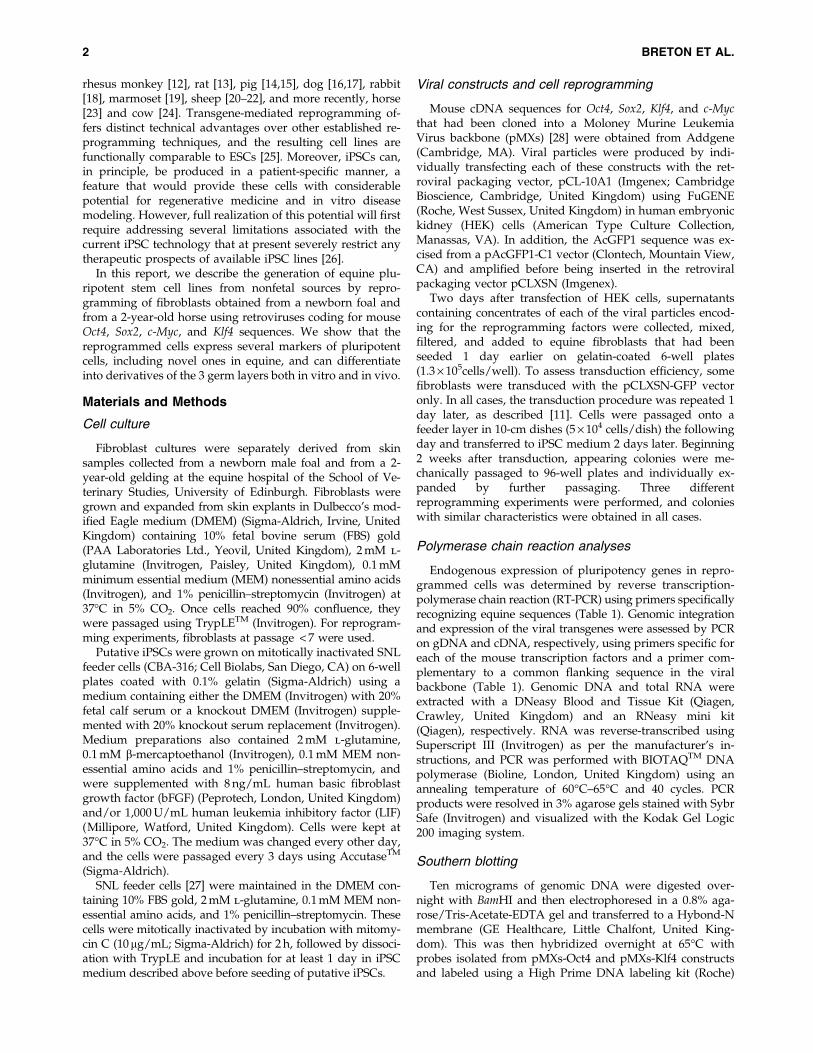
Table 1. List of Primers Used for Polymerase Chain Reaction Analyses
Primer Sequence Product (bp)
OCT4 (For) ATTGAGACCCGAGTGAGAGG 74OCT4 (Rev) CTGATCTGCTGCAGTGTGGSOX2 (For)a CACCCACAGCAAATGACAGC 252SOX2 (Rev)a TTTCTGCAAAGCTCCTACCGNANOG (For)b TCCTCAATGACAGATTTCAGAGA 323NANOG (Rev)b GAGCACCAGGTCTGACTGTTDNMT3b (For) CTTCTGCGTGGAGTGTCTGG 169DNMT3b (Rev) GGTGTCGCTGGTAAAGAAGGLIN28 (For) CATGGGCTCTGTGTCAAACC 167LIN28 (Rev) CGGTCATGGACAGGAAGCREX1 (For) GACGGGAAAGGCCTGGATAGAAG 297REX1 (Rev) GGCGGTAAGAAGCTGTTGAGAAAGGGAPDH (For) CATCATCCCTGCTTCTACTGG 117GAPDH (Rev) TCCACGACTGACACGTTAGGViral Common (For) GGATCCCAGTGTGGTGGTACGViral Oct4 (Rev) CTGTAGGGAGGGCTTCGGGCACTT 850Viral Sox2 (Rev) TCACATGTGCGACAGGGGCAG 1,000Viral Klf4 (Rev) TTAGGCTGTTCTTTTCCGGGGCCACGA 1,200Viral c-Myc (Rev) TTATGCACCAGAGTTTCGAAGCTGTTC 1,400
aFrom Sumer et al. [24].bFrom Nagy et al. [23].
Table 2. List of Antibodies Used
for Immunocytochemistry
Marker Supplier Product no. Dilution
Oct4 Santa Cruz Sc-5279 1/300Sox2 Santa Cruz Sc-17320 1/200SSEA1 DSHB MC480 1/100SSEA4 Abcam ab16287 1/300Nanog Abcam ab80892 1/200Lin28 Abcam ab63740 1/200TRA1-60 Cell Signalling 9656 1/200Rex 1 Millipore MAB4316 1/200TUJ-1 R&D Systems MAB1195 1/100AFP R&D Systems MAB1368 1/200Troponin I Abcam ab19615 1/100Vimentin Abcam ab8978 1/300Pan-cytokeratin Sigma C5992 1/200Nestin Millipore 07-449 1/100GATA4 Abcam ab5694 1/300ASMA Abcam ab56944 1/100
EQUINE INDUCED PLURIPOTENT STEM CELLS 3
7 days in the DMEM containing 10% FBS. The resultingembryoid bodies (EBs) were then transferred onto gelatin-coated 6-well plates in the same medium and allowed todifferentiate for 3 weeks, after which they were fixed beforeimmunostaining.
Induction of cell differentiation in vivo
Putative equine iPSCs were harvested with TrypLE, and5 · 106 cells resuspended in 100mL of the DMEM mediumcontaining 10 mM HEPES, 10% FBS, and 200 nM l-glutamine.Fifteen microliters of the resulting cell slurry was surgicallygrafted under the kidney capsule of NOD/SCID mice. Ani-mals were euthanized between 5 and 8 weeks after injection,and tumors recovered and processed for histological analysis.All animal procedures were carried out under the UnitedKingdom Home Office Animals (Scientific Procedures) Act1986, after approval by the Ethics Review Committee, Uni-versity of Edinburgh (Project License no. 60/3715).
Results
Reprogramming of equine fibroblasts
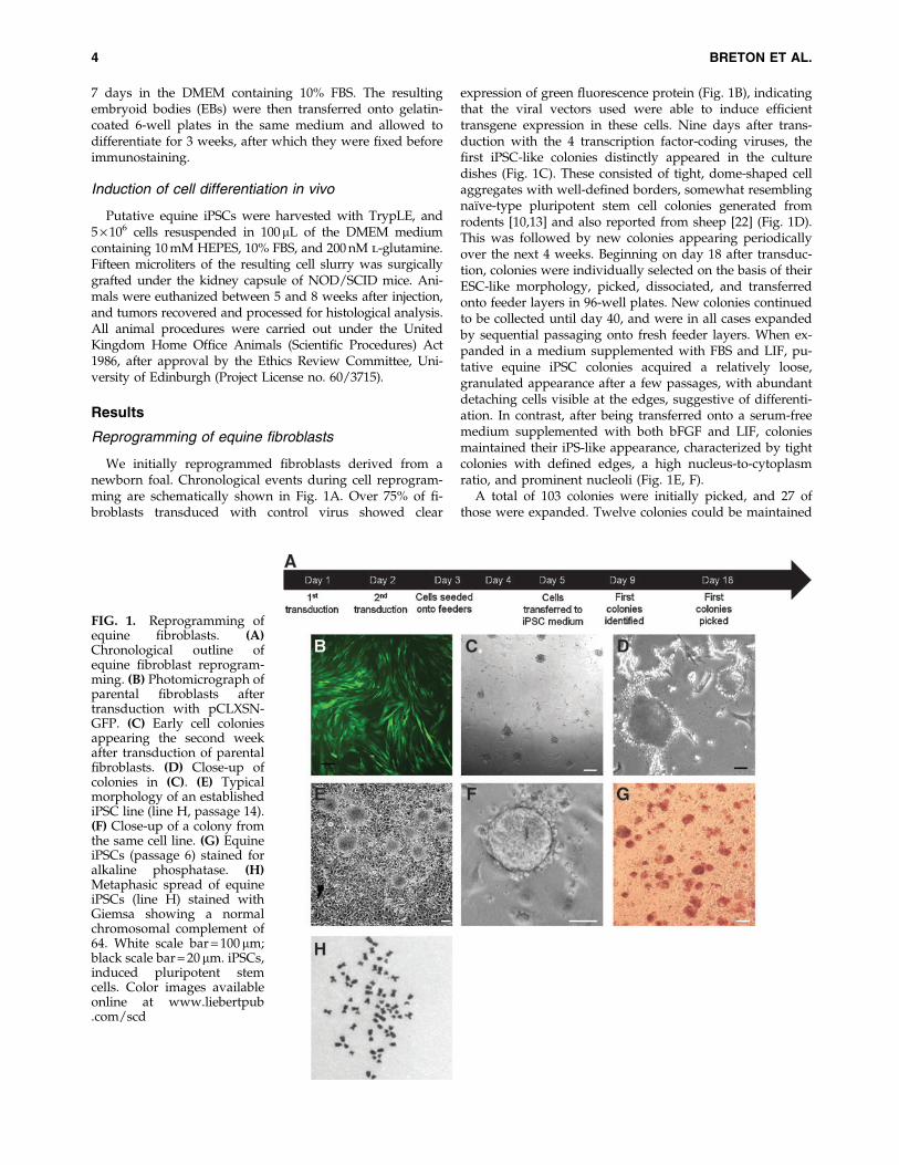
We initially reprogrammed fibroblasts derived from anewborn foal. Chronological events during cell reprogram-ming are schematically shown in Fig. 1A. Over 75% of fi-broblasts transduced with control virus showed clear
expression of green fluorescence protein (Fig. 1B), indicatingthat the viral vectors used were able to induce efficienttransgene expression in these cells. Nine days after trans-duction with the 4 transcription factor-coding viruses, thefirst iPSC-like colonies distinctly appeared in the culturedishes (Fig. 1C). These consisted of tight, dome-shaped cellaggregates with well-defined borders, somewhat resemblingnaı̈ve-type pluripotent stem cell colonies generated fromrodents [10,13] and also reported from sheep [22] (Fig. 1D).This was followed by new colonies appearing periodicallyover the next 4 weeks. Beginning on day 18 after transduc-tion, colonies were individually selected on the basis of theirESC-like morphology, picked, dissociated, and transferredonto feeder layers in 96-well plates. New colonies continuedto be collected until day 40, and were in all cases expandedby sequential passaging onto fresh feeder layers. When ex-panded in a medium supplemented with FBS and LIF, pu-tative equine iPSC colonies acquired a relatively loose,granulated appearance after a few passages, with abundantdetaching cells visible at the edges, suggestive of differenti-ation. In contrast, after being transferred onto a serum-freemedium supplemented with both bFGF and LIF, coloniesmaintained their iPS-like appearance, characterized by tightcolonies with defined edges, a high nucleus-to-cytoplasmratio, and prominent nucleoli (Fig. 1E, F).
A total of 103 colonies were initially picked, and 27 ofthose were expanded. Twelve colonies could be maintained
FIG. 1. Reprogramming ofequine fibroblasts. (A)Chronological outline ofequine fibroblast reprogram-ming. (B) Photomicrograph ofparental fibroblasts aftertransduction with pCLXSN-GFP. (C) Early cell coloniesappearing the second weekafter transduction of parentalfibroblasts. (D) Close-up ofcolonies in (C). (E) Typicalmorphology of an establishediPSC line (line H, passage 14).(F) Close-up of a colony fromthe same cell line. (G) EquineiPSCs (passage 6) stained foralkaline phosphatase. (H)Metaphasic spread of equineiPSCs (line H) stained withGiemsa showing a normalchromosomal complement of64. White scale bar = 100 mm;black scale bar = 20 mm. iPSCs,induced pluripotent stemcells. Color images availableonline at www.liebertpub.com/scd
4 BRETON ET AL.
for up to at least 6 passages and all showed alkaline phos-phatase staining (Fig. 1G). Results from characterization of 4of these cell lines (H, U, E, B) are shown below. These 4 lineshave now been robustly expanded (over 30 passages in thecase of lines H and U), and they display a typical equinekaryotype (Fig. 1H).
Expression of pluripotency markersby reprogrammed cells
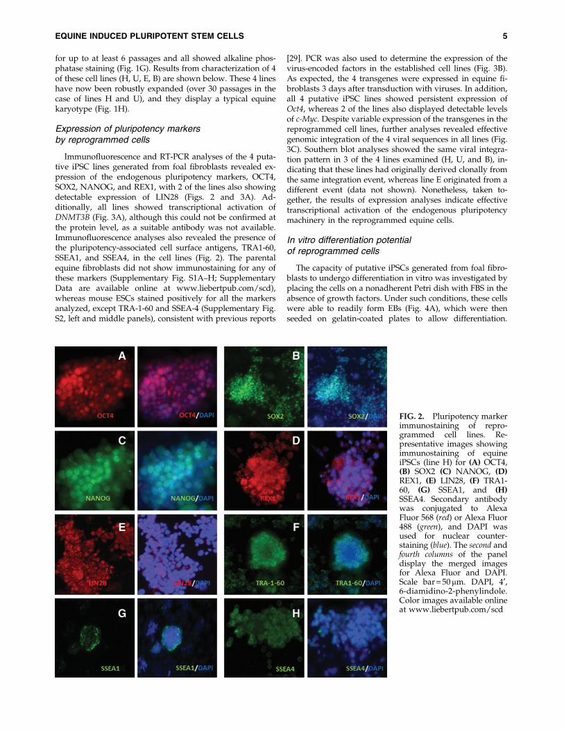
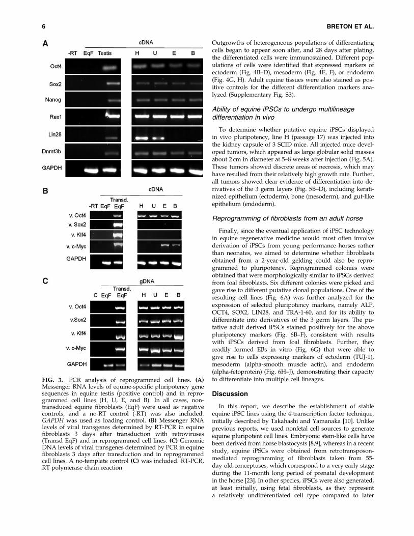
Immunofluorescence and RT-PCR analyses of the 4 puta-tive iPSC lines generated from foal fibroblasts revealed ex-pression of the endogenous pluripotency markers, OCT4,SOX2, NANOG, and REX1, with 2 of the lines also showingdetectable expression of LIN28 (Figs. 2 and 3A). Ad-ditionally, all lines showed transcriptional activation ofDNMT3B (Fig. 3A), although this could not be confirmed atthe protein level, as a suitable antibody was not available.Immunofluorescence analyses also revealed the presence ofthe pluripotency-associated cell surface antigens, TRA1-60,SSEA1, and SSEA4, in the cell lines (Fig. 2). The parentalequine fibroblasts did not show immunostaining for any ofthese markers (Supplementary Fig. S1A–H; SupplementaryData are available online at www.liebertpub.com/scd),whereas mouse ESCs stained positively for all the markersanalyzed, except TRA-1-60 and SSEA-4 (Supplementary Fig.S2, left and middle panels), consistent with previous reports
[29]. PCR was also used to determine the expression of thevirus-encoded factors in the established cell lines (Fig. 3B).As expected, the 4 transgenes were expressed in equine fi-broblasts 3 days after transduction with viruses. In addition,all 4 putative iPSC lines showed persistent expression ofOct4, whereas 2 of the lines also displayed detectable levelsof c-Myc. Despite variable expression of the transgenes in thereprogrammed cell lines, further analyses revealed effectivegenomic integration of the 4 viral sequences in all lines (Fig.3C). Southern blot analyses showed the same viral integra-tion pattern in 3 of the 4 lines examined (H, U, and B), in-dicating that these lines had originally derived clonally fromthe same integration event, whereas line E originated from adifferent event (data not shown). Nonetheless, taken to-gether, the results of expression analyses indicate effectivetranscriptional activation of the endogenous pluripotencymachinery in the reprogrammed equine cells.
In vitro differentiation potentialof reprogrammed cells
The capacity of putative iPSCs generated from foal fibro-blasts to undergo differentiation in vitro was investigated byplacing the cells on a nonadherent Petri dish with FBS in theabsence of growth factors. Under such conditions, these cellswere able to readily form EBs (Fig. 4A), which were thenseeded on gelatin-coated plates to allow differentiation.
FIG. 2. Pluripotency markerimmunostaining of repro-grammed cell lines. Re-presentative images showingimmunostaining of equineiPSCs (line H) for (A) OCT4,(B) SOX2 (C) NANOG, (D)REX1, (E) LIN28, (F) TRA1-60, (G) SSEA1, and (H)SSEA4. Secondary antibodywas conjugated to AlexaFluor 568 (red) or Alexa Fluor488 (green), and DAPI wasused for nuclear counter-staining (blue). The second andfourth columns of the paneldisplay the merged imagesfor Alexa Fluor and DAPI.Scale bar = 50mm. DAPI, 4¢,6-diamidino-2-phenylindole.Color images available onlineat www.liebertpub.com/scd
EQUINE INDUCED PLURIPOTENT STEM CELLS 5
Outgrowths of heterogeneous populations of differentiatingcells began to appear soon after, and 28 days after plating,the differentiated cells were immunostained. Different pop-ulations of cells were identified that expressed markers ofectoderm (Fig. 4B–D), mesoderm (Fig. 4E, F), or endoderm(Fig. 4G, H). Adult equine tissues were also stained as pos-itive controls for the different differentiation markers ana-lyzed (Supplementary Fig. S3).
Ability of equine iPSCs to undergo multilineagedifferentiation in vivo
To determine whether putative equine iPSCs displayedin vivo pluripotency, line H (passage 17) was injected intothe kidney capsule of 3 SCID mice. All injected mice devel-oped tumors, which appeared as large globular solid massesabout 2 cm in diameter at 5–8 weeks after injection (Fig. 5A).These tumors showed discrete areas of necrosis, which mayhave resulted from their relatively high growth rate. Further,all tumors showed clear evidence of differentiation into de-rivatives of the 3 germ layers (Fig. 5B–D), including kerati-nized epithelium (ectoderm), bone (mesoderm), and gut-likeepithelium (endoderm).
Reprogramming of fibroblasts from an adult horse
Finally, since the eventual application of iPSC technologyin equine regenerative medicine would most often involvederivation of iPSCs from young performance horses ratherthan neonates, we aimed to determine whether fibroblastsobtained from a 2-year-old gelding could also be repro-grammed to pluripotency. Reprogrammed colonies wereobtained that were morphologically similar to iPSCs derivedfrom foal fibroblasts. Six different colonies were picked andgave rise to different putative clonal populations. One of theresulting cell lines (Fig. 6A) was further analyzed for theexpression of selected pluripotency markers, namely ALP,OCT4, SOX2, LIN28, and TRA-1-60, and for its ability todifferentiate into derivatives of the 3 germ layers. The pu-tative adult derived iPSCs stained positively for the abovepluripotency markers (Fig. 6B–F), consistent with resultswith iPSCs derived from foal fibroblasts. Further, theyreadily formed EBs in vitro (Fig. 6G) that were able togive rise to cells expressing markers of ectoderm (TUJ-1),mesoderm (alpha-smooth muscle actin), and endoderm(alpha-fetoprotein) (Fig. 6H–J), demonstrating their capacityto differentiate into multiple cell lineages.
Discussion
In this report, we describe the establishment of stableequine iPSC lines using the 4-transcription factor technique,initially described by Takahashi and Yamanaka [10]. Unlikeprevious reports, we used nonfetal cell sources to generateequine pluripotent cell lines. Embryonic stem-like cells havebeen derived from horse blastocysts [8,9], whereas in a recentstudy, equine iPSCs were obtained from retrotransposon-mediated reprogramming of fibroblasts taken from 55-day-old conceptuses, which correspond to a very early stageduring the 11-month long period of prenatal developmentin the horse [23]. In other species, iPSCs were also generated,at least initially, using fetal fibroblasts, as they representa relatively undifferentiated cell type compared to later
FIG. 3. PCR analysis of reprogrammed cell lines. (A)Messenger RNA levels of equine-specific pluripotency genesequences in equine testis (positive control) and in repro-grammed cell lines (H, U, E, and B). In all cases, non-transduced equine fibroblasts (EqF) were used as negativecontrols, and a no-RT control (-RT) was also included.GAPDH was used as loading control. (B) Messenger RNAlevels of viral transgenes determined by RT-PCR in equinefibroblasts 3 days after transduction with retroviruses(Transd EqF) and in reprogrammed cell lines. (C) GenomicDNA levels of viral transgenes determined by PCR in equinefibroblasts 3 days after transduction and in reprogrammedcell lines. A no-template control (C) was included. RT-PCR,RT-polymerase chain reaction.
6 BRETON ET AL.

developmental stages, and therefore they are, in principle,more readily reprogrammable [10,14,15,21]. Consistent withthis, it has been shown that, relative to adult mesenchymalcells, equine fetal fibroblasts express high levels of key re-programming factors [30] that very likely facilitate the re-programming of these cells. In contrast, in the present study,we could not detect expression of any of the pluripotencyfactors analyzed in parental fibroblasts, either by immuno-cytochemistry or by PCR, consistent with their postfetal or-igin. The present results represent a step forward toward thegeneration and biomedical application of reprogrammedcells from clinical equine patients.
The equine cell lines generated in this study showed nu-merous features associated with pluripotency. These includetranscriptional reactivation of endogenous OCT4 and ex-pression of 3 different cell surface antigens, all of which havebeen reported in the inner cell mass of equine blastocysts [31]as well as in equine ESC-like cells [8,9,32] and iPSCs [23].Equine iPSCs in the present report also expressed NANOG,in agreement with the earlier iPSC report by Nagy et al. [23],
as well as other ESC-associated factors, namely SOX2, REX1,LIN28, and DNMT3B, which are transcriptionally activatedduring reprogramming of human, mouse, and pig cells[10,15,33], but have not been previously reported in equinepluripotent cells. The reactivation of endogenous pluri-potency markers is well known to be associated with latestages of reprogramming [34]. Furthermore, Chan et al. [35]demonstrated that 3 of the markers found in our equineiPSCs, namely TRA-1-60, REX1, and DNMT3B, were bona fideindicators of fully reprogrammed human iPSCs, a conclusionthat is consistent with the ability of the equine iPSCs re-ported in the present study to readily produce differentiatedteratomas upon injection into SCID mice. In a previous study[23], equine cells reportedly resembling primed-type PSCswere generated using an inducible transposon-based ex-pression system and culture conditions that included LIF andbFGF as well as a combination of different signaling path-way inhibitors that are reportedly necessary to generate na-ı̈ve-type PSCs [36]. In the present study, stable equinepluripotent cell lines could be generated and maintained
FIG. 4. In vitro differentia-tion potential of repro-grammed cell lines. (A)Embryoid bodies forming 5days after equine iPSCs (lineH, passage 12) were placed insuspension culture. (B–H)Immunostaining of differenti-ated cells for markers of ec-toderm (B–D), mesoderm (E,F), or endoderm (G, H). Sec-ondary antibody was conju-gated to Alexa Fluor 568 (red)or Alexa Fluor 488 (green),and DAPI was used for nu-clear counterstaining (blue).The second and fourth columnsof the panel display themerged images for AlexaFluor and DAPI. Scale bar =20 mm. Color images avail-able online at www.liebertpub.com/scd
EQUINE INDUCED PLURIPOTENT STEM CELLS 7

without the need to use signaling inhibitors. Compared withthe cells reported by Nagy et al. [23], in general, our cell linesdid not display obvious morphological features of typicalprimed-type PSCs, but they predominantly grew as rela-tively tight, dome-like shaped cell aggregates, which is moretypical of naı̈ve-type PSCs. These apparent differences in thecells obtained between the 2 studies may have been derivedfrom differences in the conditions used to generate andmaintain the reprogrammed cells and/or from differences inthe criteria used to select ESC-like colonies for expansion.Similar discrepancies in the morphology of iPSCs from thesame species have been reported for sheep and pig[21,22,37,38], and they could be attributed to the use of hu-man ESC versus mouse ESC culture conditions in somestudies [38], but not in others [21,22]. Clearly, greater un-derstanding of the molecular pathways involved in plur-ipotency in these species, as well as of the differentpluripotent states that may be potentially generated in vitro[38], is required to reconcile these discrepancies.
The MMLV vector used for reprogramming in this studywas similar to the one used to generate the first mouse andhuman iPSC lines [10,11]. Because, in principle, such vectorsbecome transcriptionally repressed during the late stages ofreprogramming [10,11], in some studies, stable iPSC linescould not be generated using MMLV vectors, but the use oflentiviral vectors, which may not undergo the same levels oftranscriptional repression, was required [13]. Further, manysuccessful attempts to derive iPSC lines from domestic spe-cies, including horse, have involved the use of lentiviral orother expression vectors under transcriptional control by aninducible promoter, which ensures sustained transgene ex-pression required to achieve and maintain the pluripotentstate [14,15,20,23]. The equine iPSCs reported in the presentstudy showed partial silencing of transgenes characterizedby sustained expression of Oct4 in all the lines examined andexpression of c-Myc in some of the lines. This pattern isconsistent with that reported in sheep iPSCs produced usingthe same vectors [22]. In addition, studies using a similarretroviral expression system in other species often reportedsilencing of one or several of the transgenes in the iPSC linesgenerated, except for Oct4, whose expression was usuallymaintained in all or most of the lines [13,33,39,40]. Thesefindings suggest that clonal populations that fail to silenceviral Oct4 may be distinctly selected for during reprogram-ming. In that context, the ectopic Oct4 may critically con-tribute, together with the reactivated endogenouspluripotency genes, to the attainment and maintenance of theinduced pluripotent state. So far, all iPSC lines reported fromdomestic species have shown to be dependent on continuoustransgene expression for long-term propagation, as demon-strated with the use of inducible reprogramming vectors[14,20,21,41,42]. Similarly, equine iPSCs described in anearlier study quickly underwent differentiation after the ex-pression of reprogramming genes from a transposon-basedvector was turned off [23]. Significant risks associated withinsertional mutagenesis and incomplete transgene silencingseverely restrict the clinical potential of iPSCs generated withcurrent virus-based technology, a limitation that will only beovercome once robust iPSC lines can be efficiently generatedusing integration-free or nongenetic approaches [34]. An in-teresting observation in the present study was that 3 of thefoal-derived iPSC lines characterized (H, U, and B) appeared
FIG. 5. In vivo differentiation potential of reprogrammedcell lines. (A) Tumor resulting from injection of equine iPSCsinto SCID mice is pictured. The tumor can be seen (whitearrow) growing adjacent to the kidney (identified by a blackarrow). Tumors obtained contained differentiated derivativesof the 3 germ layers (stained with hematoxylin and eosin),including keratinized epithelium (B), bone (C), and gut-likeepithelium (D) structures (identified in each photo by blackarrows). Color images available online at www.liebertpub.com/scd
8 BRETON ET AL.

to be clonally derived from the same integration event, yetthese 3 lines were not phenotypically identical as showed bydifferences in the expression of pluripotency genes (LIN28)and viral c-Myc (Fig. 3); since the 3 lines were expandedusing the same culture conditions, the observed differenceslikely reflect the stochastic nature of iPSCs derivation.
In the study by Nagy et al. [23], a lack of suitable anti-bodies prevented the authors from assessing the in vitropluripotency of the reported equine iPSCs. In the presentstudy, we used a panel of antibodies for various differenti-ation markers, which we validated in adult equine tissues(Supplementary Fig. S3), to demonstrate the capacity of re-programmed equine cells to undergo differentiation intoderivatives of the 3 germ layers in vitro. The pluripotency ofputative equine iPSCs in vivo was demonstrated by injectingthese cells into the kidney capsule of SCID mice, a route thatis technically more demanding than subcutaneous or intra-muscular injection, but that facilitates discrimination be-tween the resulting tumor and host tissues. In previousstudies, equine ESC-like cells failed to produce tumorswhen injected into the testes of SCID mice [9] whereassubcutaneous injection of putative equine iPSCs resulted ingrowth of tumors after 4 months and only after injectedmice had been temporarily fed with doxycycline to main-tain expression of the reprogramming transgenes, a strategythat was reportedly necessary to avoid premature cell dif-ferentiation in vivo [23]. Equine iPSCs in the present studywere able to spontaneously maintain their pluripotencyin vivo and generated tumors that grew over a period, 5–8weeks, which was intermediate between periods normallyreported for teratomas derived from mouse and humaniPSCs [10,11]. Outgrowths resulting from the injected cellline in our study were confirmed to be teratomas, asthey contained differentiated derivatives of the 3 germlayers, constituting to this date the most stringent proof ofpluripotency for equine iPSCs.
Finally, we also showed that adult equine fibroblasts canbe reprogrammed to generate cell lines that are morpho-logically similar to iPSCs produced from neonatal fibro-blasts, express similar pluripotency factors, and readilydifferentiate into derivatives of the 3 germ layers in vitro.The age of the donor animal in this experiment, 2 years,corresponds to the career peak of most racing horses, pro-viding support to the prospect of using iPSCs for equineregenerative medicine in the future.
The potential of pluripotent stem cells in veterinarymedicine, and particularly in equine health, is similar to thatin human medicine. Although significant technical advancesstill need to be made to eliminate constraints associated withgenetic, epigenetic, and immunogenic aspects of iPSCs thatat the moment severely restrict their clinical potential [43],huge progress has already been made in the application ofiPSC technology for in vitro modeling of diseases and ther-apeutics [26]. In that regard, there are a number of equinediseases that would be amenable to experimental modelingusing iPSCs. Further, horses could be used as preclinicalmodels for human stem cell-based therapies [1]. Our resultsprovide an important step toward that goal by demonstrat-ing that nonfetal equine somatic cells can be reprogrammedto cells that are pluripotent both in vitro and in vivo. Theseestablished cell lines should facilitate the realization of theveterinary potential of the iPSC technology.
Acknowledgments
We thank Drs. Simon Lilico, Chiara Sartori, and AlexandraDiDomenico for assisting with viral preparations and So-thern blotting, and for providing reagents for gene expres-sion analyses. We are also grateful to Drs. Joe Mee, RosaRabanal, and Christopher Palgrave, and Prof. Dolors Fon-devila for assistance and advice during in vivo work andtissue analyses, to Matt Hanks, Stephanie Schauer, and Ben
FIG. 6. Characteristics of reprogrammed cell lines from adult equine fibroblasts. (A) Colonies derived from reprogrammingof fibroblasts from a 2-year-old gelding and which stained positively for (B) alkaline phosphatase, (C) OCT4, (D) SOX2, (E)LIN28, and (F) TRA-1-60. (G) These cells readily generated embryoid bodies in vitro, which produced cells expressingmarkers of (H) ectoderm, (I) mesoderm, and ( J) endoderm. Secondary antibody was conjugated to Alexa Fluor 568 (red) orAlexa Fluor 488 (green), and DAPI was used for nuclear counterstaining (blue). Merged images for Alexa Fluor and DAPI areshown. Scale bar = 20 mm. Color images available online at www.liebertpub.com/scd
EQUINE INDUCED PLURIPOTENT STEM CELLS 9
Wentink for providing equine tissues, and to Drs. TomBurdon and Alison Thomson for thoughtful commentsthroughout the project and during preparation of this man-uscript. This work was supported by grants from theHorserace Betting Levy Board (Prj 744), the Royal Collegeof Veterinary Surgeons Trust (grant no. 707), and theBiotechnology and Biological Sciences Research Council(BBSRC ISPG) to F.X.D.
Author Disclosure Statement
The authors have declared that no competing interests exist.
References
1. Tecirlioglu RT and AO Trounson. (2007). Embryonic stemcells in companion animals (horses, dogs and cats): presentstatus and future prospects. Reprod Fertil Dev 19:740–747.
2. Perkins NR, SW Reid and RS Morris. (2005). Risk factors forinjury to the superficial digital flexor tendon and suspensoryapparatus in Thoroughbred racehorses in New Zealand. N ZVet J 53:184–192.
3. Fortier LA, HG Potter, EJ Rickey, LV Schnabel, LF Foo, LRChong, T Stokol, J Cheetham and AJ Nixon. (2010). Con-centrated bone marrow aspirate improves full-thicknesscartilage repair compared with microfracture in the equinemodel. J Bone Joint Surg Am 92:1927–1937.
4. Ribitsch I, J Burk, U Delling, C Geissler, C Gittel, H Julke andW Brehm. (2010). Basic science and clinical application ofstem cells in veterinary medicine. Adv Biochem Eng Bio-technol 123:219–263.
5. Vidal MA, GE Kilroy, JR Johnson, MJ Lopez, RM Mooreand JM Gimble. (2006). Cell growth characteristics anddifferentiation frequency of adherent equine bone marrow-derived mesenchymal stromal cells: adipogenic and osteo-genic capacity. Vet Surg 35:601–610.
6. Martins-Taylor K and RH Xu. (2010). Determinants ofpluripotency: from avian, rodents, to primates. J Cell Bio-chem 109:16–25.
7. Talbot NC and A Blomberg Le. (2008). The pursuit of ES celllines of domesticated ungulates. Stem Cell Rev 4:235–254.
8. Saito S, H Ugai, K Sawai, Y Yamamoto, A Minamihashi, KKurosaka, Y Kobayashi, T Murata, Y Obata and K Yo-koyama. (2002). Isolation of embryonic stem-like cells fromequine blastocysts and their differentiation in vitro. FEBSLett 531:389–396.
9. Li X, SG Zhou, MP Imreh, L Ahrlund-Richter and WR Allen.(2006). Horse embryonic stem cell lines from the prolifera-tion of inner cell mass cells. Stem Cells Dev 15:523–531.
10. Takahashi K and S Yamanaka. (2006). Induction of pluri-potent stem cells from mouse embryonic and adult fibro-blast cultures by defined factors. Cell 126:663–676.
11. Takahashi K, K Tanabe, M Ohnuki, M Narita, T Ichisaka, KTomoda and S Yamanaka. (2007). Induction of pluripotentstem cells from adult human fibroblasts by defined factors.Cell 131:861–872.
12. Liu H, F Zhu, J Yong, P Zhang, P Hou, H Li, W Jiang, J Cai,M Liu, et al. (2008). Generation of induced pluripotent stemcells from adult rhesus monkey fibroblasts. Cell Stem Cell3:587–590.
13. Liao J, C Cui, S Chen, J Ren, J Chen, Y Gao, H Li, N Jia, LCheng, H Xiao and L Xiao. (2009). Generation of inducedpluripotent stem cell lines from adult rat cells. Cell Stem Cell4:11–15.
14. Ezashi T, BP Telugu, AP Alexenko, S Sachdev, S Sinha andRM Roberts. (2009). Derivation of induced pluripotent stemcells from pig somatic cells. Proc Natl Acad Sci U S A106:10993–10998.
15. Esteban MA, J Xu, J Yang, M Peng, D Qin, W Li, Z Jiang, JChen, K Deng, et al. (2009). Generation of induced pluri-potent stem cell lines from Tibetan miniature pig. J BiolChem 284:17634–17640.
16. Vaags AK, S Rosic-Kablar, CJ Gartley, YZ Zheng, A Ches-ney, DA Villagomez, SA Kruth and MR Hough. (2009).Derivation and characterization of canine embryonic stemcell lines with in vitro and in vivo differentiation potential.Stem Cells 27:329–340.
17. Luo J, ST Suhr, EA Chang, K Wang, PJ Ross, LL Nelson, PJVenta, JG Knott and JB Cibelli. (2011). Generation of leuke-mia inhibitory factor and basic fibroblast growth factor-dependent induced pluripotent stem cells from canine adultsomatic cells. Stem Cells Dev 20:1669–1678.
18. Honda A, M Hirose, M Hatori, S Matoba, H Miyoshi, KInoue and A Ogura. (2010). Generation of induced pluripo-tent stem cells in rabbits: potential experimental models forhuman regenerative medicine. J Biol Chem 285:31362–31369.
19. Wu Y, Y Zhang, A Mishra, SD Tardif and PJ Hornsby.(2010). Generation of induced pluripotent stem cells fromnewborn marmoset skin fibroblasts. Stem Cell Res 4:180–188.
20. Bao L, L He, J Chen, Z Wu, J Liao, L Rao, J Ren, H Li, H Zhu,et al. (2011). Reprogramming of ovine adult fibroblasts topluripotency via drug-inducible expression of defined fac-tors. Cell Res 21:600–608.
21. Li Y, M Cang, AS Lee, K Zhang and D Liu. (2011). Repro-gramming of sheep fibroblasts into pluripotency under adrug-inducible expression of mouse-derived defined factors.PLoS One 6:e15947.
22. Sartori C, AI Didomenico, AJ Thomson, E Milne, SG Lillico,TG Burdon and CB Whitelaw. (2012). Ovine-induced plu-ripotent stem cells can contribute to chimeric lambs. CellReprogram 14:8–19.
23. Nagy K, HK Sung, P Zhang, S Laflamme, P Vincent, S Agha-Mohammadi, K Woltjen, C Monetti, IP Michael, LC Smithand A Nagy. (2011). Induced pluripotent stem cell linesderived from equine fibroblasts. Stem Cell Rev 7:693–702.
24. Sumer H, J Liu, LF Malaver Ortega, ML Lim, K Khodadadiand PJ Verma. (2011). NANOG is a key factor for inductionof pluripotency in bovine adult fibroblasts. J Anim Sci89:2708–2716.
25. Hochedlinger K and R Jaenisch. (2006). Nuclear repro-gramming and pluripotency. Nature 441:1061–1067.
26. Saha K and R Jaenisch. (2009). Technical challenges in usinghuman induced pluripotent stem cells to model disease. CellStem Cell 5:584–595.
27. Pan C, A Hicks, X Guan, H Chen and CE Bishop. (2010).SNL fibroblast feeder layers support derivation and main-tenance of human induced pluripotent stem cells. J GenetGenomics 37:241–248.
28. Kitamura T, Y Koshino, F Shibata, T Oki, H Nakajima, TNosaka and H Kumagai. (2003). Retrovirus-mediated genetransfer and expression cloning: powerful tools in functionalgenomics. Exp Hematol 31:1007–1014.
29. De Miguel MP, S Fuentes-Julian and Y Alcaina. (2010).Pluripotent stem cells: origin, maintenance and induction.Stem Cell Rev 6:633–649.
30. Hackett CH, L Greve, KD Novakofski and LA Fortier.(2011). Comparison of gene-specific DNA methylation pat-
10 BRETON ET AL.

terns in equine induced pluripotent stem cell lines with cellsderived from equine adult and fetal tissues. Stem Cells Dev21:1803–1811.
31. Guest DJ and WR Allen. (2007). Expression of cell-surfaceantigens and embryonic stem cell pluripotency genes inequine blastocysts. Stem Cells Dev 16:789–796.
32. Saito S, K Sawai, A Minamihashi, H Ugai, T Murata and KKYokoyama. (2006). Derivation, maintenance, and inductionof the differentiation in vitro of equine embryonic stem cells.Methods Mol Biol 329:59–79.
33. Lowry WE, L Richter, R Yachechko, AD Pyle, J Tchieu, RSridharan, AT Clark and K Plath. (2008). Generation of hu-man induced pluripotent stem cells from dermal fibroblasts.Proc Natl Acad Sci U S A 105:2883–2888.
34. Amabile G and A Meissner. (2009). Induced pluripotentstem cells: current progress and potential for regenerativemedicine. Trends Mol Med 15:59–68.
35. Chan EM, S Ratanasirintrawoot, IH Park, PD Manos, YHLoh, H Huo, JD Miller, O Hartung, J Rho, et al. (2009). Livecell imaging distinguishes bona fide human iPS cells frompartially reprogrammed cells. Nat Biotechnol 27:1033–1037.
36. Buehr M, S Meek, K Blair, J Yang, J Ure, J Silva, R McLay, JHall, QL Ying and A Smith. (2008). Capture of authenticembryonic stem cells from rat blastocysts. Cell 135:1287–1298.
37. Telugu BP, T Ezashi and RM Roberts. (2010). The promise ofstem cell research in pigs and other ungulate species. StemCell Rev 6:31–41.
38. Telugu BP, T Ezashi and RM Roberts. (2010). Porcine in-duced pluripotent stem cells analogous to naive and primedembryonic stem cells of the mouse. Int J Dev Biol 54:1703–1711.
39. Li W, W Wei, S Zhu, J Zhu, Y Shi, T Lin, E Hao, A Hayek, HDeng and S Ding. (2009). Generation of rat and human in-duced pluripotent stem cells by combining genetic repro-gramming and chemical inhibitors. Cell Stem Cell 4:16–19.
40. Aasen T, A Raya, MJ Barrero, E Garreta, A Consiglio, FGonzalez, R Vassena, J Bilic, V Pekarik, et al. (2008). Efficientand rapid generation of induced pluripotent stem cells fromhuman keratinocytes. Nat Biotechnol 26:1276–1284.
41. Li W and S Ding. (2010). Generation of novel rat and humanpluripotent stem cells by reprogramming and chemical ap-proaches. Methods Mol Biol 636:293–300.
42. Wu Z, J Chen, J Ren, L Bao, J Liao, C Cui, L Rao, H Li, Y Gu,et al. (2009). Generation of pig induced pluripotent stem cellswith a drug-inducible system. J Mol Cell Biol 1:46–54.
43. Barrilleaux B and PS Knoepfler. (2011). Inducing iPSCs toescape the dish. Cell Stem Cell 9:103–111.
Address correspondence to:Dr. Francesc Xavier Donadeu
The Roslin InstituteUniversity of Edinburgh
Easter BushMidlothian EH25 9RG
United Kingdom
E-mail: [email protected]
Received for publication February 1, 2012Accepted after revision August 16, 2012
Prepublished on Liebert Instant Online XXXX XX, XXXX
EQUINE INDUCED PLURIPOTENT STEM CELLS 11