Department of Orthopaedic Surgery, Johns Hopkins...
31
1 CITED2 Modulates Breast Cancer Metastatic Ability Through Effects on IKKα. Swaathi Jayaraman, Michele Doucet, Wen Min Lau and Scott L. Kominsky Department of Orthopaedic Surgery, Johns Hopkins University School of Medicine, Baltimore, MD. Running title: CITED2 modulates human breast cancer metastasis Keywords: CITED2, IKKα, invasion, metastasis, cancer Financial support: Research reported in this publication was supported by the National Cancer Institute of the National Institutes of Health under Award Number R01CA157687. The content is solely the responsibility of the authors and does not necessarily represent the official views of the National Institutes of Health. Address correspondence to: Scott L. Kominsky, Ph.D., 720 Rutland Avenue, Ross 232, Baltimore, MD 21205. Fax: 410-502-6414; E-mail: [email protected] The authors disclose no potential conflicts of interest. on September 6, 2018. © 2016 American Association for Cancer Research. mcr.aacrjournals.org Downloaded from Author manuscripts have been peer reviewed and accepted for publication but have not yet been edited. Author Manuscript Published OnlineFirst on May 23, 2016; DOI: 10.1158/1541-7786.MCR-16-0081
Transcript of Department of Orthopaedic Surgery, Johns Hopkins...

1
CITED2 Modulates Breast Cancer Metastatic Ability Through Effects on IKKα.
Swaathi Jayaraman, Michele Doucet, Wen Min Lau and Scott L. Kominsky
Department of Orthopaedic Surgery, Johns Hopkins University School of Medicine, Baltimore,
MD.
Running title: CITED2 modulates human breast cancer metastasis
Keywords: CITED2, IKKα, invasion, metastasis, cancer
Financial support: Research reported in this publication was supported by the National Cancer
Institute of the National Institutes of Health under Award Number R01CA157687. The content
is solely the responsibility of the authors and does not necessarily represent the official views of
the National Institutes of Health.
Address correspondence to: Scott L. Kominsky, Ph.D., 720 Rutland Avenue, Ross 232,
Baltimore, MD 21205. Fax: 410-502-6414; E-mail: [email protected]
The authors disclose no potential conflicts of interest.
on September 6, 2018. © 2016 American Association for Cancer Research. mcr.aacrjournals.org Downloaded from
Author manuscripts have been peer reviewed and accepted for publication but have not yet been edited. Author Manuscript Published OnlineFirst on May 23, 2016; DOI: 10.1158/1541-7786.MCR-16-0081

2
Abstract
Previously, we identified the transcriptional co-activator CITED2 as a potential facilitator
of bone metastasis using a murine mammary cancer model. Extending these studies to human
breast cancer, it was observed that CITED2 mRNA expression was significantly elevated in
patient specimens of metastatic breast cancer relative to primary tumors, with highest levels in
metastasis to bone relative to non-bone sites. To further evaluate CITED2 functions in breast
cancer metastasis, CITED2 expression was stably reduced in the human breast cancer cell lines
MDA-MB-231 and MDA-MB-468, which are metastatic in animal models. While CITED2
knockdown had no effect on cell proliferation, cell migration and invasion were significantly
reduced, as was the establishment of metastasis following intra-cardiac administration in athymic
nude mice. To explore the mechanism behind these effects, gene expression following CITED2
knockdown in MDA-MB-231 cells by cDNA microarray was performed. As confirmed at the
mRNA and protein levels in both MDA-MB-231 and MDA-MB-468 cells, expression of the NF-
κB regulator IKKα was significantly reduced along with several NF-κB targets with known roles
in metastasis (OPN, MMP9, uPA, SPARC, IL-11 and IL-1β). Further, ChIP assay revealed
recruitment of CITED2 to the promoter of IKKα, indicating a direct role in regulating its
expression. Consistent with reduced IKKα expression, CITED2 knockdown inhibited both
canonical and non-canonical NF-κB signaling. Finally, restoration of IKKα expression following
CITED2 knockdown in MDA-MB-231 and MDA-MB-468 cells rescued their invasive ability.
Collectively, these data demonstrate that CITED2 modulates metastatic ability in human breast
cancer cells, at least in part, through the regulation of IKKα.
on September 6, 2018. © 2016 American Association for Cancer Research. mcr.aacrjournals.org Downloaded from
Author manuscripts have been peer reviewed and accepted for publication but have not yet been edited. Author Manuscript Published OnlineFirst on May 23, 2016; DOI: 10.1158/1541-7786.MCR-16-0081
3
Implications: The current study highlights the role of CITED2 in facilitating breast cancer
metastasis, partly via regulation of IKKα.
Introduction
Breast cancer is the most frequently diagnosed cancer in women worldwide and the
second most commonly occurring cancer overall. While primary tumors may be effectively
treated when detected early, metastatic disease is largely incurable and represents the ultimate
cause of mortality in breast cancer patients. It is estimated that ~6% of patients already have
metastatic disease at the time of diagnosis while ~20-50% of patients who are initially diagnosed
with early stage breast cancer will eventually develop metastasis (1). Sadly, the median survival
time for patients with metastatic breast cancer is only 18-30 months. Despite recent research
efforts, elucidation of the critical drivers of metastasis and their mechanism of action is lacking.
Filling this knowledge gap is essential to the development of novel therapeutic modalities and
improving the clinical management of this disease.
The Cbp/p300-interacting transactivator with Glu/Asp-rich carboxy-terminal domain-2
(CITED2) is a non-DNA binding transcriptional co-activator that was originally discovered for
its role in development (2-5). As a transcriptional co-activator, CITED2 interacts with several
transcription factors such as p300/CBP, Lhx2, TFAP2, Smad2/Smad3, PPARγ and estrogen
receptor, modulating their ability to activate gene transcription (6-11). Beyond its involvement in
development, CITED2 has also been reported to play a role in cancer, including that of the skin,
colon and lung (12-14). Recently, we identified CITED2 as a potential facilitator of breast cancer
bone metastasis using a murine mammary cancer model (15). While our preliminary analysis of
primary human breast tumor tissues revealed significantly higher levels of CITED2 mRNA
on September 6, 2018. © 2016 American Association for Cancer Research. mcr.aacrjournals.org Downloaded from
Author manuscripts have been peer reviewed and accepted for publication but have not yet been edited. Author Manuscript Published OnlineFirst on May 23, 2016; DOI: 10.1158/1541-7786.MCR-16-0081

4
relative to normal mammary epithelium (15), its expression pattern in metastatic lesions and
functional contribution to human breast cancer metastasis remain unclear.
In this study, we investigated the role of CITED2 in human breast cancer metastasis.
Here, we show that in breast cancer patients, CITED2 expression is significantly elevated in
metastatic lesions relative to primary tumors, with highest levels in bone metastasis. Further,
utilizing two highly invasive breast cancer cell lines, we show that stable knockdown of CITED2
significantly reduces tumor migration and invasion in vitro and the establishment of metastasis in
vivo. Lastly, we provide evidence that CITED2 mediates metastatic ability in human breast
cancer cells, at least in part, by regulating the expression of IKKα.
Materials and Methods
Cell lines, tissues and treatment
The human breast cancer cell lines MDA-MB-231 and MDA-MB-468 were obtained
from American Type Culture Collection, Rockville, MD (2014) and were authenticated using
DNA profiling and cytogenetic analysis by the cell bank. Cells were utilized for the experiments
within 6 months from the time of resuscitation. MDA-MB-231 cells were maintained in RPMI
medium (Gibco) supplemented with 10% fetal bovine serum (FBS, Atlanta Biologicals) and
MDA-MB-468 cells were maintained in DMEM medium (Gibco) supplemented with 10% FBS
and 1% L-Glutamine (Gibco). To identify genes that are regulated by the NF-κB pathway, cells
were treated with 10 µM of PS1145 (Sigma-Aldrich) for 16 hours at 37°C.
Normal mammary epithelium samples, kindly provided by Dr. Saraswati Sukumar (Johns
Hopkins University School of Medicine, Baltimore, MD), were prepared from reduction
mammoplasty specimens of women with no breast abnormalities. Normal and tumor tissues were
on September 6, 2018. © 2016 American Association for Cancer Research. mcr.aacrjournals.org Downloaded from
Author manuscripts have been peer reviewed and accepted for publication but have not yet been edited. Author Manuscript Published OnlineFirst on May 23, 2016; DOI: 10.1158/1541-7786.MCR-16-0081

5
obtained from the Surgical Pathology Division of the Johns Hopkins Hospital following the
approval of the institutional review board (IRB) of the Johns Hopkins University School of
Medicine. For all specimens, required written informed patient consents were obtained as
approved by the IRB.
Transfection
To study the effects of CITED2 in human breast cancer metastasis, MDA-MB-231 and
MDA-MB-468 cells were infected with the lentiviral shRNA expression vector pLKO.1-puro
(Addgene plasmid 8453) containing siRNA sequence specific for scrambled or CITED2. The
CITED2 siRNA sequence has been described previously (9, 16). Stable cells were selected in the
presence of 1 µg/ml puromycin (Sigma-Aldrich) for one week and utilized for subsequent
experiments.
For experiments involving re-expression of IKKα, shCITED2-expressing cells were
transiently transfected with a 3:1 ratio of Xtreme gene HP DNA transfection reagent (Roche) and
pCR3.1-FLAG-IKKα vector [a kind gift from Hiroyasu Nakano (Addgene plasmid 15467) (17)]
or empty vector in OPTI-MEM medium (Gibco) for 24, 48 or 72 hours.
Quantitative (q)RT-PCR
Total RNA from tissue samples and cell lines was extracted using Trizol (Invitrogen) and
cDNA was generated using a reverse transcription system (Promega). The qRT-PCR parameters
have been described previously (11). Amplification of 36B4 was used as an internal control.
Relative expression between samples was calculated by the comparative CT method. The primer
sequences used were: CITED2 (sense) 5’-ACCATCACCCTGCCCACC-3’, (antisense)
on September 6, 2018. © 2016 American Association for Cancer Research. mcr.aacrjournals.org Downloaded from
Author manuscripts have been peer reviewed and accepted for publication but have not yet been edited. Author Manuscript Published OnlineFirst on May 23, 2016; DOI: 10.1158/1541-7786.MCR-16-0081

6
CGTAGTGTATGTGCTCGCCCA; IKKα (sense) 5’-GGCTTCGGGAACGTCTGTC-3’,
(antisense) 5’-TTTGGTACTTAGCTCTAGGCGA-3’; OPN (sense) 5’-
GAAGTTTCGCAGACCTGACAT-3’, (antisense) 5’-GTATGCACCATTCAACTCCTCG-3’;
MMP9 (sense) 5’-GGGACGCAGACATCGTCATC-3’, (antisense) 5’-
TCGTCATCGTCGAAATGGGC-3’; uPA (sense) 5’-GGGAATGGTCACTTTTACCGAG-3’,
(antisense) 5’-GGGCATGGTACGTTTGCTG-3’; SPARC (sense) 5’-
AGCACCCCATTGACGGGTA-3’, (antisense) 5’-GGTCACAGGTCTCGAAAAAGC-3’; IL-11
(sense) 5’-CGAGCGGACCTACTGTCCTA-3’, (antisense) 5’-
GCCCAGTCAAGTGTCAGGTG-3’; IL-1β (sense) 5’-ATGATGGCTTATTACAGTGGCAA-
3’, (antisense) 5’-GTCGGAGATTCGTAGCTGGA-3’; 36B4 (sense) 5’-
GAAGGCTGTGGTGCTGATGG-3’, (antisense) 5’-CCCCTGGAGATTTTAGTGGT-3’.
Immunohistochemistry
Formalin-fixed and paraffin-embedded tissue sections were deparaffinized in xylene
(Fisher Scientific) and rehydrated through a graded series of ethanol (Pharmco-AAPER).
Sections were immersed in antigen retrieval solution (Dako) and heated in a steamer for 20
minutes. Cooled sections were washed with phosphate buffer saline (PBS, Gibco) and
endogenous peroxidase activity was quenched by immersing sections in 3% hydrogen peroxide
(Fischer Scientific) for 12 minutes and washed with PBS. Sections were blocked by incubation
with protein block solution (Dako) for 30 minutes at room temperature and incubated at 4°C for
18 hours with goat anti-CITED2 (1:500; Everest Biotech). Sections were then sequentially
incubated for 15 minutes at room temperature with streptavidin-biotin complex, Tyramide
amplification reagent and streptavidin-horse radish peroxidase (HRP) from the DACO CSA Kit
on September 6, 2018. © 2016 American Association for Cancer Research. mcr.aacrjournals.org Downloaded from
Author manuscripts have been peer reviewed and accepted for publication but have not yet been edited. Author Manuscript Published OnlineFirst on May 23, 2016; DOI: 10.1158/1541-7786.MCR-16-0081

7
(Vector Laboratories). To visualize proteins, the chromogen 3, 3-diaminobenzamindine (DAB;
Open Biosystems) was added for two minutes at room temperature. Sections were subsequently
washed in water and counterstained with hematoxylin Gill No. 3 (Sigma-Aldrich).
Western analysis
Total protein extracts from cell lines were obtained as previously described (15).
Cytoplasmic and nuclear extracts was processed using NE-PER cytoplasmic and nuclear
extraction reagents (Thermo Scientific) according to manufacturer’s instructions. Conditioned
medium was collected by maintaining the cells in serum free medium. Samples were resolved
using SDS-PAGE, transferred to nitrocellulose membrane (Bio-Rad) and probed with sheep anti-
CITED2 (1:250; R&D Systems), rabbit anti-IKKα, anti-IκBα, anti-p65, anti-RelB, anti-HDAC1
(1:1000; Cell Signaling Technology), mouse anti-GAPDH (1:10,000; kindly provided by Dr.
Shanmugasundaram Ganapathy Kanniappan, Johns Hopkins University School of Medicine,
Baltimore, MD) or Actin (1:1000; Sigma-Aldrich) antibodies. Membranes were incubated with
horseradish peroxidase-conjugated antibody against sheep (1:2000; R&D Systems), rabbit or
mouse (1:2000; GE HealthCare) IgG and binding was revealed by chemiluminescence detection
(Millipore).
Proliferation assay
The in vitro proliferation of cells was determined by MTS assay using 0.2 mg/ml MTS
reagent (Promega) as previously described (15). Data for each time point was obtained in
triplicate per experimental condition.
on September 6, 2018. © 2016 American Association for Cancer Research. mcr.aacrjournals.org Downloaded from
Author manuscripts have been peer reviewed and accepted for publication but have not yet been edited. Author Manuscript Published OnlineFirst on May 23, 2016; DOI: 10.1158/1541-7786.MCR-16-0081

8
Migration and invasion assay
A 24 well plate containing either 8.0 μm pore cell culture insert (BD Falcon) or 8.0 μm
pore transwell-inserts pre-coated with 100 µl Matrigel (BD Falcon) was utilized for the
migration and invasion assays, respectively. Tumor cells (2.5 x 10⁴ cells) were seeded in the
upper chamber in medium containing 0% FBS and the bottom chamber filled with medium
containing either 0% or 20% FBS as the chemo-attractant. After 16 hours (in case of migration)
or 48 hours (in case of invasion), cells in the upper chamber were removed with cotton swabs.
Cells that migrated or invaded to the lower surface of the insert were fixed in 100% cold
methanol (Fischer Scientific), washed in PBS and stained with 2% crystal violet (Harleco). Three
representative images from each well were captured at 100X magnification by light microscopy
and the total number of migrated or invaded cells per image was counted using ImageJ imaging
software (National Institute of Health, Bethesda, MD). Data are representative of at least two
independent experiments performed in triplicates per experimental condition.
In vivo assessment of breast cancer metastasis
Tumor cells (1 x 10⁵ cells) from each group were injected into the left cardiac ventricle of
five week old athymic nude mice (Taconic) [For MDA-MB -231 cells, n = 9 (scramble) and 10
(shCITED2); For MDA-MB-468 cells, n = 10 (scramble) and n = 7 (shCITED2)]. Two weeks
later, the establishment of tumor-induced osteolysis in the bone was analyzed by obtaining
digital radiographic images of the femur and tibia twice a week using a Faxitron MX-20 X-ray
unit (Faxitron X-ray Corp.) until termination of the experiment. The experiment was terminated
when animals became moribund. The osteolytic area in the radiographic images was measured
using MetaMorph image analysis software (Meta Imaging Series version 6.1, Universal Imaging
on September 6, 2018. © 2016 American Association for Cancer Research. mcr.aacrjournals.org Downloaded from
Author manuscripts have been peer reviewed and accepted for publication but have not yet been edited. Author Manuscript Published OnlineFirst on May 23, 2016; DOI: 10.1158/1541-7786.MCR-16-0081

9
Corp.). Tumor lesions within the bone were analyzed by H&E staining of bone sections
decalcified in 10% EDTA (Sigma-Aldrich). Brain, liver and lungs were harvested and
maintained in Bouin’s fixative (RICCA chemical) for 24 hours and counted for the total number
of macro-metastatic lesions.
All animal experiments were carried out in accordance with the National Research
Council’s ‘‘Guide to the Care and Use of Laboratory Animals’’. Animal use was approved by
the Johns Hopkins Animal Care and Use Committee, animal welfare assurance #A3272-01,
protocol #MO10M450.
Microarray analysis
cDNA expression between scramble and shCITED2 MDA-MB-231 cells was compared
using Agilent Human GE 4x44K v2 microarray (G4845A). Log2 transformed signal intensities,
without background subtraction were imported into GeneSpring GX 10 software (Agilent
Technologies) and (quantile) normalized within the sample type. Differentially expressed genes
in shCITED2-expressing cells relative to scramble cells were identified based on ≥ two-fold
change in gene expression. Quality assessment of samples and microarray analysis were
conducted at the Sidney Kimmel Cancer Center Microarray Core Facility at Johns Hopkins
University School of Medicine, Baltimore, MD (supported by NIH grant P30 CA006973 entitled
Regional Oncology Research Center). Microarray data are deposited in the Array Express
database www.ebi.ac.uk/arrayexpress under accession number E-MTAB-4267.
ELISA analysis
on September 6, 2018. © 2016 American Association for Cancer Research. mcr.aacrjournals.org Downloaded from
Author manuscripts have been peer reviewed and accepted for publication but have not yet been edited. Author Manuscript Published OnlineFirst on May 23, 2016; DOI: 10.1158/1541-7786.MCR-16-0081

10
OPN and IL-11 ELISA immunoassay (R&D Systems) were performed on serum free
tumor-conditioned media obtained from cell lines according to manufacturer’s instructions.
Electrophoretic mobility shift assay
Electrophoretic mobility shift assay (EMSA) was performed on nuclear cell lysates using
the LightShift EMSA and Chemiluminescent detection kit (Thermo Scientific) based on
manufacturer’s instructions using NF-κB (5’-Biotin-AAGTTGAGGGGACTTTCCCAGGCT-3’
and 5’-Biotin-AGCCTGGGAAAGTCCCCTCAACTT-3’) oligonucleotides. Oct1 (5’-Biotin-
TGTCGAATGCAAATCACTAGAA-3’ and 5’-Biotin-TTCTAGTGATTTGCATTCGACA-3’)
was used as the loading control.
Chromatin immunoprecipitation
Chromatin immunoprecipitation (ChIP) was performed on nuclear cell lysates using the
SimpleChIP Enyzmatic Chromatin IP Kit (Cell Signaling Technology) based on manufacturer’s
instructions. The promoter primer sequence used for IKKα was: (sense) 5’-
GTGGTTCCGTTCAGCCCT-3’, (antisense) 5’-TGCTCGCGCGTCTTTG-3’.
Statistical analysis
Differences in the migratory and invasive ability, average tumor area and osteolytic area,
and protein expression between experimental conditions were compared by unpaired Student’s t-
test. Differences in the mRNA expression of pro-metastatic genes in the shCITED2-expressing
cells relative to scramble cells normalized to 1.0, was compared by one sample t-test. CITED2
mRNA expression in tissues and the results of the invasion assay upon IKKα re-expression were
on September 6, 2018. © 2016 American Association for Cancer Research. mcr.aacrjournals.org Downloaded from
Author manuscripts have been peer reviewed and accepted for publication but have not yet been edited. Author Manuscript Published OnlineFirst on May 23, 2016; DOI: 10.1158/1541-7786.MCR-16-0081

11
analyzed by ANOVA and Tukey’s multiple comparison test. p-values below 0.05 were
considered statistically significant. For all figures, (*) denotes p < 0.05, (**) denotes p < 0.01
and (***) denotes p < 0.001.
Results
CITED2 expression is elevated in breast cancer metastasis
Previously, we presented evidence that CITED2 expression is significantly elevated in
primary human breast tumors relative to normal mammary epithelium, and is negatively
correlated with survival (11, 15). Extending our analysis to metastatic lesions, CITED2 mRNA
expression was significantly higher in human breast cancer metastases relative to primary breast
tumors by qRT-PCR analysis (Fig. 1A). This difference appeared to be due to the fact that
CITED2 levels in metastases were more frequently elevated beyond those observed in normal
mammary epithelium as compared to primary tumors, many of which displayed CITED2 levels
equivalent to that in normal. Consistent with mRNA results, this expression pattern was also
appreciated at the protein level in a limited subset of samples by immunohistochemical analysis
(Fig. 1B). Lastly, among metastases, higher expression of CITED2 mRNA was observed in
metastasis to bone relative to non-bone sites. Taken together, these data demonstrate that
CITED2 expression is frequently elevated in metastatic lesions of breast cancer patients, with
highest levels in bone metastasis.
Down-regulation of CITED2 inhibits breast cancer metastasis
To explore the role of CITED2 in breast cancer metastasis, we utilized the human breast
cancer cell lines MDA-MB-231 and MDA-MB-468. These cell lines are highly invasive in vitro,
on September 6, 2018. © 2016 American Association for Cancer Research. mcr.aacrjournals.org Downloaded from
Author manuscripts have been peer reviewed and accepted for publication but have not yet been edited. Author Manuscript Published OnlineFirst on May 23, 2016; DOI: 10.1158/1541-7786.MCR-16-0081

12
readily establish metastases following systemic administration in animal models (18-22), and as
we have shown previously, express elevated levels of CITED2 relative to human mammary
epithelial cells and breast cancer cell lines that are non-metastatic in animal models (15). MDA-
MB-231 and MDA-MB-468 cells were stably infected with a lentiviral expression vector
containing either shRNA specific for CITED2 (shCITED2) or scrambled shRNA (scramble) and
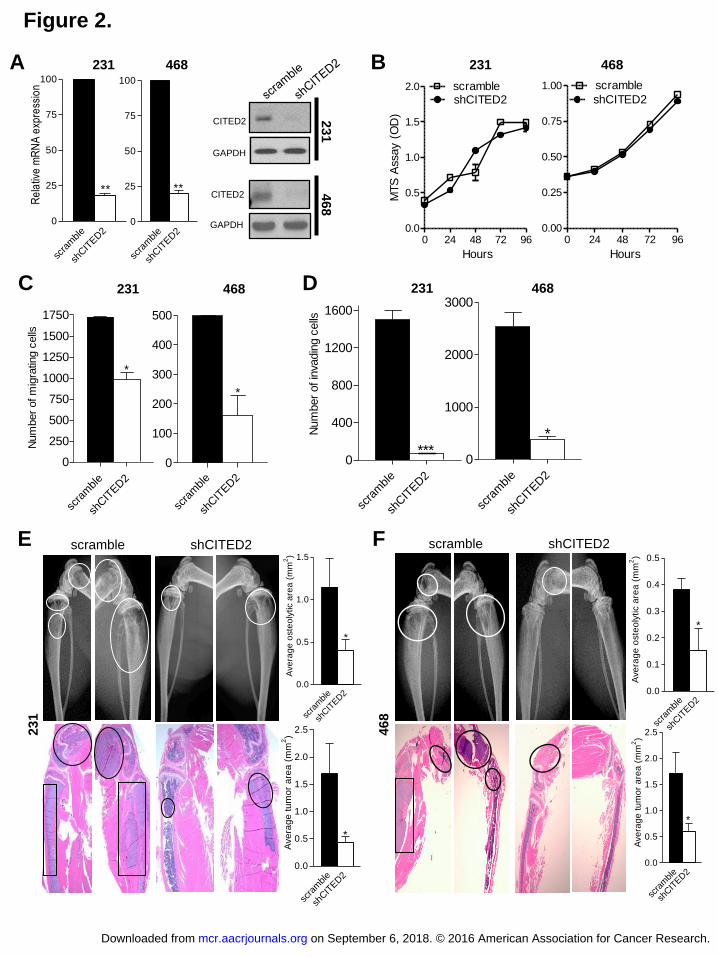
levels of CITED2 were assessed at both the mRNA and protein levels (Fig. 2A). Stable
expression of shCITED2 resulted in a greater than 75% reduction in CITED2 expression in both
MDA-MB-231 and MDA-MB-468 cells by qRT-PCR and Western analysis. Prior to examining
the effect of CITED2 on metastatic progression, we first examined whether reducing CITED2
expression altered the rate of cell proliferation in vitro. As determined by MTS assay,
shCITED2 cells exhibited a similar growth rate to that of scramble cells, indicating that
knockdown of CITED2 did not affect cell growth in either cell line (Fig. 2B). To begin exploring
CITED2 involvement in metastatic progression, we evaluated the effects of CITED2 knockdown
on the migratory and invasive ability of MDA-MB-231 and MDA-MB-468 cells using in vitro
trans-well migration and invasion assays, respectively. CITED2 knockdown significantly
reduced the migratory and invasive ability of both MDA-MB-231 and MDA-MB-468 cells (Fig.
2C and D). Next, we assessed the effects of CITED2 down-regulation on the establishment of
metastasis following intra-cardiac administration of MDA-MB-231 and MDA-MB-468 cells
stably expressing shCITED2 or scramble in athymic nude mice. While this model does not
replicate the entire metastatic cascade, it effectively assesses the ability of tumor cells to
extravasate from the vasculature, invade and colonize secondary organ sites, and establish
vascularized metastatic lesions. Mice administered with shCITED2-expressing cells displayed
significantly reduced metastasis to bone relative to the scramble group. As evidenced by digital
on September 6, 2018. © 2016 American Association for Cancer Research. mcr.aacrjournals.org Downloaded from
Author manuscripts have been peer reviewed and accepted for publication but have not yet been edited. Author Manuscript Published OnlineFirst on May 23, 2016; DOI: 10.1158/1541-7786.MCR-16-0081

13
radiography, osteolytic area was significantly lower in the shCITED2 group relative to the
scramble group (Fig. 2E and F, top). Consistent with reduced osteolysis, the shCITED2 group
also displayed a significant reduction in tumor area relative to the scramble group, as measured
on histological sections (Fig. 2E and F, bottom). Additionally, mice injected with shCITED2-
expressing MDA-MB-231 cells developed fewer brain metastases relative to those injected with
scramble cells, although no differences were observed using MDA-MB-468 cells
(Supplementary Fig. S1A). Further, metastasis to the lung and liver did not appear to be affected
by shCITED2 expression in either MDA-MB-231 or MDA-MB-468 cells (Supplementary Fig.
S1B and S1C). Taken together, these observations indicate that CITED2 may play a role in the
establishment of breast cancer metastasis, particularly to the bone.
Inhibition of CITED2 reduces expression of IKKα and pro-metastatic NF-κB target genes
To explore the mechanism through which CITED2 influences metastatic ability, we
examined the effect of CITED2 knockdown on gene expression in MDA-MB-231 cells by
cDNA microarray analysis (Supplementary Table 1). Notably, IKKα, a critical mediator of the
NF-κB signaling cascade (23) was found to be down-regulated along with several downstream
targets having reported roles in promoting metastasis, including osteopontin (OPN), matrix
metalloproteinase 9 (MMP9), urokinase type plasminogen activator (uPA), secreted protein,
acidic, cysteine-rich (SPARC), interleukin-11 (IL-11) and interleukin-1β (IL-1β). While the
extracellular matrix protein SPARC and proteases MMP9 and uPA have been shown to promote
tumor invasion (24-28), evidence suggests that the cytokines IL-11 and IL-1β facilitate the
establishment of osteolytic metastasis (18, 29). Additionally, the multi-functional protein OPN
reportedly affects numerous steps in the metastatic cascade (30). Regulation of these genes by
on September 6, 2018. © 2016 American Association for Cancer Research. mcr.aacrjournals.org Downloaded from
Author manuscripts have been peer reviewed and accepted for publication but have not yet been edited. Author Manuscript Published OnlineFirst on May 23, 2016; DOI: 10.1158/1541-7786.MCR-16-0081

14
NF-κB signaling was confirmed in MDA-MB-231 cells, wherein treatment with the IKK
inhibitor PS1145, which prevents IκBα degradation (31), reduced both basal NF-κB signaling by
Western analysis and expression of OPN, MMP9, uPA, SPARC, IL-11 and IL-1β by qRT-PCR
(Supplementary Fig. S2A and S2B). Moreover, using qRT-PCR and Western/ELISA analyses,
we confirmed down-regulation of IKKα along with the aforementioned NF-κB target genes in
shCITED2-expressing MDA-MB-231 and MDA-MB-468 cells compared to scramble cells at
both the mRNA and protein levels (Fig. 3A-F). Further, by ChIP assay in MDA-MB-231 cells,
CITED2 was found to localize to the promoter of IKKα indicating a potentially direct role for
CITED2 in the regulation of its expression. (Fig. 3G). Collectively, these data indicate that
CITED2 knockdown reduces the expression of IKKα and several downstream pro-metastatic
genes in breast cancer cells.
Down-regulation of CITED2 attenuates NF-κB signaling
NF-κB signaling is constitutively active in breast cancer (32) and occurs via both
canonical and non-canonical pathways, each of which involves IKKα. In the canonical pathway
the trimeric IKKα/β/γ complex phosphorylates the NF-κB inhibitor IκBα, inducing its
degradation and concomitantly releasing p65/p50 transcription factors to translocate into the
nucleus for regulation of gene expression (33). In the non-canonical pathway, phosphorylation of
p100 by IKKα triggers nuclear translocation of the RelB/p52 transcription factors for regulating
gene expression (33). Since CITED2 knockdown resulted in the down-regulation of IKKα
expression along with several downstream targets of NF-κB, we next investigated the effect of
CITED2 on basal NF-κB signaling in breast cancer cells. Examining the effects of CITED2
knockdown on the expression levels and localization of NF-κB signaling factors by Western
on September 6, 2018. © 2016 American Association for Cancer Research. mcr.aacrjournals.org Downloaded from
Author manuscripts have been peer reviewed and accepted for publication but have not yet been edited. Author Manuscript Published OnlineFirst on May 23, 2016; DOI: 10.1158/1541-7786.MCR-16-0081

15
analysis revealed increased levels of IκBα and reduced nuclear levels of p65 and RelB in
shCITED2-expressing MDA-MB-231 and MDA-MB-468 cells relative to scramble cells (Fig.
4A), indicating that both the canonical and non-canonical pathways were affected. Consistent
with reduced nuclear p65 and RelB levels, NF-κB DNA binding activity of the p65/p50
heterodimer was also markedly reduced in shCITED2-expressing MDA-MB-231 and MDA-MB-
468 cells relative to scramble cells as determined by EMSA (Fig. 4B). Together, these data
demonstrate the ability of CITED2 to influence NF-κB signaling.
Restoration of IKKα expression reverses effects of CITED2 knockdown on breast cancer
cell invasion
Since CITED2 knockdown reduced levels of IKKα along with numerous downstream
factors with reported roles in promoting metastatic dissemination (Fig. 3), we next investigated
the possibility that the reduced metastatic ability of shCITED2-expressing breast cancer cells
was related to the down-regulation of IKKα expression. To address this question, we transiently
restored IKKα expression in shCITED2-expressing MDA-MB-231 and MDA-MB-468 cells
(Fig. 5A) and examined invasive ability by in vitro trans-well invasion assay. Notably, restoring
IKKα expression in shCITED2-expressing cells significantly increased invasive ability relative
to empty vector-transfected shCITED2 cells, returning invasiveness to levels commensurate with
those observed in scramble cells (Fig. 5B and C). These data support a role for IKKα in
mediating the effects of CITED2 on the metastatic ability of breast cancer cells.
Discussion
on September 6, 2018. © 2016 American Association for Cancer Research. mcr.aacrjournals.org Downloaded from
Author manuscripts have been peer reviewed and accepted for publication but have not yet been edited. Author Manuscript Published OnlineFirst on May 23, 2016; DOI: 10.1158/1541-7786.MCR-16-0081

16
Despite current treatment efforts, the vast majority of patients diagnosed with metastatic
breast cancer ultimately succumb to this disease, highlighting the need for clearer understanding
of the drivers of metastasis and their mechanism of action. In this study, we have shown that
expression of the non-DNA binding transcriptional co-activator CITED2 is significantly elevated
in metastatic lesions of breast cancer patients relative to primary tumors. Further, we have
demonstrated that down-regulation of CITED2 significantly attenuates invasive and metastatic
ability in two human breast cancer cell lines (MDA-MB-231 and MDA-MB-468). Lastly, we
provide evidence that the effects of CITED2 on metastatic ability are mediated, at least in part,
by controlling expression of the NF-κB regulator IKKα, the levels of which have been shown to
negatively correlate with relapse-free survival in breast cancer patients (Supplementary Fig. S3).
While elevated levels of CITED2 were found in patient samples of metastatic disease
relative to primary tumors, highest levels were noted in metastases to bone (Fig. 1A). Further,
the inhibition of metastatic colonization observed following knockdown of CITED2 in breast
cancer cell lines (Fig. 2E and F; Supplementary Fig. S1) was largely limited to skeletal disease.
These findings are in agreement with our previous data demonstrating that reducing CITED2
expression in the murine mammary tumor cell line NT2.5 significantly reduces bone metastasis
in vivo (15), highlighting the potential importance of CITED2 as a critical mediator of bone
metastasis in breast cancer. Although the mechanism by which CITED2 mediates this effect
remains unclear, CITED2 knockdown in MDA-MB-231 and MDA-MB-468 cells resulted in
reduced expression of IL-11 and IL-1β, both of which are reported mediators of bone metastasis
and osteolysis (18, 29), thus warranting further investigation into their potential contribution
towards the metastatic effects of CITED2.
on September 6, 2018. © 2016 American Association for Cancer Research. mcr.aacrjournals.org Downloaded from
Author manuscripts have been peer reviewed and accepted for publication but have not yet been edited. Author Manuscript Published OnlineFirst on May 23, 2016; DOI: 10.1158/1541-7786.MCR-16-0081

17
In addition to the impairment of bone metastasis, CITED2 knockdown in MDA-MB-231
cells also inhibited the establishment of brain metastasis. This effect was not observed in MDA-
MB-468 cells, possibly due to the fact that this cell line colonized the brain less frequently than
MDA-MB-231 in the control condition. While the lesser ability of MDA-MB-468 cells to
colonize the brain could be due to various differences between these two cell lines, it is
interesting to note that canonical TGF-β signaling is absent in MDA-MB-468 cells due to the
lack of Smad4 in this cell line (34). Combined with the reported role of CITED2 in regulating
TGF-β signaling through Smad interactions (9), it is tempting to speculate that the divergent
ability of MDA-MB-231 and MDA-MB-468 cells to establish brain metastasis may be related to
TGF-β responsiveness. Moreover, it should be noted that the lack of canonical TGF-β signaling
in MDA-MB-468 cells did not impact the effect of CITED2 knockdown on invasion or the
establishment of bone metastasis. This indicates that these effects were mediated in a TGF-β-
independent manner, further supporting a role for NF-κB signaling, which was attenuated in both
the MDA-MB-231 and MDA-MB-468 cell lines.
Despite the ability of CITED2 to directly regulate expression of the NF-κB pathway
regulator IKKα, it is not yet clear how CITED2 modulates NF-κB signaling and transcriptional
activity in breast cancer cells. Although we did not observe changes in the mRNA expression of
NF-κB signaling intermediates downstream of IKKα, upstream mediators also exist whose
expression could be impacted by CITED2. Additionally, CITED2 is known to interact with
CREB-binding protein (CBP) and p300, reported co-activators of p65-mediated gene
transcription (35), suggesting that CITED2 could also regulate the activity of NF-κB as part of
the transcriptional complex. Although the ability of IKKα to restore NF-κB signaling (data not
shown) and rescue tumor invasion in MDA-MB-231 and MDA-MB-468 cells following CITED2
on September 6, 2018. © 2016 American Association for Cancer Research. mcr.aacrjournals.org Downloaded from
Author manuscripts have been peer reviewed and accepted for publication but have not yet been edited. Author Manuscript Published OnlineFirst on May 23, 2016; DOI: 10.1158/1541-7786.MCR-16-0081

18
knockdown (Fig. 5B and C) implicates involvement of the NF-κB pathway, it should be noted
that IKKα can also exert NF-κB-independent effects (36). Thus, further investigation is required
not only to assess the mechanism whereby CITED2 modulates NF-κB activity, but also to
determine the extent of its contribution to the pro-metastatic effects of CITED2, as well as the
ultimate effectors of its action. Addressing these questions will not only further our
understanding of CITED2 action in breast cancer, but may also provide new avenues for the
prevention and treatment of metastatic spread.
References
1. Lu J, Steeg PS, Price JE, Krishnamurthy S, Mani SA, Reuben J, et al. Breast Cancer
Metastasis: Challenges and Opportunities. Cancer Res 2009;69:4951-3.
2. Chen Y, Doughman YQ, Gu S, Jarrell A, Aota S, Cvekl A, et al. Cited2 is required for the
proper formation of the hyaloid vasculature and for lens morphogenesis. Development
2008;135:2939-48.
3. Qu X, Lam E, Doughman YQ, Chen Y, Chou YT, Lam M, et al. Cited2, a coactivator of
HNF4alpha, is essential for liver development. EMBO J 2007;26:4445-56.
4. Xu B, Qu X, Gu S, Doughman YQ, Watanabe M, Dunwoodie SL, et al. Cited2 is required for
fetal lung maturation. Dev. Biol 2008;317:95-105.
5. Yin Z, Haynie J, Yang X, Han S, Kiatchoosakun S, Restivo J, et al. The essential role of
Cited2, a negative regulator for HIF-1alpha, in heart development and neurulation. Proc. Natl.
Acad. Sci. U.S.A 2002;99:10488-93.
on September 6, 2018. © 2016 American Association for Cancer Research. mcr.aacrjournals.org Downloaded from
Author manuscripts have been peer reviewed and accepted for publication but have not yet been edited. Author Manuscript Published OnlineFirst on May 23, 2016; DOI: 10.1158/1541-7786.MCR-16-0081

19
6. Bhattacharya S, Michels CL, Leung MK, Arany ZP, Kung AL, Livingston DM. Functional
role of p35srj, a novel p300/CBP binding protein, during transactivation by HIF-1. Genes Dev
1999;13: 64–75.
7. Glenn DJ, Maurer RA. MRG1 binds to the LIM domain of Lhx2 and may function as a
coactivator to stimulate glycoprotein hormone alpha-subunit gene expression. J. Biol. Chem
1999;274:36159-67.
8. Bragança J, Eloranta JJ, Bamforth SD, Ibbitt JC, Hurst HC, Bhattacharya S. Physical and
functional interactions among AP-2 transcription factors, p300/CREB-binding protein, and
CITED2. J. Biol. Chem 2003;278:16021-29.
9. Chou YT, Want H, Chen Y, Danielpour D, Yang YC. Cited2 modulates TGFbeta-mediated
upregulation of MMP9. Oncogene 2006;25:5547-60.
10. Tien ES, Davis JW, Vanden Heuvel JP. Identification of the CREB-binding protein/p300-
interacting protein CITED2 as a peroxisome proliferator activated receptor alpha coregulator. J.
Biol. Chem 2004;279:24053-63.
11. Lau WM, Doucet M, Huang D, Weber KL, Kominsky SL. CITED2 modulates estrogen
receptor transcriptional activity in breast cancer cells. Biochem. Biophys. Res. Commun
2013;437:261-6.
12. Sun HB, Zhu YX, Yin T, Sledge G, Yang YC. MRG1, the product of a melanocyte-specific
gene related gene, is a cytokine-inducible transcription factor with transformation activity. Proc.
Natl. Acad. Sci. U.S.A 1998;95:13555-60.
13. Bai L, Merchant JL. A role for CITED2, a CBP/p300 interacting protein, in colon cancer cell
invasion. FEBS Lett 2007;581:5904-10.
on September 6, 2018. © 2016 American Association for Cancer Research. mcr.aacrjournals.org Downloaded from
Author manuscripts have been peer reviewed and accepted for publication but have not yet been edited. Author Manuscript Published OnlineFirst on May 23, 2016; DOI: 10.1158/1541-7786.MCR-16-0081

20
14. Chou YT, Hsieh CH, Chiou SH, Hsu CF, Kao YR, Lee CC, et al. CITED2 functions as a
molecular switch of cytokine-induced proliferation and quiescence. Cell Death Differ
2012;19:2015-28.
15. Lau WM, Weber KL, Doucet M, Chou YT, Brady K, Kowalski J, et al. Identification of
prospective factors promoting osteotropism in breast cancer: a potential role for CITED2, Int J
Cancer 2010;126:876-84.
16. Chou YT and Yang YC. Post-transcriptional control of CITED2 by transforming growth
factor beta. Regulation via Smads and CITED2 coding region. J Biol. Chem 2006;281:18451-62.
17. Nakano H, Shindo M, Sakon S, Nishinaka S, Mihara M, Yagita H, et al. Differential
regulation of IkappaB kinase alpha and beta by two upstream kinases, NF-kappaB-inducing
kinase and mitogen-activated protein kinase/ERK kinase kinase-1. Proc. Natl. Acad. Sci. U.S.A
1998;95:3537-42.
18. Kang Y, Siegel PM, Shu W, Drobnjak M, Kakonen SM, Cordόn-Cardo C, et al. A multigenic
program mediating breast cancer metastasis to bone. Cancer Cell 2003;3:537-49.
19. Minn AJ, Gupta GP, Siegel PM, Bos PD, Shu W, Giri DD, et al. Genes that mediate breast
cancer metastasis to lung. Nature 2005;436:518-24.
20. Bos PD, Zhang XH, Nadal C, Shu W, Gomis RR, Nguyen DX, et al. Genes that mediate
breast cancer metastasis to the brain. Nature 2009;459:1005-9.
21. Lau WM, Doucet M, Stadel R, Huang D, Weber KL, Kominsky SL. Enpp1: a potential
facilitator of breast cancer bone metastasis. PLoS One 2013;8:e66752.
22. Vantyghem SA, Allan AL, Postenka CO, Al-Katib W, Keeney M, Tuck AB, et al. A new
model for lymphatic metastasis: development of a variant of the MDA-MB-468 human breast
on September 6, 2018. © 2016 American Association for Cancer Research. mcr.aacrjournals.org Downloaded from
Author manuscripts have been peer reviewed and accepted for publication but have not yet been edited. Author Manuscript Published OnlineFirst on May 23, 2016; DOI: 10.1158/1541-7786.MCR-16-0081

21
cancer cell line that aggressively metastasize to lymph nodes. Clin Exp Metastasis 2005;22:351-
61.
23. Adli M, Merkhofer E, Cogswell P, Baldwin AS. IKKalpha and IKKbeta each function to
regulate NF-kappaB activation in the TNF-induced/canonical pathway. PLoS One 2010;5:e9428.
24. Seno T, Harada H, Kohno S, Teraoka M, Inoue A, Ohnishi T. Downregulation of SPARC
expression inhibits cell migration and invasion in malignant gliomas. Int J Dev Neurosci
1999;17:463-72.
25. Golembieski WA, Ge S, Nelson K, Mikkelsen T, Rempel SA. Increased SPARC expression
promotes U87 glioblastoma invasion in vitro. Int J Oncol 2009;34:707-15.
26. Chen J, Wang M, Xi B, Xue J, He D, Zhang J, et al. SPARC is a key regulator of
proliferation, apoptosis and invasion in human ovarian cancer. PLoS One 2012;7:e42413.
27. Mehner C, Hockla A, Miller E, Ran S, Radisky DC, Radisky ES. Tumor-cell produced
matrix metalloproteinase 9 (MMP-9) drives malignant progression and metastasis of basal-like
triple negative breast cancer. Oncotarget 2014;5:2736-49.
28. Tang L, Han X. The urokinase plasminogen activator system in breast cancer invasion and
metastasis. Biomed Pharmacother 2013;67:179-82.
29. Liu Q, Russell MR, Shahriari K, Jernigan DL, Lioni MI, Garcia FU, et al. Interleukin-1β
promotes skeletal colonization and progression of metastatic prostate cancer cells with
neuroendocrine features. Cancer Res 2013;73:1-9.
30. Shevde LA, Samant RS. Role of osteopontin in the pathophysiology of cancer. Matrix
Biology 2014;37:131-41.
on September 6, 2018. © 2016 American Association for Cancer Research. mcr.aacrjournals.org Downloaded from
Author manuscripts have been peer reviewed and accepted for publication but have not yet been edited. Author Manuscript Published OnlineFirst on May 23, 2016; DOI: 10.1158/1541-7786.MCR-16-0081

22
31. Yemelyanov A, Gasparian A, Lindholm P, Dang L, Pierce JW, Kisselijov F, et al. Effects of
IKK inhibitor PS1145 on NF-κB function, proliferation, apoptosis and invasion activity in
prostate carcinoma cells. Oncogene 2006;25:387-98.
32. Yamaguchi N, Ito T, Azuma S, Ito E, Honma R, Yanagisawa Y, et al. Constitutive activation
of nuclear factor-kappaB is preferentially involved in the proliferation of basal-like subtype
breast cancer cell lines. Cancer Sci 2009;100:1668-74.
33. Hoesel B, Schmid JA. The complexity of NF-κB signaling in inflammation and cancer. Mol
Cancer 2013;12:86.
34. Schutte M, Firuban RH, Hedrick L, Cho KR, Nadasdy GM, Weinstein CL, et al. DPC4 gene
in various tumor types. Cancer Res 1996;56:2527-30.
35. Gerritsen ME, William AJ, Neish AS, Moore S, Shi Y, Collins T. CREB-binding
protein/p300 are transcriptional coactivators of p65. Proc. Natl. Acad. Sci. U.S.A 1997;94:2927-
32.
36. Huang WC, Hung MC. Beyond NF-κB activation: nuclear functions of IκB kinase α. J
Biomed Sci 2013;20:3.
Figure legends
Figure 1.
CITED2 expression is elevated in human breast cancer metastasis. A, CITED2 mRNA
expression was determined by qRT-PCR in human normal mammary epithelium (n=12), primary
breast tumor tissues (invasive ductal carcinoma) from patients surviving greater than (n=11) and
less than (n=8) five years from the time of diagnosis, and metastatic lesions obtained from non-
bone (n=19) and bone (n=6) sites. (p* < 0.05; p** < 0.01). B, Immunohistochemical analysis
on September 6, 2018. © 2016 American Association for Cancer Research. mcr.aacrjournals.org Downloaded from
Author manuscripts have been peer reviewed and accepted for publication but have not yet been edited. Author Manuscript Published OnlineFirst on May 23, 2016; DOI: 10.1158/1541-7786.MCR-16-0081
23
was performed on paraffin-embedded tissue sections of normal mammary epithelium (n = 5),
invasive ductal carcinoma (IDC) (n = 5) and breast cancer bone metastasis (BBM) (n = 8) using
goat anti-CITED2 antibody. Protein was visualized using DAB (brown). Sections were
counterstained with hematoxylin and visualized by light microscopy. N: Normal, T: Tumor and
B: Bone. Magnification: 400X.
Figure 2.
CITED2 knockdown in breast cancer cells inhibits tumor migration and invasion in vitro and
bone metastasis in vivo. A, CITED2 mRNA expression (left) was determined by qRT-PCR. Data
are representative of triplicate experiments. CITED2 protein expression (right) was determined
by Western analysis performed on equal amounts of protein from total cell lysates. GAPDH
serves as the loading control. B, Cell proliferation was determined by MTS assay. C and D, The
migratory and invasive capability of the tumors was determined by migration (C) and invasion
(D) assays, respectively. E and F, scramble or shCITED2-expressing MDA-MB-231 (E) and
MDA-MB-468 (F) cells were injected into the left cardiac ventricle of athymic nude mice. Top:
Digital radiographic imaging of the femur and tibia showing areas of osteolysis marked by white
circles. The adjacent histograms represent the average osteolytic area measured on the
radiographic images between the experimental groups. Bottom: H&E staining of decalcified bone
sections indicating presence of tumor lesions as shown by black rectangles and circles. The
adjacent histograms represent the average tumor area measured on histological images between
the experimental groups. (p* <0.05, p** < 0.01, p*** < 0.001).
Figure 3.
on September 6, 2018. © 2016 American Association for Cancer Research. mcr.aacrjournals.org Downloaded from
Author manuscripts have been peer reviewed and accepted for publication but have not yet been edited. Author Manuscript Published OnlineFirst on May 23, 2016; DOI: 10.1158/1541-7786.MCR-16-0081

24
CITED2 knockdown in breast cancer cells reduces expression of IKKα and pro-metastatic NF-
κB target genes. A and D, mRNA expression was determined by qRT-PCR. Data are
representative of triplicate experiments. B and E, Protein expression of IKKα was determined by
Western analysis of total cell lysates (CE). GAPDH serves as the loading control. Protein
expression of MMP9 and uPA was determined by Western analysis of serum free condition
medium (CM). C and F, protein expression of IL-11 and OPN (MDA-MB-231 only) was
analyzed by ELISA. G, Localization of CITED2 or IgG to the IKKα promoter was assessed by
ChIP assay using anti-sheep CITED2 or sheep IgG antibodies in wild-type MDA-MB-231 cells.
(p* <0.05, p** < 0.01, p*** < 0.001).
Figure 4.
CITED2 regulates NF-κB signaling. A, Western analysis of cytoplasmic IκBα and nuclear p65
and RelB proteins was performed on equal amounts of protein obtained from cytoplasmic (CE)
and nuclear (NE) cell lysates. GAPDH and HDAC1 serve as the cytoplasmic and nuclear loading
controls, respectively. B, Non-radioactive EMSA analysis was performed on equal amounts of
nuclear cell lysate using NF-κB and Oct-1 oligonucleotides. Oct-1 serves as the loading control.
(p* <0.05, p** < 0.01).
Figure 5.
Restoration of IKKα expression following CITED2 knockdown in breast cancer cells rescues
cell invasiveness. A, IKKα expression was determined by Western analysis of equal amounts of
total protein lysates obtained from scramble cells or shCITED2-expressing cells transfected with
either empty vector (EV) or IKKα. GAPDH serves as the loading control. B and C, Invasive
on September 6, 2018. © 2016 American Association for Cancer Research. mcr.aacrjournals.org Downloaded from
Author manuscripts have been peer reviewed and accepted for publication but have not yet been edited. Author Manuscript Published OnlineFirst on May 23, 2016; DOI: 10.1158/1541-7786.MCR-16-0081

25
ability of scramble cells and shCITED2 cells transfected with either empty vector (EV) or IKKα
was determined by in vitro trans-well invasion assay. (B) Representative images of trans-well
inserts following staining of invading cells with crystal violet (purple). (C) Quantification of
invading cells showing the average number per experimental condition. (p*** < 0.001).
on September 6, 2018. © 2016 American Association for Cancer Research. mcr.aacrjournals.org Downloaded from
Author manuscripts have been peer reviewed and accepted for publication but have not yet been edited. Author Manuscript Published OnlineFirst on May 23, 2016; DOI: 10.1158/1541-7786.MCR-16-0081

Figure 1.
A
B
0
250
500
750
1000
Primary tumors
*
1000
3000
5000
Metastases
**
Normal Mammary
Epithelium
> 5 yr
Survival
< 5 yr
SurvivalNon-Bone Bone
*R
ela
tive m
RN
A E
xp
ressio
n
Mammary epithelium IDC (high CITED2) IDC (low CITED2) BBM
N
T
T T
T
T
N B
T
N
N
T
T
on September 6, 2018. © 2016 American Association for Cancer Research. mcr.aacrjournals.org Downloaded from
Author manuscripts have been peer reviewed and accepted for publication but have not yet been edited. Author Manuscript Published OnlineFirst on May 23, 2016; DOI: 10.1158/1541-7786.MCR-16-0081
0 24 48 72 960.0
0.5
1.0
1.5
2.0 scrambleshCITED2
Hours
MTS
Assay (
OD
)
scra
mble
shCIT
ED2
0
250
500
750
1000
1250
1500
1750
*
Num
ber
of m
igra
ting c
ells
scra
mble
shCIT
ED2
0
100
200
300
400
500
*
scra
mble
shCIT
ED2
0
400
800
1200
1600
***
Num
ber
of in
vadin
g c
ells
scra
mble
shCIT
ED2
0
1000
2000
3000
*
C D
Figure 2.
A B
231 468
231 468
231 468
scra
mble
shCIT
ED2
0.0
0.5
1.0
1.5
*
Ave
rag
e o
ste
oly
tic a
rea
(m
m2)
scra
mble
shCIT
ED2
0.0
0.5
1.0
1.5
2.0
2.5
*
Ave
rag
e t
um
or
are
a (
mm
2)
E F scramble shCITED2
2
31
scramble shCITED2
4
68
scra
mble
shCIT
ED2
0.0
0.5
1.0
1.5
2.0
2.5
*
Ave
rag
e t
um
or
are
a (
mm
2)
scra
mble
shCIT
ED2
0.0
0.1
0.2
0.3
0.4
0.5
*A
ve
rag
e o
ste
oly
tic a
rea
(m
m2)
shCITED2
0 24 48 72 960.00
0.25
0.50
0.75
1.00 scramble
Hoursscra
mble
shCIT
ED2
0
25
50
75
100
**
Re
lativ
e m
RN
A e
xpre
ssio
n
231 468
CITED2
GAPDH
CITED2
GAPDH
23
1 4
68
scra
mble
shCIT
ED2
0
25
50
75
100
**
on September 6, 2018. © 2016 American Association for Cancer Research. mcr.aacrjournals.org Downloaded from
Author manuscripts have been peer reviewed and accepted for publication but have not yet been edited. Author Manuscript Published OnlineFirst on May 23, 2016; DOI: 10.1158/1541-7786.MCR-16-0081

IKK
MM
P9uP
A
SPARC
IL11
IL1
-100
-50
-7.5
-5.0
-2.5
0.0
*
** ***
*
*
Rela
tive m
RN
A e
xpre
ssio
n2
31
46
8
Figure 3.
A B C
D E F
MMP9
uPA
CE
C
M
IKKα
GAPDH
MMP9
CM
uPA
IKKα
CE
GAPDH
scra
mble
shCIT
ED2
0.0
0.1
0.2
0.3
0.4
0.5
*
OP
N e
xpre
ssi
on (
ng/m
l)
scra
mble
shCIT
ED2
0
10000
20000
***
30000
40000
50000
IL11 e
xpre
ssio
n (
pg/m
l)
scra
mble
shCIT
ED2
0
10000
15000
17500
20000
22500
25000
**
IL11 e
xpre
ssio
n (
pg/m
l)
G
23
1
IgG
CIT
ED2
0.00
0.05
0.10
0.15
0.20
0.25
*
% I
nput
IKK
OPN
MM
P9
uPA
SPAR
CIL
11IL
1-25
-15
-5-5.0
-2.5
0.0
***
**
**
*
*
Rela
tive m
RN
A e
xpre
ssio
n
on September 6, 2018. © 2016 American Association for Cancer Research. mcr.aacrjournals.org Downloaded from
Author manuscripts have been peer reviewed and accepted for publication but have not yet been edited. Author Manuscript Published OnlineFirst on May 23, 2016; DOI: 10.1158/1541-7786.MCR-16-0081

Figure 4.
IkBα
GAPDH
CE
p65
RelB
HDAC1
NE
231 468 A
B
46
8 2
31
NF
-B
Oct1
p65.p50
p50.p50
p65.p50
p50.p50
Oct1
NF
-B
on September 6, 2018. © 2016 American Association for Cancer Research. mcr.aacrjournals.org Downloaded from
Author manuscripts have been peer reviewed and accepted for publication but have not yet been edited. Author Manuscript Published OnlineFirst on May 23, 2016; DOI: 10.1158/1541-7786.MCR-16-0081

A
231 468
IKKα
GAPDH
Figure 5.
IKKα
GAPDH
B
scra
mbl
e
shCIT
ED2
(EV) )
shCIT
ED2
(IKK
0
200
400
600
800 ***
***
Num
ber
of
inva
din
g c
ells
scra
mbl
e
shCIT
ED2
(EV) )
shCIT
ED2
(IKK
0
100
200
300
400***
***
Num
ber
of
inva
din
g c
ells
scramble
shCITED2
(EV)
shCITED2
(IKKα)
scramble
shCITED2
(EV)
shCITED2
(IKKα)
C
on September 6, 2018. © 2016 American Association for Cancer Research. mcr.aacrjournals.org Downloaded from
Author manuscripts have been peer reviewed and accepted for publication but have not yet been edited. Author Manuscript Published OnlineFirst on May 23, 2016; DOI: 10.1158/1541-7786.MCR-16-0081

Published OnlineFirst May 23, 2016.Mol Cancer Res Swaathi Jayaraman, Michele Doucet, Wen Min Lau, et al. Effects on IKKalphaCITED2 Modulates Breast Cancer Metastatic Ability Through
Updated version
10.1158/1541-7786.MCR-16-0081doi:
Access the most recent version of this article at:
Material
Supplementary
http://mcr.aacrjournals.org/content/suppl/2016/06/25/1541-7786.MCR-16-0081.DC1
Access the most recent supplemental material at:
Manuscript
Authoredited. Author manuscripts have been peer reviewed and accepted for publication but have not yet been
E-mail alerts related to this article or journal.Sign up to receive free email-alerts
Subscriptions
Reprints and
To order reprints of this article or to subscribe to the journal, contact the AACR Publications
Permissions
Rightslink site. Click on "Request Permissions" which will take you to the Copyright Clearance Center's (CCC)
.http://mcr.aacrjournals.org/content/early/2016/05/21/1541-7786.MCR-16-0081To request permission to re-use all or part of this article, use this link
on September 6, 2018. © 2016 American Association for Cancer Research. mcr.aacrjournals.org Downloaded from
Author manuscripts have been peer reviewed and accepted for publication but have not yet been edited. Author Manuscript Published OnlineFirst on May 23, 2016; DOI: 10.1158/1541-7786.MCR-16-0081
![THE The JOHNS HOPKINS CLUB Events JOHNS HOPKINS … [4].pdf · Club Herald July / August 2015 Events THE The JOHNS HOPKINS CLUB JOHNS HOPKINS UNIVERSITY 3400 North Charles Street,](https://static.fdocuments.in/doc/165x107/5fae1ad08ad8816d2e1aaabe/the-the-johns-hopkins-club-events-johns-hopkins-4pdf-club-herald-july-august.jpg)