
DEPARTMENT OF MEDICAL RESEARCH ENCES No. 5, Ziwaka … Vol 1-24 PDF/MHSRJ V… · Khin Saw Aye, Aye...
56
MYANMAR H H E E A A L L T T H H S S C C I I E E N N C C E E S S RESEARCH JOURNAL Volume 18, No. 1, 2006 DEPARTMENT OF MEDICAL RESEARCH (Lower Myanmar) No. 5, Ziwaka Road Yangon 11191, Myanmar CONTENTS Sea snakebite in Myanmar: epidemiology and treatment seeking behaviour …………..... 1 Tun Pe, Aye Aye Myint, Sann Mya & Maung Maung Toe Diagnostic significance of fibrin(-ogen) degradation products in cerebrospinal fluid in childhood meningitis …………………………………………………………………… 6 Ne Win, Khine Hla Myint, Thein Thein Myint, Than Nu Shwe, Aye Maung Han, Aye Aye Lwin, Minn Minn Myint Thu & Tin Htar Nwe Isolation of anaerobic Gram-positive bacilli and detection of Clostridium perfringens alpha toxin from some foods…………………………………………………………… 13 Thin Thin Maw, Mar Mar Nyein, Mi Mi Htwe & Aye Aye Maw Experimental production of goat russell’s viper antivenom……………………………… 17 Aye Aye Myint & Tun Pe Evaluation of Polymerase Chain Reaction (PCR) Amplification of Mycobacterium leprae in biopsy specimens from leprosy patients…………………………………….. 21 Khin Saw Aye, Yin Min Htun, Aye Aye Win, Tin Zar Maw & Kyaw Kyaw Antibacterial activity of some plants and formulations and determination of Minimum Inhibitory Concentration (MIC) by microtitre plate dilution method………………… 26 Mar Mar Nyein, Mi Mi Htwe, Ba Han & Ei Ei Khine Compartmental syndrome following a green pit viper (Trimeresurus erythrurus) bite………………………………………………………... 31 Tun Pe & Tin Tin Aung Comparison of Polymerase Chain Reaction, immunohistochemistry and conventional histopathology in the diagnosis of leprosy in Myanmar………………… 34 Khin Saw Aye, Aye Aye Win, Tin Zar Maw, Kyaw Kyaw & Khine San Yin Prevalence, awareness, correlates, treatment and control of hypertension in a rural community of Waw Township, Bago Division……………………………………….. 41 Han Win, Aung Thu, Khin Myat Tun, Khin Khin Swe Myat, Than Than Lwin, Sandar Kyi, Myat Myat Thu, Tin Htar Lwin & Aye Hnin Phyu
Transcript of DEPARTMENT OF MEDICAL RESEARCH ENCES No. 5, Ziwaka … Vol 1-24 PDF/MHSRJ V… · Khin Saw Aye, Aye...
MMYYAANNMMAARR
HHEEAALLTTHH SSCCIIEENNCCEESS RREESSEEAARRCCHH JJOOUURRNNAALL
Volume 18, No. 1, 2006
DEPARTMENT OF MEDICAL RESEARCH
(Lower Myanmar)
No. 5, Ziwaka Road
Yangon 11191, Myanmar
CONTENTS
Sea snakebite in Myanmar: epidemiology and treatment seeking behaviour …………..... 1 Tun Pe, Aye Aye Myint, Sann Mya & Maung Maung Toe Diagnostic significance of fibrin(-ogen) degradation products in cerebrospinal fluid in
childhood meningitis …………………………………………………………………… 6 Ne Win, Khine Hla Myint, Thein Thein Myint, Than Nu Shwe, Aye Maung Han,
Aye Aye Lwin, Minn Minn Myint Thu & Tin Htar Nwe Isolation of anaerobic Gram-positive bacilli and detection of Clostridium perfringens
alpha toxin from some foods…………………………………………………………… 13 Thin Thin Maw, Mar Mar Nyein, Mi Mi Htwe & Aye Aye Maw Experimental production of goat russell’s viper antivenom……………………………… 17 Aye Aye Myint & Tun Pe
Evaluation of Polymerase Chain Reaction (PCR) Amplification of Mycobacterium leprae in biopsy specimens from leprosy patients…………………………………….. 21
Khin Saw Aye, Yin Min Htun, Aye Aye Win, Tin Zar Maw & Kyaw Kyaw
Antibacterial activity of some plants and formulations and determination of Minimum Inhibitory Concentration (MIC) by microtitre plate dilution method………………… 26
Mar Mar Nyein, Mi Mi Htwe, Ba Han & Ei Ei Khine Compartmental syndrome following a green pit viper
(Trimeresurus erythrurus) bite………………………………………………………... 31 Tun Pe & Tin Tin Aung
Comparison of Polymerase Chain Reaction, immunohistochemistry and
conventional histopathology in the diagnosis of leprosy in Myanmar………………… 34Khin Saw Aye, Aye Aye Win, Tin Zar Maw, Kyaw Kyaw & Khine San Yin Prevalence, awareness, correlates, treatment and control of hypertension in a rural
community of Waw Township, Bago Division……………………………………….. 41Han Win, Aung Thu, Khin Myat Tun, Khin Khin Swe Myat, Than Than Lwin, Sandar Kyi, Myat Myat Thu, Tin Htar Lwin & Aye Hnin Phyu
Distribution of coliforms , faecal coliforms and enteropathogenic E. coli (EPEC)
in fried rice and water samples from street vendors of Yangon and detection of bacterial toxins……………………………………………………………………. 48
Mar Mar Nyein, Yee Yee Aung, Mi Mi Htwe & Tin Nwe Short Report : Determination of anticardiolipin antibodies in recurrent abortion………. 53 Ne Win, Ye Naing Oo, Minn Minn Myint Thu, Yin Min Htun, Mu Mu Shwe &
Thein Myint Thu
Editor-in-Chief
Professor Dr. Paing Soe
---------------------------------------------------------------------------------------------------------------- Editor
Dr. Soe Thein
Associate Editors Daw Nyunt Nyunt Swe Dr. Ni Thet Oo
---------------------------------------------------------------------------------------------------------------- Editorial Board
Prof. Dr. Aye Maung Han Prof. Dr. U Kyaw Prof. Dr. Saw Naing Prof. Dr. Nyunt Wai
Prof. Dr. Myo Win Prof. Dr. Kyi Kyi Thinn
Prof. Col. Marlar Than Prof. Dr. Tracy Sein
Dr.Tun Pe Dr. Aye Kyaw Dr. Than Tun Sein Dr. W.Tun Lin Dr. Kyaw Moe Dr. Myo Khin Dr. Khin Myat Tun Dr. Khin Pyone Kyi
----------------------------------------------------------------------------------------------------------------
Business Manager
Dr. Kyaw Min
Editorial Manager
U Aung Myo Min
Production Manager
U Ye Thway
---------------------------------------------------------------------------------------------------------------- Printed at the Publications Division, Department of Medical Research (Lower Myanmar)
---------------------------------------------------------------------------------------------------------------
Restricted for Internal Use Only
The Myanmar Health Sciences Research Journal,Vol. 18, No. 1, 2006
Sea snakebite in Myanmar : epidemiology and treatment seeking behaviour
*Tun Pe, *Aye Aye Myint, **Sann Mya & ***Maung Maung Toe
*Venom Research Laboratory, Department of Medical Research (LM)
**Department of Medicine, Institute of Medicine (2), Yangon, ***Department of Medical Research (LM)
Although sea snake bite occurs among fishermen in Myanmar, the incidence has not been documented. In order to determine the incidence, case fatality and treatment seeking behaviour of the victims, a community based study was conducted in three fishing communities namely Letkokekone (LKK) (Yangon Division), Kyaikkami (KKM) (Mon State) and Daedaye-Pyarpone-Bogalay (DPB) (Ayeyawady Division). A house-to-house visit was conducted and the victims were asked structured questionnaires. The cumulative incidence of sea snake bite for 4 years (1999-2002) in LKK was 318/100000, in KKM was 75/100000 and for 5 years (1999-2003) in DPB was 118.9/100000. Case fatality rate ranged from 8.5 to 20.2%. The mean age of the victims was 35.64 years (age range 10 - 87 yr). Majority of them were fishermen, being bitten during day time on legs (LKK), hands (KKM) and on both hands and legs (DPB) while engaged in fishing activity. Majority sought treatment with local healers (LKK and KKM) and home remedy (DPB). Hospital treatment was sought in less than 10% of the cases and incidence based on hospital data was underestimated. None of them used prophylaxis against snakebite. Wound incision and local application or ingestion of herbs were widely practiced among the victims. Health education on the use of prophylaxis at work and correct first aid should be promoted. Practice on harmful unscientific wound treatment and local and home remedy should be discouraged.
INTRODUCTION
Sea snakebite is an important occupational hazard of fishermen. Accidental bite occurs while sorting fish at sea especially under insufficient light [1]. According to Reid [2] 80% of the accidental bites fail to envenom victims. Severe envenomed cases present with neuromuscular paralysis and renal failure secondary to myoglobinuria. Specific antivenom is not available in Myanmar. Traditional practice in fishing community is that only very sick victims are advised to seek hospital treatment. The incidence of sea snake bite based on hospital data is 0.4% [3]. There were few reports on sea snake bite cases in Myanmar [1,4,5]. Recent community-based study of snakebite in Myanmar [6] and other countries [7-9] suggested that incidence based on hospital data was underestimated. Community-based
study of sea snake bite in two townships [10] highlighted that because of variations in traditional belief and fishing technique between communities, incidence, case fatality rate and treatment seeking behaviour may vary. In order to verify the statement, epidemiological study of sea snakebite was further extended to fishing communities in Ayeyawady Division and findings were presented in this communication.
MATERIALS AND METHODS
Community-based study of incidence, case fatality rate and treatment seeking behaviour of sea snake bite victims was carried out in three fishing communities namely Letkokekone (LKK) (Yangon Division), Kyaikkami (KKM) (Mon State) and Daedaye – Pyarpon - Bogalay (DPB) (Ayeyawady Division) with the help of
basic health staffs of the respective township from January to May 2003 in LKK and KKM and February to May 2004 in DPB. A house to house visit was undertaken by assigned midwife. Set proforma including size of the household, age, sex of sea snake bite victims within last 4 years (LKK and KKM) and five years (DPB) was asked to the head of household and the victim was identified. Structured questionaires designed to cover circum-stances of the bite, fatality and treatment seeking behaviour, use of first aid and prophylaxis were asked to the victims or next of kin if the victim was dead. For children, guardians or parents were asked. Coded data were entered and analyzed using Epi info version (6.04d) software.
RESULTS
Incidence/case fatality rate
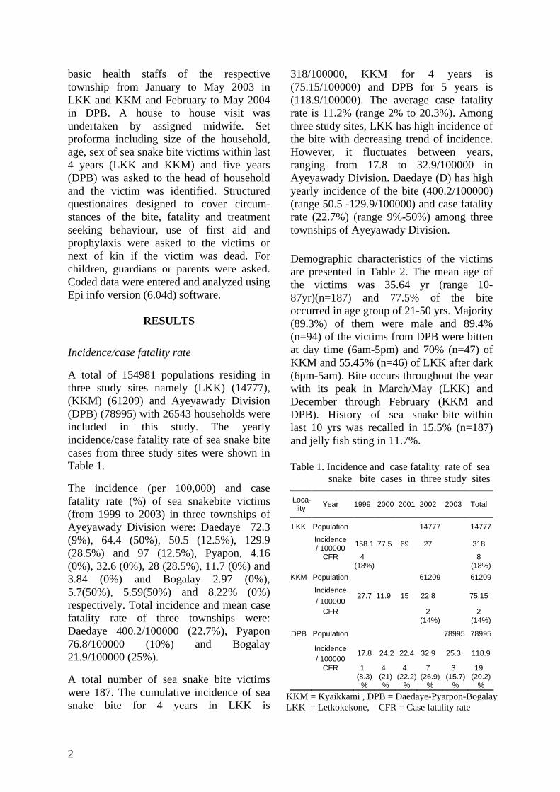
A total of 154981 populations residing in three study sites namely (LKK) (14777), (KKM) (61209) and Ayeyawady Division (DPB) (78995) with 26543 households were included in this study. The yearly incidence/case fatality rate of sea snake bite cases from three study sites were shown in Table 1.
The incidence (per 100,000) and case fatality rate (%) of sea snakebite victims (from 1999 to 2003) in three townships of Ayeyawady Division were: Daedaye 72.3 (9%), 64.4 (50%), 50.5 (12.5%), 129.9 (28.5%) and 97 (12.5%), Pyapon, 4.16 (0%), 32.6 (0%), 28 (28.5%), 11.7 (0%) and 3.84 (0%) and Bogalay 2.97 (0%), 5.7(50%), 5.59(50%) and 8.22% (0%) respectively. Total incidence and mean case fatality rate of three townships were: Daedaye 400.2/100000 (22.7%), Pyapon 76.8/100000 (10%) and Bogalay 21.9/100000 (25%).
A total number of sea snake bite victims were 187. The cumulative incidence of sea snake bite for 4 years in LKK is
318/100000, KKM for 4 years is (75.15/100000) and DPB for 5 years is (118.9/100000). The average case fatality rate is 11.2% (range 2% to 20.3%). Among three study sites, LKK has high incidence of the bite with decreasing trend of incidence. However, it fluctuates between years, ranging from 17.8 to 32.9/100000 in Ayeyawady Division. Daedaye (D) has high yearly incidence of the bite (400.2/100000) (range 50.5 -129.9/100000) and case fatality rate (22.7%) (range 9%-50%) among three townships of Ayeyawady Division. Demographic characteristics of the victims are presented in Table 2. The mean age of the victims was 35.64 yr (range 10-87yr)(n=187) and 77.5% of the bite occurred in age group of 21-50 yrs. Majority (89.3%) of them were male and 89.4% (n=94) of the victims from DPB were bitten at day time (6am-5pm) and 70% (n=47) of KKM and 55.45% (n=46) of LKK after dark (6pm-5am). Bite occurs throughout the year with its peak in March/May (LKK) and December through February (KKM and DPB). History of sea snake bite within last 10 yrs was recalled in 15.5% (n=187) and jelly fish sting in 11.7%.
Table 1. Incidence and case fatality rate of sea snake bite cases in three study sites
Loca-lity Year 1999 2000 2001 2002 2003 Total
LKK Population 14777 14777
Incidence / 100000 158.1 77.5 69 27 318
CFR 4 (18%) 8
(18%)KKM Population 61209 61209
Incidence
/ 100000 27.7 11.9 15 22.8 75.15
CFR 2 (14%) 2
(14%)
DPB Population 78995 78995
Incidence / 100000
17.8 24.2 22.4 32.9 25.3 118.9
CFR 1
(8.3)%
4 (21)%
4 (22.2)
%
7 (26.9)
%
3 (15.7)
%
19 (20.2)
% KKM = Kyaikkami , DPB = Daedaye-Pyarpon-Bogalay LKK = Letkokekone, CFR = Case fatality rate
2
Circumstances of the bites Majority 84.49% (n=187) were fisherman and 86.6% were bitten while engaged in fishing activities: setting up/drawing conical net (34.7%), stake net (19.25%), bag net (9%), casting net (8.5%), fish sorting (11.76%) and carrying fish baskets (3.2%). Bite at sea shore accounts for 13.4%: while walking/sitting on sand bank of LKK (3.7%) and in shallow sea water of DPB while doing washing up/working (9.6%) (Table 3).
Site of bite
Majority of the victims are bitten on legs 81 % (n=47) in LKK, right more than left (21 and 17) and on hands 70% (n=46), left
more than right (21 and 11) in KKM. Both legs 47.9% (n=45) and hands 52.1% (n=49), right more than left (35 and 14) were equally bitten in DPB.
Site of bite and circumstances of the bite
Legs are bitten more than hands while setting up stake net (18 and 5), catching fish with a bag net (11 and 6), getting into shallow sea (18 and 0) and walking/sitting at sea shore (7 and 0). In contrast setting up of conical net carries equal risk on both hands and legs(6 and 6), however, hands are bitten more than legs in sorting fish (20 and 2), while drawing /wrapping up conical net (34 and 19) and casting net (10 and 6). Both legs and hands are equally at risk while carrying fish baskets from boat to sea shore (3 and 3) and drawing stake net (7 and 6).
First aid/prophylaxis
None of the victims used prophylaxis against sea snake bite. About 80% (n=187) of the sea snakebite victims carried out wound treatments such as incision 23.5%, local herbal application 16%, application of tourniquet 11.2% and coagulation of the wound 11.2%. Ingestion and or local
Table 2. Demographic characteristics of the sea snake bite victims
Age Mean 35.64yr (Range 10-87yr)
10-20yr 17 (9%) 21-30yr 56 (29.9%) 31-40yr 51 (27.3%) 41-50yr 38 (20.3%) 51-60yr 21 (11.2%) 61-70yr 3 (1.6%) 81-90yr (87) 1 (0.5%) Sex Male 167 (89.3%) Female 20 (11.7%) Male: female ratio 167:20 (2.6:1) The bite Throughout the year
Peak -March-May LKK
KKM
-Dec-Feb
DPB
Day (6am-5pm) 84 (89.4%) -DPB
Night (6pm-5am) 32 (70%) -KKM
Time bite
26 (55.4%) -LKK Site of bite Legs 38/47 (81%)-LKK Hands 32/46 (70%)-KKM
45 and 49 -DPB
Legs and hands
(47.9% and 52.1%) Occupation Fishing (n=187) 158 (84.49%) Street vender 12 (6.4%) Ad hoc. 11 (5.8%) Farmer 4 (2.1%) Dependent 1 (0.53%) Carpenter 1 (0.53%)
LKK =Letkokekone KKM=Kyaikkami DPB=Daedaye-Pyarpon-Bogalay
Table 3. Circumstances of the bites
Activity Number(%)
1 Fishing activities 162/187 (86.6%)
Conical net (34.7%)
Setting up 12 (6.4%)
Drawing 53 (28.3%)
Stake net (19.25%)
Setting up 23 (12.29%)
Drawing 13 (6.95%)
Bag net 17 (9%)
Casting net 16 (8.5%)
Fish sorting 22 (11.76%)
Carrying fish baskets 6 (3.2%) 2 At seashore 25/187 (13.4%)
Walking (LKK) 6 (3.2%)
Sitting (LKK) 1 (0.5%)
Getting into sea (DPB) 3 (1.6%)
Activities at seashore 15 (8.02%)
Lkk =Letkokekone DPB=Daedaye-Pyarpon-Bogalay
3
application of herbs on incised wound was recalled in 46% (n=47) victims from KKM.
Treatment seeking behavior
Seeking treatment with local healers was a common practice in LKK 66% (n=46) and in KKM 57% (n=47) and home remedy in DPB 73.4% (n=94). Hospital treatment was sought in 9% (n=187) and at local clinic 6.4% (n=187) (Table 4).
Treatment provided by traditional healers consists of either ingestion and or local application of herbs to the wound in KKM and wound incision and suction in LKK. Home remedy practiced in LKK (to take coconut flesh with jaggery) differs from KKM (to take coconut juice and rub the wound with lime) and DPB (ingestion of local herbs, to take coconut flesh/juice with jaggery or herbs and drink meditated water).
Clinical features of the victims
The common clinical features recalled by the victims (n=187) are drowsiness (78.6%), muscle ache (71.6%), muscle stiffness (62.5%), heavy upper eye lid (56.6%) and passing dark urine (myoglobinuria) (31.5%) denoting systemic envenoming. Passing dark urine was recalled in 71% (n=47) of the victim from KKM and 22.3% (n=94) in DPB.
DISCUSSION
Incidence/case fatality rate The study highlights that sea snake bite is an occupational hazard of fishermen. Yearly
incidence of sea snake bite in LKK in earlier years is 3 to 8 times more than that of KKM and DPB. Recent decrease in incidence of the bite in LKK could be attributed to recent introduction of modified fish catching technique and advancement of sand bank in sea shore creating unfavourable condition for catching fish and prawn in sea shore. Although yearly incidence of the bite is decreasing, the figures are still high. It is likely that inclusion of a proportion of systemic envenomed cases (myoglobinuria 22.3%) in home remedy group is responsible for high fatality rate (20.2%) in DPB. In contrast low mortality rate (2%) in KKM with 71% passing dark urine must be misdiagnosed by the victims for passing high coloured urine in dehydrated victims since they refused to drink after bites. Passing dark urine (myoglobinuria) signifies systemic envenoming and carries a bad prognosis.
Table 4. Treatment seeking behaviour of the victims
Sources Letkoke-
kone
Kyaik- kami
Daedaye- Pyapon- Bogalay (DPB)
Total
Local healers 31(66%) 26 (57%) 14 (14.9%) 71 (37.9%)
Home remedy 11(23%) 3 ( 6.5%) 69 (73.4%) 83 (44.3%)
Hospital 5 (11%) 9 (19.6%) 3 ( 2.2%) 17 ( 9%)
Clinic 0 8 (17.4%) 4 ( 4.3%) 12 ( 6.4%)
No treatment 0 0 4 ( 4.3%) 4 ( 2.1%)
Since 9% of the victims sought hospital treatment, the true incidence of sea snakebite based on hospital data was underestimated. In order to get a true incidence of the bite it should be made a notifiable disease.
Prophylaxis/ first aid
Use of no longer recommended wound treatments should be discouraged. Since none of the victim used prophylaxis against sea snake, health education on use of prophylaxis such as wearing protective gloves, taking precaution at work and provision of proper illumination at work site after dark should be given to fishing communities. Two accidental sea snake bite cases admitted to Yangon General Hospital occurred after dark while sorting fish under insufficient light [1]. Health education on use of the correct first aid for neurotoxic envenoming i.e. compression immobili-zation using crepe bandage [11] should be promoted. Treatment seeking behaviour
Majority of the victims from DPB prefer home remedy in KKM and local healers in
4
5
LKK. Their treatments have no scientific values and give a false sense of security and their uses should be discouraged. According to Reid [2], 80% of sea snake bite victims are not envenomed. Majority of the cases treated by local healers and home remedy fall into this group and will recover with conservative treatment. This favourable outcome has attracted fishing communities to seek treatment from them. Only very sick victims were advised to seek treatment at hospital. It is likely that wrong treatment seeking behaviour of the victims leads to high fatality rate in DPB. Less than 10% of the victims sought treatment at hospital. It is recommended that all sea snake bite cases should be kept under observation and if no myoglobinuria (passing dark urine) developed in 4-6 hours after the bite, the victim will not be systemic envenoming [12]. Health education on such information should be given to fishing communities, so that they will be well informed about prognosis of the illness and where to seek treatment.
Early referral to hospitals with capability of performing renal dialysis and assisted ventilatory support to treat renal failure and neurotoxic respiratory paralysis in severe envenomed cases should be practiced. In summary, health education leading to changes in behaviour on use of prophylaxis and correct first aid should be given to fishing communities and encourage them to seek treatment at hospital.
REFERENCES
1. Tin Myint, Tun Pe & Sann Mya. Sea snake bite cases admitted to Yangon General Hospital.
Myanmar Health Research Congress abstract 2002; p 11.
2. Reid, H.A. Sea snake bites. British Medical Journal 1956; 2:73-78.
3. Aye Aye Myint, Tun Pe & Tin Zar Maw. An epidemiological study of snakebite and venomous snake survey in Myanmar. In: Management of Snakebite and Research, WHO/SEARO publication,12-16.
4. Sann Mya, Theingie Yin & Tun Pe. Study of snakebite cases admitted to Yangon general Hospital. Myanmar Health Sciences Research Journal, 2004;16:42-45.
5. Sann Mya, Theingie Yin & Tun Pe. Study of snakebite cases referred to Yangon general Hospital. Myanmar Health Sciences Research Journal, 2004;16:14 -16.
6. Tun Pe, Aye Aye Myint, Sunn Htut, Khin Aye Kyu & Maung Maung Toe. Prevalence, case fatality rate and treatment seeking behaviour of the snakebite victims from two townships of Myanmar. Myanmar Health Research Congres abstract s, 2003; p 13.
7. Coombs, M.D., Dunachie, S.J., Brooker,S., Haynes, J., Church, J., & Warrell, D.A. Snakebites in Kenya: a preliminary survey of four areas. Transactions of Royal Society of Tropical Medicine and Hygiene 1997; 91:319-321.
8. Hati, A.K, Mandal, M., De, M.K., Mukherjee, H. & Hati, R.N. Epidemiology of snakebite in the district of Burdwan, West Bengal. Journal of Indian Medical Association,. 1991; 90:145-147.
9. Snow, R.W., Bronzan, R., Roques, T., Nyamawi, C., Murphy, S., & Marsh, K. The prevalence and morbidity of snakebite and treatment seeking behaviour among a rural Kenya population. Annals of Tropical Medicine and Parasitology 1994; 88:665-671.
10. Tun Pe, Aye Aye Myint, Sann Mya, Min Tin Htay & Maung Maung Toe. Prevalence, case fatality rate and treatment seeking behaviour of sea snakebite victims from two fishing communities. Myanmar Health Research Congress abstract 2003; p 14 .
11. Sutherland, S.K., Coulter, A.R., & Harris, R.D. Rationalisation of first-aid measure for elapid snake bites. Lancet 1979;1:183-186.
12 White, J. Clinical toxicology of sea snakebite in handbook of clinical toxicology of animal venoms and poisons. Eds. J. Meier & J. White Boca Ratom, FL: CRC Press, 1995.
The Myanmar Health Sciences Research Journal, Vol. 18, No. 1, 2006
Diagnostic significance of fibrin(-ogen) degradation products in cerebrospinal fluid in childhood meningitis
*Ne Win, **Khine Hla Myint, **Thein Thein Myint, **Than Nu Shwe,
**Aye Maung Han, *Aye Aye Lwin,*Minn Minn Myint Thu & ***Tin Htar Nwe
*Pathology Research Division, DMR (LM) **Yangon Children Hospital, Institute of Medicine 1
***Clinical Pathology Laboratory, Yangon Children Hospital
Fibrin(-ogen) degradation products (FDP) was determined in cerebrospinal fluid (CSF) of 106 children suffering from meningitis (35 tuberculous, 37 pyogenic, and 34 non-bacterial meningitis) by using a locally developed test kit (DMR-FDP test kit). Generally diagnosis of different meningitis is made clinically, supported by routine examination of CSF (CSF-RE). The sensitivity of the test kit is 2 µg/ml fibrinogen equivalent. FDP content is determined semi-quantitatively by the doubling dilution of CSF and described as 0, 2, 4, 8, 16, 32, 64, 128, 256, and 512 µg/ml. Overall FDP content in CSF was ranging from 0-512 µg/ml. It varied with the type of meningitis: 0-8 µg/ml in non-bacterial meningitis, and ≥16 µg/ml in bacterial meningitis. CSF-FDP content was always ≤8 µg/ml in all of non-bacterial meningitis cases (34/34; 100%). In 35 TBM cases, 34 cases have 16-64 µg/ml, 1 case 128 µg/ml. In 37 pyogenic meningitis cases, 10 have 64 µg/ml and 27 have ≥128 µg/ml. Statistically, CSF-FDP level of 0-8 µg/ml has 100% sensitivity for non-bacterial meningitis; 16-64 µg/ml has 97.1% sensitivity for TB meningitis; and ≥128 µg/ml has 72.9% sensitivity for pyogenic meningitis. This study could define a cut-off point: CSF-FDP content, >8 µg/ml for bacterial meningitis and ≤8 µg/ml for non-bacterial meningitis and could also differentiate a meningitis case into different types: (i) nonbacterial meningitis when CSF-FDP ≤8 µg/ml and bacterial when ≥16µg/ml, (ii) tuberculous when 16-64 µg/ml, and (iii) pyogenic meningitis when ≥128 µg/ml. In conclusion, determination of FDP content in CSF by DMR-FDP test kit greatly facilitates the differential diagnoses of meningitis in children and it will be of great benefit to the clinicians, particularly at the health centers where and/or when laboratory facilities for CSF-RE are inefficient. The CSF-FDP should be measured routinely in children with meningitis and is suggested to be included in CSF-RE as an additional biochemical parameter to other conventional tests.
INTRODUCTION
Routine examination of cerebrospinal fluid (CSF-RE) is a common laboratory procedure in day-to-day clinical practice. It is particularly useful in meningitis for the etiological diagnosis as bacterial (pyogenic or tuberculous) and nonbacterial (viral, fungal, others) to be followed by an appropriate and prompt treatment. Provisional diagnosis of meningitis is made clinically and etiologic diagnosis follows
after CSF-RE. Etiological confirmation by microbiological, immunological and DNA techniques is done only when importantly indicated in selected cases for various constraints.
The presence of FDP in CSF has been reported in meningococcemia, fulminant pyogenic and viral meningitis, sub-arachnoid hemorrhage and intraventricular hemorrhage [1, 2, 3, 4, 5]. In these studies the emphases were made only from the aspects of the clinical severity and associated DIC being
6
occurred in these cases. Reports on CSF-FDP in relation to CSF-RE findings are very few and it was never been described from diagnostic aspect in meningitis worldwide. Moreover, CSF-FDP has never been studied in tuberculous meningitis. The reasons for such knowledge gaps are many but scarcity of cases available and expensiveness of FDP determination are commonly and impor-tantly included. We have been previously reported the presence of FDP in CSF in association with CSF-RE findings [6]. That report high-lighted that abnormality in CSF-RE (such as increased protein content, decreased sugar and chloride content and increased cell counts) is associated with presence of FDP in CSF. We have also suggested CSF-FDP to be used as an alternative approach when CSF-RE is not feasible or available for any reasons. Based on the previous findings the present study is conducted with the following main objectives: (i) To determine FDP content in CSF from children with meningitis either pyogenic, tuberculous or others (non-bacterial meningitis such as viral, fungal, parasitic) diagnosed clinically and supported by CSF-RE findings; and (ii) To evaluate the role of CSF-FDP deter-mination in the diagnosis of different forms of meningitis.
MATERIALS AND METHODS
Study design
A cross-sectional, analytical, hospital- based, collaborative study was conducted between Pathology Research Division, DMR (Lower Myanmar), and Medical Units and Clinical Pathology Laboratory of Yangon Children Hospital (YCH).
Methodology
One-hundred and six children admitted for signs and symptoms of meningitis were entered into the study in one year study period. CSF-RE was indicated and lumbar puncture was done as routines after getting an informed consent. One millilitre of
venous blood was also collected in a clean test tube and separated for serum.
Preparation of DMR-FDP test kit DMR-FDP test kit was prepared as described previously at the Pathology Research Division, DMR (Lower Myanmar) [7].
In brief:
Coagulase positive strain Staphylococcus aureus is subcultured on tryptic soy agar overnight. Bacterial colonies are collected from the plate by rinsing with normal saline in a large test tube. The bacterial suspension is washed twice with saline and thrice with distilled water. Then it is deep-freezed and dried by lyophilization to make powdered form. Each test kit contains 10 mg bacterial powder (clumping factor) which is reconstituted with one ml of distilled water. The sensitivity of the test kit is 2 μg/ml of FDP.
Routine examination of CSF (CSF-RE) CSF-RE was done conventionally [8]. It contains tension at the time of tapping, colour or appearance visually, visual formation of coagulum on overnight standing, total and differential white cell count by special counting chamber using white cell pipette, biochemical tests for sugars (copper reduction method), proteins (turbidimetric method) and chloride content (colourimetric method).
Determination of total FDP in CSF FDP was determined in the CSF as described previously [6, 9]. Briefly: A drop of CSF is placed on a black tile (or glass slide) and mixed thoroughly with a drop of suspension from the test kit. Clumping reaction was observed in a few seconds in positive samples indicating presence of FDP >2 μg/ml (i.e., sensitivity of the test).
Doubling dilution was done in positive samples into 1:2, 1:4, 1:8, 1:16, 1:32, 1:64, 1:128, 1:256, and so on by using distilled water or normal saline. The highest dilution which shows clumping was taken and it was multiplied by 2 μg/ml. The result is the FDP
7
content of CSF.E.g., When the highest dilution with clumping reaction is 1:16, the FDP content is 16x2 = 32 μg / ml.
Determination of total FDP in serum
FDP in serum was determined similarly as described above [10, 11, 12, 13].
Diagnosis of meningitis
The clinical diagnosis of meningitis was made by thorough history taking and physical examination. The aetiological diagnosis was based on the CSF-RE findings and CxR evidences.
(a) Pyogenic meningitis:
- Straw colour / turbid in appearance - Total WBC count is greatly
increased (50 - numerous / cu.mm) - All polymorph or polymorph
predominance in differential count - Total protein content is greatly
increased (100 - >200 mg%) - Sugar content is greatly reduced
(<50% to absent) - Chloride content reduced (about 120
mg% or less than 100 mg%) - A septic foci may or may not be
present
(b) Tuberculous meningitis
- Clear colourless / opalescent in appearance
- Total WBC count increased (50-500 / cu. mm)
- Lymphocyte predominance and some polymorphs in differential count
- Total protein content is moderately increased (100->200 mg%)
- Sugar content reduced (<50% to absent)
- Chloride content reduced (about 120 mg% or less than 100 mg%)
- CxR finding may or may not be positive for tuberculosis
- Tuberculin test may or may not be positive
- History of TB contact present or not
(c) Viral and other meningitis
- Clear and colourless in appearance - Total WBC count increased (about 5
per cu.mm) - Lymphocyte predominance in dif-
ferential count - Total protein content slightly
increased (about 10 mg%) - Sugar content normal (>50%) - Chloride content normal (120+ mg%)
RESULTS
Clinical and CSF-RE findings have established the etiological diagnosis in a total of 106 children as follows:
(i) 37 pyogenic (ii) 35 tuberculous (iii) 34 non-bacterial
CSF-FDP was detected in 97 cases, not in 9 cases. It ranged from 4–512 μg /ml and distributed as follows:
FDP content
Total cases
Pyo- genic
Tuber- culous
Non- bacterial
0 µg/ml 9 0 0 9 4µg/ml 7 0 0 7 8 µg/ml 18 0 0 18 16 µg/ml 20 2 18 0 32 µg/ml 6 2 4 0 64 µg/ml 18 6 12 0 128 µg/ml 17 16 1 0 256 µg/ml 10 10 0 0 512 µg/ml 1 1 0 0
Total 106 37 35 34
i CSF-FDP ≤ 8 μg/ml was found only in nonbacterial meningitis cases (n=34)
ii CSF-FDP 16-64 μg/ml was found only in bacterial meningitis cases (n=44) 34 tuberculous; 10 pyogenic;
iii CSF-FDP 128-512 μg/ml was found only in bacterial meningitis cases (n=28) 27 pyogenic; 1 tuberculous;
8
Table 1. CSF-FDP content in different menin- gitis
FDP content (µg/ml) Meningitis
Total 0 4 8 16 32 64 128 256 512
Pyogenic 37 0 0 0 2 2 6 16 10 1
Tuberculous 35 0 0 0 18 4 12 1 0 0
Nonbacterial 34 9 7 18 0 0 0 0 0 0
Total 106 9 7 18 20 6 18 17 10 1
Table 3. CSF-FDP vs serum FDP in different meningitis
Serum FDP Meningitis 0-8
(μg/ml) 16-64
(μg/ml) 128+ (μg/ml)
P T NB P T NB P T NB
P 0 0 0 0
T 0 0 0-8
(μg/ml) NB 31 3 0 P 9 1 T 17 10 7 0
16-64
(μg/ml) NB 0 0 0 P 15 5 0 3 2 2 T 1
CSF FDP
128+ (μg/ml)
NB 0 0 0
Total 24 22 31 11 10 3 2 3 0
(1) All of 34 nonbacterial meningitis cases have CSF-FDP ≤ 8 μg/ml.
(2) 34 of 35 tuberculous meningitis cases have CSF-FDP 16-64 μg/ml; one case has 128 μg/ml.
(3) 27 of 37pyogenic meningitis cases have CSF-FDP≥ 128 μg/ml; ten cases have 16-64 μg/ml.
Table 2. Serum FDP in different meningitis
FDP content (μg/ml) Meningitis
0 2 4 8 16 32 64 128 256 512
Pyogenic 4 5 9 6 6 4 1 1 1 0
Tuberculous 6 2 5 9 4 4 2 1 2 0
Nonbacterial 9 5 15 2 2 1 0 0 0 0
Total 19 12 29 17 12 9 3 2 3 0
CSF-FDP and serum FDP level has no significant correlation (p>.7631)
P = Pyogenic meningitis T = Tuberculous meningitis NB = Non-bacterial meningitis
77/106 cases (72.6%) have serum FDP within normal range (2-10 μg/ml); 16-64 μg/ml was seen in 24/106 cases (22.6%); 5/106 cases (4.8%) have serum FDP 128-256 μg/ml. 32 μg/ml is maximum for non-bacterial meningitis (Table2).
Summary of the findings
1. CSF-FDP is significantly correlated with CSF-appearance, -WBC count, and –protein.
2. CSF-FDP 8 μg/ml is the cut-off point for bacterial meningitis either pyogenic or tuberculous.
3. CSF-FDP <8 μg/ml predicts non-bacterial meningitis.
4. CSF-FDP 16-64 μg/ml predicts tuberculous meningitis.
5. CSF-FDP >128 μg/ml predicts pyogenic meningitis.
6. CSF-FDP 0-8 μg/ml has 100% sensitivity for non-bacterial menin-gitis; 16-64 μg/ml has 97.1% sensitivity for TB meningitis; >128 μg/ml has 72.9% sensitivity for pyogenic meningitis.
7. Sensitivity of CSF-FDP >128+ μg/ml for pyogenic meningitis will be increased to 98.2% when CSF-appearance is considered together. All cases of pyogenic cases in this group have CSF-appearance opales-cent / turbid; no xanthochromic.
DISCUSSION
No study has been described the usefulness of FDP determination in CSF in the diagnosis of different forms of meningitis worldwide. Previous studies reported the presence of small or late fragments of fibrin degradation such as D- and E-fragments, and D-dimers since the emphases were made only from the clinical severity, prognosis and DIC (disseminated intravascular coagulation) aspects; never from the diagnostic emphasis [14, 15, 16, 17, 18].
9
This study could describe diagnostic implication of determination of total FDP in CSF in different forms of meningitis since DMR-FDP kit used in this study detects all fragments of fibrin degradation (not only late fragments but also early fragments like fragments A, B, X, Y, fibrinopeptides, fibrin monomers, and fibrin polymers) [19, 20, 21].
CSF-RE is a common laboratory practice for various disorders involving nervous system including meningitis. Patients suspected of having meningitis always should have a specimen of CSF (usually by lumbar puncture) and examined in the laboratory as soon as possible. Prompt identification of the causal organism is important because until an exact aetiological diagnosis has been made the proper antimicrobial therapy cannot be prescribed. A small range of biochemical tests is usually undertaken for CSF-RE. Hence, biochemical investigation of the CSF is usually less important diagnostically than simple inspection for appearance (colour, turbidity, spontaneous clotting or coagulum) and cytological examination (red cells, white cells, others) [22]. Only when appropriate and indicated, microbiological investigations and serological tests for syphilis are carried out.
Although CSF-RE is a common, easy, important and useful laboratory procedure, it has some laboratory and diagnostic pitfalls and disadvantages such as:
(A) The specimen must be dispatched to the laboratory at once; delay may result in the death of delicate pathogen such as meningococci, disintegration of leucocytes and the reduction in the CSF-sugar. Specimen should not be kept in the refrigerator, which kills H. influenzae.
(B) The presence of blood is the main cause of an abnormal colour. Normally no red blood cells should be present. Some may be introduced as a result of trauma whilst obtaining the fluid. Xanthochromia (yellow
colour) may be due to altered hemoglobin several days after a subarachnoid hemorrhage, large amount of pus, a very high protein content and jaundice. Small numbers of red cells also give fluids an opalescent appearance. If traces of substances such as alcohol are mixed with the fluid during its collection some opalescence may result. Spontaneous clotting (coagulum) occurs when there is an excess of fibrinogen in the specimen, usually associated with a very high protein concentration.
(C) CSF-cell count is done using a wbc count pipette in a special counting chamber. Leishman's stain is used for differential count when cell count is increased. CSF-wbc count may be undetectable in some cases of bacterial meningitis, particularly in children, in immunocompromised patients, and if antibiotics have been given before lumbar puncture.
(D) CSF-sugar is carried out by any of the usual blood sugar methods. If obvious pus is present, being indicated for bacterial meningitis, CSF-sugar content provides little additional information. CSF-sugar determination does not reliably distinguish between different forms of infective meningitis, because the result may be normal in any form. More importantly, CSF which has become contaminated during laboratory sampling may show a fall in CSF-sugar content if kept at room temperature. Streptomycin given intrathecally shortly before CSF sampling may interfere with copper reduction methods.
(E) The CSF-protein is increased in the presence of blood and pus. If either of these is apparent on visual inspection or by microscopical examination of the specimen no further information is provided by
10
CSF-protein content and the laboratory staff should not be unnecessarily exposed to potentially dangerous infected material. Although turbidimetric method used for CSF-protein is simple and quick, turbidity is affected by temperature, time, presence of red cells and bacteria.
(F) Normal CSF-chloride content is
usually affected by plasma concentration. If there is much vomiting, particularly in TBM before specific treatment became available, the plasma chloride falls and the CSF value follows it.
As it has been described elsewhere [6, 7, 9, 11, 12], DMR-FDP test kit has many advantages. It needs neither special skill nor equipment to perform, is simple and easy to interpret, and rapid enough to be accomplished even at the bed-side or in lumbar puncture room, and more importantly, highly economical. This study has clearly demonstrated that CSF-FDP: (i) is detected in more than 95% of
meningitis cases (ii) 8 μg/ml is a cut-off point for
bacterial and non-bacterial meningitis
(iii) 16-64 μg/ml predicts tuberculous meningitis
(iv) 128 μg/ml and above predict pyogenic meningitis
(v) has overall sensitivity 90% (vi) has sensitivity 100% for non-
bacterial meningitis: 97% for TBM and 73% for pyogenic meningitis
(vii) was formed mainly by local fibrinolysis, not by simple diffusion from the plasma since serum FDP level is within normal range in most of the cases.
Thus CSF-FDP content measured by DMR-FDP test kit strongly and reliably predicts aetiologic diagnosis of meningitis occurring in the children. In conclusion, determination of CSF-FDP is suggested in children with meningitis particularly in health centers where or when laboratory facilities for CSF-RE lack and it should be included as an additional test to other routine biochemical tests of conventional CSF-RE.
ACKNOWLEDGEMENT Gram-positive strains of Staphylococcus aureus for subcultures to prepare DMR-FDP test kit were supported by Bacteriology Research Division, DMR (Lower Myanmar) and Bacteriology Department, National Health Laboratory. Clumping factor in powdered form was prepared by lyophilization using a lyophilizer from Biochemistry Research Division, DMR (Lower Myanmar). The investigators are greatly indebted to Dr. U Tun Pe, Director (Research), DMR (Lower Myanmar) who has suggested to conduct this type of study while discussing on a paper presented in the previous Myanmar Health Research Congress 1999, Yangon, Myanmar. Sincere thanks are due to Dr. Maung Maung Toe, Deputy Director, Department of Medical Research (Lower Myanmar) for his kind assistance in statistical analysis.
REFERENCES 1. Brueton, M.J, Tugwell, P., Whittle, H.C.&
Greenwood, B.M. Fibrinogen degradation products in cerebrospinal fluid of patients with group A meningococcal meningitis. Journal of Clinical Pathology 1974; 27(5): 402-4.
2. Lang, D.T., Berberian, L.B., Lee, S.& Ault, M. Rapid differentiation of subarachnoid hemorrhage from traumatic lumbar puncture using D-dimer assay. Journal of Clinical Pathology 1990; 93(3): 403-405.
3. Whitelaw, A., Creighton, L., Gaffney, P. Fibrinolysis in cerebrospinal fluid after intraventricular hemorrhage. Archives of Diseases of Children 1991; 66: 808-810.
11
Research Journal 1997; 9(3): 127-132 . 4. Brueton, M.J., Breeze, G.R., Stuart, J. Fibrin- fibrinogen degradation products in cerebrospinal fluid. Journal of Clinical Pathology 1976; 29(4): 341-4 .
13. Than Than Aye. Coagulation profiles in complicated Russell’s viper bites. Thesis. MMedSc (Pathology). IM I. Yangon. 1997.
5. Li, F., Zhang, G., Zhao, W. Coagulation and fibrinolytic activity in patients with acute cerebral infarction. China Medical Journal (Eng) 2003; 116(3): 475-7.
14. Kastenbauer, S.& Pfister, H.W., Pneumococcal meningitis in adults: spectrum of complications and prognostic factors in a series of 87 cases. Brain 2003; 126(Pt 5): 1015-25.
6. Ne Win, Khin Khin Cho Naing, Kyi Kyi Han, Myint Aung, Khin Saw Aye, Thaung Hla, Maung Maung Toe, Tin Nwe. A breakthrough in laboratory examination: determination of fibrin (-ogen) degradation products in cerebrospinal fluid. Myanmar Health Research Congress. Abstract. Department of Medical Research (Lower Myanmar) 1999; p 24.
15. Barshtein, IuA, Kononenko VV, Iarosh OA. Disseminated intravascular coagulation and its pathogenetic significance in meningoence-phalitis. Zh Nevropatol Psikhiatr Im S S Korsakova 1989; 89(2): 21-6.
7. Ne Win, Kyaw Htwe, Thi Thi Naing, Ni Win, Hla Pe. Development of a test kit for fibrin / fibrinogen degradation products. Abstract. Myanmar Health Research Congress. Department of Medical Research (Lower Myanmar) Yangon 1993; pp 23.
16. Anisimova IuN. The pathological anatomy of pneumococcal meningo-encephalitis in adults. Arkh Patol 1990; 52(1): 11-7.
17. Hansen, B., Black, F.T.& Andersen, P.L. Purulent meningitis at the Marselisborg Hospital 1980-1990. Ugeskr Laeger 1994; 156(4): 7049-57.
8. Practical Clinical Biochemistry. In: Cerebrospinal fluids, miscellaneous fluids. Eds: Harold Varley, Alan H Gowenlock, Maurice Bell. Fifth edn. Volume 1. William Heinemann Medical Books Limited. London. 1980. pp 1197-1218.
18. Bajo, R., Vaca, R., Elduayen, R., Zarallo, L., Cardesa, J.J.& Perez-Miranda, M., Fibrin-fibrinogen degradation products in cerebrospinal fluid of patients with miningococcal infections (author's transl). Med Clin (Barc) 1980; 75 (8): 338-4.
19. Hawiger, J., Niewiarowski, S., Gurewich, V.& Thomas, D.P. Measurement of fibrinogen and fibrin degradation products in serum by staphylococcal clumping test. Journal of Laboratory Clinical Medicine 1970; 75: 98-112.
9. Khin Khin Cho Naing. Determination of fibrin(-ogen) degradation products in cerebrospinal fluid by staphylococcal clumping test. Thesis. MSc (Zoology). Department of Zoology. Yangon University. 1998.
20. Lipinski, B., Hawiger, J.& Jeljaszewicz, J., Staphylococcal clumping with soluble fibrin monomer complexes. Journal of Experimental Medicine 1967; 126: 979-992 .
10. Ne Win, Moe San, Than Than Tin. Urinary fibrin(-ogen) degradation products in pregnancy associated hypertension. Myanmar Health Science Research Journal 1998; 10(3); 135-138.
21. Marder, V.J., Matcheett, M.O.,& Sherry S. Detection of serum fibrinogen and fibrin degradation products. Comparison of six techniques using purified products and application in clinical studies. Ameican Journal of Medicine 1971; 51: 71-82.
11. Ne Win, Cho Mar Lwin, Thein Thein Myint, Than Nu Shwe, Aye Maung Han, et al. Disseminated intravascular coagulation has no role in the pathogenesis of grade IV dengue shock syndrome. Abstract. Myanmar Health Research Congress. 2002. pp 21.
22. The cerebrospinal fluid. In Clinical Chemistry in Diagnosis and Treatment. Eds: Joan F Zilva, Peter R Pannall, Philip D Mayne. Fifth edn. Edward Arnold, A Division of Hodder and
12. May Emerald, Khin Aye Kyi, Aye Aye Myint, Ne Win. Coagulation profiles in common malignancies. Myanma r Health Sciences Stoughton, London. 1989; pp 426-432.
12
The Myanmar Health Sciences Research Journal,Vol. 18, No.1, 2006
Isolation of anaerobic Gram-positive bacilli and detection of Clostridium perfringens alpha toxin from some foods
*Thin Thin Maw, *Mar Mar Nyein, *Mi Mi Htwe & *Aye Aye Maw
*Bacteriology Research Division Department of Medical Research (Lower Myanmar)
Isolation of anaerobic Gram-positive bacilli and detection of Clostridium perfringens alpha toxin were done on 120 samples of different food collected from shops available around Yangon area during 2003. After culturing in Trypticase Glucose Yeast (TGY) media, Clostridium perfringens alpha toxin was detected by using ELISA kit obtained from Bio-X Diagnostics, Belgium. Anaerobic Gram- positive bacilli was isolated from 31 samples (25.83%). They were isolated from 16.67%, 25%, 29.17%, 20.83% and 37.5% of noodles, fermented vegetables, fermented fish and prawns, salted fish paste (ngapi) and pickled tea respectively. The toxins were detected from 20 samples of food: 4.17% (1/24 samples of noodles), 45.80% (11/24 samples of fermented vegetables such as salted/fermented beans) and 4.17% (1/24 samples of fermented fish & prawns), 29.19% (7/24 from ngapi) respectively. Thus, this study indicates that toxin could be elaborated in protein rich preserved foods (fish / prawn paste).
INTRODUCTION
Food and beverages sold in streets which are affordable sources of nourishment for students, low income workers and others contain substantial amount of valuable nutrients. However, accessibility, availabi-lity and quality need to be maintained for the control of foodborne transmission via street vendors. A number of cases of foodborne bacterial infection and intoxication had been transmitted through street foods. Cholera, typhoid, staphy-lococcal food poisoning, hepatitis and other diseases can be transmitted through such foods [1-5].
Food poisoning is the general description, although two recognized types of illnesses are caused by two distinct metabolites. The diarrhoeal type of illness is caused by a large molecular weight protein, while the vomiting (emetic) type of illness is believed to be caused by a low molecular weight, heat stable peptide. Enterotoxemia due to Clostridium perfringens is intestinal and involves types A, B, C or D. Type A has
been implicated in rare outbreaks of gastritis and haemolytic disease of ruminants (enterotoxigenic jaundice, the yellows, yellow lamb disease) and the haemorrhagic enteritis in cattle, horses, dogs, and infant alpacas. Clostridium perfringens type A causes necrotic enteritis in poultry and a mild form of food poisoning in humans. Demonstration of alpha toxin in the contents of the small intestine is the only way to definitely diagnose enterotoxemia.. By using ELISA method, it is possible to detect alpha toxin in biological fluid or in culture filtrates. Thus, this study was undertaken to isolate Gram-positive bacilli and detect Clostridium perfringens alpha toxin from some food available around Yangon area.
One hundred and twenty four samples of different kinds of food including noodles (wheat and rice) fermented fish and prawns, fermented vegetables, fish paste from Pabedan, Mayangone, Kyauktada and
MATERIALS AND METHODS
Food samples
13
Botahtaung were randomly collected and used for isolation of anaerobic Gram-positive species.
Study period
January to September, 2003.
Study design
Random descriptive study
Bacteriological cultural methods
For culture and isolation of anaerobic Gram positive bacilli, Trypticase Glucose Yeast extract (TGY) with L-cysteine broth and agar was used. The plates were incubated at 37oC for 5 days in an anaerobic jar supplemented with Gaspak (BBL). Biochemical tests, Gram, acid fast and spore staining were carried out whenever necessary.
Detection of Clostridium perfringens alpha toxin from culture supernatant The 96-well microtitration plates sensitized by specific antibodies for the alpha-toxin were used for the test. The culture supernatants were added and incubated for 60 minutes at room temperature. The plates were washed and incubated for 60 minutes with the conjugate which was a peroxidase labeled anti-alpha-toxin specific polyclonal antibody. After the second incubation, the plates were washed again and the enzyme substrate (hydrogen peroxide) and the chromogen (tetramethyl benzidine, TMB) were added. If the alpha-toxin is present in the tested samples, the conjugate remains bound to the corresponding microwells and the enzyme catalyses the transformation of the colourless chromogen into a pigmented compound. The intensity of the resulting blue colour is proportionate to the titre of alpha-toxin in the sample. Enzymatic reaction was stopped by acidification and resulting optical density was measured at 450nm using the microplate reader. the signals recorded for the negative control wells were substracted from the corresponding positive microwells (Bio-X Diagnostics, Belgium). The optical density
at wave length 450nm with the cut-off value in controls was calculated after substracting the optical density of corresponding negative control results. The limit of positivity for the antigen is 0.150. Any sample that yields a difference in optical density that is greater than or equal to 0.150 is considered positive. Conversely, any sample that yields a difference in the optical density that is less than 0.150 is considered negative.
RESULTS
Based on the morphology and cultural appearances, they were roughly identified as Clostridium species when they fall into the following categories. After culturing anaerobically on Trypticase Glucose Yeast agar, small, compact, shiny and transluscent colonies which are similar to the features of Clostridium perfringens were picked and stained. They were Gram-positive rods in young cultures and not acid fast, catalase negative and produced spores that were heat resistant. The motility of anaerobes was easy to detect. The shape and position of spores when present, calls for subjective judgement and it may well be making a distinction to describe one spore as subterminal and another as central. In Cowan and Steel, they avoided this issue by indicating only those strains that produce terminal spores, and assumed that all others produced central or subterminal spores. Isolation of Gram-positive bacilli rate is high (16.67% to 37.5% (Table 1).
Detection of Clostridium perfringens alpha toxin from culture supernatants of different kinds of food
Alpha toxin from culture supernatants of different kinds of food is shown in Table 2. Out of 24 samples of each category of food tested, 1 , 11 , 1 and 7 samples of noodles, fermented vegetables, fermented fish and prawn; and ngapi (shrimp) produced Clostridium perfringens toxin (alpha toxin) respectively.
14
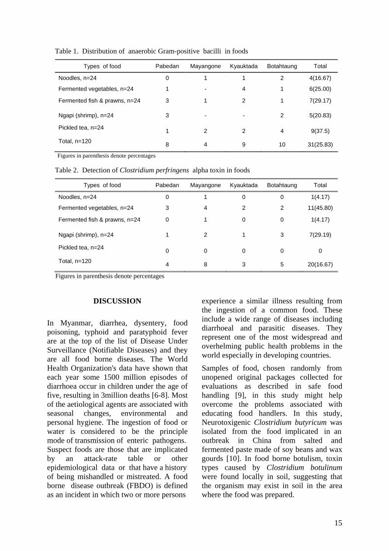
Table 1. Distribution of anaerobic Gram-positive bacilli in foods
Types of food Pabedan Mayangone Kyauktada Botahtaung Total
Noodles, n=24 0 1 1 2 4(16.67)
Fermented vegetables, n=24 1 - 4 1 6(25.00)
Fermented fish & prawns, n=24 3 1 2 1 7(29.17)
Ngapi (shrimp), n=24 3 - - 2 5(20.83)
Pickled tea, n=24 1 2 2 4 9(37.5)
Total, n=120 8 4 9 10 31(25.83)
Figures in parenthesis denote percentages
Table 2. Detection of Clostridium perfringens alpha toxin in foods
Types of food Pabedan Mayangone Kyauktada Botahtaung Total
Noodles, n=24 0 1 0 0 1(4.17)
Fermented vegetables, n=24 3 4 2 2 11(45.80)
Fermented fish & prawns, n=24 0 1 0 0 1(4.17)
Ngapi (shrimp), n=24 1 2 1 3 7(29.19)
Pickled tea, n=24 0 0 0 0 0
Total, n=120 4 8 3 5 20(16.67)
Figures in parenthesis denote percentages
DISCUSSION
In Myanmar, diarrhea, dysentery, food poisoning, typhoid and paratyphoid fever are at the top of the list of Disease Under Surveillance (Notifiable Diseases) and they are all food borne diseases. The World Health Organization's data have shown that each year some 1500 million episodes of diarrhoea occur in children under the age of five, resulting in 3million deaths [6-8]. Most of the aetiological agents are associated with seasonal changes, environmental and personal hygiene. The ingestion of food or water is considered to be the principle mode of transmission of enteric pathogens. Suspect foods are those that are implicated by an attack-rate table or other epidemiological data or that have a history of being mishandled or mistreated. A food borne disease outbreak (FBDO) is defined as an incident in which two or more persons
experience a similar illness resulting from the ingestion of a common food. These include a wide range of diseases including diarrhoeal and parasitic diseases. They represent one of the most widespread and overhelming public health problems in the world especially in developing countries.
Samples of food, chosen randomly from unopened original packages collected for evaluations as described in safe food handling [9], in this study might help overcome the problems associated with educating food handlers. In this study, Neurotoxigenic Clostridium butyricum was isolated from the food implicated in an outbreak in China from salted and fermented paste made of soy beans and wax gourds [10]. In food borne botulism, toxin types caused by Clostridium botulinum were found locally in soil, suggesting that the organism may exist in soil in the area where the food was prepared.
15
In this study, isolation of Gram-positive bacilli rate is high (16.67% to 37.5%). Detection of alpha toxin is also recorded (4.17 to 45.80%). Among them, the fermented vegetables, fish and prawn are usually eaten raw in this locality.
The results indicated that food tested in this study, heavily contaminated with bacilli and with alpha toxin highlights the importance of food safety in this locality. Thus, preventive measures should be carried out through health education to consumers as well as to the sellers.
ACKNOWLEDGEMENTS
The authors would like to express their gratitude to Director-General Professor Dr Paing Soe and Deputy Director General Dr Soe Thein for their suggestions and guidance for performing this research, to Director Dr Tun Pe for his encouragement and advice and to the staff of Bacteriology Research Division for their cooperation. This research was supported by a research grant from the World Health Organization under MMR FOS 002(4.4) Foodsafety (Ger:4.4.5), APW.
REFERENCES
1. Abdussalam, M. & Kaferstein, F., K. Safety of street foods. World Health Forum 1993; 14: 191-193.
2. Mensah, P., Yeboah-Manu, D., Owusu-Darko, K. & Ablordey, A. Street foods in Accra, Ghana: how safe are they? Bulletin o f the World Health Organization 2002; 80 7, 546-554.
3. Lim-Quizon, M. C., Benabaye, R. M., White, F. M., Dayrit, M. M. & Whgite, M. E. Cholera in metropolitan Manila: foodborne transmission via strret vendors. Bulletin of the World Health Organization 1994; 72(5): 745-749.
4. Echeverria, P., Taylor, D. N. Seriwatana, J., Leksomboon, U., Chaicumpa, W., Tirapat, C. & Rowe, B. Potential sources of enterotoxigenic Escherichia coli in homes of children with diarrhea in Thailand. Bulletin of the World Health Organization 1987; 65(2): 207-215.
5. Andrews, W. Manual of food quality control 4. Rev. 1. Microbiological analysis. FAO Food
and Nutrition papers 1992. Food and Agricul-ture Organization of the United Nations, Rome.
6. Ko Ko. Overview of food safety in South-East Asia region. The World Health Organization Report 2000, p 1-6.
7. Snyder, J. D. & Merson, M. H.. The magnitude of the global problem of acute diarrhea disease: a review of active surveillance data. Bulletin of the World Health Organization 1982; 60: 605-613.
8. Editors. Global Forum for Health Research. The 10/90 Report on Health Research, 2000.
9. Jacob, M. Safe food handling. A training guide for managers of food service establishments.
The World Health Organization, Geneva, 1989.
10. Meng, X., Karasawa, T., Zou, K., Kuang, X., Wang, X., Lu, C., Wang, C., Yamakawa, K. & Nakamura, S. Characterization of a neuro- toxingenic Clostridium butyricum strain isolated from the food implicated in an outbreak of food-borne type E. botulism. Journal of Clinical Microbiology 2002; 35(8): 2160-2162.
16
The Myanmar Health Sciences Research Journal ,Vol. 18, No. 1, 2006
Experimental production of goat russell’s viper antivenom
*Aye Aye Myint & *Tun Pe
*Venom Research Laboratory Department of Medical Research (Lower Myanmar)
Snakebite is an occupational hazard of farmers. Specific antidote for management of snakebite is the timely administration of an adequate dose of potent specific antivenom. Because of increased demand of antivenom requirement, ways and means to increase production of antivenom in an alternative host is sought for. Feasibility of raising Russell’s viper antivenom in goats was attempted by giving a monthly intradermal injection with a total dose of 2 mg/ml of venom adjuvant mixture at six sites per goat for two years. Antivenom level was monitored on samples collected at 8 days following each boosting by indirect enzyme immunoassay method. Results indicated that antibody reached its peak six months after immunization and sustained at its peak throughout the study. Efficacy of the salt precipitated antivenom was assessed by performing mouse protection test. ED50(s) of 5LD50 of the antivenom (6, 12 and 18 months after immunization) were 15, 15.8 and 16.2 µl respectively. This study highlights that commercial antivenom could be raised in goat, an alternative to horse and it will help in fulfilling antivenom requirement.
INTRODUCTION
Russell’s viper bite is an occupational hazard of farmers. Yearly incidence of Russell’s viper bite in Myanmar is 7710 with a case fatality rate of 7.2% [1]. Russell’s viper antivenom is used throughout the country for treating Russell’s viper bite cases. Myanmar Pharmaceutical Factory is the sole manufacturer of the antivenom in Myanmar. Horses are used for raising antivenom. Since Russell’s viper antivenom is used for treating suspected and specific bites in township hospitals throughout the country [2], production of antivenom could not meet the demand. In order to fulfill it, ways and means of production of antivenom other than from horses has become an urgent necessity. Feasibility of raising Russell’s viper antivenom in goats was attempted and its neutralizing potency assessed for possible commercial use.
MATERIALS AND METHODS
Immunisation method Two male goats each weighing 20kg were immunized intradermally at six sites on the dorsum of the goats with a total dose of 2 mg/ml Russell’s viper venom mixed with an equal volume of Complete Freund’s adjuvant at monthly intervals. Incomplete Freund’s adjuvant was used in immunogen mixture in subsequent immunizations. The goats were bled 8 days after each boosting. Serum obtained following centrifugation at 1500 rpm for 10 min at 4°C was stored at -80°C until use. Immunisation of the goats lasted for 2 years. Monitoring of reactions Local reactions such as swelling, ulceration, abscess formation and general constitutional symptoms such as alertness, eating habit,
17
general well- being were recorded daily for a week after each boosting in goats. Characterisation of immunogen (venom) Determination of Median Lethal Dose
The lethal toxicity of the venom was assessed by intravenous injection of 0.2 ml of venom in physiological saline into the tail vein of 18-20 gm male Institute of Cancer Research (ICR) strain mice. Six mice were used for each venom dose. For control, six mice were injected with normal saline. A wide range of venom (3µg-9.15µg) was selected. Death following 24 hours after injection was noted. The LD50 (intravenous) of the venom was calculated by probit analysis [3].
Determination of neutralising efficacy of goat antivenom (ED50) Neutralisation of lethal activity of the venom by the antivenom (ED50) was performed according to WHO recom-mended standard test of neutralizing activity. Pooled sulphate precipitated goat antivenom was used for the assay. Peak sera collected from 6-12 month, 13-18 month and 19-24 month durations were pooled into 3 groups and precipitated with ammonium sulphate. Briefly, 100µl of a fixed amount (5LD50=24 µg) of Russell’s viper venom was incubated with an equal volume of varying amount of sulphate precipitated goat antivenom for 30 minutes at 37°C.Each mixture (0.2ml) was injected intravenously into groups of five ICR mice (18~20gm) and deaths were recorded within 24 hours. Controls received 5LD50 of Russell’s viper venom in PBS. Results were analyzed by probit test and neutralization is expressed as effective dose 50% (ED50), the minimum amount of antivenom that will save 50% of the test animals in 24 hours after injection [4]. Ammonium sulphate precipitation Ammonium sulphate precipitation was carried out according to Wiyada (1995)
method [5]. Briefly, 30 gm of ammonium sulphate was gradually added to 100 ml of goat serum. The mixture was stirred vigorously for 1 hour at 22-24°C and centrifuged at 3,000 rpm for 15 minutes at 4°C. After decanting the supernatant, precipitate was dissolved in 10ml of 0.15M PBS (PH 7.2) and dialyzed against the same buffer for 3 days at 4° C. Afterwards, the preparation was filtered through 0.45µm Millipore filter and the filtrate was stored at -80° C until use. Determination of antibody level Antibody level in goat was monitored by indirect enzyme linked immuno assay (EIA) [6]. In brief, 96-well microtitre plate (Nunc-Immuno U) was coated overnight at 4ºC with 100µl of 1µg/ml of Russell’s viper venom in 0.05 M carbonate buffer, pH 9.6 in moist chamber. After five washings with PBS/Tween 20, remaining unbound free binding sites were blocked with 3% BSA-PBS for one hour at 37˚C. The plate was washed five times with PBS-Tween and 100µl of goat sera were added and incubated at 37˚C for one hour. The plate was washed again in PBS/Tween 20 and 100µl of 1: 2000 dilution of peroxidase conjugated rabbit anti-goat IgG (Dakkopatt) was added and incubated for one hour at 37˚C. The wells were then washed and the final reaction revealed by adding 100 µl of substrate containing 0.03% H2O2 and 2.5mg/ml dihydrochloride O-phenylene-diamine diluted in 0.1M pH 5.0 citric acid buffer solution. The reaction was interrupted after 15-minute incubation at room temperature by adding 50µl/well of 2.5M H2SO4. The reaction was read in EIA reader (Dynatech Lab) at 490 nm wavelength.
Determination of protein concentration of goat antivenom Protein concentration of the goat antivenom was determined by measurement of absorbance at 280nm.
18
RESULT
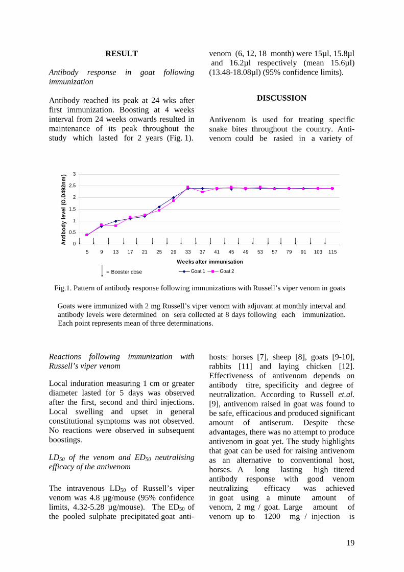
Antibody response in goat following immunization Antibody reached its peak at 24 wks after first immunization. Boosting at 4 weeks interval from 24 weeks onwards resulted in maintenance of its peak throughout the study which lasted for 2 years (Fig. 1).
Reactions following immunization with Russell’s viper venom
Local induration measuring 1 cm or greater diameter lasted for 5 days was observed after the first, second and third injections. Local swelling and upset in general constitutional symptoms was not observed. No reactions were observed in subsequent boostings.
LD50 of the venom and ED50 neutralising efficacy of the antivenom
The intravenous LD50 of Russell’s viper venom was 4.8 µg/mouse (95% confidence limits, 4.32-5.28 µg/mouse). The ED50 of the pooled sulphate precipitated goat anti-
venom (6, 12, 18 month) were 15µl, 15.8µl and 16.2µl respectively (mean 15.6µl) (13.48-18.08µl) (95% confidence limits).
DISCUSSION
Antivenom is used for treating specific snake bites throughout the country. Anti-venom could be rasied in a variety of
0
0.5
1
1.5
2
2.5
3
5 9 13 17 21 25 29 33 37 41 45 49 53 57 79 91 103 115
Weeks after immunisation
Antib
ody
leve
l (O
.D49
2nm
)
Goat 1 Goat 2= Booster dose
According to Russell et al [9], antivenom
Fig.1. Pattern of antibody response following immunizations with Russell’s viper venom in goats Goats were immunized with 2 mg Russell’s viper venom with adjuvant at monthly interval and
antibody levels were determined on sera collected at 8 days following each immunization. Each point represents mean of three determinations.
hosts: horses [7], sheep [8], goats [9-10], rabbits [11] and laying chicken [12]. Effectiveness of antivenom depends on antibody titre, specificity and degree of neutralization. According to Russell et.al. [9], antivenom raised in goat was found to be safe, efficacious and produced significant amount of antiserum. Despite these advantages, there was no attempt to produce antivenom in goat yet. The study highlights that goat can be used for raising antivenom as an alternative to conventional host, horses. A long lasting high titered antibody response with good venom neutralizing efficacy was achieved in goat using a minute amount of venom, 2 mg / goat. Large amount of venom up to 1200 mg / injection is
19
required in hyperimmunization of horses [7]. Although serum yield from each bleed in goat is less than horses, they are cheaper to maintain than horses and are suitable for tropical climate.
The ED50 of sulphate precipitated goat antivenom was 15.6µl (1 ml neutralize 0.307 mg of Russell’s viper venom) and is comparable to that of current Russell’s viper antivenom (Batch no: C98011. expiry 4/2001) 12µl (1 ml neutralize 0.383 mg of RVV) [13].However, high neutralization efficacy of antivenom could be achieved by
further concentrating the antivenom.
We demonstrated that goat immunized with a low dose of venom produced high titer antibody with venom neutralising capability. Owing to its efficacy and simplicity, this method can be used in raising other antivenoms. In conclusion, the goat antivenom has comparable venom neutralizing efficacy to that of current Russell’s viper antivenom manufactured by Myanmar Pharmaceutical Factory and this technology could be used in addition to current antivenom production in horses in order to step up antivenom production.
REFERENCES
1. Personal communication. Yearly incidence and
case fatality rate of snakebite cases from 14 states and divisions of Myanmar, 1998-2003. Statistics Division, Department of Health Planning, Ministry of Health, Myanmar.
2. Tun Pe, Aye Aye Myint and Nu Nu Aung Antivenom abuse: a review of antivenom policy in management of Russell’s viper bite cases of six snakebite endemic township hospitals. Myanmar Health Sciences Research Journal 1991; 11,1-3:33-37.
3. Finney,D.J. Probit analysis, 3rded. Cambridge , Cambridge University Press, 1971.
4. Theakston, R.D.G. & Reid, H. A. The development of simple standard assay procedures for characterization of snake venoms. Bulletin of W.H.O 1983; 61:949-956.
5. Wiyada, Choroensiriwatana & RI laboratory staff Regional training course on advanced methods of local reagent production of hepatitis B markers, 1995; p 21.
6. Tun Pe, Aye Aye Myint and Maung Chit. Humoral response following traditional active immunization against King Cobra venom. The snake 1994; 26:61-65.
7. Russell, F . E. Snake venom immunology:
Historical and practical considerations. Journal of Toxicology 1988; 7: 1-82.
8. Al-Asmari, A.K., Al-Abdulla, I.H., Crouch, R.G., Smith, D.C. & Sjostrom, L. Assessment of ovine antivenom raised against venom from the desert black cobra (Walterinnessia aegyptia). Toxicon 1997; 15, 1: 141-145.
9. Kochalty, W. F., Bowl-Ledford, E., DalyJ, G. & Billings, T.A. Preparation of coral snake antivenom from goat serum. Toxicon 1971; 297-298.
10. Russell, F.E., Timmerman, W.F.,& Meadows, P. Clinical use of antivenin prepared from goat serum . Toxicon 1970; 8: 63-65.
11. Russell, F.E., Use of Crotalus monovalent antivenom from rabbit serum. C Ther Res 1961; 3:438-440.
12. Carroll, S.B., Thalley, B.S., Theakston, R.D.G. & Laing, G. Comparison of the purity and efficacy of affinity purified avian antivenoms with commercial equine crotalid antivenoms. Toxicon 1992; 30, 9:1017-1025.
13. Tun Pe, Aye Aye Myint and Kyi May Htwe Efficacy of the new batches of monospecific Russellis viper (Daboia russelii siamensis) and cobra (Naja kaouthia) antivenom manufactured by Myanmar Pharmaceutical Factory. Myanmar Health Science Research Journal 2003; 15, 1-3:12-15.
20
The Myanmar Health Sciences Research Journal, Vol. 18, No. 1, 2006
Evaluation of Polymerase Chain Reaction (PCR) Amplification of Mycobacterium leprae in biopsy specimens from leprosy patients
*Khin Saw Aye, *Yin Min Htun, *Aye Aye Win, *Tin Zar Maw &**Kyaw Kyaw
*Immunology Research Division, Department of Medical Research (LM), **Central Special Skin Clinic, Yangon General Hospital
Eighty two skin biopsy specimens from leprosy patients attending at Central Special Skin Clinic, Yangon General Hospital were collected before taking chemotherapy. They were examined by a two-step polymerase chain reaction assay using a set of four nested oligonucleotide primers for the detection and identification of Mycobacterium leprae. It did not require the use of radioactively labelled hybridization probes. The nested-primer procedure amplified a 347 base-pair product from M. leprae genomic DNA. The PCR results were then compared with bacterial indices (BI) of slit-skin smears. PCR was positive in 8 (80%) of 10 biopsy specimens with BI of 0 determined for the slit-skin smear from the same patients. PCR also gave positive results for 71 (98.6%) of 72 BI positive cases. The agreement rate of these two tests is 89% but the false negative rate of BI for diagnosis of leprosy compared to PCR method is 10.39%. According to these results, PCR has an advantage over microscopic examination in detecting M. leprae in biopsy specimens negative for acid-fast bacilli, and is a useful tool for laboratory diagnosis.
INTRODUCTION
The recent development of polymerase chain reaction (PCR) has brought an un-precedented opportunity for sensitive, specific, and rapid detection of Mycobacterium leprae in clinical specimens. In the literature, there have been various target sequences for PCR and DNA probes specific for M. leprae, such as genes encoding the 36-kDa antigen [1, 2], the 18-kDa antigen [3], or the 65-kDa antigen [4] and repetitive sequences of M. leprae [5, 6]. Most of the reports showed that PCR with or without DNA probes seemed very sensitive, so that even 1 to 100 organisms are detectable by the method. In addition, PCR provided virtually 100% specificity in detecting the organism in clinical samples. In the study of De Wit et al 1991 with biopsy specimens from leprosy patients, PCR gave a positive result in about 80% of biopsies from leprosy patients negative for acid-fast bacilli (AFB), thus indicating that PCR is a useful tool for the laboratory
diagnosis of leprosy [1-3]. In Myanmar, there is no information about PCR based on other target sequences and protocol was available for M. leprae. In this study, therefore, we attempted to evaluate PCR using primers amplifying a 347 base-pair product from M. leprae genomic DNA [4] in skin biopsy specimens from untreated leprosy patients, and the results were then compared with microscopic findings.
MATERIALS AND METHODS
Ethical clearance was approved by Department of Medical Research (Lower Myanmar). After taken inform consent, biopsy specimens were obtained from 82 untreated leprosy patients attended at the Central Special Skin Clinic, Yangon General Hospital. Bacterial indices (BI) were determined microscopically for skin slit smear samples before skin biopsy. For each patient, slit-skin smears from three sites depending on the clinical type of leprosy were prepared also, as WHO described [7] which consist of two
21
categories, paucibacillary (PB) and multibacillary (MB). PB leprosy is defined as five or fewer skin lesions with no bacilli in skin smears, and MB leprosy cases have six or more lesions and may be skin smear positive. The collected skin scrap smears were examined by Z-N stain to check Bacillary Index (BI) and Morphological Index (MI). The average BI was then calculated for each patient before analysis. Tissue samples from persons with dermatologic problems other than leprosy also were included as controls. Biopsy specimens were cut in half; one half was used for paraffin embedding, and the other half was preserved in 70% ethanol used for PCR. Preparation of M. leprae DNAs from biopsy specimens were done by QIAGEN, Germany kit. Amplification of M. leprae DNA was done by nested PCR. The primers amplifying a 347 base-pair product from M. leprae genomic DNAs are as follow.
L1 1236-1253 GTGGCTCAGATCCGTACC
L21813-1792(C) ATGCCACCGGTCGGGTCGCTCG
L3 1458-1476 CTACAGGCTGCTCCGGCTC
L4 1804-1782 (C) GTCGGGTCGCTCGCCGGAGCTGC
The 25 μl reaction mixture contained 2.5 μl of template solution prepared from biopsy samples, 0.1 μl of Ex Taq DNA polymerase (Takara Shuzo Co., Shiga, Japan), oligode oxyribonucleotide primer 0.25 μl of each (L1 and L2) 20 μM stock solution, 2.5 μl of 10x DNA PCR buffer, 0.5 μl of dNTP solution and 18.9 μl of water. As a positive control, DNA purified from Thai-53 strain of M. leprae was provided by Dr. M. Matsuoka, NIID, Japan, and distilled water was included as negative control in each experiment.
The amplifications were carried out in a programmable thermal Mastercycler personal eppendrof USA in a two-step cycle of 20 s at 98°C followed by 1 min at 68°C. The samples were usually amplified through 25 cycles by using the outside pair of primers (L1 and L2); 10% of the amplified mixture was then transferred to a fresh tube containing reaction mixture with the inside
primers (L3 and L4). The second amplification was usually allowed to proceed through 30 cycles. Ten microliters of the reaction mixture was electrophoresed on 2% agarose gels. After electrophoresis, the gel was stained with ethidium bromide, and the 347 bp DNA band was examined under UV illumination.
RESULTS
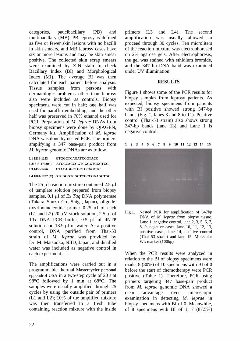
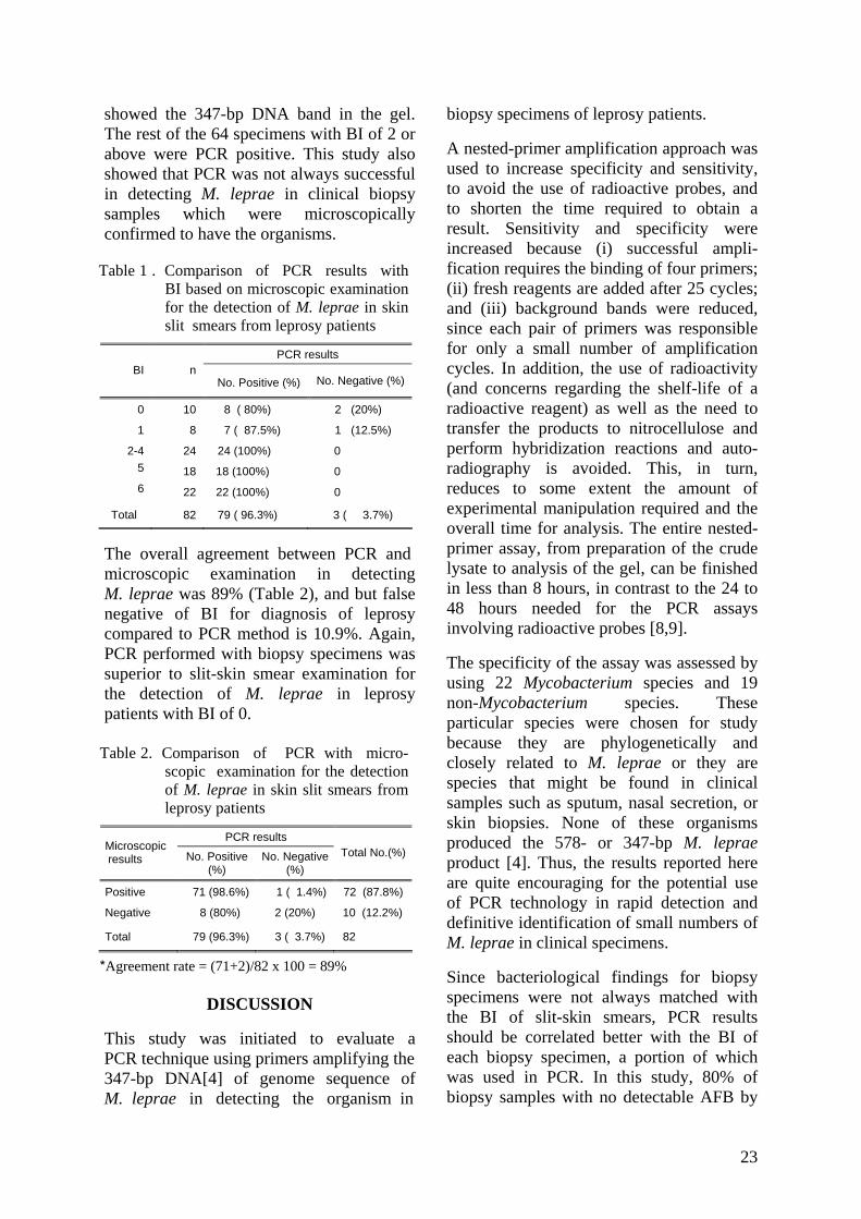
Figure 1 shows some of the PCR results for biopsy samples from leprosy patients. As expected, biopsy specimens from patients with BI positive showed strong 347-bp bands (Fig. 1, lanes 3 and 8 to 11). Positive control (Thai-53 strain) also shows strong 347-bp bands (lane 13) and Lane 1 is negative control.
1 2 3 4 5 6 7 8 9 10 11 12 13 14 15
Fig.1. Nested PCR for amplification of 347bp DNA of M. leprae from biopsy tissue. Lane 1, negative control, lane 2, 3, 5, 6, 7, 8, 9, negative cases, lane 10, 11, 12, 13, positive cases, lane 14, positive control (Thai 53 strain) and lane 15, Molecular Wt: marker (100bp)
When the PCR results were analyzed in relation to the BI of biopsy specimens were made, 8 (80%) of 10 specimens with BI of 0 before the start of chemotherapy were PCR positive (Table 1). Therefore, PCR using primers targeting 347 base-pair product from M. leprae genomic DNA showed a clear advantage over microscopic examination in detecting M. leprae in biopsy specimens with BI of 0. Meanwhile, of 8 specimens with BI of 1, 7 (87.5%)
22
showed the 347-bp DNA band in the gel. The rest of the 64 specimens with BI of 2 or above were PCR positive. This study also showed that PCR was not always successful in detecting M. leprae in clinical biopsy samples which were microscopically confirmed to have the organisms. The overall agreement between PCR and
DISCUSSION
This study was in aluate a
M. leprae in detecting the organism in
ivity,
and 19
tched with
microscopic examination in detecting M. leprae was 89% (Table 2), and but false negative of BI for diagnosis of leprosy compared to PCR method is 10.9%. Again, PCR performed with biopsy specimens was superior to slit-skin smear examination for the detection of M. leprae in leprosy patients with BI of 0.
itiated to evPCR technique using primers amplifying the 347-bp DNA[4] of genome sequence of
biopsy specimens of leprosy patients.
A nested-primer amplification approach was used to increase specificity and sensitto avoid the use of radioactive probes, and to shorten the time required to obtain a result. Sensitivity and specificity were increased because (i) successful ampli-fication requires the binding of four primers; (ii) fresh reagents are added after 25 cycles; and (iii) background bands were reduced, since each pair of primers was responsible for only a small number of amplification cycles. In addition, the use of radioactivity (and concerns regarding the shelf-life of a radioactive reagent) as well as the need to transfer the products to nitrocellulose and perform hybridization reactions and auto-radiography is avoided. This, in turn, reduces to some extent the amount of experimental manipulation required and the overall time for analysis. The entire nested-primer assay, from preparation of the crude lysate to analysis of the gel, can be finished in less than 8 hours, in contrast to the 24 to 48 hours needed for the PCR assays involving radioactive probes [8,9].
The specificity of the assay was assessed by using 22 Mycobacterium species
T ble 1 . Comparison of PCR results with
non-Mycobacterium species. These particular species were chosen for study because they are phylogenetically and closely related to M. leprae or they are species that might be found in clinical samples such as sputum, nasal secretion, or skin biopsies. None of these organisms produced the 578- or 347-bp M. leprae product [4]. Thus, the results reported here are quite encouraging for the potential use of PCR technology in rapid detection and definitive identification of small numbers of M. leprae in clinical specimens.
Since bacteriological findings for biopsy specimens were not always mathe BI of slit-skin smears, PCR results should be correlated better with the BI of each biopsy specimen, a portion of which was used in PCR. In this study, 80% of biopsy samples with no detectable AFB by
aBI based on microscopic examination for the detection of M. leprae in skin slit smears from leprosy patients
PCR results BI n
egative (%) No. Positive (%) No. N
0 10 2 8 ( 80%) (20%)
1 8 7 ( 87.5%) )
2 2
tal 3.7%)
1 (12.5%
-4 4 24 (100%) 0 5 18 18 (100%) 0 6 22 22 (100%) 0
To 82 79 ( 96.3%) 3 (
Table 2. Comparison of PCR with micro-
lts
scopic examination for the detection of M. leprae in skin slit smears from leprosy patients
PCR resu Microscopic
No. Po gative Total No.(%)results sitive No. Ne(%) (%)
Positive 71 (98.6%) %) 72 (87.8%) 1 ( 1.4
Negative
8 (80%) 2 (20%) 10 (12.2%)
Total 79 (96.3%) 3 ( 3.7%) 82
*Agreement rat 8 9e = (71+2)/ 2 x 100 = 8 %
23
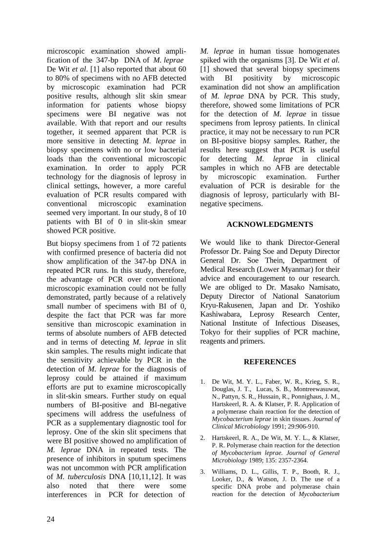
microscopic examination showed ampli-fication of the 347-bp DNA of M. leprae De Wit et al. [1] also reported that about 60 to 80% of specimens with no AFB detected by microscopic examination had PCR positive results, although slit skin smear information for patients whose biopsy specimens were BI negative was not available. With that report and our results together, it seemed apparent that PCR is more sensitive in detecting M. leprae in biopsy specimens with no or low bacterial loads than the conventional microscopic examination. In order to apply PCR technology for the diagnosis of leprosy in clinical settings, however, a more careful evaluation of PCR results compared with conventional microscopic examination seemed very important. In our study, 8 of 10 patients with BI of 0 in slit-skin smear showed PCR positive.
But biopsy specimens from 1 of 72 patients with confirmed presence of bacteria did not show amplification of the 347-bp DNA in
We would like to thank Director-General Professo irector General Dr. Soe Thein, Department of
1. De Wit, M. Y. L., Faber, W. R., Krieg, S. R., Douglas, J. ntreewasuwat, N., Pattyn, S. R., Hussain, R., Ponnighaus, J. M.,
eneral
ase chain reaction for the detection of Mycobacterium
repeated PCR runs. In this study, therefore, the advantage of PCR over conventional microscopic examination could not be fully demonstrated, partly because of a relatively small number of specimens with BI of 0, despite the fact that PCR was far more sensitive than microscopic examination in terms of absolute numbers of AFB detected and in terms of detecting M. leprae in slit skin samples. The results might indicate that the sensitivity achievable by PCR in the detection of M. leprae for the diagnosis of leprosy could be attained if maximum efforts are put to examine microscopically in slit-skin smears. Further study on equal numbers of BI-positive and BI-negative specimens will address the usefulness of PCR as a supplementary diagnostic tool for leprosy. One of the skin slit specimens that were BI positive showed no amplification of M. leprae DNA in repeated tests. The presence of inhibitors in sputum specimens was not uncommon with PCR amplification of M. tuberculosis DNA [10,11,12]. It was also noted that there were some interferences in PCR for detection of
M. leprae in human tissue homogenates spiked with the organisms [3]. De Wit et al. [1] showed that several biopsy specimens with BI positivity by microscopic examination did not show an amplification of M. leprae DNA by PCR. This study, therefore, showed some limitations of PCR for the detection of M. leprae in tissue specimens from leprosy patients. In clinical practice, it may not be necessary to run PCR on BI-positive biopsy samples. Rather, the results here suggest that PCR is useful for detecting M. leprae in clinical samples in which no AFB are detectable by microscopic examination. Further evaluation of PCR is desirable for the diagnosis of leprosy, particularly with BI-negative specimens.
ACKNOWLEDGMENTS
r Dr. Paing Soe and Deputy D
Medical Research (Lower Myanmar) for their advice and encouragement to our research. We are obliged to Dr. Masako Namisato, Deputy Director of National Sanatorium Kryu-Rakusenen, Japan and Dr. Yoshiko Kashiwabara, Leprosy Research Center, National Institute of Infectious Diseases, Tokyo for their supplies of PCR machine, reagents and primers.
REFERENCES
T., Lucas, S. B., Mo
Hartskeerl, R. A. & Klatser, P. R. Application of a polymerase chain reaction for the detection of Mycobacterium leprae in skin tissues. Journal of Clinical Microbiology 1991; 29:906-910.
2. Hartskeerl, R. A., De Wit, M. Y. L., & Klatser, P. R. Polymerase chain reaction for the detection of Mycobacterium leprae. Journal of GMicrobiology 1989; 135: 2357-2364.
3. Williams, D. L., Gillis, T. P., Booth, R. J., Looker, D., & Watson, J. D. The use of a specific DNA probe and polymer
24
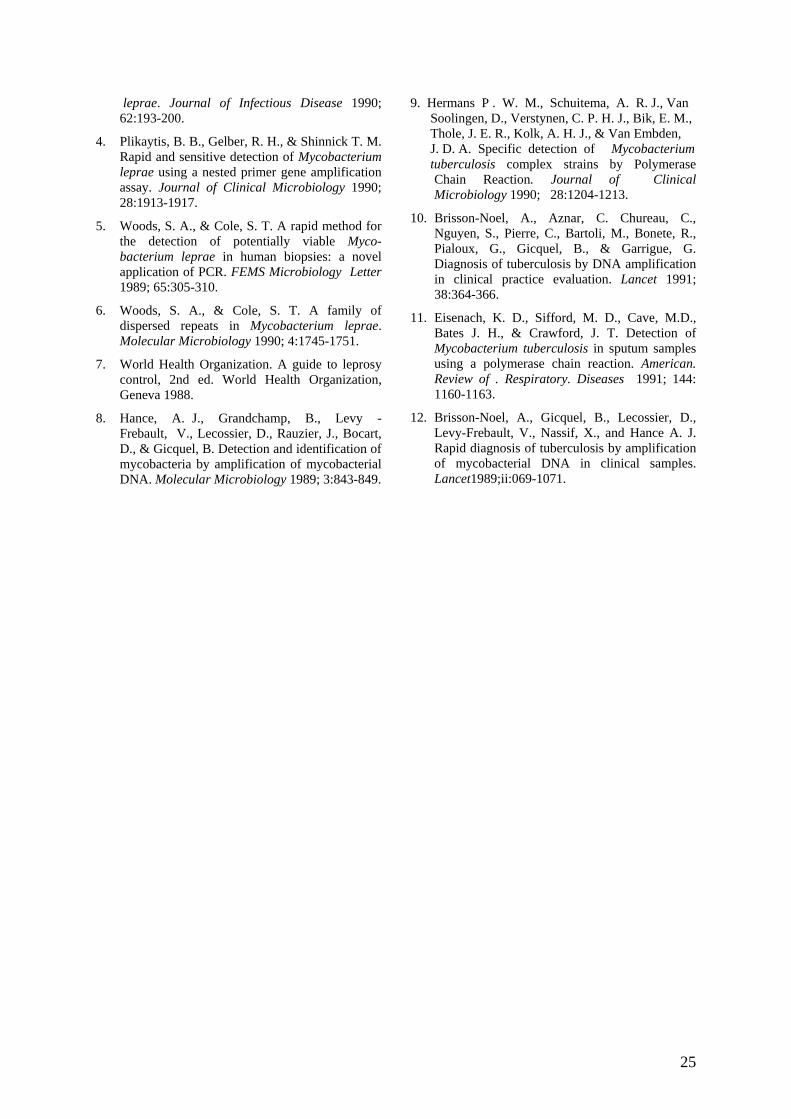
leprae. Journal of Infectious Disease 1990; 62:193-200.
4. Plikaytis, B. B., Gelber, R. H., & Shinnick T. M. Rapid and sensitive detection of Mycobacterium leprae using a nested primer gene amplification assay. Journal of Clinical Microbiology 1990;
CR. FEMS Microbiology Letter
nization. A guide to leprosy
y amplification of mycobacterial
9. Hermans P . W. M., Schuitema, A. R. J., Van olingen, D., Verstynen, C. P. H. J., Bik, E. M., ole, J. E. R., Kolk, A. H. J., & Van Embden,
J. D. A. Specific detection of Mycobacterium
plification
11.
merase chain reaction. American.
12.
terial DNA in clinical samples.
SoTh
tuberculosis complex strains by Polymerase
Chain Reaction. Journal of Clinical Microbiology 1990; 28:1204-1213. 28:1913-1917.
5. Woods, S. A., & Cole, S. T. A rapid method for the detection of potentially viable Myco-bacterium leprae in human biopsies: a novel application of P
10. Brisson-Noel, A., Aznar, C. Chureau, C., Nguyen, S., Pierre, C., Bartoli, M., Bonete, R., Pialoux, G., Gicquel, B., & Garrigue, G. Diagnosis of tuberculosis by DNA amin clinical practice evaluation. Lancet 1991; 38:364-366.
Eisenach, K. D., Sifford, M. D., Cave, M.D., Bates J. H., & Crawford, J. T. Detection of Mycobacterium tuberculosis in sputum samples using a poly
1989; 65:305-310.
6. Woods, S. A., & Cole, S. T. A family of dispersed repeats in Mycobacterium leprae. Molecular Microbiology 1990; 4:1745-1751.
7. World Health OrgaReview of . Respiratory. Diseases 1991; 144: 1160-1163.
Brisson-Noel, A., Gicquel, B., Lecossier, D., Levy-Frebault, V., Nassif, X., and Hance A. J. Rapid diagnosis of tuberculosis by amplification of mycobac
control, 2nd ed. World Health Organization, Geneva 1988.
8. Hance, A. J., Grandchamp, B., Levy -Frebault, V., Lecossier, D., Rauzier, J., Bocart, D., & Gicquel, B. Detection and identification of mycobacteria b
Lancet1989;ii:069-1071.DNA. Molecular Microbiology 1989; 3:843-849.
25
The Myanmar Health Sciences Research Journal, Vol. 18, No. 1, 2006
Antibacterial activity of some plants and formulations and determination of Minimum Inhibitory Concentration (MIC) by microtitre plate dilution method
*Mar Mar Nyein *Mi Mi Htwe, **Ba Han & **Ei Ei Khine
*Bacteriology Research Division , Department Medical Research (LM)
**Chemistry Department, Yangon University
Traditional Medicine Formulation (TMF-01) [mixture of Cyperus rotundus Linn. (_rufrkefnif;), Alpinia galangal Walld (y'JaumjuD;) and Piper betle Linn. (uGrf;)] and plants [Alpinia galangal Walld. (y'JaumjuD;) Acorus calamus Linn. (vif;ae), and Piper longum Linn. (ydwfcsif;)] were tested for their antibacterial activity on 20 different types of bacteria (Escherichia coli=6 types, Proteus morganii, Shigella boydii, S. dysenteriae, S. flexneri, S. sonnei, S. derby, S. krefeld, Staphylococcus aureus; S. epidermidis, Vibrio cholerae O1, V. cholerae O139, V. cholerae Inaba, V. cholerae Ogawa and V. fluvialis) from clinical sources. The bacteriostatic and bactericidal activities of extracts were tested by microtitre plate dilution method and the optical density was determined by microplate reader. It was found that water extract of TMF had antibacterial activity on 20 different types of bacteria with various inhibition zone sizes ranging from 14 to 26 mm. Similarly, water extract of ash and water extract of ash from water extract of Alpinia galangal Walld (y'JaumjuD;) possess antibacterial activity with the zone size of 7-18 mm. The asarone and methyl piperate compound obtained from Acorus calamus Linn. (vif;ae), and Piper longum Linn. (ydwfcsif;) respectively also showed antibacterial activity on E. coli, S. flexneri, S. aureus and P. aeruginosa. The Minimum Inhibitory Concentration (MIC) of water extracts of TMF -01 was ranging from 0.16 to 0.32mg/ml. Similarly, the MIC of water extract of ash of Alpinia galangal Walld (y'JaumjuD;) was 0.16 mg/ml and water extract of ash from water extract of Alpinia galangal Walld (y'JaumjuD;) ranged from 0.078 to 0.625mg/ml. Moreover, the MIC of asarone from Acorus calamus Linn. (vif;ae) was 0.06-0.12mg/ml and that of methyl piperate from Piper longum Linn. (ydwfcsif;) was 0.03mg/ml.
INTRODUCTION
Herbal medicines, which have formed the basis of health care throughout the world since the earliest days of mankind, are still widely used and are of considerable importance in international trade. Recognition of their clinical, pharmaceutical and economic values is not only widespread but popularity is also maintained for his-torical and cultural reasons [1,2]. Plants, Alpina galangal Walld. (y'JaumjuD;), Acorus calamus Linn. (vif;ae) and Piper longum Linn. (ydwfcsif;) [3] and Traditional Medicine Formulation 01 are well-known
traditionally and are used by traditional medicine practitioners . Thus, this study is to determine the antibacterial activity of different preparations of extracts and to determine the minimum inhibitory concentration by microtitreplate dilution method.
MATERIALS AND METHODS
Traditional Medicine Formulation(TMF-01): It contains Cyperus rotundus Linn. (_rufrkefnif;), Alpinia galangal Walld. (y'JaumjuD;), and Piper betle Linn. (uGrf;).
26
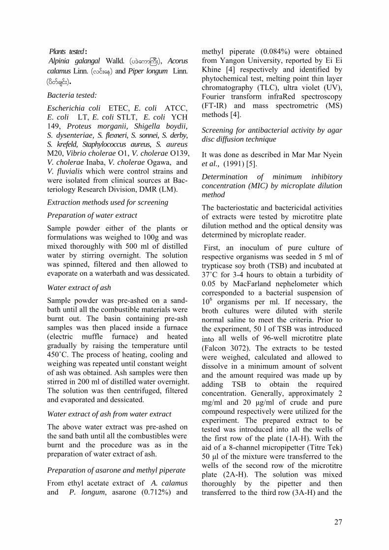
Plants tested : Alpinia galangal Walld. (y'JaumjuD;), Acorus calamus Linn. (vif;ae) and Piper longum Linn. (ydwfcsif;). Bacteria tested:
Escherichia coli ETEC, E. coli ATCC, E. coli LT, E. coli STLT, E. coli YCH 149, Proteus morganii, Shigella boydii, S. dysenteriae, S. flexneri, S. sonnei, S. derby, S. krefeld, Staphylococcus aureus, S. aureus M20, Vibrio cholerae O1, V. cholerae O139, V. cholerae Inaba, V. cholerae Ogawa, and V. fluvialis which were control strains and were isolated from clinical sources at Bac-teriology Research Division, DMR (LM).
Extraction methods used for screening
Preparation of water extract
Sample powder either of the plants or formulations was weighed to 100g and was mixed thoroughly with 500 ml of distilled water by stirring overnight. The solution was spinned, filtered and then allowed to evaporate on a waterbath and was dessicated.
Water extract of ash Sample powder was pre-ashed on a sand-bath until all the combustible materials were burnt out. The basin containing pre-ash samples was then placed inside a furnace (electric muffle furnace) and heated gradually by raising the temperature until 450˚C. The process of heating, cooling and weighing was repeated until constant weight of ash was obtained. Ash samples were then stirred in 200 ml of distilled water overnight. The solution was then centrifuged, filtered and evaporated and dessicated.
Water extract of ash from water extract The above water extract was pre-ashed on the sand bath until all the combustibles were burnt and the procedure was as in the preparation of water extract of ash.
Preparation of asarone and methyl piperate From ethyl acetate extract of A. calamus and P. longum, asarone (0.712%) and
methyl piperate (0.084%) were obtained from Yangon University, reported by Ei Ei Khine [4] respectively and identified by phytochemical test, melting point thin layer chromatography (TLC), ultra violet (UV), Fourier transform infraRed spectroscopy (FT-IR) and mass spectrometric (MS) methods [4].
Screening for antibacterial activity by agar disc diffusion technique
It was done as described in Mar Mar Nyein et al., (1991) [5].
Determination of minimum inhibitory concentration (MIC) by microplate dilution method
The bacteriostatic and bactericidal activities of extracts were tested by microtitre plate dilution method and the optical density was determined by microplate reader.
First, an inoculum of pure culture of respective organisms was seeded in 5 ml of trypticase soy broth (TSB) and incubated at 37˚C for 3-4 hours to obtain a turbidity of 0.05 by MacFarland nephelometer which corresponded to a bacterial suspension of 106 organisms per ml. If necessary, the broth cultures were diluted with sterile normal saline to meet the criteria. Prior to the experiment, 50 l of TSB was introduced into all wells of 96-well microtitre plate (Falcon 3072). The extracts to be tested were weighed, calculated and allowed to dissolve in a minimum amount of solvent and the amount required was made up by adding TSB to obtain the required concentration. Generally, approximately 2 mg/ml and 20 μg/ml of crude and pure compound respectively were utilized for the experiment. The prepared extract to be tested was introduced into all the wells of the first row of the plate (1A-H). With the aid of a 8-channel micropipetter (Titre Tek) 50 μl of the mixture were transferred to the wells of the second row of the microtitre plate (2A-H). The solution was mixed thoroughly by the pipetter and then transferred to the third row (3A-H) and the
27
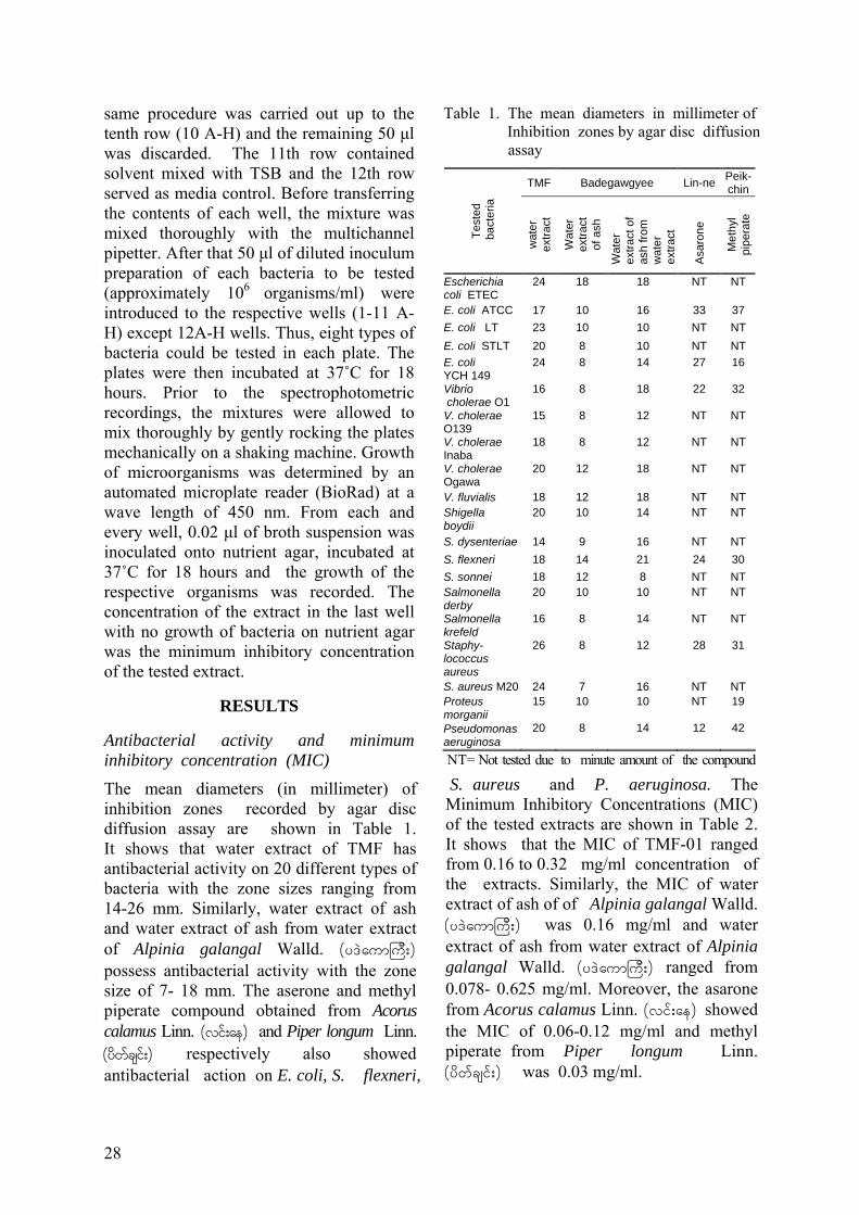
same procedure was carried out up to the tenth row (10 A-H) and the remaining 50 μl was discarded. The 11th row contained solvent mixed with TSB and the 12th row served as media control. Before transferring the contents of each well, the mixture was mixed thoroughly with the multichannel pipetter. After that 50 μl of diluted inoculum preparation of each bacteria to be tested (approximately 106 organisms/ml) were introduced to the respective wells (1-11 A-H) except 12A-H wells. Thus, eight types of bacteria could be tested in each plate. The plates were then incubated at 37˚C for 18 hours. Prior to the spectrophotometric recordings, the mixtures were allowed to mix thoroughly by gently rocking the plates mechanically on a shaking machine. Growth of microorganisms was determined by an automated microplate reader (BioRad) at a wave length of 450 nm. From each and every well, 0.02 μl of broth suspension was inoculated onto nutrient agar, incubated at 37˚C for 18 hours and the growth of the respective organisms was recorded. The concentration of the extract in the last well with no growth of bacteria on nutrient agar was the minimum inhibitory concentration of the tested extract.
RESULTS
Antibacterial activity and minimum inhibitory concentration (MIC)
The mean diameters (in millimeter) of inhibition zones recorded by agar disc diffusion assay are shown in Table 1. It shows that water extract of TMF has antibacterial activity on 20 different types of bacteria with the zone sizes ranging from 14-26 mm. Similarly, water extract of ash and water extract of ash from water extract of Alpinia galangal Walld. (y'JaumjuD;) possess antibacterial activity with the zone size of 7- 18 mm. The aserone and methyl piperate compound obtained from Acorus calamus Linn. (vif;ae) and Piper longum Linn. (ydwfcsif;) respectively also showed antibacterial action on E. coli, S. flexneri,
S. aureus and P. aeruginosa. The Minimum Inhibitory Concentrations (MIC) of the tested extracts are shown in Table 2. It shows that the MIC of TMF-01 ranged from 0.16 to 0.32 mg/ml concentration of the extracts. Similarly, the MIC of water extract of ash of of Alpinia galangal Walld. (y'JaumjuD;) was 0.16 mg/ml and water extract of ash from water extract of Alpinia galangal Walld. (y'JaumjuD;) ranged from 0.078- 0.625 mg/ml. Moreover, the asarone from Acorus calamus Linn. (vif;ae) showed the MIC of 0.06-0.12 mg/ml and methyl piperate from Piper longum Linn. (ydwfcsif;) was 0.03 mg/ml.
Table 1. The mean diameters in millimeter of Inhibition zones by agar disc diffusion
assay
TMF Badegawgyee Lin-ne Peik-chin
Test
ed
bac
teria
wat
er
extra
ct
Wat
er
extra
ct
of a
sh
Wat
er
extra
ct o
f as
h fro
m
wat
er
extra
ct
Asa
rone
Met
hyl
pipe
rate
Escherichia coli ETEC
24 18 18 NT NT
E. coli ATCC 17 10 16 33 37 E. coli LT 23 10 10 NT NT E. coli STLT 20 8 10 NT NT E. coli YCH 149
24 8 14 27 16
Vibrio cholerae O1
16 8 18 22 32
V. cholerae O139
15 8 12 NT NT
V. cholerae Inaba
18 8 12 NT NT
V. cholerae Ogawa
20 12 18 NT NT
V. fluvialis 18 12 18 NT NT Shigella boydii
20 10 14 NT NT
S. dysenteriae 14 9 16 NT NT S. flexneri 18 14 21 24 30 S. sonnei 18 12 8 NT NT Salmonella derby
20 10 10 NT NT
Salmonella krefeld
16 8 14 NT NT
Staphy- lococcus aureus
26 8 12 28 31
S. aureus M20 24 7 16 NT NT Proteus morganii
15 10 10 NT 19
Pseudomonas aeruginosa
20 8 14 12 42
NT= Not tested due to minute amount of the compound
28
29
Table 2. Minimum Inhibitory Concentrations of TMF and some plant extracts in mg/ml
TMF Badegawgyee Lin-ne
Extraction method plays an important role in determination of antibacterial testing. Moreover, the MICs of asarone from Acorus calamus Linn. (vif;ae) was 0.06-0.12 mg/ml and methyl piperate from Piper longum Linn. (ydwfcsif;) was 0.03 mg/ml and these plants were known to possess antibacterial activity [6]. These plants were used for various ailments by Myanmar traditional medicine practioners especially for gastrointestinal disorders [7, 8].
Peikchin
Tested bacteria Water
extract
Water extract of ash
Water extract of ash from water extract
Asarone Methyl piperate
Escherichia coli ATCC
0.16
0.16
0.078
0.12
0.03
Escherichia coli STLT
NT NT NT 0.63 0.03
Phytochemical testing showed that water extract of TMF 01 contains α-amino acids, glycosides and saponins. Water extract of Alpinia galangal Walld. (y'JaumjuD;)
contains alkaloids, α-amino acids, glycosides, phenolic compounds, saponins,steroids, terpenoids, tannins and reducing sugar. Sodium (9.74%), magnesium (1.52%), potassium (1.01%), calcium (1.23%) were found as major elements in TMF-01. In Alpinia galangal Walld. (y'JaumjuD;), sodium (0.07%), magnesium (0.34%), potassium (2.24%), and calcium (0.08%) were found. Iron was found as a minor element; chromium, zinc, and arsenic were found as trace elements in both TMF and Alpinia galangal Walld. (y'JaumjuD;) [9].
Escherichia coli LT
0.16 0.16 0.078 0.06 0.03
Proteus morganii
0.16 0.16 0.625 NT 0.03
Salmonella derby
0.16 0.16 0.625 NT 0.03
Salmonella krefeld
0.32 0.16 0.625 NT 0.03
Staphylo-coccus aureus ATCC
0.32 0.16 0.625 0.12 0.03
Staphylo-coccus aureus
0.32 0.16 0.625 0.06 0.03
Vibrio fluvialis
0.32 0.16 0.625 NT NT
NT= Not tested
The minimum inhibitory concentration of Alpinia galangal Walld. (y'JaumjuD;)
extracts AW (water extract of ash) and WAW (water extract of ash from water According to their study, these plants
possess not only the antibacterial activity but also salts and nutrients that show antisecretory activity in gastrointestinal infections.
extract) tested on Escherichia coli ETEC is shown in Fig. 1 .
DISCUSSION The microplate dilution method also elaborates the specificity, sensitivity and the least amount required for media, reagents and glassware. It also saves time and working space in conducting the experiments.
In this study by agar disc diffusion technique, antibacterial activity was found in the water extract of ash of TMF-01. Water extract of ash and water extract of ash from water extract of Alpinia galangal Walld. (y'JaumjuD;) also showed a significant inhibition zone. In determination of MIC, it showed that the MIC of water extract of ash from water extract of Alpinia galangal Walld (y'JaumjuD;) was 0.078 mg/ml.
ACKNOWLEDGEMENTS
The author would like to be grateful to Director - General Professor Dr. Paing Soe and Deputy Director-general Dr. Soe Thein
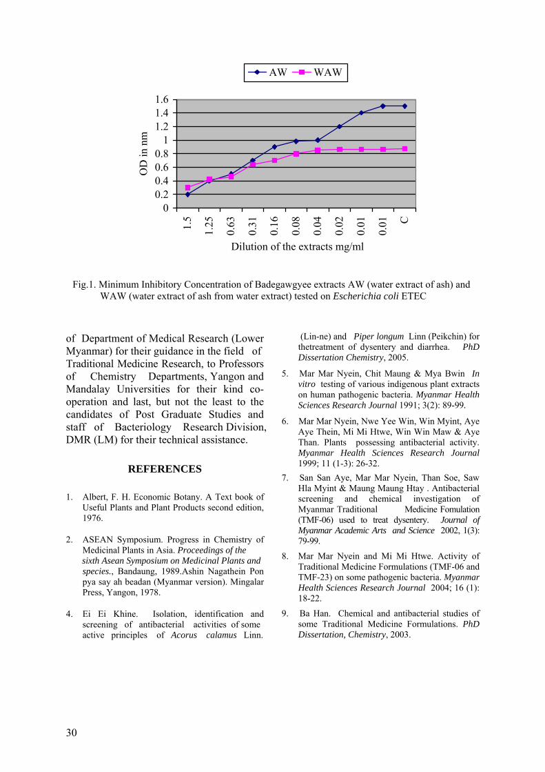
of Department of Medical Research (Lower Myanmar) for their guidance in the field of Traditional Medicine Research, to Professors of Chemistry Departments, Yangon and Mandalay Universities for their kind co-operation and last, but not the least to the candidates of Post Graduate Studies and staff of Bacteriology Research Division, DMR (LM) for their technical assistance.
REFERENCES
1. Albert, F. H. Economic Botany. A Text book of Useful Plants and Plant Products second edition, 1976.
2. ASEAN Symposium. Progress in Chemistry of Medicinal Plants in Asia. Proceedings of the
sixth Asean Symposium on Medicinal Plants and 8. Mar Mar Nyein and Mi Mi Htwe. Activity of Traditional Medicine Formulations (TMF-06 and TMF-23) on some pathogenic bacteria. Myanmar Health Sciences Research Journal 2004; 16 (1): 18-22.
species., Bandaung, 1989.Ashin Nagathein Pon pya say ah beadan (Myanmar version). Mingalar Press, Yangon, 1978.
4. Ei Ei Khine. Isolation, identification and screening of antibacterial activities of some active principles of Acorus calamus Linn.
00.20.40.60.8
11.21.41.6
1.5
1.25
0.63
0.31
0.16
0.08
0.04
0.02
0.01
0.01 C
Dilution of the extracts mg/ml
OD
in n
m
AW WAW
Fig.1. Minimum Inhibitory Concentration of Badegawgyee extracts AW (water extract of ash) and WAW (water extract of ash from water extract) tested on Escherichia coli ETEC
(Lin-ne) and Piper longum Linn (Peikchin) for thetreatment of dysentery and diarrhea. PhD Dissertation Chemistry, 2005.
5. Mar Mar Nyein, Chit Maung & Mya Bwin In vitro testing of various indigenous plant extracts on human pathogenic bacteria. Myanmar Health Sciences Research Journal 1991; 3(2): 89-99.
6. Mar Mar Nyein, Nwe Yee Win, Win Myint, Aye Aye Thein, Mi Mi Htwe, Win Win Maw & Aye Than. Plants possessing antibacterial activity. Myanmar Health Sciences Research Journal 1999; 11 (1-3): 26-32.
7. San San Aye, Mar Mar Nyein, Than Soe, Saw Hla Myint & Maung Maung Htay . Antibacterial screening and chemical investigation of Myanmar Traditional Medicine Fomulation (TMF-06) used to treat dysentery. Journal of Myanmar Academic Arts and Science 2002, 1(3): 79-99.
9. Ba Han. Chemical and antibacterial studies of some Traditional Medicine Formulations. PhD Dissertation, Chemistry, 2003.
30
The Myanmar Health Sciences Research Journal, Vol. 18, No. 1, 2006
Compartmental syndrome following a green pit viper (Trimeresurus erythrurus) bite
*Tun Pe & **Tin Tin Aung
*Department of Medical Research (Lower Myanmar)
**Department of Medicine, Institute of Medicine (2)
Green snake bite is common in Myanmar and majority of the victims do not seek hospital treatment because of lack of mortality and morbidity. However, in the present case, a 17 yr old girl bitten by a big green snake with a dry tail who applied tight tourniquets developed massive local swelling (diagnosed as a compartmental syndrome) and bleeding from the wound. Fasciotomy performed on the patient with incoagulable blood leads to uncontrolled local bleeding, shock and wound sepsis almost killing the patient was rescued by replacement of 26 units of blood, intensive care treatment to combat shock and sepsis and ventilatory support for 6 days. The patient landed up with wrist drop and sensory loss of the limb and has spent 6 months in hospital for reconstructive surgery. The massive swelling and wrist drop could be secondary to the use of tight tourniquets and compartmental syndrome. The study highlights that surgical intervention in snake bite patient with incoagulable blood should be delayed until specific antivenom or clotting factor substitutes are given to correct the coagulation defect.
INTRODUCTION
Green snake bite occurs through out Myanmar. Green snake known as Mwe-sein is well known in Myanmar. Very few victims brought the snake for identification. It is believed that its bite leads to massive local swelling with no casualty. Not all green snake bite cases seek medical treatment at hospital and prevalence rate of 4% based on hospital data is underestimated [1]. However, green snake bite accounts for 16% of the snake bite cases admitted to Yangon General Hospital between January 1999 to April 2001 and 64% of them were bitten in Bahan Township [2]. Community based study of epidemiology of snakebite carried out in Taungdwingyi and Kyaukpadaung shows that prevalence rate of green pit viper bites varies from 5.4-5.8% [3]. Of 7 species known to inhabit in Myanmar [4], Trimeresurus erythrurus is the most frequently encountered species and is responsible for most bites [4-5]. Clinical features and development of
antibody following green pit viper (Tr. erythrurus) bite has been reported earlier [4]. In this communication, development of compartmental syndrome in a green pitviper (Tr. erythrurus) bite victim is described.
MATERIALS AND METHODS Case report
A 17 year-old girl from Golden Valley, Yangon, was bitten by a 29 x 2 inches long green snake with a dry tail, at lower 1/3 of radial border of right forearm while moving roofing sheets in house compound at 10 am on 6 June 2002. The snake hanged on to the site of bite had to be removed. A tight cotton tourniquet was applied at mid right upper arm following the bite. She was transported to emergency unit, Yangon General Hospital 2.5h following the bite. On arrival, she had massive local swelling of the whole limb below the tourniquet with dark brown discolouration of the skin below
31
elbow joint, heaviness and numbness of the whole limb. Another cotton bandage of moderate tightness was applied below the first at emergency unit. Both were removed on arrival at the medical ward, Yangon General Hospital 1.5h later (4hr after the bite). Local swelling spread to right anterior chest wall following release of the tourniquets.
At admission, the whole limb was swollen up to right half of the anterior chest wall. Her blood pressure measured was 130/80 mmHg and she had a pulse rate of 100 per minute. Blood was coagulable on admission and found to be prolonged on retesting at 3hr after the bite.
Eight hours after the bite, there was marked swelling of the whole limb and discolouration of the skin overlying mid 1/3 of the arm. The limb was cold, tender with loss of sensory function. Radial, ulner and axillary pulses were not palpable. Blisters and necrosis developed at the site of bite. Bleeding from the wound resulting in soaking of 3 blankets and was covered with oozing blood.
Sixteen hours later, the patient was pale, restless and bleeding from the local wound continued. Her blood pressure fell to 60/ 0 by palpation and pressure agents, steroid and blood were given to combat shock. Fasciotomy was carried out on right forearm in flexor and extensor compartments, carpel tunnel, dorsum of hand and right arm. Necrosis of muscles was noted in extensor (severely), flexor (moderate) and in triceps, biceps and post deltoid. Radial pulse was not palpable.
Twenty one hours after the bite, she was pale and had a temperature of 99º F, blood pressure 80/50mmHg and heart rate of 120/min. Another unit of blood was given.
Twenty four hours after the bite, her blood pressure fell again (BP 60mmHg by palpation); local bleeding continued and became restless. Resuscitation measures were given along with transfusion of
another unit of blood. Her blood pressure remained labile and pressure agents were given in order to combat shock. Another unit of fresh blood was given at 38hr after the bite.
Sixty one hours after the bite, the patient was referred to intensive care unit with shock and gasping. She was put on respirator/ventilator and measures to combat shock including replacement of fresh blood were given. Spontaneous ventilation returned 140hr (day 6) after the bite. Wound debridgement was carried out on day 12 which disclosed black discoloration of skin extending from dorsum of hand to forearm, black bullae on dorsum of 2nd and 4th finger and discoloration of muscle in the wound. Her condition became stable (18 days after the bite) following transfusion of a total 26 units of fresh blood. Skin grafting of the wound was attempted 10 times during 6 months following the bite. Wrist drop with loss of sensory function was noted. The patient was still in hospital waiting to undergo further skin grafting. Investigations showed prothrombin time and activated partial thrombin time returned to normal values on 8 days after the bite.
DISCUSSION
Green snake bite is common in Myanmar and its bite rarely leads to severe morbidity and mortality. (One green snake bite patient developed internal bleeding following massive massage of abdomen in Mandalay hospital, Personal communication, Dr. Sann Mya). In Yangon, most green snake bite cases come from Golden Valley and Shwe-gondine localities, Bahan Township [2, 4].
Green pit viper bite results in massive local swelling with defibrination leading to incoagulable blood [4, 5]. In this case, the patient has massive local swelling, skin discolouration below the tourniquets and loss of sensation and radial pulse suggesting development of ill effects of tourniquets and excessive extravasations of plasma and fluid
32
into limb and necrosis of extensor muscles of the right hand.
Development of coagulopathy leads to profuse bleeding from the wound and haemorrhagic hypovolumic shock. No specific antivenom is available in Myanmar. Time taken for restoration of clotting defect depends on the amount of circulating venom procoagulant (fibrinogen activator) and fibrinogen turns over of the liver. Specific antivenom has been shown to play a vital role in correcting clotting defect [6]. In this case clot restoration occured about 8 days after the bite following replacement of several units of fresh blood. In our earlier studies, clot restoration occured around 8 days without replacement of clotting factors and it could take in the absence of anti-venom as long as 28 days after the bite [4].
Massive local swelling is due to spreading factor(s) of the venom. However, in the present case, massive swelling leading to compartmental syndrome is a compound effect of venom and tight tourniquet(s). Wrist drop in this case is secondary to prolong application of tight tourniquets and compartmental syndrome. Ill effects following application of tight tourniquets have been documented [7]. This case demonstrates that the surgical intervention should be delayed until normal clot restoration has occurred either by giving specific antivenom to neutralise circulating venom procoagulant or replacement of the clotting factors to correct the clotting defect. A similar distarous persistent bleeding leading to development of haemorrhagic shock despite transfusion of 20 units of blood occurred in a Malayan pit viper bite victim following fasciotomy before correction of coagulation defect has been reported [8]. A profuse bleeding following fasciotomy in a green pit viper (Trimeresurus albolabris) bite case had also been reported [8]. It is fortunate that the life of the patient was saved by energetic efforts of the medical personnel and infusion of 26units of blood to combat shock and septicemia.
The study highlights that green pit viper bite cases presented with coagulopathy should be corrected with specific antivenom or replacement of the clotting factors first before performing any surgical intervention. A limited stock of specific antivenom for green pit viper should be made available for these cases in the hospitals.
REFERENCES
1. Aye Aye Myint, Tun Pe and Tin Zar Maw. An epidemiological study of snakebite and venomous snake survey in Myanmar. In: Management of snakebite and research, WHO/SEARO Publication 2002.
2. Sann Mya, Theingyi Yin and Tun Pe. Study of snakebite cases admitted to Yangon General Hospital. Myanmar Health Sciences Research Journal 2004; 16 (1-3):42-45.
3. Tun Pe, Aye Aye Myint, Sunn Htut, Khin Aye Kyu and Maung Maung Toe. Prevalence, case fatality rate and treatment seeking behavior of the snakebite victims from two townships of Myanmar. Myanmar Health Research Congress abstract 2003;p 13.
4. Tun Pe, Aye Aye Myint and Nu Nu Aung. Green pit viper (Trimeresurus erythrurus) bite in Myanmar: clinical features, venom antigen levels and development of natural antibodies. Myanmar Health Sciences Research Journal 2001;2 (1-3):1-6.
5. Maung-Maung-Aye. Some experience in the management of snake-bite. Burma Medical Journal 1972; 20:33-40.
6. Warrell, D.A., Davidson, N. McD., Greenwood, R.M., Ormerod, L.D., Pope, H. M., Watkins, B.J. & Prentice, C.R.M.. Poisoning by bites of the saw-scaled or carpet viper (Echis carinatus) in Nigeria. Quarterly Journal of Medicine 1977; 46: 33-62.
7. Tun Pe, Tin Nu Swe, Myint Lwin, Warrell, DA., &Than Win. The efficacy of tourniquets as a first aid measure for Russell's viper bite in Burma. Transactions of Royal Society of Tropical Medicine & Hygiene 1987; 81: 403-405.
8. Warrell, DA. WHO/SEARO guidelines for the Clinical Management of Snakebites in the Southeast Asian Region. Supplement to the Southeast Asian Journal of Tropical Medicine and Public Health, 30 Supplement 1, 1999.
33
The Myanmar Health Sciences Research Journal, Vol. 18, No. 1, 2006
Comparison of Polymerase Chain Reaction, immunohistochemistry and conventional histopathology in the diagnosis of leprosy in Myanmar
*Khin Saw Aye, *Aye Aye Win, *Tin Zar Maw,
**Kyaw Kyaw & ***Khine San Yin
*Immunology Research Division, Department of Medical Research (L M) **Central Special Skin Center, Yangon General Hospital
***Department of Pathology, Institute of Medicine 1 and Yangon General Hospital
Skin biopsy specimens were obtained from 69 leprosy patients attending at Central Special Skin Clinic, Yangon General Hospital. All biopsy samples were examined by polymerase chain reaction (PCR) using the primers amplifying the 130 base-pair fragment of the gene from the 16S ribosomal RNA of Mycobacterium leprae, hematoxylin and eosin (H&E) staining, modified Fite-Faraco (FF) technique for M. leprae and immunostaining with the antibody against the phenolic glycolipid-1 (PGL-1) and Bacille Calmette-Guerin (BCG) using ABC (Avidin Biotin Complex) method. PCR was positive for 49 (71%) of 69 specimens. In 22 (31.8%) cases, only PCR was positive for M. leprae and all other tests were negative. AFB was positive for 19 (27.5%) of 69, PGL-1 was positive for 20 (29%) of 69, BCG was positive for 27 (39%) of 69. Epithelioid cells granuloma was detected in 17 (24.6%) patients and peripheral nerve inflammatory infiltration in 42 (60%) of 69. Comparison of PCR with other method showed statistically significant difference (p<0.001). PCR has an advantage over microscopic examination in detecting M. leprae in biopsy specimens which are negative for acid-fast bacilli.
INTRODUCTION
Mycobacterium leprae, the etiologic agent of leprosy remains one of the few pathogens that can not be cultivated in vitro. The diagnosis of leprosy is still based upon principles used a century ago: clinical examination of the patient lesions, demonstration of acid-fast bacilli (AFB) in slit-skin smears, and histopathology. Leprosy is most easily diagnosed when Mycobacterium leprae is demonstrable in diseased tissues, but this is often difficult in the indeterminated and in the tuberculoid types of leprosy in which M. leprae is rarely detected. Such a histopathological diagnostic procedure is relatively insensitive and does not give a definitive identification of the infecting organism like M. leprae.
Several attempts have been made in recent years to improve the sensitivity and specificity of the detection of M. leprae with immunology, biochemistry and nucleic acid probes [1]. For example, Clark-Curtiss and Docherty have described a DNA probe that can be used in a dot blot hybridization assay to detect as little as 1pg of purified DNA, the amount in approximately 300 bacilli [2].
Recently, several investigators have studied the use of polymerase chain reaction (PCR) to detect mycobacteria. This method has been used to detect extremely low number of M. leprae in fresh unfixed human skin biopsy specimens, providing a powerful direct and unequivocal test for M. leprae infection [3,4]. Since routine biopsy specimens for histopathological analysis are prepared in standard fixative, fresh tissues
34
are generally not available for PCR analysis. Adaptation of PCR for detecting M. leprae in fixed tissue would give clinicians the option of testing biopsy specimens for the presences of M. leprae, potentially aiding in the diagnosis of difficult cases. Recent data have shown that PCR can be used to detect genes from eukaryotic and viral genomic DNA derived from formalin-fixed paraffin-embedded tissues [5,6,7]. M. leprae DNA has also been detected in extracts from paraffin-embedded skin biopsy [8]. In the literature, there have been various target sequences for PCR and DNA probes specific for M. leprae, such as genes encoding the 36-kDa antigen [3,8], 18-kDa antigen [4], 65-kDa antigen [9] or repetitive sequences of M. leprae [10,11]and 16S ribosomal RNA of M. leprae [12]. Most of the reports showed that PCR with DNA probes seemed very sensitive, so that even 1 to 100 organisms were detectable by the method. In addition, PCR provided virtually 100% specificity in detecting the organism in clinical samples.
However, PCR tools have not been fully evaluated for detecting M. leprae in clinical specimens from leprosy patients. In a study with biopsy specimens from leprosy patients, PCR gave a positive result in about 61% of biopsies from leprosy patients who were negative for AFB [8], thus indicating that PCR is a useful tool for the laboratory diagnosis of leprosy. We use the primers for 16S rRNA of M. leprae to amplify 130-base-pair product of M. leprae genomic DNA extracted from paraffin-embedded sections of biopsy specimens from untreated leprosy patients and the results were then compared with microscopic findings.
At the same time, an investigation on the demonstration of phenolic glycolipid-1 (PGL-1) as a M. leprae-specific antigen of M. leprae, Bacille Calmette-Guerin (BCG) as a common antigen in all species of Mycobacterium in skin biopsies by immunostaining using Avidin Biotin Complex (ABC) Technique was done. In this study, we attempt to correlate the
clinically diagnosed or suspected cases having leprosy with the finding of M. leprae using immunostaining method and PCR to evaluate whether this test for demonstrating the presence of M. leprae will enhance our ability to diagnose early leprosy.
MATERIALS AND METHODS
Biopsy specimens After taken informed consent, biopsy specimens were obtained from 82 untreated leprosy patients presented to the Central Special Skin Clinic (CSSC), Yangon General Hospital (YGH) from June 2004 to June 2005. Biopsy specimens were cut in half; one half was used for paraffin embedding, and the other half was preserved in 70% ethanol used for PCR. Three-micron thick sections were cut serially and air dried for the study. Staining methods The following methods were applied to serial section: hematoxylin and eosin (H&E), Fite's acid-fast method, immunohisto-chemical staining by using ABC technique [14]. The primary antibody against PGL-1 monoclonal was provided by Dr. M. Makino, NIID, Japan using dilution of 1:1000 [15] and antibody to BCG polyclonal derived from M.tuberculosis (DAKO-B 012402), used at a dilution of 1:2000. They were incubated for 30 minutes at room temperature. Counter stain was done with Mayer’s hematoxylin. Immnohistochemical assessment Direct visual assessment was done using Nikon X 100 dry objective lens. The histological localization of recognized M. leprae was determined by comparing the sections incubated with the antibody against PGL-1 and BCG to parallel serial section stained with H&E.
35
DNA extraction
Preparation of M. leprae DNA from biopsy specimens was done by QIAGEN (Germany) kit and DNA extraction from paraffin-embedded skin biopsy samples by DEXPART (TAKARA, Japan).
Specificity of the primers
In order to examine the specificity of primers amplifying the 130bp of a sequence of 16SrRNA of M. leprae, genomic DNA purified from M. leprae, 16 other Mycobacterium species (M. scrofulaceum, M. kansasii, M. nonchromogenicum, M. malmoense, M. microti, M. intracellulare, M. triviale, M. tuberculosis, M. fortuitum, M. africanum, M. simiae, M. gastri, M. terrae, M. chelonae subsp. chelonae, M. marinum, M. ulcerans), and DNA extracted from healthy human skin were used for PCR. The 130bp DNA was amplified by PCR only in DNA from M . leprae and not in DNA from human and other mycobacterium species which were examined in the study [12]. This indicated that the 130-bp DNA amplified with primes ML16S and ML16SA is specific to M. leprae.
Amplification of M. leprae DNA by PCR
The primers corresponding to portions of the sequence of 16SrRNA of M. leprae and generate a130-bp fragment. The sequences of primers were: ML16S: 5' AAAAAATCTTTTTTAGAGAT 3'(Forward)
ML16SA: 5′TTCAAGGCGCATGTCTTG 3′(Reverse)
The 50 μl reaction mixture contained 10 μl of template solution, 0.2 μl of Ex Taq DNA polymerase (Takara Shuzo Co., Shiga, Japan), 0.5 μl 1 μM of each primer, 5μl of 10x DNA PCR buffer, 8 μl of dNTP solution and 25.8 μl of water. The reaction was performed with a Mastercycler personal
Eppendof AG, Hamburg, Germany as follows: First, the reaction mixture was heated to 94°C for 1 min. One round of amplification consisted of a 30 second denaturation step at 94°C, a 2 min annealing step at 44°C and a 3 min elongation step at 72°C and run for 45 cycles. Then 10μl of the PCR product was electrophoresed on 2% agarose gels for half an hour. After electrophoresis, the gel was stained with ethidium bromide, and visualized under UV illumination.
RESULTS
Histopathological finding
AFB was detected in 19 (27.5%) of 69 located mainly in the superficial dermis. Most of the bacilli showed solid or nonsolid, lymphohistiocytic infiltrate in skin adnexa in 12 (17.4%) of 69 and early granuloma formation with a small number of epithelioid cells surrounded by collection of lymphocytes in 17(24.6%) patients. PGL-1 antigen was positive in 20 (29%) of 69, BCG antigen was positive in 27 (39%) of 69. PGL-1 antigens and BCG were positive in 17 (25%) of 69. Both AFB and PGL-1 were positive in 18 (26.1%) of 69. Peripheral nerve inflammatory infiltration was seen in 42 (60%) of 69.
PCR studies
Detection of the M. leprae-specific 130-bp fragment by PCR , indicating the presence of M. leprae, was obtained from 49(71%) of 69 specimens. In 22 (31.8%) among them, the only PCR were positive for M. leprae and all other tests were negative. Twenty- nine (42.02%) of 69 AFB negative specimens were PCR positive. Therefore, PCR using primers targeting the 130bp fragment of M. leprae showed a clear advantage over microscopic examination in detecting M. leprae in biopsy specimens with AFB negative. All 21 AFB positive cases by Fite’s method were PCR positive. This study also showed that PCR was always successful in detecting M. leprae in clinical biopsy samples which were
36
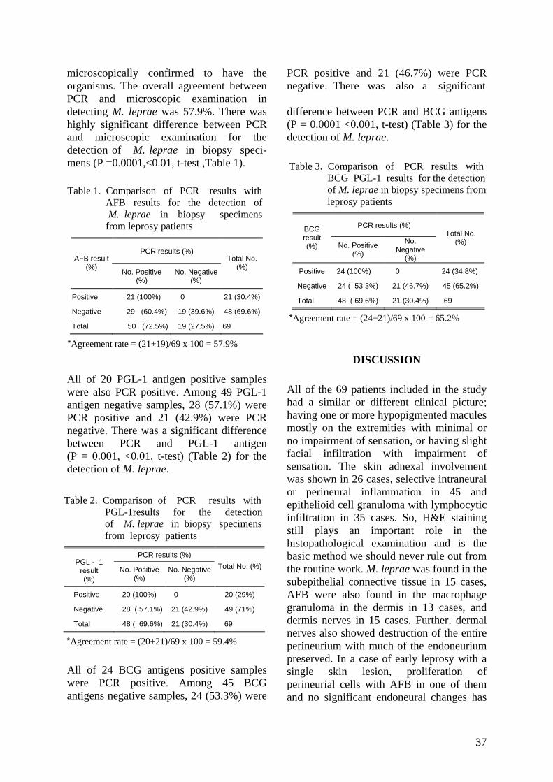
microscopically confirmed to have the organisms. The overall agreement between PCR and microscopic examination in detecting M. leprae was 57.9%. There was highly significant difference between PCR and microscopic examination for the detection of M. leprae in biopsy speci-mens (P =0.0001,<0.01, t-test ,Table 1).
All of 20 PGL-1 antigen positive samples were also PCR positive. Among 49 PGL-1 antigen negative samples, 28 (57.1%) were PCR positive and 21 (42.9%) were PCR negative. There was a significant difference between PCR and PGL-1 antigen (P = 0.001, <0.01, t-test) (Table 2) for the detection of M. leprae.
All of 24 BCG antigens positive samples were PCR positive. Among 45 BCG antigens negative samples, 24 (53.3%) were
PCR positive and 21 (46.7%) were PCR negative. There was also a significant difference between PCR and BCG antigens (P = 0.0001 <0.001, t-test) (Table 3) for the detection of M. leprae.
Table 3. Comparison of PCR results with BCG PGL-1 results for the detection of M. leprae in biopsy specimens from leprosy patients
PCR results (%) BCG result (%) No. Positive
(%)
No. Negative
(%)
Total No. (%)
Positive 24 (100%) 0 24 (34.8%)
Negative 24 ( 53.3%) 21 (46.7%) 45 (65.2%)
Total 48 ( 69.6%) 21 (30.4%) 69
*Agreement rate = (24+21)/69 x 100 = 65.2%
Table 1. Comparison of PCR results with AFB results for the detection of M. leprae in biopsy specimens from leprosy patients
PCR results (%) AFB result
(%) No. Positive (%)
No. Negative (%)
Total No. (%)
Positive 21 (100%) 0 21 (30.4%)
Negative 29 (60.4%) 19 (39.6%) 48 (69.6%)
Total 50 (72.5%) 19 (27.5%) 69
*Agreement rate = (21+19)/69 x 100 = 57.9% DISCUSSION
All of the 69 patients included in the study had a similar or different clinical picture; having one or more hypopigmented macules mostly on the extremities with minimal or no impairment of sensation, or having slight facial infiltration with impairment of sensation. The skin adnexal involvement was shown in 26 cases, selective intraneural or perineural inflammation in 45 and epithelioid cell granuloma with lymphocytic infiltration in 35 cases. So, H&E staining still plays an important role in the histopathological examination and is the basic method we should never rule out from the routine work. M. leprae was found in the subepithelial connective tissue in 15 cases, AFB were also found in the macrophage granuloma in the dermis in 13 cases, and dermis nerves in 15 cases. Further, dermal nerves also showed destruction of the entire perineurium with much of the endoneurium preserved. In a case of early leprosy with a single skin lesion, proliferation of perineurial cells with AFB in one of them and no significant endoneural changes has
Table 2. Comparison of PCR results with PGL-1results for the detection of M. leprae in biopsy specimens from leprosy patients
PCR results (%) PGL - 1
result (%)
No. Positive (%)
No. Negative (%)
Total No. (%)
Positive 20 (100%) 0 20 (29%)
Negative 28 ( 57.1%) 21 (42.9%) 49 (71%)
Total 48 ( 69.6%) 21 (30.4%) 69
*Agreement rate = (20+21)/69 x 100 = 59.4%
37
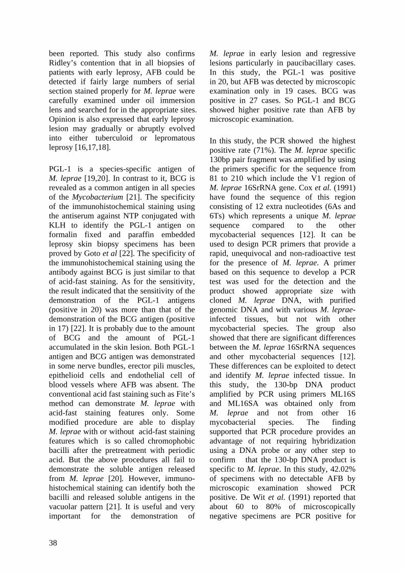
been reported. This study also confirms Ridley’s contention that in all biopsies of patients with early leprosy, AFB could be detected if fairly large numbers of serial section stained properly for M. leprae were carefully examined under oil immersion lens and searched for in the appropriate sites. Opinion is also expressed that early leprosy lesion may gradually or abruptly evolved into either tuberculoid or lepromatous leprosy [16,17,18].
PGL-1 is a species-specific antigen of M. leprae [19,20]. In contrast to it, BCG is revealed as a common antigen in all species of the Mycobacterium [21]. The specificity of the immunohistochemical staining using the antiserum against NTP conjugated with KLH to identify the PGL-1 antigen on formalin fixed and paraffin embedded leprosy skin biopsy specimens has been proved by Goto et al [22]. The specificity of the immunohistochemical staining using the antibody against BCG is just similar to that of acid-fast staining. As for the sensitivity, the result indicated that the sensitivity of the demonstration of the PGL-1 antigens (positive in 20) was more than that of the demonstration of the BCG antigen (positive in 17) [22]. It is probably due to the amount of BCG and the amount of PGL-1 accumulated in the skin lesion. Both PGL-1 antigen and BCG antigen was demonstrated in some nerve bundles, erector pili muscles, epithelioid cells and endothelial cell of blood vessels where AFB was absent. The conventional acid fast staining such as Fite’s method can demonstrate M. leprae with acid-fast staining features only. Some modified procedure are able to display M. leprae with or without acid-fast staining features which is so called chromophobic bacilli after the pretreatment with periodic acid. But the above procedures all fail to demonstrate the soluble antigen released from M. leprae [20]. However, immuno-histochemical staining can identify both the bacilli and released soluble antigens in the vacuolar pattern [21]. It is useful and very important for the demonstration of
M. leprae in early lesion and regressive lesions particularly in paucibacillary cases. In this study, the PGL-1 was positive in 20, but AFB was detected by microscopic examination only in 19 cases. BCG was positive in 27 cases. So PGL-1 and BCG showed higher positive rate than AFB by microscopic examination.
In this study, the PCR showed the highest positive rate (71%). The M. leprae specific 130bp pair fragment was amplified by using the primers specific for the sequence from 81 to 210 which include the V1 region of M. leprae 16SrRNA gene. Cox et al. (1991) have found the sequence of this region consisting of 12 extra nucleotides (6As and 6Ts) which represents a unique M. leprae sequence compared to the other mycobacterial sequences [12]. It can be used to design PCR primers that provide a rapid, unequivocal and non-radioactive test for the presence of M. leprae. A primer based on this sequence to develop a PCR test was used for the detection and the product showed appropriate size with cloned M. leprae DNA, with purified genomic DNA and with various M. leprae-infected tissues, but not with other mycobacterial species. The group also showed that there are significant differences between the M. leprae 16SrRNA sequences and other mycobacterial sequences [12]. These differences can be exploited to detect and identify M. leprae infected tissue. In this study, the 130-bp DNA product amplified by PCR using primers ML16S and ML16SA was obtained only from M. leprae and not from other 16 mycobacterial species. The finding supported that PCR procedure provides an advantage of not requiring hybridization using a DNA probe or any other step to confirm that the 130-bp DNA product is specific to M. leprae. In this study, 42.02% of specimens with no detectable AFB by microscopic examination showed PCR positive. De Wit et al. (1991) reported that about 60 to 80% of microscopically negative specimens are PCR positive for
38
AFB [8]. The results from this study showed a slightly lower positive rate, but higher than that of Cox et al. (1991). In the latter, only 11% of paucibacillary disease showed the positive results. It was apparent that PCR is more sensitive in detecting M. leprae in biopsy specimens with no or low bacterial loads than the conventional microscopic examination.
In summary, PCR using primers of 16SrRNA of M. leprae, to amplify the 130 fragment of M. leprae DNA showed a clear advantage over microscopic examination and immunostaining in detecting M. leprae in tissue with negative AFB. It is recommended that PCR studies to detect M. leprae should be done wherever possible in conjunction with histopathological examination and immunohistochemical staining in order to obtain the precise diagnosis of early leprosy.
ACKNOWLEDGMENTS
We would like to thank Director-General Professor Dr. Paing Soe and Deputy Director General Dr. Soe Thein, Department of Medical Research (Lower Myanmar) for their advice and encouragement to our research. We are obliged to Dr. Masako Namisato, Deputy Director of National Sanatorium Kryu-Rakusenen, Japan and Dr. Yoshiko Kashiwabara, Leprosy Research Center, National Institute of Infectious Diseases, Tokyo for their supplies of PCR machine, reagents and primers. We acknowledge to Dr. M. Makino from Leprosy Research Center, National Institute of Infectious Diseases, Tokyo, Japan for supply of monoclonal antibody against PGL-1.
REFERENCES
1. Clark-Curtiss, J.E.. Benefits of recombinant DNA technology for the study of Mycobacterium leprae. Curren. Topics in Microbiology & Immunology 1988;138:61-79.
2. Clark-Curtiss, J.E., & Docherty, M.A. A species-specific repetitive sequence in Mycobacterium leprae. Journal of Infectious Diseases 1989;159:7-15.
3. Hartskeerl, R.A., DeWit, M.Y.L. & Klatser, P.R.. Polymerase chain reaction for the detection of Mycobacterium leprae. Journal of General Microbiology 1989; 135:2375-2364.
4. Willams, D.L., Gillis, T.P, Booth, R.J. Looker, D. & Watson, J.D. The use of a specific DNA probe and polymerase chain reaction for the detection of Mycobacterium leprae. Journal of infectious diseases 1990;162:193-200
5. Dubeau,L., Chandier, L.A. Gralow, J.R., Nichols, P.W. & Jones,.P.A. Southern blot analysis of DNA extracted formalin-fixed pathology specimens. Cancer Research 1986;46: 2964-2969.
6. Goelz, S.E., Hamilton, S.R. & Vogelstein, B.Purification of DNA from formaldehyde-fixed and paraffin-embedded human tissue. Biochemstry & Biophysics. Research Communication 1985;130 :118-126.
7. Shibata, D., Arnheium, N. & Martin,W.J. Detection of human papillomar virus in paraffin-embedded tissue using the polymerase chain reaction. Journal of Experimental Medicine 1988;167:225-230.
8. DeWit, M.Y.L., Faber, W.R., Krieg, S.R., Douglas, J.T., Lucas, S.B., Montreewasuwat, N., Patttyn, S.R., Hussain, R., Ponnighaus, J.M., Hartskeerl, R.A. & Klatser, P.R. Application of a polymerase chain reaction for the detection of Mycobacterium leprae in skin tissue. Journal of Clinical Microbiology 1991;29:906-910.
9. Plikaytis, B.B.,R., Gelber, H. & Shinnick, T.M .Rapid and sensitive detection of Mycobacterium leprae using a nested-primer gene amplification assay. Journal of Clinical Microbiology 1990; 28:1913-1917.
10. Woods, S.A. & Cole, S.T. A rapid method for the detection of potentially viable Mycobacterium leprae in human biopsies; a novel application of PCR. FEMS microbiology Letter 1989;65:305-310.
11. Woods, S.A., & Cole, S.T. A family of dispersed repeats in Mycobacterium leprae. Molecular Microbiology 1990;4:1745-1751.
12. Cox, R.A., Kempsell, K., Fairclough, L. & Colston, M.J.. The 16S ribosomal RNA of Mycobacterium leprae contains a unique sequence which can be used for identification by the polymerase chain reaction. Journal of Medical Microbiology 1991 ; 35:284-290.
39
19. Brennan, P.J. & Barrow, W.W. Evidence for species-specific lipid antigens in Mycobacterium leprae. Journal of Leprosy 1980; 48: 382-387.
13. Wang, T., Izumi S., Butt, K.I., Kawatsu, K.& Maeda, Y. Demonstration of PGL-1 & LAM-B antigen in paraffin section of leprosy skin lesion. Japan Journal of leprosy 1992; 61:165-174. 20. Hunter, S.W. & Brennan, P.J. A novel phenolic
glycolipid from Mycobacterium leprae possibility involved in immunogenicity and pathogenicity. Journal of Bacteriology 1981; 147:728-735.
14. Lawrence, D. T. Atlas of diagnostic immunohistopathology, 1st ed., J.B. Lippincott Company, Philadelphia 1990; pp2-16.
15. Fujiwara, T. & Izumi, S. Synthesis of the negolycocojugates of phenolic glycolipid-related trisaccharides for the serodiagnosis of leprosy. Agric . Biology. Chemistry 1987; 51:2539.
21. Hunter, S.W. el al. Structure and antigenicity of the phosphorylated lipopolysaccharide antigens from the leprosy and tubercle bacillus. Journal of Biological Chemistry 1986; 261:12345-12351.
16. Ridley, D.S. The early skin lesion of leprosy. In: Pathogenesis of Leprosy and Related Diseases London : Wrirht, Butterworth & Co.Ltd,1988; pp.61-70.
22. Goto, M. & Izumi, S. Light and electromicro-scopic immunohistochemistry using anti-PGL-1 antibody specific for Mycobacterium leprae. International Journal of Leprosy 1991; 59(1): 195.
17. Murray, K.A.,mclelland, B.A. & Job, C.K. Early perineural proliferation. Arc, Dermatology 1984; 120: 360-361.
23. Fleury, R .N. & Bacchi, C.E.S-100 protein and immunoperoxidase technique as aid in the histopathology diagnosis of leprosy. Interna-tional Journal of Leprosy 1987; 55: 338-334.
18. Ridley, D.S. Histological diagnosis. In; Pathogenesis of Leprosy and Related Diseases. London: Wrirht, Butterworth & Co.Ltd,1988; pp.146-151.
40
The Myanmar Health Sciences Research Journal, Vol. 18, No. 1, 2006
Prevalence, awareness, correlates, treatment and control of hypertension in a rural community of Waw Township, Bago Division
*Han Win, *Aung Thu, *Khin Myat Tun, *Khin Khin Swe Myat, *Than Than Lwin,
*Sandar Kyi, *Myat Myat Thu, *Tin Htar Lwin & *Aye Hnin Phyu
*Clinical Research Division Department of Medical Research (Lower Myanmar)
Hypertension is an important cause of cardiovascular morbidity and mortality. A cross-sectional survey was conducted on 644 subjects (20 years and above, mean age 48.9 years) in Thuyethamain Village in Waw Township, Bago Division. The objectives were to determine the prevalence of hypertension, assess the degree of awareness, taking treatment and control of high blood pressure, and to identify the correlates of hypertension awareness. Hypertension was defined as a mean systolic blood pressure ≥ 140 mm Hg, diastolic blood pressure ≥ 90 mm Hg, and/or use of antihypertensive medications. The overall prevalence of hypertension was 29% (95% confidence interval (95% CI): 25.5%–32.7%). Among hypertensives, 43.9% were aware of the condition, while 28.9% were being treated and 16.6% were under control. Multivariate analysis showed that women have better awareness of hypertension (odds ratio (OR) = 5.1, 95% CI: 1.9–13.7). Other factors independently associated with increased awareness of hypertension were higher education status (OR for high school and above level compared to no schooling = 3.3, 95% CI: 1.2-9.5), and dependents compared to manual workers (OR = 3.5, 95% CI: 1.2-9.7). In this study, over half of hypertensive subjects were unaware of the condition and less than a third was under treatment. These observations highlight the need for regular screening coupled with educational programs to promote hypertension awareness in the community.
INTRODUCTION
Uncontrolled hypertension (HT) is an established risk factor for the development of vascular diseases. Cardiovascular diseases (CVD) account for nearly a third of all deaths worldwide [1]. CVD are increasing in developing countries [2,3] and it has been estimated that CVD will be the major cause of morbidity and mortality in these countries by the year 2020 [4]. Consequently, the prevention of risk factors for CVD is a public health priority worldwide [1]. Hypertension is a premier risk factor for CVD, which is easily recognized if sought, and can be treated effectively . Treatment of high blood
pressure (BP) has been consistently reported to reduce the risk of CVD [5].
In Myanmar, as in most other countries, hypertension is a major public health problem. According to a previous CVD survey, the prevalence of HT in rural areas was 12.4% [6]. But the prevalence varies in different communities with reported lowest prevalence in Laydaunggan (2.1%) and the highest prevalence in Thonegwa (38.4%) [7, 8].
The prevalence, degree of awareness, treatment, and control of high BP in rural communities of Waw Township were previously unknown. So this study was conducted with the following objectives.
41
♦ To determine the prevalence of hyper-tension
♦ To assess the degree of awareness, taking treatment, and control of hyper-tension
♦ To identify the correlates of awareness of high blood pressure
MATERIALS AND METHODS
Study area and population
Community survey was carried out in Thuyethamain village, Waw Township in Bago Division during November 2004. Cross-sectional study design was employed. A total of 644 persons aged 20 years and over participated in the study.
Sample size and sampling procedure
Assuming that a crude prevalence of hypertension was 20% and desired precision 0.03, the calculated sample size was 640 at 95% confidence level. Thuyethamain village in Waw Township was purposely selected for the study of hypertension. For recruitment of the study subjects, a list of eligible persons (aged 20 years and above currently living in the village) was taken before the survey. From this list of 987 persons (sampling frame), random numbers were generated using EPITABLE program to have a required sample size.
Data collection
The survey had two principal components: (a) interviewing the subjects using a questionnaire and (b) measurement of BP and anthropometry. Informed written consent was obtained from all the participants before the interview.
Study questionnaire
Before the data collection commenced, team members were trained on the use of research instruments and methods of data collection. Investigators administered a pre-tested structured questionnaire, which collected information on demographic, socioeconomic, medical history, and
information on lifestyle habits such as smoking and alcohol consumption.
Measurement of BP and anthropometry
Before the BP was measured, the investigator made sure that the subjects had not consumed any hot beverages, such as tea or coffee or smoked/chewed tobacco or undertaken vigorous physical activity within 30 minutes preceding the household visit. If they had, then the measurements were postponed by 30 minutes. Medical officers measured the BP of all the participants twice using a mercury column sphygmomanometer and a standard protocol. Both BP readings were obtained from the seated subject using a cuff of an appropriate size, with the arm supported and the sphygmomanometer at the level of the heart [9]. Blood pressure was taken twice (5 minutes apart) only after the interview which usually lasted about 10 minutes. The BP cuff was inflated to 30 mmHg above the pressure at which the radial pulse disappeared, and then deflated slowly. Phases 1 and 5 of the Korotkoff sound were taken as indicatators for systolic blood pressure (SBP) and diastolic blood pressure (DBP) respectively [9]. Both SBP and DBP readings were recorded to the nearest 5 mmHg. The average of the two readings of SBP and DBP was taken as the BP of the participant.
Body weight and height of all the participants were also measured. Weight was recorded to the nearest 0.5 kg, while height was measured to the nearest 0.5 cm. Body mass index (BMI) was computed as the weight in kilograms divided by the square of the height in meters.
Working definitions
Hypertension
The HT status of the subjects was assessed based on the criteria formulated by the World Health Organization–International Society of HT (WHO–ISH) and the US Seventh Joint National Committee (JNC VII) report on the prevention, detection,
42
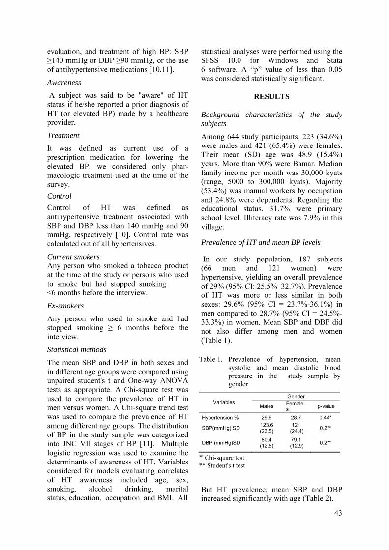
evaluation, and treatment of high BP: SBP >140 mmHg or DBP >90 mmHg, or the use of antihypertensive medications [10,11].
Awareness
A subject was said to be "aware" of HT status if he/she reported a prior diagnosis of HT (or elevated BP) made by a healthcare provider.
Treatment
It was defined as current use of a prescription medication for lowering the elevated BP; we considered only phar-macologic treatment used at the time of the survey. Control Control of HT was defined as antihypertensive treatment associated with SBP and DBP less than 140 mmHg and 90 mmHg, respectively [10]. Control rate was calculated out of all hypertensives.
Current smokers Any person who smoked a tobacco product at the time of the study or persons who used to smoke but had stopped smoking <6 months before the interview.
Ex-smokers
Any person who used to smoke and had stopped smoking ≥ 6 months before the interview.
Statistical methods
The mean SBP and DBP in both sexes and in different age groups were compared using unpaired student's t and One-way ANOVA tests as appropriate. A Chi-square test was used to compare the prevalence of HT in men versus women. A Chi-square trend test was used to compare the prevalence of HT among different age groups. The distribution of BP in the study sample was categorized into JNC VII stages of BP [11]. Multiple logistic regression was used to examine the determinants of awareness of HT. Variables considered for models evaluating correlates of HT awareness included age, sex, smoking, alcohol drinking, marital status, education, occupation and BMI. All
statistical analyses were performed using the SPSS 10.0 for Windows and Stata 6 software. A “p” value of less than 0.05 was considered statistically significant.
RESULTS Background characteristics of the study subjects
Among 644 study participants, 223 (34.6%) were males and 421 (65.4%) were females. Their mean (SD) age was 48.9 (15.4%) years. More than 90% were Bamar. Median family income per month was 30,000 kyats (range, 5000 to 300,000 kyats). Majority (53.4%) was manual workers by occupation and 24.8% were dependents. Regarding the educational status, 31.7% were primary school level. Illiteracy rate was 7.9% in this village.
Prevalence of HT and mean BP levels
In our study population, 187 subjects (66 men and 121 women) were hypertensive, yielding an overall prevalence of 29% (95% CI: 25.5%–32.7%). Prevalence of HT was more or less similar in both sexes: 29.6% (95% CI = 23.7%-36.1%) in men compared to 28.7% (95% CI = 24.5%-33.3%) in women. Mean SBP and DBP did not also differ among men and women (Table 1).
But HT prevalence, mean SBP and DBP
Table 1. Prevalence of hypertension, mean systolic and mean diastolic blood pressure in the study sample by gender
Gender Variables
Males Females p-value
Hypertension % 29.6 28.7 0.44*
SBP(mmHg) SD 123.6 (23.5)
121 (24.4) 0.2**
DBP (mmHg)SD 80.4 (12.5)
79.1 (12.9) 0.2**
* Chi-square test ** Student's t test
But HT prevalence, mean SBP and DBP increased significantly with age (Table 2).
43
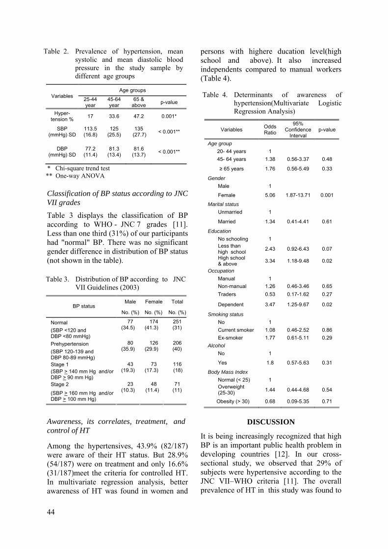
Classification of BP status according to JNC VII grades
Table 3 displays the classification of BP according to WHO - JNC 7 grades [11]. Less than one third (31%) of our participants had "normal" BP. There was no significant gender difference in distribution of BP status (not shown in the table).
Awareness, its correlates, treatment, and control of HT
Among the hypertensives, 43.9% (82/187) were aware of their HT status. But 28.9% (54/187) were on treatment and only 16.6% (31/187)meet the criteria for controlled HT. In multivariate regression analysis, better awareness of HT was found in women and
persons with highere ducation level(high school and above). It also increased independents compared to manual workers (Table 4). Kalavathy and colleagues found that 51.8% of the elderly in South India had HT [19]. The Hypertension Study Group, in their
DISCUSSION
It is being increasingly recognized that high BP is an important public health problem in developing countries [12]. In our cross-sectional study, we observed that 29% of subjects were hypertensive according to the JNC VII–WHO criteria [11]. The overall prevalence of HT in this study was found to
Table 3. Distribution of BP according to JNC VII Guidelines (2003)
Male Female Total BP status
No. (%) No. (%) No. (%)
Normal (SBP <120 and DBP <80 mmHg)
77 (34.5)
174 (41.3)
251 (31)
Prehypertension (SBP 120-139 and DBP 80-89 mmHg)
80 (35.9)
126 (29.9)
206 (40)
Stage 1 (SBP > 140 mm Hg and/or DBP > 90 mm Hg)
43 (19.3)
73 (17.3)
116 (18)
Stage 2
(SBP > 160 mm Hg and/or DBP > 100 mm Hg)
23 (10.3)
48 (11.4)
71 (11)
Table 4. Determinants of awareness of hypertension(Multivariate Logistic Regression Analysis)
Variables Odds Ratio
95% Confidence
Interval p-value
Age group 20- 44 years 1 45- 64 years 1.38 0.56-3.37 0.48
≥ 65 years 1.76 0.56-5.49 0.33
Gender Male 1
Female 5.06 1.87-13.71 0.001
Marital status Unmarried 1
Married 1.34 0.41-4.41 0.61
Education No schooling 1
Less than high school 2.43 0.92-6.43 0.07
High school & above 3.34 1.18-9.48 0.02
Occupation Manual 1 Non-manual 1.26 0.46-3.46 0.65 Traders 0.53 0.17-1.62 0.27
Dependent 3.47 1.25-9.67 0.02
Smoking status No 1 Current smoker 1.08 0.46-2.52 0.86 Ex-smoker 1.77 0.61-5.11 0.29 Alcohol No 1
Yes 1.8 0.57-5.63 0.31
Body Mass Index Normal (< 25) 1 Overweight (25-30) 1.44 0.44-4.68 0.54
Obesity (> 30) 0.68 0.09-5.35 0.71
Table 2. Prevalence of hypertension, mean systolic and mean diastolic blood pressure in the study sample by different age groups
Age groups Variables 25-44
year 45-64 year
65 & above p-value
Hyper-tension % 17 33.6 47.2 0.001*
SBP (mmHg) SD
113.5 (16.8)
125 (25.5)
135 (27.7) < 0.001**
DBP (mmHg) SD
77.2 (11.4)
81.3 (13.4)
81.6 (13.7) < 0.001**
* Chi-square trend test ** One-way ANOVA
44
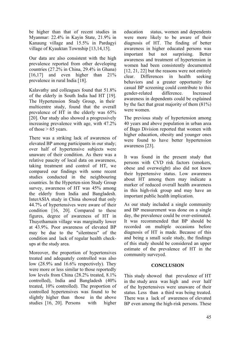
be higher than that of recent studies in Myanmar: 22.4% in Kayin State, 21.9% in Kanaung village and 15.5% in Pardagyi village of Kyauktan Township [13,14,15].
Our data are also consistent with the high prevalence reported from other developing countries (27.2% in China, 29.4% in Ghana) [16,17] and even higher than 21% prevalence in rural India [18].
Kalavathy and colleagues found that 51.8% of the elderly in South India had HT [19]. The Hypertension Study Group, in their multicentre study, found that the overall prevalence of HT in the elderly was 65% [20]. Our study also showed a progressively increasing prevalence with age, with 47.2% of those > 65 years.
There was a striking lack of awareness of elevated BP among participants in our study; over half of hypertensive subjects were unaware of their condition. As there was a relative paucity of local data on awareness, taking treatment and control of HT, we compared our findings with some recent studies conducted in the neighbouring countries. In the Hyperten-sion Study Group survey, awareness of HT was 45% among the elderly from India and Bangladesh. InterASIA study in China showed that only 44.7% of hypertensives were aware of their condition [16, 20]. Compared to these figures, degree of awareness of HT in Thuyethamain village was marginally lower at 43.9%. Poor awareness of elevated BP may be due to the "silentness" of the condition and lack of regular health check-ups at the study area.
Moreover, the proportion of hypertensives treated and adequately controlled was also low (28.9% and 16.6% respectively). They were more or less similar to those reportedly low levels from China (28.2% treated, 8.1% controlled), India and Bangladesh (40% treated, 10% controlled). The proportion of controlled hypertensives was found to be slightly higher than those in the above studies [16, 20]. Persons with higher
education status, women and dependents were more likely to be aware of their diagnosis of HT. The finding of better awareness in higher educated persons was important but not surprising. Better awareness and treatment of hypertension in women had been consistently documented [12, 21, 22] but the reasons were not entirely clear. Differences in health seeking behaviors and a greater opportunity for casual BP screening could contribute to this gender-related difference. Increased awareness in dependents could be explained by the fact that great majority of them (81%) were women.
The previous study of hypertension among 40 years and above population in urban area of Bago Division reported that women with higher education, obesity and younger ones were found to have better hypertension awareness [23]. It was found in the present study that persons with CVD risk factors (smokers, obese and overweight) also did not know their hypertensive status. Low awareness about HT among them may indicate a marker of reduced overall health awareness in this high-risk group and may have an important public health implication.
As our study included a single community and BP measurement was done on a single day, the prevalence could be over-estimated. It was recommended that BP should be recorded on multiple occasions before diagnosis of HT is made. Because of this and being a small scale study, the findings of this study should be considered an upper estimate of the prevalence of HT in the community surveyed.
CONCLUSION
This study showed that prevalence of HT in the study area was high and over half of the hypertensives were unaware of their status. Less than a third was being treated. There was a lack of awareness of elevated BP even among the high-risk persons. These
45
observations highlight the need for regular blood pressure screening coupled with educational programs to promote hypertension awareness in the community.
ACKNOWLEDGEMENT
This study was funded by WHO. We would like to express our sincere thanks to Professor Dr Paing Soe, Director-General of Department of Medical Research (Lower Myanmar), for allowing us to conduct the study. We are also grateful to Dr Kyaw Swa Lwin, Station Medical Officer, and health staff of Thuyethamain village and all subjects who participated in the study.
REFERENCES 1. World Health Organization. The World Health
Report. Reducing risks, promoting healthy life. Geneva: WHO 2002.
2. Chockalingam, A, Balaguer-Vinto I (eds). Impending global pandemic of cardiovascular diseases: challenges and opportunities for the prevention and control of of cardiovascular diseases in developing countries and economiesin transition. World Heart Federation. Barcelona: Prous Science 1999.
3. Reddy, K.S. & Yusuf, S. Emerging epidemic of cardiovascular disease in developing countries. Circulation 1998; 97: 596–601
4. Murray, C.J. & Lopez, A,D. The global burden of disease: a comprehensive assessment of mortality and disability from disease, injuries and risk factors in 1990 and projected to 2020. Cambridge, Mass: Harvard School of Public Health 1996.
5. Staessen, J.A., Wang, J.G. & Thijs, L. Cardiovascular protection and blood pressure reduction: a meta-analysis. Lancet 2001; 358:00201305–1315.
6. Ministry of Health, Myanmar: National Health Plan, 1996-2000.
7. Tin Nu Swe, Than Pe, Tin Zaw, Khin Saw, Khin Thet Wai, Phyu Phyu Aung et al. An investigation of hypertension in Thonegwa Township. Myanmar Health Research Con-gress, Programme and abstracts 1998: pp. 90.
8. El-Zawahry, M. et al. Laydaungan community health survey. Burma Research Congress., Abstract of papers 1971: pp. 76-77.
9. Frohlich, E.D. Blood pressure measurement. Canadian Journal of Cardiology 1995; 11 (Suppl): 35H–37H.
10. World Health Organization–International Society of Hypertension Guidelines for the Management of Hypertension. Guidelines Subcommittee. Journal of Hypertensions 1999; 17: 151–183.
11. Chobanian, A.V., Bakris, G.L., Black, H.R., Cushman ,W.C., Green, L.A., Izzo, JL. Jr. et al. National Heart, Lung, and Blood Institute Joint National Committee on Prevention, Detection, Evaluation, and Treatment of High Blood Pressure. National High Blood Pressure Education Program Coordinating Committee. The Seventh Report of the Joint National Committee on Prevention, Detection, Evaluation, and Treatment of High Blood Pressure: the JNC 7 report. JAMA 2003; 289: 2560–2572.
12. Fuentesc, R., Ilmaniemi, N., Laurikainen, E., Tuomilehto, J. & Nissinen, A. Hypertension in developing economies: a review of population-based studies carried out from 1980 to 1998. Journal of Hypertension 2000; 18:521–529.
13. San Shwe, Ohnmar, Kyu Kyu Than, Than Tun Sein, Aung Thu, et al. Prevalence of hypertension in two selected villages of Kayin State, Myanmar. Regional Health Forum 2004; 8: 104-112.
14. Report of the hypertension status of people in Kanaung village, Kyauktan Township by DMR, Lower Myanmar, 2001 (Myanmar language).
15. Han Win, Khin Myat Tun, Maung Maung Toe, San Hla Mu, et al. Prevalence of hypertension in Pardagyi village, Kyauktan Township. Myanmar Health Sciences Research Journal 2005;17(1): 22-26.
16. Dongfeng, Gu, Kristi Reynolds, Xigui Wu, Jing Chen, et al. Prevalence, awareness, treatment and control of hypertension in China. Hypertension 2002; 40: 920-927.
17. Agyemang, C., Bruijnzeels, M.A. & Owusu-Dabo, E. Factors associated with hypertension awareness, treatment and control in Ghana, West Africa. Journal of Human Hypertension 2005; advance online publication.
18. Gupta, R., Sharma, A.K, Prevalence of hyper- tension and subtypes in an Indian rural population: clinical and electrocardiographic correlates. Journal of Human Hypertension 1994; 8 (11):823-9.
19. Kalavathy, M.C., Thankappan, K.R., Sarma, P.S. & Vasan, R.S. Prevalence, awareness, treatment and control of hypertension in an elderly community-based sample in Kerala, India.
46

National Medical Journal of India 2000; 13(1): 9-15.
Hypertension 1997; 15: 591-600. 22. Jose, R.B., Fernando, R.A. et al. Blood
PressureSpain; Distribution, Awareness, Control, and Benefits of a reduction in Average Pressure. Hypertension, 1998; 32: 998-1002.
20. Hypertension Study Group. Prevalence, awareness, treatment and control of hypertension among the elderly in Bangladesh and India: a multicentre study. Bulletin of World Health Organization 2001; 79: 490–500.
21. Klungel, O.H. et al. Sex differences in the pharmacological treatment of hypertension:
23. Thet Thet Mu. Factors associated with hypertension and hypertension awareness among the 40-years-and-above age group of urban population in Bago Division. M.Med.Sc Thesis, 1997. a review of population-based studies. Journal of
47
The Myanmar Health Sciences Research Journal, Vol. 18, No. 1, 2006
Distribution of coliforms , faecal coliforms and enteropathogenic E. coli (EPEC) in fried rice and water samples from street vendors of Yangon
and detection of bacterial toxins
*Mar Mar Nyein ,**Yee Yee Aung ,*Mi Mi Htwe & ***Tin Nwe
*Bacteriology Research Division, DMR (LM) **Dagon University
***Yangon University
The contamination of coliforms, faecal coliforms and enteropathogenic Escherichia coli (EPEC) was tested in 45 fried rice samples collected from different street vendors of three townships from Yangon area. Toxin produced by E. coli was also studied. High total bacterial count of 104 to >106 was recorded from 42 samples of fried rice. Among them, coliforms, faecal coliforms and EPEC were isolated from 32 (71.11%) samples, 15 (33.33) samples and 29 (64.44 %) samples respectively. The serogroup O55K59 was the commonest pathogen isolated from 11 samples (24.44 %). The other serogroups encountered were O1K51 (from 4 samples), O26K60 (from 3 samples), O25K+ and O125K70 (from 2 samples each). Other serogroups isolated were O27K+, O44K74, O86K62, O111K58, O114K90, O128K70 and O136K78 (from one sample each). Enterotoxigenic E. coli–heat-labile (ETEC-LT) toxin and verotoxin (VTEC) were detected from 2 samples (4.44%) and from one sample respectively. Similarly, coliforms, faecal coliforms and EPEC were isolated respectively from 39 (86.66%), 21 (46.67%) and 23 (51.11 %) water samples used for multipurpose in those shops. The serogroup O55K59 was commonly found with the isolation rate of 11 (24.44%); O119K69 was isolated from 2 samples and the other serogroups such as O1K51, O26K60, O44K74, O86K62, and O128K67 were also detected . ETEC-LT toxin was detected from one sample (2.22%) of water only.
INTRODUCTION
Many microorganisms previously unrecog-nized as food-borne or harmful are emerging as human pathogens transmitted by food. Within the past decade, the epidemiology of microbial food-borne diseases has changed, not only because of human population’s increasingly sus-ceptibility to diseases and of changing life styles (including more adventurous eating, more convenience foods, and less time devoted to food preparation) but also because of the emergence of newly recognized food-borne pathogens [1, 2]. In developing countries, a great number of ready-to-eat food is sold on the streets. The
term "street food" refers to a wide variety of ready-to-eat foods and beverages sold, and sometimes prepared, in public places. As with fast food, the final preparation occurs when meals are ordered by customers. Street food may be consumed where it is purchased or can be taken away and eaten elsewhere. The consumption of street food is common in many countries where unemployment is high, salaries are low, work opportunities and social programs are limited, and where urbanization is taking place. Street food vendors benefit from a positive cash flow, often evade taxation, and can determine their own working hours. By selling snacks, complete meals and refreshments at relatively low prices, they
48
provide an essential service to workers, shoppers, travelers, and people with low incomes. People who depend on such food are often more interested in its convenience than in questions of its safety, quality and hygiene. The hygienic aspects of vending operations are a major source of concern for food control officers. For example, stands are often crude structures, and running water may not be readily available. Also toilets and adequate washing facilities are rarely available. The washing of hands, utensils, and dishes is often done in buckets or bowls. Disinfection is not usually carried out, and insects and rodents may be attracted to sites where there is no organized sewage disposal [3,4]. Finally food is not adequately protected from flies and refrigeration is usually unavailable. The presence of coliforms and faecal coliforms in food and water is an indicator for contamination. Water and food always serve as vehicles in transmission of contaminated bacteria. Thus, this study was carried out to investigate the contaminated bacteria in food and water from street vendors of Yangon area.
Objective
To investigate the presence of coliforms, faecal coliforms and enteropathogenic Escherichia coli (EPEC) in fried rice and water sold by street vendors of Yangon area.
Specific objectives
1. To determine the presence of coliforms and faecal coliforms in fried rice and water
2. To isolate Escherichia coli from fried rice and water
3. To identify enteropathogenic Escheri-chia coli (EPEC)
4. To define the enterotoxin produced by E. coli isolates.
MATERIALS AND METHODS Tested samples
Forty five samples of fried rice (approx.
500 grams) sold in street vendors and water samples (one litre) used for multipurpose from respective shops were collected aseptically from three townships of Latha, Yankin and Dagon new satellite town from 25-1-05 to 16-5-05.
Sample preparation
Twenty grams of fried rice were soaked in phosphate buffer (PBS) and homogenized with the aid of stainless steel homogenizer (Ace homogenizer, Nihonseiki kaisha Ltd.). Serial 10-fold dilutions were carried out in 90 ml PBS to obtain up to 105 dilutions aseptically [5].
Total bacterial count
It was carried out by the method of Miles and Misra, 1930 [6].
Determination of coliforms and faecal coliforms
It was carried out by the multiple tube method as well as by Millipore membrane filtration method [7]. Isolation, identi-fication and characterization of bacteria from fried rice were carried out by the methods described in WHO, 1982 [8] and by Lennette, Balows, Hausler and Shadomy, 1985 [9] and serological tests by Ewing, 1986 [10].
Enterotoxin assays
They were carried out for Escherichia coli heat labile enterotoxin (LT) and verotoxin detection using VET-RPLA kit (Oxoid TD-920) and VET-RPLA kit (Oxoid, TD- 0960A) which are reverse passive latex agglutination test. The isolates were cultured in Casamino Acid Yeast Extract (CA-YE) broth at 37˚C overnight in a shaking waterbath. The culture supernatant was used by mixing with polymer latex particles sensitized with purified rabbit antiserum which is reactive with E. coli heat labile toxin or with polymer latex particles sensitized with purified rabbit antiserum which is reactive with E. coli VT1 or VT2.
49
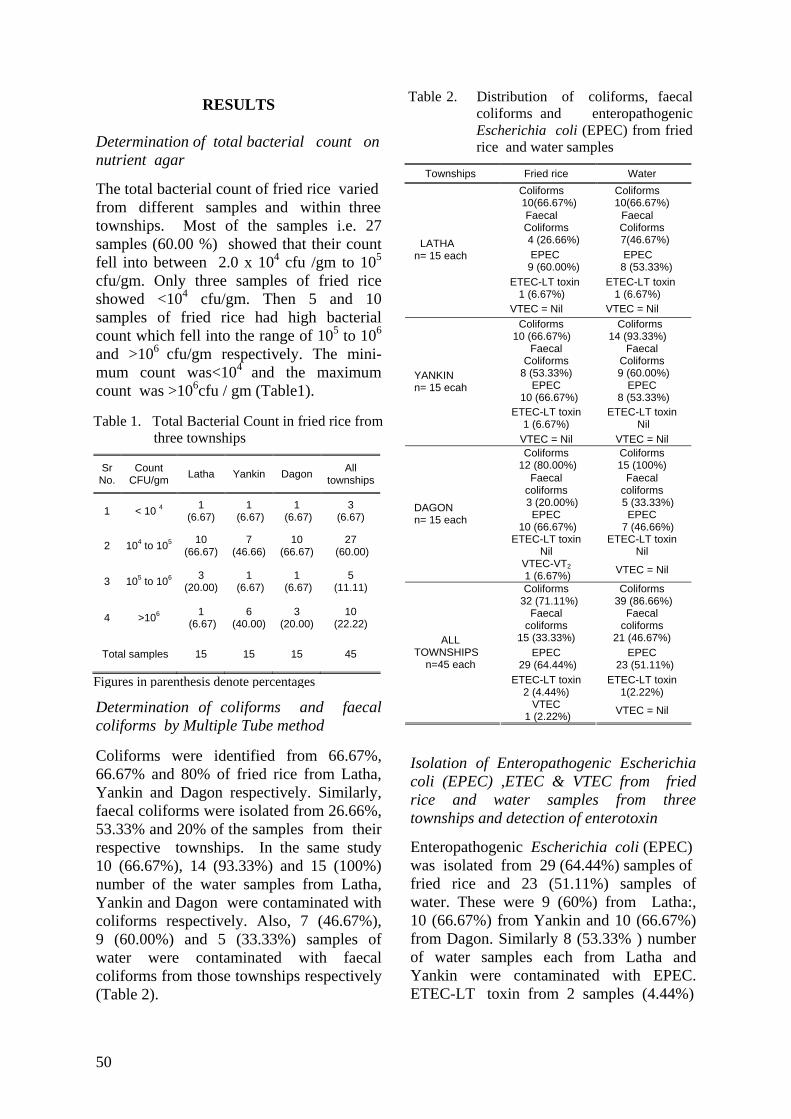
RESULTS
Determination of total bacterial count on nutrient agar
The total bacterial count of fried rice varied from different samples and within three townships. Most of the samples i.e. 27 samples (60.00 %) showed that their count fell into between 2.0 x 10⁴ cfu /gm to 105 cfu/gm. Only three samples of fried rice showed <104 cfu/gm. Then 5 and 10 samples of fried rice had high bacterial count which fell into the range of 105 to 106 and >106 cfu/gm respectively. The mini-mum count was<104 and the maximum count was >106cfu / gm (Table1).
Determination of coliforms and faecal coliforms by Multiple Tube method
Table 2. Distribution of coliforms, faecal coliforms and enteropathogenic Escherichia coli (EPEC) from fried rice and water samples
Townships Fried rice Water Coliforms 10(66.67%)
Coliforms 10(66.67%)
Faecal Coliforms
4 (26.66%)
Faecal Coliforms
7(46.67%) EPEC 9 (60.00%)
EPEC 8 (53.33%)
ETEC-LT toxin 1 (6.67%)
ETEC-LT toxin 1 (6.67%)
LATHA n= 15 each
VTEC = Nil VTEC = Nil Coliforms 10 (66.67%)
Coliforms 14 (93.33%)
Faecal Coliforms
8 (53.33%)
Faecal Coliforms
9 (60.00%) EPEC
10 (66.67%) EPEC
8 (53.33%) ETEC-LT toxin
1 (6.67%) ETEC-LT toxin
Nil
YANKIN n= 15 ecah
VTEC = Nil VTEC = Nil Coliforms
12 (80.00%) Coliforms 15 (100%)
Faecal coliforms
3 (20.00%)
Faecal coliforms
5 (33.33%) EPEC
10 (66.67%) EPEC
7 (46.66%) ETEC-LT toxin
Nil ETEC-LT toxin
Nil
DAGON n= 15 each
VTEC-VT2 1 (6.67%) VTEC = Nil
Coliforms 32 (71.11%)
Coliforms 39 (86.66%)
Faecal coliforms
15 (33.33%)
Faecal coliforms
21 (46.67%) EPEC
29 (64.44%) EPEC
23 (51.11%) ETEC-LT toxin
2 (4.44%) ETEC-LT toxin
1(2.22%)
ALL TOWNSHIPS
n=45 each
VTEC 1 (2.22%) VTEC = Nil
Table 1. Total Bacterial Count in fried rice from three townships
Sr No.
Count CFU/gm Latha Yankin Dagon All
townships
1 < 10 4 1 (6.67)
1 (6.67)
1 (6.67)
3 (6.67)
2 104 to 105 10 (66.67)
7 (46.66)
10 (66.67)
27 (60.00)
3 105 to 106 3 (20.00)
1 (6.67)
1 (6.67)
5 (11.11)
4 >106 1 (6.67)
6 (40.00)
3 (20.00)
10 (22.22)
Total samples 15 15 15 45
Figures in parenthesis denote percentages
Coliforms were identified from 66.67%, 66.67% and 80% of fried rice from Latha, Yankin and Dagon respectively. Similarly, faecal coliforms were isolated from 26.66%, 53.33% and 20% of the samples from their respective townships. In the same study 10 (66.67%), 14 (93.33%) and 15 (100%) number of the water samples from Latha, Yankin and Dagon were contaminated with coliforms respectively. Also, 7 (46.67%), 9 (60.00%) and 5 (33.33%) samples of water were contaminated with faecal coliforms from those townships respectively (Table 2).
Isolation of Enteropathogenic Escherichia coli (EPEC) ,ETEC & VTEC from fried rice and water samples from three townships and detection of enterotoxin
Enteropathogenic Escherichia coli (EPEC) was isolated from 29 (64.44%) samples of fried rice and 23 (51.11%) samples of water. These were 9 (60%) from Latha:, 10 (66.67%) from Yankin and 10 (66.67%) from Dagon. Similarly 8 (53.33% ) number of water samples each from Latha and Yankin were contaminated with EPEC. ETEC-LT toxin from 2 samples (4.44%)
50
and VTEC (VT2) from one sample of fried rice (2.22%) were also detected (Table 2). The serogroup O55K59 was the commonest pathogen isolated from 11 samples (24.44%). The other serogroups encountered were O1K51 (from 4 samples), O26K60 ( from 3 samples ), O25K + and O125K70 (from 2 samples each). Other serogroups isolated were O27K+, O44K74, O86K62, O111K58, O114K90, O128K67 and O136K78 (from one sample each) (Table 3). Similarly, 39 (86.66%), 21 (46.67%) and 23 (51.11 %) number of coliforms, faecal coliforms and EPEC respectively were isolated from water samples used for multipurpose in those shops. Similar serogroups such as O55K59 was found to be common with the isolation rate of 11 (24.44%). O119K69 was isolated from 2 samples and the other serogroups such as O1K51, O26K60, O44K74, O86K62, and O128K67 were also detected (Table 3). ETEC–LT toxin was detected from one sample (2.22%) of water.
Distribution of coliforms and faecal coliforms in fried rice and water samples
This study showed that coliforms were solated from 71.11 percent and 86.67 per- cent of fried rice and water samples
respectively. Faecal coliforms were identified from 33.33 percent and 46.67 percent of fried rice and water samples respectively (Table 2).
DISCUSSION
Microbial pathogenicity has been defined as the structural and biochemical mechanisms whereby microorganisms cause disease. Infection may imply colonization, multiplication, invasion or adhesion of a pathogen on or within a host, but disease (infectious disease) is used to describe an infection that causes significant overt damage of the host. Escherichia coli isolates that bear mannose-resistant hemagglutinins and related adhesions, are associated with two common diseases: urinary tract infection and diarrhea. Epidemiological studies suggest that children and pregnant women are prone to recurrent or persistant infections caused by these organisms. Diffuse-adhering Escherichia coli (DAEC) is a putative cause of diarrhea in Mayan children in Mexico [11]. It was noted that E. coli was isolated from 50%, 50% and 30 % of children with cystitis, protracted diarrhea and pyelonephritis in pregnant
Table 3. Isolation of different serogroups from three townships
Latha Yankin Dagon Total Sero- groups Food Water Food Water Food Water Food Water O1 K51 1 1 2 0 1 3 4 4 O25 K+ 1 0 1 0 0 0 2 0 O26 K60 0 0 1 1 2 0 3 1 O27 K+ 0 0 0 0 1 0 1 0 O44 K74 0 0 0 0 1 1 1 1 O55K59 6 6 3 3 2 2 11 11 O86 K62 0 0 1 1 0 0 1 1 O111 K58 0 0 0 0 1 0 1 0 O114 K90 0 0 0 0 1 0 1 0 O119 K69 0 0 0 1 0 1 0 2 O125 K70 0 0 2 2 0 0 2 2 O128 K67 0 1 0 0 1 0 1 1 O136 K78 1 0 0 0 0 0 1 0
Total 9 (60.00)
8 (53.33)
10 (66.67)
8 (53.33)
10 (66.67)
7 (46.66)
29 (64.44)
23 (51.11)
Figures in parenthesis denote percentages
51
women respectively. The relationship between the presence of bacterial virulence and the severity of urinary tract infection (UTI) was shown by Blanco et al., 1996 [12]. It was reported that bottled water may cause 10% of food poisoning cases [13]. A close relationship between presence of CFAs and determinate serotypes was reported [14]. In our study, E. coli and EPEC were isolated from both food and water in three areas. The study revealed the importance of the contaminated bacteria either in food or water with high risk to infect children and adults. The pathogenic bacteria can cause gastroenteritis and urinary tract infection. Thus, safety of food and water is always essential.
ACKNOWLEDGEMENTS
We would like to thank our Director- General Prof. Dr. Paing Soe, Deputy Director-General Dr. Soe Thein and Director Dr. Kyaw Moe for their keen interest and support to carry out this environmental research.
REFERENCES
1. Altekruse, S., F. & Swerdlow, D., L. The changing epidemiology and food borne diseases. American Journal of Medical Science 1996; 311: 23-29.
2. Berkelman, R. L. Emerging infectious diseases in the United States,. Journal of Infectious Diseases 1993; 170: 272-277.
3. Mensah, P.,Owusu-Darko, K., Yeboah-Manu, Ablordey, A., Nkrumah, F. K., & Kamiya, H. The role of street food vendors in the transmission of enteric pathogens. Ghana Medical Journal 1999 ; 33: 19-29.
4. Mensah, P., Yeboah-Manu, D., Owusu-Darko, K., & Ablordey, A. Street foods in Accra, Ghana: how safe are they? (Research). Bulletin
of World Health Organization 2000: 80 (7): 546-553.
5. Andrews, W. Manual of food quality control. 4 Rev 1. Microbiological analysis. Food and Agriculture Organization of the United Nations, Rome. 1992.
6.. Miles, A.A. & Misra, S.S. The estimation of the bactericidal power of the blood. Journal of Hygiene (London) 1938; p. 732-749.
7. WHO Guidelines for drinking-water quality. second edition. Volume 3. Surveillance and Control of community supplies. WHO, Geneva, 1997.
8. WHO. Manual of diagnostics laboratory procedures for acute enteric infection. Simplified methods. Geneva: WHO, 1983.
9. Lennette, E. H., Balows, A., Hausler W. J., & Shadomy, H. J. Manual of Clinical Microbiology. Fourth edition. American Society for Microbiology, Washington, D. C. 1149. 1985.
10. Ewing,W.H. Edwards and Ewing's Identification of Enterobacteriaceae. Fourth edition. Elsevier Science Publishing Company Incorporation. New York. 1986.
11. Giron, J. A., Jones, T. Millan-Velasco., F.. Castro-Munoz,, Zarate,L. et al. Diffuse-adhering Escherichia coli (DAEC) as a putative cause of diarrhea in Mayan children in Mexico. Journal of Infectious Diseases 1991;163:507-513. http://www.foodaccp.com/memberonly/newsletter85.html
12. Blanco, M., Blanco, J. E., Aonso, M. P & Blanco, J. Virulence factors and O groups of Escherichia coli isolates from patients with acute pyelonephritis, cystitis and asymptomatic bacteriuria. European Journal of Epidemiology 1996; 12 (2): 191-198.
13. Laing, A. Bottled water may cause 10% of food poisoning cases. Food safety Newsletter Food HAACCP.com. 2003; p.2-3.
14. Blanco, J., Blenco, M., Gonzalez, E. A., Blanco, J. E., Alanso, M. P., Garabal, J. I., & Jonsen, W. H. Serotypes and colonization factors of enterotoxigenic Escherichia coli isolated from various countries. European Journal of Epidemiology 1993; 9: 489-496.
.
52
The Myanmar Health Sciences Research Journal, Vol. 18, No. 1, 2006
SHORT REPORT
Determination of anticardiolipin antibodies in recurrent abortion
*Ne Win, **Ye Naing Oo, *Minn Minn Myint Thu, *Yin Min Htun, *Mu Mu Shwe & **Thein Myint Thu
*Pathology Research Division, DMR (Lower Myanmar)
**Department of Obstetrics and Gynecology, Defense Services Medical Academy
Miscarriage is the most common complication of pregnancy. Spontaneous miscarriage occurs in approximately 15-20% of all pregnancies according to hospital figures while it may be up to 30% as many cases remain unreported to hospital. Between 1% and 2% of fertile women will experience the recurring loss of pregnancy. No apparent cause is found in approximately 50% of cases [1, 2]. Anti-cardiolipin antibody (ACLA) (an anti-phospholipid antibody-APLA) is strongly linked with fetal loss [3, 4]. In developed and some developing countries, it has been suggested that before planning a subsequent pregnancy, the presence of ACLA should be tested in all women with poor pregnancy outcomes [5, 6]. Although the incidence of APLA in recurrent pregnancy loss has been studied worldwide, there is not a single study yet in Myanmar. We report the ACLA of both IgG and IgM subtypes content in women with the history of recurrent abortion.
Twenty women with the history of recurrent abortion were recruited in this study. Those women with known Systemic Lupus Erythematosus and other connective tissue disorders, chronic malaria, diabetes, chronic renal diseases, reactive Venereal Disease Research Laboratory (VDRL) test, suspected induced abortion, traumatic pregnancy losses, uterine anomalies and haemoglobin H diseases are not included in this study. Serum ACLA-IgG and ACLA- IgM were measured by Enzyme Linked
Immuno Sorbent Assay (ELISA) method. Cardiolipin (Sigma) was used as an antigen, goat-anti-human IgG and IgM were used as primary antibodies and alkaline phosphatase-conjugated rabbit-anti-goat IgG (Sigma) was used as secondary labeling antibody Di-amino Benzidine (DAB) was used as a substrate for color development and the OD was read with automatic ELISA reader (18500/1, HUMAREADER, HUMAN, Germany) at the wave-length of 405 nm.
Serial dilution of sera from patients and controls was carried out starting from Neat, 1:100, 1:500, 1:1000, 1:2000, up to > 1:8000 dilution. For IgG, samples which are positive with neat and 1:100 dilution are taken as low (normal) titre, 1:500 and 1:1000 dilution as medium titre, and 1:2000 and above are taken as high titre. For IgM, samples positive to <1:2000 are taken as low (normal) titre, to 1:8000 as medium titre and >1:8000 as high titre. Twenty apparently healthy individuals were used as controls. Ninety-six well Titertek polystyrene plate was used and the blank value, estimated in triplicate on each plate was obtained by identical treatment of wells using phosphate buffered saline/fetal calf serum (PBS/FCS), in the place of serum. The ACLA antibodies are distributed as follows;
ACLA-IgG titre is low and comparable with normal controls in 12/20 (60%) of the cases with recurrent abortion. It is found to be
53
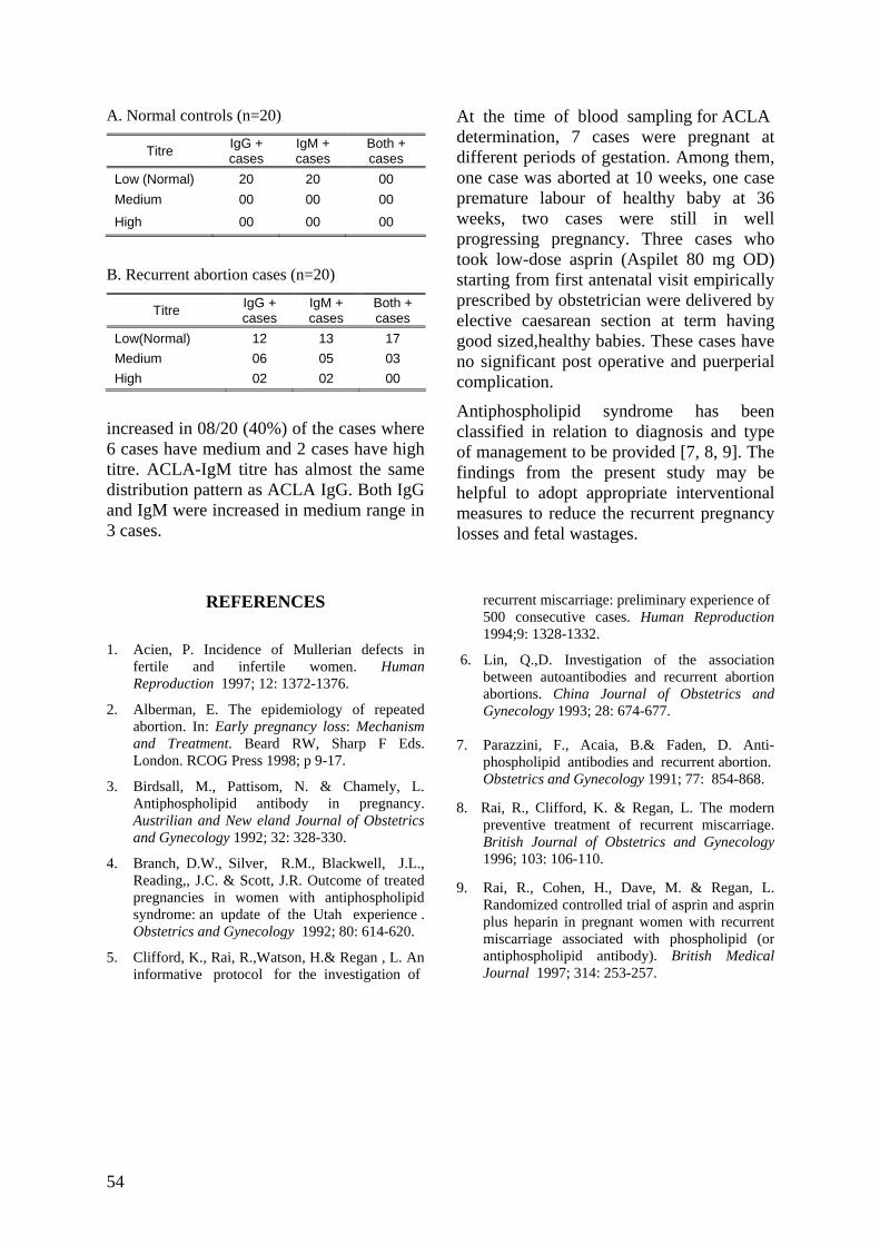
A. Normal controls (n=20)
Titre IgG + cases
IgM + cases
Both + cases
Low (Normal) 20 20 00 Medium 00 00 00
High 00 00 00
B. Recurrent abortion cases (n=20)
Titre IgG + cases
IgM + cases
Both + cases
Low(Normal) 12 13 17 Medium 06 05 03 High 02 02 00
increased in 08/20 (40%) of the cases where 6 cases have medium and 2 cases have high titre. ACLA-IgM titre has almost the same distribution pattern as ACLA IgG. Both IgG and IgM were increased in medium range in 3 cases.
At the time of blood sampling for ACLA determination, 7 cases were pregnant at different periods of gestation. Among them, one case was aborted at 10 weeks, one case premature labour of healthy baby at 36 weeks, two cases were still in well progressing pregnancy. Three cases who took low-dose asprin (Aspilet 80 mg OD) starting from first antenatal visit empirically prescribed by obstetrician were delivered by elective caesarean section at term having good sized,healthy babies. These cases have no significant post operative and puerperial complication.
Antiphospholipid syndrome has been classified in relation to diagnosis and type of management to be provided [7, 8, 9]. The findings from the present study may be helpful to adopt appropriate interventional measures to reduce the recurrent pregnancy losses and fetal wastages.
REFERENCES
1. Acien, P. Incidence of Mullerian defects in
fertile and infertile women. Human Reproduction 1997; 12: 1372-1376.
2. Alberman, E. The epidemiology of repeated abortion. In: Early pregnancy loss: Mechanism and Treatment. Beard RW, Sharp F Eds. London. RCOG Press 1998; p 9-17.
3. Birdsall, M., Pattisom, N. & Chamely, L. Antiphospholipid antibody in pregnancy. Austrilian and New eland Journal of Obstetrics and Gynecology 1992; 32: 328-330.
4. Branch, D.W., Silver, R.M., Blackwell, J.L., Reading,, J.C. & Scott, J.R. Outcome of treated pregnancies in women with antiphospholipid syndrome: an update of the Utah experience . Obstetrics and Gynecology 1992; 80: 614-620.
5. Clifford, K., Rai, R.,Watson, H.& Regan , L. An informative protocol for the investigation of
recurrent miscarriage: preliminary experience of 500 consecutive cases. Human Reproduction 1994;9: 1328-1332.
6. Lin, Q.,D. Investigation of the association between autoantibodies and recurrent abortion abortions. China Journal of Obstetrics and Gynecology 1993; 28: 674-677.
7. Parazzini, F., Acaia, B.& Faden, D. Anti-
phospholipid antibodies and recurrent abortion. Obstetrics and Gynecology 1991; 77: 854-868.
8. Rai, R., Clifford, K. & Regan, L. The modern preventive treatment of recurrent miscarriage. British Journal of Obstetrics and Gynecology 1996; 103: 106-110.
9. Rai, R., Cohen, H., Dave, M. & Regan, L. Randomized controlled trial of asprin and asprin plus heparin in pregnant women with recurrent miscarriage associated with phospholipid (or antiphospholipid antibody). British Medical Journal 1997; 314: 253-257.
54


![BURMA PRESS SUMMARYKyaw Zan [Rakine]; Yebaw Aye Thwin [Rangoon]; Yebaw Sai Tun Kyi [Shan]; Yebaw Saw Naing [Irrawaddy]; and Yebaw Kyaw Tint [Tatmadaw]. (WPD 7/23) Congress Opening](https://static.fdocuments.in/doc/165x107/60b7b1ddf647ea32cd592c7e/burma-press-summary-kyaw-zan-rakine-yebaw-aye-thwin-rangoon-yebaw-sai-tun.jpg)