DEPARTMENT OF LABORATORY MEDICINE DIVISION OF …
25
St.Michael’s DEPARTMENT OF LABORATORY MEDICINE DIVISION OF PATHOLOGY Document Name: Pathology Specimen Collection Manual Document #: 58134 Status: Current Uncontrolled When Printed Authority for Issue: Dr. Catherine Streutker Authorized Date: 10/29/2021 Version: 3.4.2 Effective Date: 10/29/2021 Any document appearing in paper form is uncontrolled and should be checked against the master electronic current version prior to use. Only original printed material with the “CONTROLLED” water mark may exist in designated locations. The controlled printed document should only be used when the electronic version is unavailable. Unauthorized photocopies or alterations of this document are uncontrolled documents. Page 1 of 25 PATHOLOGY SPECIMEN COLLECTION MANUAL
Transcript of DEPARTMENT OF LABORATORY MEDICINE DIVISION OF …

St.Michael’s
DEPARTMENT OF LABORATORY MEDICINE
DIVISION OF PATHOLOGY
Document Name: Pathology Specimen Collection
Manual
Document #: 58134
Status: Current
Uncontrolled When Printed
Authority for Issue: Dr. Catherine Streutker Authorized Date: 10/29/2021
Version: 3.4.2 Effective Date: 10/29/2021 Any document appearing in paper form is uncontrolled and should be checked against the master electronic current version prior to use. Only original printed material with the “CONTROLLED” water mark may exist in designated locations. The controlled printed document should only be used when the electronic version is unavailable. Unauthorized photocopies or alterations of this document are uncontrolled documents.
Page 1 of 25
PATHOLOGY SPECIMEN
COLLECTION MANUAL

St.Michael’s
DEPARTMENT OF LABORATORY MEDICINE
DIVISION OF PATHOLOGY
Document Name: Pathology Specimen Collection
Manual
Document #: 58134
Status: Current
Uncontrolled When Printed
Authority for Issue: Dr. Catherine Streutker Authorized Date: 10/29/2021
Version: 3.4.2 Effective Date: 10/29/2021 Any document appearing in paper form is uncontrolled and should be checked against the master electronic current version prior to use. Only original printed material with the “CONTROLLED” water mark may exist in designated locations. The controlled printed document should only be used when the electronic version is unavailable. Unauthorized photocopies or alterations of this document are uncontrolled documents.
Page 2 of 25
INDEX
General Information ……………………………………………………………….. 3
Specimen Collection ………………………………………………………………....4
Requisitions …………………………………………………………………………..5
Labels …………………………………………………………………………………7
Consent Forms for Autopsy …………………………………………………………7
Specimen Transport ………………………………………………………………….7
Verbal Requests for RUSH Specimens ……………………………………………..8
Specimen Rejection …………………………………………………………………..8
Table 1 Specimen Collection - Gynaecological Cytology ……………………….10
Table 2 Specimen Collection - Non-gynaecological Cytology …………………..13
Table 3 Specimen Collection - Surgical …………………………………………..19
Information Sheets (Appendices):
Out-Patient Sputum Collection for Cytology ……………………………….23
Hologic Cervical Cancer Screening (ThinPrep) Quick Reference Guide….24

St.Michael’s
DEPARTMENT OF LABORATORY MEDICINE
DIVISION OF PATHOLOGY
Document Name: Pathology Specimen Collection
Manual
Document #: 58134
Status: Current
Uncontrolled When Printed
Authority for Issue: Dr. Catherine Streutker Authorized Date: 10/29/2021
Version: 3.4.2 Effective Date: 10/29/2021 Any document appearing in paper form is uncontrolled and should be checked against the master electronic current version prior to use. Only original printed material with the “CONTROLLED” water mark may exist in designated locations. The controlled printed document should only be used when the electronic version is unavailable. Unauthorized photocopies or alterations of this document are uncontrolled documents.
Page 3 of 25
PATHOLOGY LABORATORY General Information
Hours of Operation:
Pathology Office - Weekdays 08:30 – 17:00
Pathology Office - Weekends and Statutory Holidays Closed
Pathologist-on-Call call locating
Surgical Receiving (CC2-070) - Weekdays 08:00 – 16:30
- Weekends and Statutory Holidays Closed
Cytology Receiving (CC2-050) – Weekdays 08:00 – 16:00
- Weekends and Statutory Holidays Closed
After working hours, Pathology specimens are left in a plastic bin located in the Microbiology Laboratory,
CC2-044 or the Core Lab, CC2-005.
Surgical specimens placed in 10% Neutral Buffered Formalin should be kept at room temperature
(refrigeration slows down the penetration rate of the fixative into the tissue).
Cytology specimens in CytoLyt or PreservCyt should be kept at room temperature..
Fresh Cytology specimens, without fixative, should be refrigerated and brought to the lab as soon as
possible.
Cytology CSF specimens must be fresh and received in the Cytology lab within 60 minutes of
collection.
Location: Cardinal Carter North Wing, 2nd floor

St.Michael’s
DEPARTMENT OF LABORATORY MEDICINE
DIVISION OF PATHOLOGY
Document Name: Pathology Specimen Collection
Manual
Document #: 58134
Status: Current
Uncontrolled When Printed
Authority for Issue: Dr. Catherine Streutker Authorized Date: 10/29/2021
Version: 3.4.2 Effective Date: 10/29/2021 Any document appearing in paper form is uncontrolled and should be checked against the master electronic current version prior to use. Only original printed material with the “CONTROLLED” water mark may exist in designated locations. The controlled printed document should only be used when the electronic version is unavailable. Unauthorized photocopies or alterations of this document are uncontrolled documents.
Page 4 of 25
SPECIMEN COLLECTION
Surgical and Cytology specimens are collected from a variety of sites for the diagnosis of malignant and
benign processes. The site from which the sample is collected dictates the method of collection. The
method of collection affects the morphology of the cellular samples. The importance of proper specimen
collection and submission is essential.
1. Verifying patient identification and labeling the specimens at the time of collection while in the
presence of the patient creates a positive link between patient and specimens. Loss of this link can
lead to medical errors, such as switched or mislabeled specimens. The identity of the patient shall
be confirmed prior to collection by the person collecting the sample. Patient identity shall be
verified using, at minimum, two identifiers and able patients shall be asked to state these
identifiers.
2. The container should be labeled during or immediately after the collection. There should be no
more than one unlabeled container at a time.
3. No more than one set of patient labels should be present in the specimen collection area. After the
collection is completed any unused labels should be discarded.
4. Collect the specimen using the proper technique, supplies and fixative.
5. Place the specimen in a specimen transport bag with a pouch in the bag. The specimen should be
placed in the sealed portion of the bag and the requisition should be placed in the pouch.
6. Transport the specimen to the laboratory promptly.
7. Dispose of material used in the collection (following international, national, regional, local and
organizational requirements).
8. Proceed to the next patient.

St.Michael’s
DEPARTMENT OF LABORATORY MEDICINE
DIVISION OF PATHOLOGY
Document Name: Pathology Specimen Collection
Manual
Document #: 58134
Status: Current
Uncontrolled When Printed
Authority for Issue: Dr. Catherine Streutker Authorized Date: 10/29/2021
Version: 3.4.2 Effective Date: 10/29/2021 Any document appearing in paper form is uncontrolled and should be checked against the master electronic current version prior to use. Only original printed material with the “CONTROLLED” water mark may exist in designated locations. The controlled printed document should only be used when the electronic version is unavailable. Unauthorized photocopies or alterations of this document are uncontrolled documents.
Page 5 of 25
All specimens should be collected in accordance with Body Substance Precautions. All specimens are
considered potentially infectious and should be handled as such.
Specimens should be collected and stored as indicated in the following tables:
Table 1. Specimen Collection - Gynaecological Cytology
Table 2. Specimen Collection - Non-gynaecological Cytology
Table 3. Specimen Collection - Surgical Specimens
Requisitions: Each specimen submitted to the Pathology Laboratories (Surgical and Cytology) must be
transported in a secondary container, which is usually the specimen transport bag and
accompanied by a completed Surgical or Cytology requisition which is inserted into the pouch
in the back of the transport bag.
Pathology and cytology specimens must be submitted with a written cytology or surgical
pathology requisition. VERBAL REQUESTS ARE NOT ACCEPTED.

St.Michael’s
DEPARTMENT OF LABORATORY MEDICINE
DIVISION OF PATHOLOGY
Document Name: Pathology Specimen Collection
Manual
Document #: 58134
Status: Current
Uncontrolled When Printed
Authority for Issue: Dr. Catherine Streutker Authorized Date: 10/29/2021
Version: 3.4.2 Effective Date: 10/29/2021 Any document appearing in paper form is uncontrolled and should be checked against the master electronic current version prior to use. Only original printed material with the “CONTROLLED” water mark may exist in designated locations. The controlled printed document should only be used when the electronic version is unavailable. Unauthorized photocopies or alterations of this document are uncontrolled documents.
Page 6 of 25
The requisition form should include the following information: (** Required information)
o ** Patient’s full name
o Patient’s Hospital J #
o ** Patient’s date of birth and sex
o Patient’s Encounter #
o ** Patient’s room number / location
o ** Requesting Physician’s name and physician most responsible for the patient
o ** Specimen source (s) (In surgical cases with multiple parts, the source of each must
be indicated on the requisition)
o ** Date and time of specimen collection
o ** Clinical diagnosis, relevant history
o If a specimen is known or suspected of having a biohazard agent (Tb, CJD, AIDS,
Hepatitis, etc) this should be documented on the requisition
o Please note that microbiological cultures cannot be performed on specimens submitted to
Pathology
Specimen Containers:
Specimen containers are provided by pathology and available in various sizes. The clinics are
provided with 10% formalin, pre-filled, 90 ml containers. For larger specimens, containers of
10% formalin are available in 500 ml., 1 Litre, 5 Litre, and 15 Litre containers. A ratio of 1:10,
tissue size to formalin volume, should be maintained to ensure adequate fixation. (10 to 20
times specimen size)
St.Michael’s
DEPARTMENT OF LABORATORY MEDICINE
DIVISION OF PATHOLOGY
Document Name: Pathology Specimen Collection
Manual
Document #: 58134
Status: Current
Uncontrolled When Printed
Authority for Issue: Dr. Catherine Streutker Authorized Date: 10/29/2021
Version: 3.4.2 Effective Date: 10/29/2021 Any document appearing in paper form is uncontrolled and should be checked against the master electronic current version prior to use. Only original printed material with the “CONTROLLED” water mark may exist in designated locations. The controlled printed document should only be used when the electronic version is unavailable. Unauthorized photocopies or alterations of this document are uncontrolled documents.
Page 7 of 25
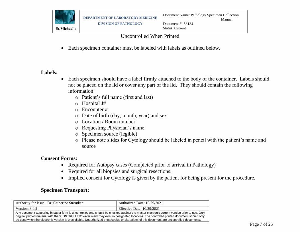
Each specimen container must be labeled with labels as outlined below.
Labels:
Each specimen should have a label firmly attached to the body of the container. Labels should
not be placed on the lid or cover any part of the lid. They should contain the following
information:
o Patient’s full name (first and last)
o Hospital J#
o Encounter #
o Date of birth (day, month, year) and sex
o Location / Room number
o Requesting Physician’s name
o Specimen source (legible)
o Please note slides for Cytology should be labeled in pencil with the patient’s name and
source
Consent Forms:
Required for Autopsy cases (Completed prior to arrival in Pathology)
Required for all biopsies and surgical resections.
Implied consent for Cytology is given by the patient for being present for the procedure.
Specimen Transport:

St.Michael’s
DEPARTMENT OF LABORATORY MEDICINE
DIVISION OF PATHOLOGY
Document Name: Pathology Specimen Collection
Manual
Document #: 58134
Status: Current
Uncontrolled When Printed
Authority for Issue: Dr. Catherine Streutker Authorized Date: 10/29/2021
Version: 3.4.2 Effective Date: 10/29/2021 Any document appearing in paper form is uncontrolled and should be checked against the master electronic current version prior to use. Only original printed material with the “CONTROLLED” water mark may exist in designated locations. The controlled printed document should only be used when the electronic version is unavailable. Unauthorized photocopies or alterations of this document are uncontrolled documents.
Page 8 of 25
All specimens must be transported in specimen transport bags. These are clear bags with a
pouch in the back and are labeled as biohazard. Only one specimen should be placed in each
bag.
Specimens are transported regularly throughout the day by the portering system to the
laboratory.
After hours, specimens are placed in Microbiology Laboratory (CC2-044) or Core Lab
(CC2-005).
Verbal Requests for Rush examination of Specimens in Pathology:
The Division of Pathology will be informed of RUSH (STAT) specimens in two manners:
o RUSH specimen is delivered to/or received at Surgical/ Cytology Receiving.
o The Division will receive a phone request by physician, resident, CLM etc to RUSH a
specimen that has been received by the Division or is in transit to the Division.
No technical staff have the authority to approve a RUSH specimen. Request for RUSH
specimens are brought to the attention of the Pathologist covering that day. In the absence of
the Pathologist, the case is brought to the immediate attention of the Director of Pathology.
Specimen Rejection:
Specimen rejection may occur for any of the following criteria:
o Specimen(s) and/or requisition(s) arrive unlabeled
o Specimen(s) arrive with a mismatched specimen/requisition
o Specimen are received grossly leaking
o Specimen requisition is contaminated by blood or leaking formalin
o Specimen arrives without a requisition
St.Michael’s
DEPARTMENT OF LABORATORY MEDICINE
DIVISION OF PATHOLOGY
Document Name: Pathology Specimen Collection
Manual
Document #: 58134
Status: Current
Uncontrolled When Printed
Authority for Issue: Dr. Catherine Streutker Authorized Date: 10/29/2021
Version: 3.4.2 Effective Date: 10/29/2021 Any document appearing in paper form is uncontrolled and should be checked against the master electronic current version prior to use. Only original printed material with the “CONTROLLED” water mark may exist in designated locations. The controlled printed document should only be used when the electronic version is unavailable. Unauthorized photocopies or alterations of this document are uncontrolled documents.
Page 9 of 25
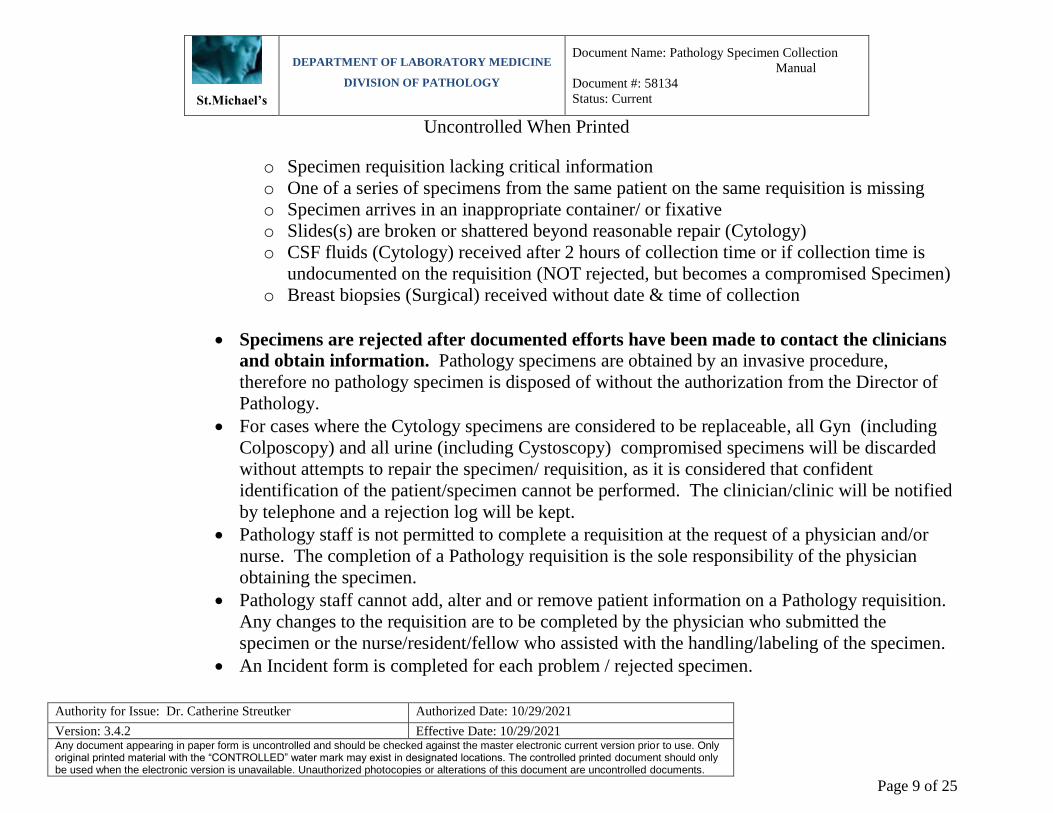
o Specimen requisition lacking critical information
o One of a series of specimens from the same patient on the same requisition is missing
o Specimen arrives in an inappropriate container/ or fixative
o Slides(s) are broken or shattered beyond reasonable repair (Cytology)
o CSF fluids (Cytology) received after 2 hours of collection time or if collection time is
undocumented on the requisition (NOT rejected, but becomes a compromised Specimen)
o Breast biopsies (Surgical) received without date & time of collection
Specimens are rejected after documented efforts have been made to contact the clinicians
and obtain information. Pathology specimens are obtained by an invasive procedure,
therefore no pathology specimen is disposed of without the authorization from the Director of
Pathology.
For cases where the Cytology specimens are considered to be replaceable, all Gyn (including
Colposcopy) and all urine (including Cystoscopy) compromised specimens will be discarded
without attempts to repair the specimen/ requisition, as it is considered that confident
identification of the patient/specimen cannot be performed. The clinician/clinic will be notified
by telephone and a rejection log will be kept.
Pathology staff is not permitted to complete a requisition at the request of a physician and/or
nurse. The completion of a Pathology requisition is the sole responsibility of the physician
obtaining the specimen.
Pathology staff cannot add, alter and or remove patient information on a Pathology requisition.
Any changes to the requisition are to be completed by the physician who submitted the
specimen or the nurse/resident/fellow who assisted with the handling/labeling of the specimen.
An Incident form is completed for each problem / rejected specimen.

St.Michael’s
DEPARTMENT OF LABORATORY MEDICINE
DIVISION OF PATHOLOGY
Document Name: Pathology Specimen Collection
Manual
Document #: 58134
Status: Current
Uncontrolled When Printed
Authority for Issue: Dr. Catherine Streutker Authorized Date: 10/29/2021
Version: 3.4.2 Effective Date: 10/29/2021 Any document appearing in paper form is uncontrolled and should be checked against the master electronic current version prior to use. Only original printed material with the “CONTROLLED” water mark may exist in designated locations. The controlled printed document should only be used when the electronic version is unavailable. Unauthorized photocopies or alterations of this document are uncontrolled documents.
Page 10 of 25
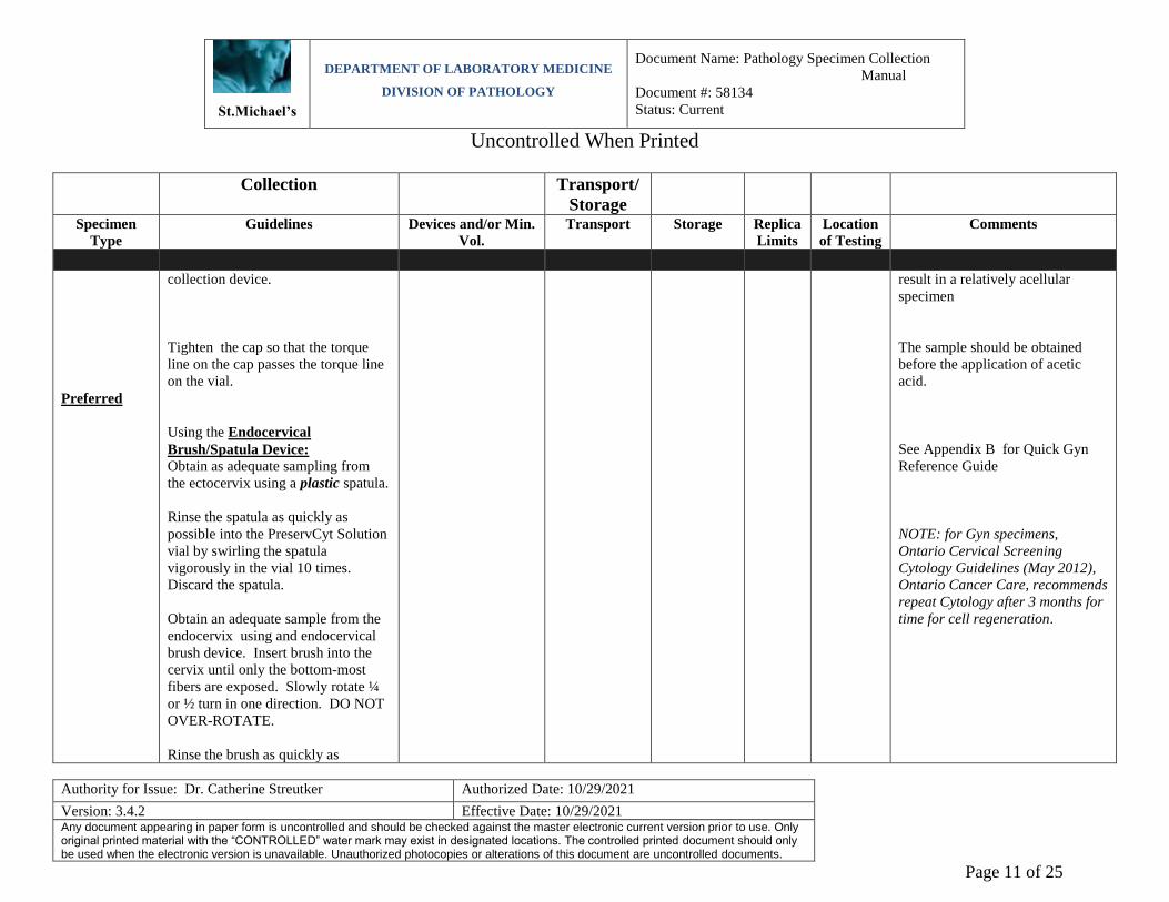
Table 1: Specimen Collection - Gynaecological Cytology
Collection Transport/
Storage
Specimen
Type
Guidelines Devices and/or Min.
Vol.
Transport Storage Replica
Limits
Location
of Testing
Comments
Pap Smear
(liquid)
Preferred
Collected by physician or authorized
personnel
Using the Broom-Like Device:
Obtain an adequate sampling from
the cervix using a broom-like device.
Insert the central bristles of the
broom into the endocervical canal
deep enough to allow the shorter
bristles to fully contact the
ectocervix. Push gently, and rotate
the broom in a clockwise direction 5
times.
Rinse the broom as quickly as
possible into the PreservCyt Solution
by pushing the broom into the bottom
of the vial 10 times, forcing the
bristles apart. As a final step, swirl
the broom vigorously to further
release material. Discard the
Gynae samples
collected using a
broom-type or
endocervical
brush/plastic spatula
combination
collection devices.
Specimen rinsed in
prefilled PreservCyt
Solution vials.
Room
temperature
N/A
SMH
The patient should be tested 2
weeks after the first day of her
menstrual period, and not when she
is menstruating.
The patient should not use vaginal
medication, vaginal contraceptives,
or douches during the 48 hours
before the pap exam.
Lubricant jellies should not be
used to lubricate the speculum.
Remove excess mucus or other
discharge present before taking the
sample. This should be gently
removed with ring forceps holding
a folded gauze pad.
The cervix should not be cleaned
by washing with saline or it may
St.Michael’s
DEPARTMENT OF LABORATORY MEDICINE
DIVISION OF PATHOLOGY
Document Name: Pathology Specimen Collection
Manual
Document #: 58134
Status: Current
Uncontrolled When Printed
Authority for Issue: Dr. Catherine Streutker Authorized Date: 10/29/2021
Version: 3.4.2 Effective Date: 10/29/2021 Any document appearing in paper form is uncontrolled and should be checked against the master electronic current version prior to use. Only original printed material with the “CONTROLLED” water mark may exist in designated locations. The controlled printed document should only be used when the electronic version is unavailable. Unauthorized photocopies or alterations of this document are uncontrolled documents.
Page 11 of 25
Collection Transport/
Storage
Specimen
Type
Guidelines Devices and/or Min.
Vol.
Transport Storage Replica
Limits
Location
of Testing
Comments
Preferred
collection device.
Tighten the cap so that the torque
line on the cap passes the torque line
on the vial.
Using the Endocervical
Brush/Spatula Device:
Obtain as adequate sampling from
the ectocervix using a plastic spatula.
Rinse the spatula as quickly as
possible into the PreservCyt Solution
vial by swirling the spatula
vigorously in the vial 10 times.
Discard the spatula.
Obtain an adequate sample from the
endocervix using and endocervical
brush device. Insert brush into the
cervix until only the bottom-most
fibers are exposed. Slowly rotate ¼
or ½ turn in one direction. DO NOT
OVER-ROTATE.
Rinse the brush as quickly as
result in a relatively acellular
specimen
The sample should be obtained
before the application of acetic
acid.
See Appendix B for Quick Gyn
Reference Guide
NOTE: for Gyn specimens,
Ontario Cervical Screening
Cytology Guidelines (May 2012),
Ontario Cancer Care, recommends
repeat Cytology after 3 months for
time for cell regeneration.

St.Michael’s
DEPARTMENT OF LABORATORY MEDICINE
DIVISION OF PATHOLOGY
Document Name: Pathology Specimen Collection
Manual
Document #: 58134
Status: Current
Uncontrolled When Printed
Authority for Issue: Dr. Catherine Streutker Authorized Date: 10/29/2021
Version: 3.4.2 Effective Date: 10/29/2021 Any document appearing in paper form is uncontrolled and should be checked against the master electronic current version prior to use. Only original printed material with the “CONTROLLED” water mark may exist in designated locations. The controlled printed document should only be used when the electronic version is unavailable. Unauthorized photocopies or alterations of this document are uncontrolled documents.
Page 12 of 25
Collection Transport/
Storage
Specimen
Type
Guidelines Devices and/or Min.
Vol.
Transport Storage Replica
Limits
Location
of Testing
Comments
possible in the PreservCyt Solution
by rotationg the device in the
solution 10 times while pushing
against the PreservCyt vial wall.
Swirl vigorously to further release
material. Discard the brush.
Tighten the cap so that the torque
line on the cap passes the torque line
on the vial.
Record the patient’s name and J
number on the vial. Record the
patient information and medical
history on the cytology requisition
form.
Pap smear
(conventional)
if liquid
PreservCyt is
not available.
Collected by physician or authorized
personnel
Specimen spread
evenly on slide
labeled, in pencil,
with patient’s name
and fixed
immediately (within
seconds) with
Cytology fixative.
Slide should not be
allowed to air dry.
Submitted on
slides and
fixed
immediately.
Slide placed in
cardboard
slide holder for
transportation
to the
Cytology lab.
Room
temperature
N/A
SMH
Spray fixatives should be held 6 –
10 inches (15 – 25 cm) from the
glass slide when applied.
Immediate fixation of the cellular
sample is necessary to prevent air-
drying which obscures cellular
detail and compromises specimen
evaluation.
St.Michael’s
DEPARTMENT OF LABORATORY MEDICINE
DIVISION OF PATHOLOGY
Document Name: Pathology Specimen Collection
Manual
Document #: 58134
Status: Current
Uncontrolled When Printed
Authority for Issue: Dr. Catherine Streutker Authorized Date: 10/29/2021
Version: 3.4.2 Effective Date: 10/29/2021 Any document appearing in paper form is uncontrolled and should be checked against the master electronic current version prior to use. Only original printed material with the “CONTROLLED” water mark may exist in designated locations. The controlled printed document should only be used when the electronic version is unavailable. Unauthorized photocopies or alterations of this document are uncontrolled documents.
Page 13 of 25
Table 2: Specimen Collection - Non-gynaecological Cytology
Collection Transport/Storage
Specimen
Type
Collection
Method
Guidelines Devices and/or
Min. Vol.
Transport Storage Replica
Limits
Location
of Testing
Comments
Body Fluid
Drainage or
aspiration
Collected by physician or
authorized personnel.
Body fluids usually collected with aseptic technique by needle
puncture and aspiration of the
body cavity fluid
Clean, dry container
Submitted in fresh state to the
Cytology lab
Room temperature, if
delay unavoidable
refrigerate up to 72 hours.
N/A
SMH
Adding 3 – 5 IU heparin/ml to a
container prior to obtaining a bloody
sample will usually inhibit clotting and not adversely affect morphology.
For small fluid accumulation – submit entire specimen for evaluation.
For large specimens, total volume 100 ml of well-mixed fluid should be sent to
the Cytology lab for examination.
Refrigeration reduces the rate of
degeneration.
CSF
Fluid
Collected by physician or
authorized personnel.
Small clean, dry tube
Submitted in fresh state to the
Cytology lab within 1 hour of
collection.
Room temperature or
refrigerated
N/A
SMH
CSF should be collected fresh and
delivered to the Cytology lab as quickly
as possible to prevent cellular deterioration.
For optimal CSF sample, the time
between collection and preparation
should be ˂ 1 hour . ˃ 1 hour, the CSF sample may be compromised.
CSF collection after hours or on the weekend, should be deferred to the next

St.Michael’s
DEPARTMENT OF LABORATORY MEDICINE
DIVISION OF PATHOLOGY
Document Name: Pathology Specimen Collection
Manual
Document #: 58134
Status: Current
Uncontrolled When Printed
Authority for Issue: Dr. Catherine Streutker Authorized Date: 10/29/2021
Version: 3.4.2 Effective Date: 10/29/2021 Any document appearing in paper form is uncontrolled and should be checked against the master electronic current version prior to use. Only original printed material with the “CONTROLLED” water mark may exist in designated locations. The controlled printed document should only be used when the electronic version is unavailable. Unauthorized photocopies or alterations of this document are uncontrolled documents.
Page 14 of 25
Collection Transport/Storage Specimen
Type
Collection
Method
Guidelines Devices and/or
Min. Vol.
Transport Storage Replica
Limits
Location
of Testing
Comments
business day, if possible.
If the CSF sample cannot be prepared
immediately, it should be refrigerated at 4˚C. If a delay of more than 48 hours is
anticipated, the sample can be preserved by adding an equal volume of
50% ethanol. A CSF sample for
cytology exam should never be frozen.
CSF (for Flow
Cytometry)
Collected by physician or authorized personnel after initial
cytologic evaluation, upon the
recommendation of the pathologist.
Submitted directly in fresh
state to Special Heamatology.
Please contact Special Haematology at
ext. 2141, for specimen requirements.
Urinary Tract
Voided Urine
Pass directly into clean, dry
container
Clean, dry container
(50 ml orange top)
Submitted in fresh state to the
Cytology lab
Room temperature or
refrigerate if delay
unavoidable
N/A
SMH
20 – 50 ml preferred amount.
The first void of the day is unsuitable
for cytological exam because urothelial cells that have been sitting in urine for
long periods will show degenerative
changes that may compromise assessment.
Bladder /
ureteral or renal pelvis
washings
Balance salt solution
Total volume
Submitted in fresh state to the
Cytology lab
Room temperature or
refrigerate if delay unavoidable
N/A
SMH
Any instrumentation or unusual
anatomic alteration should be noted on the requisition (e.g. Presence of on ileal
conduit).
GI Tract
(including:
esophagus, intestine,
pancreas, bile
duct)
Brushings
Collected by physician or authorized personnel.
After the brushing is performed, the brush is rolled across the slide
in an area approximately 2.5
centimeters in diameter (size of a quarter) to produce a thin evenly
layered smear. The slide is spray
20 ml
If brush tip submitted, it must be
covered with saline.
Alternatively, the tip
may be swirled in the
Submitted in saline or CytoLyt
fixative to the Cytology lab.
Cytology fixative has an expiry
date. This date should be
checked to ensure optimal effectiveness.
Room temperature or
refrigerate if delay
unavoidable
N/A
SMH
Spray fixatives should be held 6 – 10
inches (15 – 25 cm) from the glass slide when applied.
Immediate fixation of the cellular sample is necessary to prevent air-
drying which obscures cellular detail

St.Michael’s
DEPARTMENT OF LABORATORY MEDICINE
DIVISION OF PATHOLOGY
Document Name: Pathology Specimen Collection
Manual
Document #: 58134
Status: Current
Uncontrolled When Printed
Authority for Issue: Dr. Catherine Streutker Authorized Date: 10/29/2021
Version: 3.4.2 Effective Date: 10/29/2021 Any document appearing in paper form is uncontrolled and should be checked against the master electronic current version prior to use. Only original printed material with the “CONTROLLED” water mark may exist in designated locations. The controlled printed document should only be used when the electronic version is unavailable. Unauthorized photocopies or alterations of this document are uncontrolled documents.
Page 15 of 25
Collection Transport/Storage Specimen
Type
Collection
Method
Guidelines Devices and/or
Min. Vol.
Transport Storage Replica
Limits
Location
of Testing
Comments
fixed immediately with Cytology fixative.
media without submitting the brush.
and compromises specimen evaluation.
Any added CytoLyt fixative should be
documented on the specimen container/requisition.
GI Tract (including:
esophagus,
intestine, pancreas, bile
duct) cont.
Washings
Collected by physician or authorized personnel.
Small aliquots of balanced saline
solution are washed over a directly visualized area and
removed immediately with
suction.
If delay is expected, washing may
be placed into CytoLyt (transport
medium). Note: CytoLyt is not
used in place of saline for the
wash.
Clean container with saline or if delay
expected, place into
CytoLyt
Submitted in saline to the Cytology lab
Room temperature
N/A
SMH
Any added CytoLyt fixative should be documented on the specimen
container/requisition.
Respiratory
Sputum
Place directly into clean, dry
container
Clean, dry container
(50 ml orange top)
Submitted in fresh state to the
Cytology lab
Room temperature
N/A
SMH
Early morning, deep cough specimens
are preferred.
Collecting multiple (3) sputum samples
over several days optimizes sensitivity.
See Appendix A: for out-patient sputum
collection.
Bronchial
brushings
Collected by physician or
authorized personnel.
After the brushing is performed,
the brush is rolled across the slide
in an area approximately 2.5 centimeters in diameter (size of a
20 ml
If brush tip submitted, it must be
covered.
Alternatively, the tip
Submitted in saline or CytoLyt
fixative to the Cytology lab.
Cytology fixative has an expiry
date. This date should be
checked to ensure optimal effectiveness.
Room temperature
N/A
SMH
Spray fixatives should be held 6 – 10
inches (15 – 25 cm) from the glass slide
when applied.
Immediate fixation of the cellular
sample is necessary to prevent air-drying which obscures cellular detail

St.Michael’s
DEPARTMENT OF LABORATORY MEDICINE
DIVISION OF PATHOLOGY
Document Name: Pathology Specimen Collection
Manual
Document #: 58134
Status: Current
Uncontrolled When Printed
Authority for Issue: Dr. Catherine Streutker Authorized Date: 10/29/2021
Version: 3.4.2 Effective Date: 10/29/2021 Any document appearing in paper form is uncontrolled and should be checked against the master electronic current version prior to use. Only original printed material with the “CONTROLLED” water mark may exist in designated locations. The controlled printed document should only be used when the electronic version is unavailable. Unauthorized photocopies or alterations of this document are uncontrolled documents.
Page 16 of 25
Collection Transport/Storage Specimen
Type
Collection
Method
Guidelines Devices and/or
Min. Vol.
Transport Storage Replica
Limits
Location
of Testing
Comments
quarter) to produce a thin evenly layered smear. The slide is spray
fixed immediately.
may be swirled in the media without
submitting the brush.
and compromises specimen evaluation.
Any added CytoLyt fixative should be
documented on the specimen container/requisition.
Bronchial washing
Collected by physician or authorized personnel.
Small aliquots of balanced saline
solution are washed over a directly visualized area and
removed immediately with
suction.
If delay is expected, washing may
be placed into CytoLyt (transport
medium). Note: CytoLyt is not
used in place of saline for the
wash.
Clean container with saline or if delay
expected, place into
CytoLyt fixative
Room temperature
N/A
SMH
The brush may be submitted in solution or discarded after vigorously removing
the adherent cellular material into the
medium.
Any added fixative should be noted on
the requisition.

St.Michael’s
DEPARTMENT OF LABORATORY MEDICINE
DIVISION OF PATHOLOGY
Document Name: Pathology Specimen Collection
Manual
Document #: 58134
Status: Current
Uncontrolled When Printed
Authority for Issue: Dr. Catherine Streutker Authorized Date: 10/29/2021
Version: 3.4.2 Effective Date: 10/29/2021 Any document appearing in paper form is uncontrolled and should be checked against the master electronic current version prior to use. Only original printed material with the “CONTROLLED” water mark may exist in designated locations. The controlled printed document should only be used when the electronic version is unavailable. Unauthorized photocopies or alterations of this document are uncontrolled documents.
Page 17 of 25
Collection Transport/Storage Specimen
Type
Collection
Method
Guidelines Devices and/or
Min. Vol.
Transport Storage Replica
Limits
Location
of Testing
Comments
Fine Needle
Aspiration
Biopsy (FNAB)
-breast -thyroid
-lung
-etc
FNAB may be performed on any
body site that can be reached with
a fine needle (defined as 22 or higher gauge).
The procedure as well as minor
complications of bruising and
bleeding should be explained to the patient.
Written consent should be obtained where the procedure is
performed.
The skin should be cleansed with an alcohol swab prior to puncture
for superficial FNAB. For
percutaneous biopsy of deep lesions, sterile or aseptic
technique is used.
Sedation is rarely used, possibly for a deep-seated FNAB when the
patient is uncomfortable or
anxious.
For solid lesions multiple passes
with separate needles are performed. Staying within the
lesion, the needle is moved in a
cutting motion needed to obtain
an adequate sample must be
adjusted for the body site and
characteristics of the lesion. These biopsies may be performed
with suction or by the “non-
suction” techniques. Once the cellular material is seen in the
needle hub, suction is released
and the needle is withdrawn.
Cellular material
smeared on slides
labeled, in pencil, with patient’s name
and fixed immediately or
needle is rinsed in
CytoLyt (transport medium) to remove
residual cellular
material
Room temperature
N/A
SMH
For cystic lesions, remove as much
fluid as possible. The cyst fluid can be
handled as a liquid specimen. If there is a residual mass, the procedure for
solid lesion should be followed.
Local pressure is usually adequate to
control post procedural bleeding for superficial sites. Patients who have
undergone deep FNAB should be
followed for complications as clinically appropriate. All patients who undergo
FNAB should be
observed during and following the
procedure until they are stable.

St.Michael’s
DEPARTMENT OF LABORATORY MEDICINE
DIVISION OF PATHOLOGY
Document Name: Pathology Specimen Collection
Manual
Document #: 58134
Status: Current
Uncontrolled When Printed
Authority for Issue: Dr. Catherine Streutker Authorized Date: 10/29/2021
Version: 3.4.2 Effective Date: 10/29/2021 Any document appearing in paper form is uncontrolled and should be checked against the master electronic current version prior to use. Only original printed material with the “CONTROLLED” water mark may exist in designated locations. The controlled printed document should only be used when the electronic version is unavailable. Unauthorized photocopies or alterations of this document are uncontrolled documents.
Page 18 of 25
Collection Transport/Storage Specimen
Type
Collection
Method
Guidelines Devices and/or
Min. Vol.
Transport Storage Replica
Limits
Location
of Testing
Comments
Tzanck Smear
Collected by physician or
authorized personnel
Spread on slide
labeled, in pencil,
with patient’s name and fixed
immediately or specimen placed in
CytoLyt
Direct scrape procedure is
preferred and slides are
submitted to Cytology smeared and fixed.
Room temperature
N/A
SMH
The suspect lesion is premoistened with
saline. If possible, a fresh vesicle
should be chosen that has not ruptured and crusted.
With a disposable needle a fresh vesicle
is carefully opened or the crust from a
ruptured lesion is removed.
Using the edge of a metal spatula,
scalpel blade, or glass slides, the margin of the lesion is scraped. The
edges of the lesion will have the best
yield of cells.
The obtained material is carefully
spread on an alcohol moistened microscopic slide and fixed. It is
imperative that the material be fixed
immediately after smearing.
Alternatively, the scraping tool may be
rinsed in CytoLyt and processed on the ThinPrep.
Synovial Fluid
(for crystals)
Collected by physician or
authorized personnel
Small, clean, dry
container (red top)
Submit in fresh state to the
Cytology lab
Room temperature
N/A
SMH
Fat Pad Aspirate
Collected by physician or authorized personnel
Small, clean, dry container
Submit in fresh state to the Cytology lab
Room temperature
N/A
MH
Do not fix specimen with alcohol.

St.Michael’s
DEPARTMENT OF LABORATORY MEDICINE
DIVISION OF PATHOLOGY
Document Name: Pathology Specimen Collection
Manual
Document #: 58134
Status: Current
Uncontrolled When Printed
Authority for Issue: Dr. Catherine Streutker Authorized Date: 10/29/2021
Version: 3.4.2 Effective Date: 10/29/2021 Any document appearing in paper form is uncontrolled and should be checked against the master electronic current version prior to use. Only original printed material with the “CONTROLLED” water mark may exist in designated locations. The controlled printed document should only be used when the electronic version is unavailable. Unauthorized photocopies or alterations of this document are uncontrolled documents.
Page 19 of 25
Table 3 Specimen Collection – Surgical
Collection Transport/Storage Specimen
Type
Guidelines Devices and/or
Min. Vol.
Transport Storage Replica
Limits
Location
of Testing
Comments
Fresh Tissue Biopsy for frozen
section
Collected by physician / surgeon
Submitted in fresh state to the Pathology OR lab (located on
CC 5) for Quick Sections
N/A
SMH
Lymph node or
core biopsy for Suspect
Lymphoma
Collected by physician/
surgeon
Call pathologist (ext 5851) for
lymphoma protocol. Specimen submitted as fresh
tissue or in normal saline.
N/A SMH
Small Tissue
Biopsy
Collected by physician /
surgeon
Appropriate size
container, to
accommodate specimen : 10% NBF
fixative (1:20) ratio
Submitted in fixed state to
Pathology
Room
temperature
N/A SMH For optimum results, ratio of specimen to
fixative should be 1:20.
Exact time of specimen collection of breast biopsies is critical on surgical pathology
requisitions.
NBF: (Neutral Buffered Formalin).
Large Tissue
Specimen
Collected by physician/
surgeon
Appropriate size
container to accommodate
specimen: 10% NBF
fixative (1:10) ratio
Surgical samples are
deposited within the 5th floor OR Pathology collection
window Lab, Room 601, in
the vented formalin cabinet
designated for specimens.
Room
temperature
N/A SMH Pathology must be made aware of any late
samples after 5 pm that do not fit into the standard formalin pre-filled containers
available. On-call pathologist to be
paged.
Large Surgical
Breast Samples
Collected by physician/
surgeon
Appropriate size
container to
accommodate
specimen : 10% NBF fixative
(1:10) ratio
Surgical samples are Placed
within the formalin cabinets
in OR Pathology Lab,
Donnelly 5-601.
Breast Tissue only - call the
Pathology office at x5915 for
pickup
Room
temperature
N/A SMH Pathology must be made aware of any late
samples after 5 pm. On-call pathologist to
be paged.

St.Michael’s
DEPARTMENT OF LABORATORY MEDICINE
DIVISION OF PATHOLOGY
Document Name: Pathology Specimen Collection
Manual
Document #: 58134
Status: Current
Uncontrolled When Printed
Authority for Issue: Dr. Catherine Streutker Authorized Date: 10/29/2021
Version: 3.4.2 Effective Date: 10/29/2021 Any document appearing in paper form is uncontrolled and should be checked against the master electronic current version prior to use. Only original printed material with the “CONTROLLED” water mark may exist in designated locations. The controlled printed document should only be used when the electronic version is unavailable. Unauthorized photocopies or alterations of this document are uncontrolled documents.
Page 20 of 25
Collection Transport/Storage Specimen
Type
Guidelines Devices and/or
Min. Vol.
Transport Storage Replica
Limits
Location
of Testing
Comments
Bone Marrow
Biopsy
Collected by physician Specimen
immediately placed
in B plus fixative (10 ml container)
Submitted in fixed state to
Pathology
Room
temperature
N/A SMH Exact time of specimen collection is critical
on surgical pathology requisition
Tissue Fragments for Uric Acid
Crystals
Collected by physician Specimen placed into absolute alcohol.
Submitted in fixed state to Pathology
Room temperature
N/A SMH
Fresh Fetus and
placenta (for genetic
testing)
Collected by physician/
surgeon
Submitted in fresh state to
Pathology collection window CC 2-072
Refrigerator
4°C
N/A
SMH
Must indicate on the Pathology requisition
that specimen is for genetic testing. The
Pathology lab sends to Mount Sinai Hospital. A Mount Sinai Hospital
Cytogenetics Requisition - Perinatal
must accompany the specimen.
Placenta/fetus
Collected by physician/
Surgeon
Submitted fresh
During work hours to
pathology collecting
window CC 2-070
After work hours either
kept in 15th floor fridge or transported to fridge
in core lab 2CC
Refrigerated
N/A
SMH
Tissue for (EM)
Electron
Microscopy Tumors- brain,
pituitary
Collected by physician
Tissue placed into
2.5% gluteraldehyde
Specimen is picked up by EM
technologist from OR
Pathology lab
Refrigerated
N/A
SMH
Tissue for
EM/LM/IF
(kidney) EM/LM/EH
Collected by physician
Submitted in fresh state.
Technologist will assess
specimen adequacy and divide specimen appropriately
Room
temperature
N/A
SMH
Must make arrangement with technologist
(ext 5915) prior to kidney specimen
collection.
St.Michael’s
DEPARTMENT OF LABORATORY MEDICINE
DIVISION OF PATHOLOGY
Document Name: Pathology Specimen Collection
Manual
Document #: 58134
Status: Current
Uncontrolled When Printed
Authority for Issue: Dr. Catherine Streutker Authorized Date: 10/29/2021
Version: 3.4.2 Effective Date: 10/29/2021 Any document appearing in paper form is uncontrolled and should be checked against the master electronic current version prior to use. Only original printed material with the “CONTROLLED” water mark may exist in designated locations. The controlled printed document should only be used when the electronic version is unavailable. Unauthorized photocopies or alterations of this document are uncontrolled documents.
Page 21 of 25
Collection Transport/Storage Specimen
Type
Guidelines Devices and/or
Min. Vol.
Transport Storage Replica
Limits
Location
of Testing
Comments
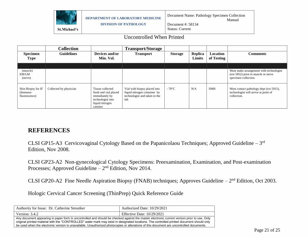
(muscle) EM/LM
(nerve)
Must make arrangement with technologist (ext 5852) prior to muscle or nerve
specimen collection.
Skin Biopsy for IF
(Immuno- fluorescence)
Collected by physician
Tissue collected
fresh and vial placed immediately by
technologist into
liquid nitrogen canister
Vial with biopsy placed into
liquid nitrogen container by technologist and taken to the
lab
- 70°C
N/A
SMH
Must contact pathology dept (ext 5915),
technologist will arrive at point of collection.
REFERENCES
CLSI GP15-A3 Cervicovaginal Cytology Based on the Papanicolaou Techniques; Approved Guideline – 3rd
Edition, Nov 2008.
CLSI GP23-A2 Non-gynecological Cytology Specimens: Preexamination, Examination, and Post-examination
Processes; Approved Guideline – 2nd Edition, Nov 2014.
CLSI GP20-A2 Fine Needle Aspiration Biopsy (FNAB) techniques; Approves Guideline – 2nd Edition, Oct 2003.
Hologic Cervical Cancer Screening (ThinPrep) Quick Reference Guide

St.Michael’s
DEPARTMENT OF LABORATORY MEDICINE
DIVISION OF PATHOLOGY
Document Name: Pathology Specimen Collection
Manual
Document #: 58134
Status: Current
Uncontrolled When Printed
Authority for Issue: Dr. Catherine Streutker Authorized Date: 10/29/2021
Version: 3.4.2 Effective Date: 10/29/2021 Any document appearing in paper form is uncontrolled and should be checked against the master electronic current version prior to use. Only original printed material with the “CONTROLLED” water mark may exist in designated locations. The controlled printed document should only be used when the electronic version is unavailable. Unauthorized photocopies or alterations of this document are uncontrolled documents.
Page 22 of 25
APPENDICES
Appendix A: Out-Patient Sputum Collection for Cytology
Appendix B: Hologic Cervical Cancer Screening (ThinPrep) Quick Reference Guide

St.Michael’s
DEPARTMENT OF LABORATORY MEDICINE
DIVISION OF PATHOLOGY
Document Name: Pathology Specimen Collection
Manual
Document #: 58134
Status: Current
Uncontrolled When Printed
Authority for Issue: Dr. Catherine Streutker Authorized Date: 10/29/2021
Version: 3.4.2 Effective Date: 10/29/2021 Any document appearing in paper form is uncontrolled and should be checked against the master electronic current version prior to use. Only original printed material with the “CONTROLLED” water mark may exist in designated locations. The controlled printed document should only be used when the electronic version is unavailable. Unauthorized photocopies or alterations of this document are uncontrolled documents.
Page 23 of 25
Appendix A
OUT-PATIENT SPUTUM COLLECTION FOR
CYTOLOGY
A series of three (3) consecutive early morning deep cough specimens, before breakfast, is recommended for cytologic evaluation. The patient is to cough up sputum from deep within the lungs and collect the specimen in the specimen container. An early morning deep cough specimen is optimal. After the third sample is collected, all three containers should be brought in together and delivered to the Cytology Laboratory (2CC Wing North, Room 2050).
30 Bond Street Toronto, Ontario
M5B 1W8 416-360-4000
www.stmichaelshospital.com

St.Michael’s
DEPARTMENT OF LABORATORY MEDICINE
DIVISION OF PATHOLOGY
Document Name: Pathology Specimen Collection
Manual
Document #: 58134
Status: Current
Uncontrolled When Printed
Authority for Issue: Dr. Catherine Streutker Authorized Date: 10/29/2021
Version: 3.4.2 Effective Date: 10/29/2021 Any document appearing in paper form is uncontrolled and should be checked against the master electronic current version prior to use. Only original printed material with the “CONTROLLED” water mark may exist in designated locations. The controlled printed document should only be used when the electronic version is unavailable. Unauthorized photocopies or alterations of this document are uncontrolled documents.
Page 24 of 25
Appendix B

St.Michael’s
DEPARTMENT OF LABORATORY MEDICINE
DIVISION OF PATHOLOGY
Document Name: Pathology Specimen Collection
Manual
Document #: 58134
Status: Current
Uncontrolled When Printed
Authority for Issue: Dr. Catherine Streutker Authorized Date: 10/29/2021
Version: 3.4.2 Effective Date: 10/29/2021 Any document appearing in paper form is uncontrolled and should be checked against the master electronic current version prior to use. Only original printed material with the “CONTROLLED” water mark may exist in designated locations. The controlled printed document should only be used when the electronic version is unavailable. Unauthorized photocopies or alterations of this document are uncontrolled documents.
Page 25 of 25