Department of Critical Care Medicine, Respiratory ...
15
Point-of-care ultrasonography for the hospitalist CLEVELAND CLINIC JOURNAL OF MEDICINE VOLUME 88 • NUMBER 6 JUNE 2021 345 H ospitalists are increasingly using point-of-care ultrasonography (POCUS), and have access to ultrasound machines that are more portable, more available, and less expensive. The numerous uses of POCUS for procedural guidance, diagnosis, and monitor- ing can add considerable value to patient care. All hospitalists should have an understand- ing of POCUS nomenclature, applications, and findings. This review highlights various uses of POCUS in hospitalized patients. ■ DIRECT CLINICIAN INVOLVEMENT Ultrasonography is low-cost, radiation-free, and noninvasive, allowing it to be repeated multiple times with little risk to patients. What sets it apart from traditional diagnostic ultrasonography is that it is wholly performed by a bedside clini- cian directly involved in patient care, without requiring a sonographer and radiologist for image acquisition and interpretation (Table 1). A hospi- talist can quickly perform a physical examination combined with goal-directed ultrasonography of various organs based on presenting signs and symptoms. Serial scans can be performed to assess progression or response to therapy. POCUS enhances patient experience and patient-clinician rapport by increasing inter- actions between the clinician and patient. 1 POCUS has become notably important in the COVID-19 pandemic, allowing protocolized ultrasonographic assessment of multiple or- gans by a bedside physician, thereby minimiz- ing exposure and the need for formal studies. 2 Recognizing the importance of POCUS, numerous medical schools have integrated training in ultrasonography in their curricula. REVIEW doi:10.3949/ccjm.88a.20141 ABSTRACT Point-of-care ultrasonography (POCUS) has emerged as a vital tool in medicine. Initially used for procedural guid- ance, POCUS is now used for diagnostics and monitoring of the lung, heart, abdomen, and deep vein thrombosis. This wide applicability makes it an essential tool for hos- pitalists in daily clinical practice. This article provides an overview of the clinical integration of POCUS and basic image interpretation. KEY POINTS Lung POCUS can help in evaluating pneumothorax, alveolar-interstitial syndrome, lung consolidation, and pleural effusions as the cause for respiratory distress. Focused cardiac ultrasonography can help in evaluating left and right ventricular function, right atrial pressure, pericardial effusion, and tamponade. Abdominal ultrasonography can aid evaluation of ascites, hemoperitoneum, hydronephrosis, acute pyelonephritis, and gallstones, and can confirm Foley catheter placement. Point-of-care compression ultrasonography can rapidly detect deep vein thrombosis with high accuracy. POCUS can guide numerous procedures, including central venous catheter insertion, peripheral intravenous catheter insertion, abdominal paracentesis, and thoracentesis. Guramrinder Singh Thind, MD Department of Critical Care Medicine, Respiratory Institute, Cleveland Clinic, Cleveland, OH Steven Fox, MD Department of Critical Care Medicine, Respiratory Institute, Cleveland Clinic, Cleveland, OH Mohit Gupta, MD Department of Hospital Medicine, Cleveland Clinic, Cleveland, OH; Clinical Assistant Professor, Cleveland Clinic Lerner College of Medicine of Case Western Reserve University, Cleveland, OH Praveen Chahar, MD Anesthesiology Institute, Cleveland Clinic, Cleveland, OH; Clinical Assistant Professor, Cleveland Clinic Lerner College of Medi- cine of Case Western Reserve University, Cleveland, OH Robert Jones, DO, FACEP Department of Emergency Medicine, Metro- Health Medical Center, Cleveland, OH; Professor, Cleveland Clinic Lerner College of Medicine of Case Western Reserve University, Cleveland, OH Siddharth Dugar, MD Department of Critical Care Medicine, Respiratory Institute, Cleveland Clinic, Cleveland, OH; Clinical Assistant Professor, Cleveland Clinic Lerner College of Medicine of Case Western Reserve University, Cleveland, OH on December 2, 2021. For personal use only. All other uses require permission. www.ccjm.org Downloaded from
Transcript of Department of Critical Care Medicine, Respiratory ...

Point-of-care ultrasonographyfor the hospitalist
CLEVELAND CLINIC JOURNAL OF MEDICINE VOLUME 88 • NUMBER 6 JUNE 2021 345
Hospitalists are increasingly using point-of-care ultrasonography (POCUS),
and have access to ultrasound machines that are more portable, more available, and less expensive. The numerous uses of POCUS for procedural guidance, diagnosis, and monitor-ing can add considerable value to patient care. All hospitalists should have an understand-ing of POCUS nomenclature, applications, and fi ndings. This review highlights various uses of POCUS in hospitalized patients.
■ DIRECT CLINICIAN INVOLVEMENT
Ultrasonography is low-cost, radiation-free, and noninvasive, allowing it to be repeated multiple times with little risk to patients. What sets it apart from traditional diagnostic ultrasonography is that it is wholly performed by a bedside clini-cian directly involved in patient care, without requiring a sonographer and radiologist for image acquisition and interpretation (Table 1). A hospi-talist can quickly perform a physical examination combined with goal-directed ultrasonography of various organs based on presenting signs and symptoms. Serial scans can be performed to assess progression or response to therapy. POCUS enhances patient experience and patient-clinician rapport by increasing inter-actions between the clinician and patient.1 POCUS has become notably important in the COVID-19 pandemic, allowing protocolized ultrasonographic assessment of multiple or-gans by a bedside physician, thereby minimiz-ing exposure and the need for formal studies.2
Recognizing the importance of POCUS, numerous medical schools have integrated training in ultrasonography in their curricula.
REVIEW
doi:10.3949/ccjm.88a.20141
ABSTRACTPoint-of-care ultrasonography (POCUS) has emerged as a vital tool in medicine. Initially used for procedural guid-ance, POCUS is now used for diagnostics and monitoring of the lung, heart, abdomen, and deep vein thrombosis. This wide applicability makes it an essential tool for hos-pitalists in daily clinical practice. This article provides an overview of the clinical integration of POCUS and basic image interpretation.
KEY POINTSLung POCUS can help in evaluating pneumothorax, alveolar-interstitial syndrome, lung consolidation, and pleural effusions as the cause for respiratory distress.
Focused cardiac ultrasonography can help in evaluating left and right ventricular function, right atrial pressure, pericardial effusion, and tamponade.
Abdominal ultrasonography can aid evaluation of ascites, hemoperitoneum, hydronephrosis, acute pyelonephritis, and gallstones, and can confi rm Foley catheter placement.
Point-of-care compression ultrasonography can rapidly detect deep vein thrombosis with high accuracy.
POCUS can guide numerous procedures, including central venous catheter insertion, peripheral intravenous catheter insertion, abdominal paracentesis, and thoracentesis.
Guramrinder Singh Thind, MDDepartment of Critical Care Medicine,Respiratory Institute, Cleveland Clinic,Cleveland, OH
Steven Fox, MDDepartment of Critical Care Medicine,Respiratory Institute, Cleveland Clinic, Cleveland, OH
Mohit Gupta, MDDepartment of Hospital Medicine, Cleveland Clinic, Cleveland, OH; Clinical Assistant Professor, Cleveland Clinic Lerner College of Medicine of Case Western Reserve University, Cleveland, OH
Praveen Chahar, MDAnesthesiology Institute, Cleveland Clinic, Cleveland, OH; Clinical Assistant Professor, Cleveland Clinic Lerner College of Medi-cine of Case Western Reserve University, Cleveland, OH
Robert Jones, DO, FACEPDepartment of Emergency Medicine, Metro-Health Medical Center, Cleveland, OH; Professor, Cleveland Clinic Lerner College of Medicine of Case Western Reserve University, Cleveland, OH
Siddharth Dugar, MDDepartment of Critical Care Medicine, Respiratory Institute, Cleveland Clinic, Cleveland, OH; Clinical Assistant Professor, Cleveland Clinic Lerner College of Medicine of Case Western Reserve University, Cleveland, OH
on December 2, 2021. For personal use only. All other uses require permission.www.ccjm.orgDownloaded from

346 CLEVELAND CLINIC JOURNAL OF MEDICINE VOLUME 88 • NUMBER 6 JUNE 2021
POCUS FOR THE HOSPITALIST
The Society of Hospital Medicine, the Ameri-can College of Physicians, and the Alliance for Academic Internal Medicine, have also endorsed its use.3–5
Billing for ultrasound-assisted procedures may provide a means to offset the costs of equipment, training, and administration.
■ IMPROPER USE AND INTERPRETATION CAN CAUSE HARM
POCUS can improve patient care but may also cause harm through improper use and interpretation.6 It needs to be applied in a deliberate and thoughtful manner: multiple views should be obtained for appropriate in-terpretation, and images must be evaluated in the clinical context. A comprehensive imag-ing study should be considered if POCUS was of limited utility and the probability of a par-ticular disorder remains high despite negative fi ndings with POCUS. The accuracy of POCUS depends on the skills and judgment of the operator. Even if basic fi ndings are understood, many nuances and potential pitfalls exist. Clinicians may be falsely reassured by seemingly normal POCUS fi ndings while the patient actually has a seri-ous disease that a radiologic study could de-tect. Conversely, incidental fi ndings may lead to unnecessary treatments and testing. But because POCUS may be used improp-erly does not mean it should not be used. In fact, the major medicolegal issue surrounding POCUS is failure to perform it in a timely fashion.7
■ LUNG AND PLEURAL ULTRASONOGRAPHY
Lung and pleural ultrasonography can nar-row the broad differential diagnosis of re-spiratory distress (Table 2)8–11 and facilitate prompt management.12 In many hospitals, no radiologist is available to perform lung ultra-sonography, making lung and pleural POCUS a critical skill for hospitalists. Training in lung and pleural POCUS is feasible with a simple curriculum consisting of didactics and limited supervised examinations.13,14
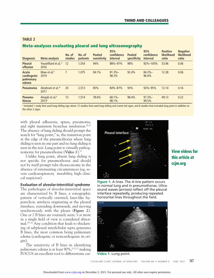
Initial lung assessmentLung assessment starts with identifying the pleural line, a shimmering hyperechoic struc-ture between the ribs (Figure 1). Respiropha-sic sliding of the pleura gives a shimmering ap-pearance, referred to as “lung sliding.” The tissue-air interface in the subpleural region of aerated lung is a strong refl ector. Ul-trasound is repeatedly refl ected between the pleura and the probe, leading to a reverberation artifact appearing as equidistant parallel echoic lines, known as A lines (Figure 1). An A-line pattern indicates normal lung, but it can also be seen with pneumothorax and in conditions with normally aerated pulmonary parenchyma, such as pulmonary embolism, chronic obstructive pulmonary disease, and asthma.15
Evaluation of pneumothoraxIn pneumothorax, the air between the parietal and visceral pleurae prevents pleural contact, giving an A-line pattern without lung sliding. Absence of lung sliding has good sensitivity (> 95%) for pneumothorax but poor specifi c-ity (60%–99%).8,16 This pattern also occurs
The absenceof lung slidingshould promptthe searchfor ‘lung point,’which isvirtually pathognomonic for pneumo-thorax
TABLE 1
Point-of-care ultrasonography workfl owcompared with traditional consultative ultrasonography
Consultativeultrasonography
POCUS
Decision to perform ultrasonography Primary clinician
Image acquisition Sonographer
Image interpretation SonographerRadiologist
Primary clinician
Clinical integration RadiologistPrimary clinician
on December 2, 2021. For personal use only. All other uses require permission.www.ccjm.orgDownloaded from
CLEVELAND CLINIC JOURNAL OF MEDICINE VOLUME 88 • NUMBER 6 JUNE 2021 347
THIND AND COLLEAGUES
View videos for this article at ccjm.org
with pleural adhesions, apnea, pneumonia, and right mainstem bronchus intubation.16,17 The absence of lung sliding should prompt the search for “lung point,” ie, the transition point at the edge of the pneumothorax where lung sliding is seen in one part and no lung sliding is seen in the rest. Lung point is virtually pathog-nomonic for pneumothorax (Video 1).17
Unlike lung point, absent lung sliding is not specifi c for pneumothorax and should not by itself prompt tube thoracostomy in the absence of extenuating circumstances (eg, se-vere cardiorespiratory instability, high clini-cal suspicion).
Evaluation of alveolar-interstitial syndromeThe pathologies of alveolar-interstitial space are characterized by B lines, a sonographic pattern of vertically oriented, laser-like hy-perechoic artefacts originating at the pleural interface, extending downwards, and moving synchronously with the pleura (Figure 2). One or 2 B lines are routinely seen; 3 or more in a single fi eld of view is considered abnor-mal.18–20 Any condition that leads to thicken-ing of subpleural interlobular septa generates B lines, the most common being pulmonary edema (cardiogenic or noncardiogenic in ori-gin). The sensitivity of B lines in identifying pulmonary edema is at least 90%,9,21,22 making POCUS an excellent tool to differentiate car-
TABLE 2
Meta-analyses evaluating pleural and lung ultrasonography
Diagnosis Meta-analysisNo. of studies
No. of patients
Pooled sensitivity
95% confi dence interval
Pooled specifi city
95% confi dence interval
Positive likelihood ratio
Negative likelihood ratio
Pleural effusion
Yousefi fard et al,11 2016
12 1,554 94% 88%–97% 98% 92%–100% 53.96 0.06
Acute cardiogenic pulmonary edema
Maw et al,9 2019
7 1,075 94.1% 81.3%–98.3%
92.4% 84.2%–96.4%
12.38 0.06
Pneumonia Alzahrani et al,10 2017
20 2,513 85% 84%–87% 93% 92%–95% 12.14 0.16
Pneumo-thorax
Alrajab et al,8 2013a
13 1,514 78.6% 68.1%–98.1%
98.4% 97.3%–99.5%
49.13 0.22
a Included 1 study that used lung sliding sign alone, 12 studies that used lung sliding and comet tail signs, and 6 studies that included lung point in addition to the other 2 signs.
Figure 1. A lines. The A-line pattern occurs in normal lung and in pneumothorax. Ultra-sound waves (arrows) refl ect off the pleural interface repeatedly, producing repeated horizontal lines throughout the fi eld.
Video 1. Lung point.
Pleural interface
on December 2, 2021. For personal use only. All other uses require permission.www.ccjm.orgDownloaded from

348 CLEVELAND CLINIC JOURNAL OF MEDICINE VOLUME 88 • NUMBER 6 JUNE 2021
POCUS FOR THE HOSPITALIST
diogenic pulmonary edema from exacerbation of chronic obstructive pulmonary disease. In addition, B lines may help guide diuresis and assess fl uid tolerance. B lines also occur in pneumonia, pulmo-nary fi brosis, acute respiratory distress syn-drome, and pneumonitis of any etiology. Careful evaluation of the pattern of B-line distribution and the pleural line, along with clinical correlation, can help distinguish these different causes (Table 3).23–25 The presence of B lines effectively rules out pneumothorax, as they are produced from subpleural lung units.17
Evaluation of consolidationUltrasound waves can traverse subpleural lung consolidation, resulting in the absence of A lines and a true 2-dimensional image of the consolidated lung (Figure 3). Almost all acute alveolar consolidations (98.5%) are found adjoining the visceral pleura, providing the necessary window for detection.26 The fi nding of subpleural consolidation or focal B lines, or both, is suggestive of pneu-monia. The sensitivity and specifi city of lung
ultrasonography for diagnosing pneumonia is just 85% or more.10 Nonetheless, supportive clinical and laboratory data with the charac-teristic ultrasound patterns can substantiate a diagnosis of pneumonia.
Evaluation of pleural effusionPortable chest radiography has a sensitiv-ity of 60% for detecting pleural effusion27; in contrast, lung ultrasonography is 94% sensi-tive and 98% specifi c.11 Lung ultrasonography can also better characterize basal opacities by distinguishing consolidation from pleural ef-fusion (Figure 4). It can also detail the fea-tures of pleural effusion, with simple effusion appearing anechoic, and complex effusions characterized by septations, loculations, and debris. The size of a pleural effusion can also be quantifi ed using lung ultrasonography.28
■ FOCUSED CARDIAC ULTRASONOGRAPHY
Focused cardiac ultrasonography (this term is preferred to “echocardiography” to high-light its focused nature) provides critical insight into hemodynamic status. It can be performed with excellent diagnostic ac-curacy for important cardiac abnormalities (Table 4).29
Focused questions, including global assess-ment of left ventricular function, presence or absence of a pericardial effusion, assessment of right ventricular size and function, and es-timation of right atrial pressure, can help nar-
Focused cardiac ultrasonog-raphy provides critical insight into hemo-dynamic status
Figure 2. B lines. The B-line pattern occurs in the setting of interstitial thickening by any cause, including cardiogenic pulmonary edema, noncardiogenic pulmonary edema, interstitial fi brosis, and interstitial pneumo-nia/pneumonitis. It is analogous to ground- glass opacity on computed tomography. It is demonstrated by vertical lines resembling the tail of a comet and extending to the bottom of the screen. In this image, con-fl uent B lines (arrow) indicate signifi cant interstitial involvement.
Figure 3. Small peripheral (subpleural) con-solidation. This is demonstrated by a small area of lung parenchyma visualized direct-ly beneath the pleura (arrow). This pattern is common in bacterial or viral pneumonia, including COVID-19 pneumonia.
on December 2, 2021. For personal use only. All other uses require permission.www.ccjm.orgDownloaded from

CLEVELAND CLINIC JOURNAL OF MEDICINE VOLUME 88 • NUMBER 6 JUNE 2021 349
THIND AND COLLEAGUES
row a differential diagnosis and guide man-agement in patients with cardiorespiratory distress.
Evaluating left ventricular functionEvaluation of left ventricular systolic function is one of the primary objectives of focused car-diac ultrasonography. As a general rule, mul-tiple views should be obtained for appropriate interpretation. Although objective methods of left ventricular systolic evaluation are avail-able and recommended, qualitative “eyeball estimation” is appropriate and feasible, with studies demonstrating high accuracy of visual estimation compared with recommended ob-jective measures.30,31 Left ventricular systolic function can be qualitatively graded as se-verely reduced, moderately reduced, mildly re-duced, normal, or hyperdynamic. Cardiology-performed echocardiography can be requested for further quantitative evaluation (Video 2, Video 3).
Evaluating right ventricular functionBetter understanding of the importance of right ventricular function has led to including
its evaluation in various protocols assessing shock and respiratory failure.32 Although ob-jectively estimating right ventricular size and function is challenging, qualitative assessment can be made at the bedside by directly com-paring the left and right ventricle. Size. The right ventricle is normally less than two-thirds the size of the left. A right ventricle-to-left ventricle ratio of 1 or higher is associated with poor outcomes in pulmo-
Evaluatingleft ventricular systolic functionis one of the primaryobjectivesof focusedcardiac ultra-sonography
TABLE 3
Characteristics of B lines based on etiology a
Cardiogenic pulmonary edema
Noncardiogenic diffuse pulmonary interstitial edema
Interstitial pneumonia or pneumonitis (bacterial, viral, or infl ammatory) Interstitial fi brosis
Distribution Diffuse
Usually bilateral and symmetric
Predominant in dependent regions
Diffuse or patchy
Often asymmetric
Focal or patchy
Usually asymmetric
Diffuse or patchy
Variable symmetry
Spared areas Absent Often present Present Often present
Number of B lines
Variable Variable Variable Variable
Pleura Smooth Irregular Irregular Irregular
Subpleural consolidations
Absent Present Present Typically absent
Reduced lung sliding
Absent May be present May be present May be present
Pleural effusion Often present Typically absent May be present Typically absenta Defi ning the terminology: diffuse = present throughout; patchy = present in many areas throughout, absent in other areas throughout; focal = present in one region but not in others; spared areas = regions of lung with A-line pattern (amid a background of B-line pattern).
Figure 4. Pleural effusion and consolidation.
Spleen
Effusion
Lung Diaphragm
on December 2, 2021. For personal use only. All other uses require permission.www.ccjm.orgDownloaded from

350 CLEVELAND CLINIC JOURNAL OF MEDICINE VOLUME 88 • NUMBER 6 JUNE 2021
POCUS FOR THE HOSPITALIST
TABLE 4
Focused cardiac ultrasonography: Basic views and key fi ndingsViews Probe position Possible fi ndings
Left 3rd to 5th intercostal space adjacent to the sternum, with probe marker pointing toward the right shoulder
Pericardial effusionSigns of tamponadeLeft ventricular size and systolic function
Mitral and aortic valvular pathologyAortic root dissection
From the PLAX view, the probe is rotated 90° clockwise. PSAX views are obtained by tilting the transducer from the base to the apex of the left ventricle.
Left ventricular systolic functionTricuspid, aortic, and mitralvalvular pathologyInterventricular septal deviationWall-motion abnormalities
With the probe marker point-ing toward the left, the probe is placed at the apex of the left ventricle. The apical impulse can be used as a guide.
Left ventricular size and functionRight ventricular size and functionPericardial effusion and signsof tamponadeValvular pathologyInterventricular septal deviationWall-motion abnormalities
The probe is placed below the xiphoid process, with the marker pointing toward the left.
From the subcostal long-axis view, the probe is rotated 90° counterclockwise and angulated slightly toward the left.
Pericardial effusion and signsof tamponade
Inferior vena cava sizeand collapsibility
AV = aortic valve; IVC = inferior vena cava; LV = left ventricle; MV = mitral valve; PLAX = parasternal long axis; PSAX = parasternal short axis; RV = right ventricle; TV = tricuspid valve
on December 2, 2021. For personal use only. All other uses require permission.www.ccjm.orgDownloaded from

CLEVELAND CLINIC JOURNAL OF MEDICINE VOLUME 88 • NUMBER 6 JUNE 2021 351
THIND AND COLLEAGUES
nary hypertension, pulmonary embolism, and other critical conditions. Septal kinetics. Assessing septal kinetics can also provide vital insights and help iden-tify the cause of right ventricular dysfunction: septal deviation occurs toward the left ventri-cle in diastole with right ventricular volume overload, and during systole with right ven-tricular pressure overload. Chronicity. It is important to distinguish acute from chronic right ventricular dysfunc-tion, as their causes differ. Distinguishing them is challenging with focused cardiac ul-trasonography, yet certain subtle fi ndings can point to the cause. Chronic dysfunction is seen in long-stand-ing cases of pulmonary hypertension. It is as-sociated with right ventricular hypertrophy with right ventricular free-wall thickness of more than 5 mm (Video 4). Acute dysfunction raises concern for mas-sive pulmonary embolism, acute respiratory
distress syndrome, and acute right ventricular infarction. In acute right ventricular dysfunc-tion, particularly pulmonary embolism, the McConnell sign (ie, right ventricular free- wall akinesis with sparing of the apex) is just 70% sensitive and 33% specifi c for diagnosing acute pulmonary embolism (positive likeli-hood ratio [PLR] 1.04, negative likelihood ratio [NLR] 0.91).33 Hence, pulmonary em-bolism cannot be defi nitively diagnosed with focused cardiac ultrasonography, with the no-table exception of detecting a visible throm-bus in the right heart (ie, a clot in transit).
Evaluating valvular abnormalitiesLimited evaluation of the mitral, tricuspid, and aortic valves can be performed using standard views. With some experience, gross abnormalities that may signifi cantly alter management (eg, fl ail leafl et, prolapse, large vegetation, chordae rupture) can be detected on visual examination and color Doppler. Dy-namic left ventricular outfl ow tract obstruc-
B lines may help guide diuresis and assess fl uid tolerance
Video 2. Normal parasternal long axis view. Video 3. Reduced ejection fraction.
Video 4. Dilated right ventricle. Video 5. Pericardial effusion and tamponade.
on December 2, 2021. For personal use only. All other uses require permission.www.ccjm.orgDownloaded from
352 CLEVELAND CLINIC JOURNAL OF MEDICINE VOLUME 88 • NUMBER 6 JUNE 2021
POCUS FOR THE HOSPITALIST
tion due to systolic anterior motion of the mitral valve can be detected visually and by using motion mode (M mode). Although sys-tolic anterior motion is classically seen with hypertrophic cardiomyopathy, it may also oc-cur in other situations that lead to worsening hemodynamics (eg, sepsis, acute hemorrhage, dehydration). Systolic anterior motion may be associated with severe mitral regurgitation, which resolves with resolution of systolic an-terior motion. However, bedside echocardiography is limited for assessing valvular pathologies. A detailed assessment of valvular lesions (espe-cially stenotic lesions) involves use of spectral Doppler in multiple views, which is not part of basic cardiac ultrasonography. Hence, a com-prehensive echocardiographic examination should be considered for evaluating valvular abnormalities and pathology.34
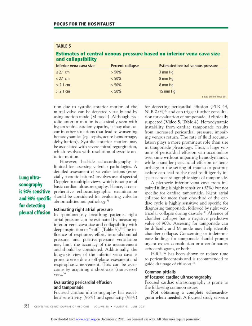
Estimating right atrial pressureIn spontaneously breathing patients, right atrial pressure can be estimated by measuring inferior vena cava size and collapsibility with deep inspiration or “sniff” (Table 5).35 The in-fl uence of respiratory effort, intra-abdominal pressure, and positive-pressure ventilation may limit the accuracy of the measurement and should be considered. Additionally, the long-axis view of the inferior vena cava is prone to error due to off-plane assessment and respirophasic movement. This can be over-come by acquiring a short-axis (transverse) view.36
Evaluating pericardial effusion and tamponadeFocused cardiac ultrasonography has excel-lent sensitivity (96%) and specifi city (98%)
for detecting pericardial effusion (PLR 48, NLR 0.04)37 and can trigger further consulta-tion for evaluation of tamponade, if clinically suspected (Video 5, Table 4). Hemodynamic instability from cardiac tamponade results from increased pericardial pressure, impair-ing venous return. The rate of fl uid accumu-lation plays a more prominent role than size in tamponade physiology. Thus, a large vol-ume of pericardial effusion can accumulate over time without impairing hemodynamics, while a smaller pericardial effusion or hem-orrhage in the setting of trauma or postpro-cedure can lead to the need to diligently in-spect echocardiographic signs of tamponade. A plethoric inferior vena cava from im-paired fi lling is highly sensitive (92%) but not specifi c for cardiac tamponade. Right atrial collapse for more than one-third of the car-diac cycle is highly sensitive and specifi c for diagnosing tamponade, followed by right ven-tricular collapse during diastole.38 Absence of chamber collapse has a negative predictive value of 90%. Assessing for tamponade can be diffi cult, and M mode may help identify chamber collapse. Concerning or indetermi-nate fi ndings for tamponade should prompt urgent expert consultation or a confi rmatory echocardiogram, or both. POCUS has been shown to reduce time to pericardiocentesis and is recommended to guide drainage of effusion.39
Common pitfalls of focused cardiac ultrasonographyFocused cardiac ultrasonography is prone to the following common issues: Not obtaining a complete echocardio-gram when needed. A focused study serves a
Lung ultra-sonography is 94% sensitive and 98% specifi c for detecting pleural effusion
TABLE 5
Estimates of central venous pressure based on inferior vena cava size and collapsibilityInferior vena cava size Percent collapse Estimated central venous pressure
≤ 2.1 cm > 50% 3 mm Hg
≤ 2.1 cm < 50% 8 mm Hg
> 2.1 cm > 50% 8 mm Hg
> 2.1 cm < 50% 15 mm HgBased on reference 35.
on December 2, 2021. For personal use only. All other uses require permission.www.ccjm.orgDownloaded from

CLEVELAND CLINIC JOURNAL OF MEDICINE VOLUME 88 • NUMBER 6 JUNE 2021 353
THIND AND COLLEAGUES
different purpose from a complete study and should not replace one. Hence, a “normal” focused cardiac ultrasonographic evaluation does not obviate the need to order a complete transthoracic echocardiogram that is clini-cally indicated. Over-relying on POCUS to manage vol-ume. POCUS fi ndings are useful as part of vol-ume status assessment, but a single POCUS fi nding in isolation should not be used to de-termine volume management (eg, giving fl u-ids for an apparently “collapsed” inferior vena cava). Findings are prone to variability and must be integrated into overall assessment, not used in isolation. Delaying POCUS during shock. Focused cardiac ultrasonography should be performed promptly in a patient with shock. Not doing so may lead to an important missed diagno-sis, such as pericardial tamponade, ventricular dysfunction, or valvular abnormality.
■ ABDOMINAL ULTRASONOGRAPHY
Evaluation of ascites and hemoperitoneumEvaluating thoracoabdominal trauma is of-ten a diagnostic challenge, prompting clini-cians to depend on ancillary tests to detect potentially life-threatening internal injuries. Ultrasonographic evaluation of free fl uid in the abdomen has been extensively studied in trauma literature for detecting hemoperi-toneum. Today, ultrasonography has virtu-ally replaced diagnostic peritoneal lavage as a primary, bedside imaging method for trauma patients.40 Numerous studies have found that examinations performed and interpreted by
treating physicians are reliably accurate com-pared with those read by radiologists.41 POCUS can also help hospitalists detect as-cites. It is more sensitive and specifi c than physi-cal examination and can guide the decision to perform paracentesis (Figure 5, Figure 6).42
Evaluation of kidney and bladderHydronephrosis, a commonly encountered and often reversible cause of acute kidney injury, can be detected with high sensitivity and specifi city by a bedside clinician using POCUS (Figure 7).43 Hydronephrosis results from urinary fl ow obstruction, which can be internal (eg, from ureteral calculus or a mass) or external (eg, from ureteral compression from structures such as an enlarged abdomi-nal aortic aneurysm, an advanced pregnancy, or a pelvic mass). Evaluation for hydrone-phrosis can be useful in cases in which uri-nary obstruction is considered. This may be particularly important in patients with acute pyelonephritis. However, mimics of hydro-nephrosis include prominent renal pyramids, prominent renal vasculature, and parapelvic cysts. Distal obstruction (eg, prostatic hypertro-phy) usually results in bilateral hydronephro-sis, so it is important to scan both kidneys. A study found more than 90% sensitivity and specifi city for detecting hydronephrosis by POCUS performed by internal medicine resi-dents given 5 hours of training compared with comprehensive radiologic ultrasonography.44 POCUS is also helpful in acute pyelone-phritis to evaluate for obstruction. Detecting large obstructive calculi would prompt urgent
Hydronephrosis can be detected with high sensitivityand specifi city with POCUS
Figure 5. Right lower quadrant with large ascites fl uid pocket; Foley catheter in bladder.
Figure 6. Ascites pocket.
on December 2, 2021. For personal use only. All other uses require permission.www.ccjm.orgDownloaded from

354 CLEVELAND CLINIC JOURNAL OF MEDICINE VOLUME 88 • NUMBER 6 JUNE 2021
POCUS FOR THE HOSPITALIST
urologic consultation. A distended bladder, being a large fl uid-fi lled structure, is easily visualized by ultraso-nography and can be distinguished from ascitic fl uid. POCUS can be used to estimate bladder volume and confi rm proper placement of a urinary catheter by visualizing a Foley balloon inside the bladder (Figure 5). This application may be particularly useful in a patient with obesity or ascites, which can make physical examination or bladder scanner determina-tions inaccurate. In patients without a urinary catheter, bladder volume estimation should be performed post-void.
Ultrasound evaluation of the biliary systemGallstones appear by ultrasonography as round hyperechoic structures in the gallblad-der or bile ducts, with posterior acoustic shad-owing. POCUS has demonstrated excellent sensitivity (89.8%) and specifi city (88.0%) for detecting cholelithiasis (PLR 7.48, NLR 0.12).45 Findings suggestive of acute cholecys-titis include gallstones, pericholecystic fl uid, gallbladder wall thickening, and sono graphic Murphy sign (ie, abdominal pain elicited by probe pressure), all of which can be assessed at the bedside with good specifi city (Figure 8).46 The common bile duct can also be mea-sured by POCUS, although it is technically challenging, especially for a novice user.47 Requesting a formal ultrasonographic study is prudent to obtain this information.
■ EVALUATION OF LOWER-EXTREMITY DEEP VEIN THROMBOSIS
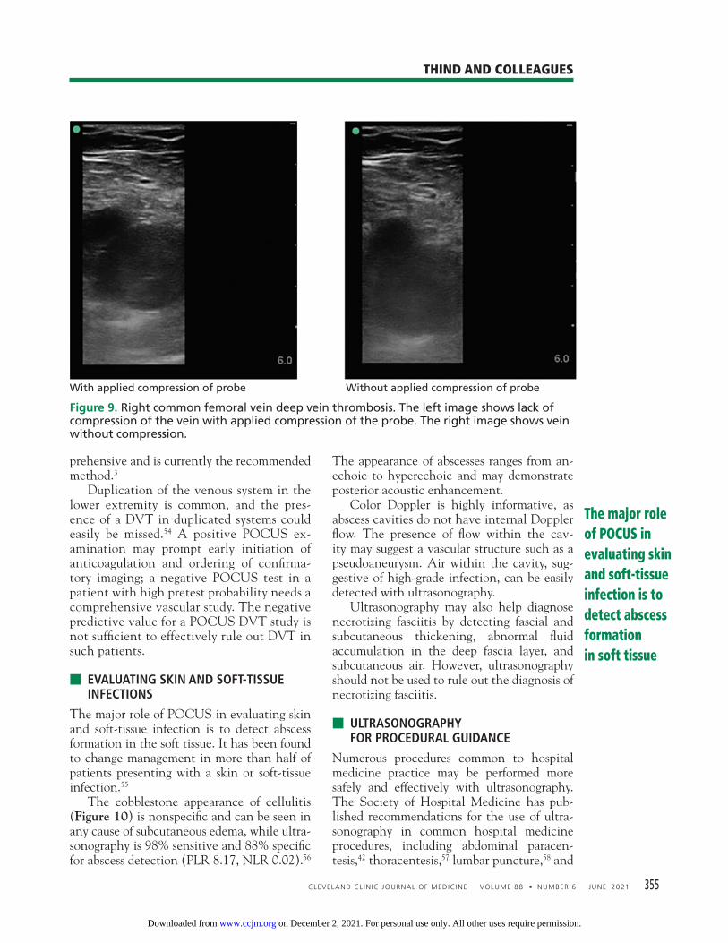
Although complete duplex ultrasonography is the standard radiological study traditionally performed to evaluate for deep vein thrombo-sis (DVT), point-of-care compression ultra-sonography can be performed rapidly with high diagnostic accuracy after limited training (Figure 9 ).48 A multicenter study of hospital-ist-performed compression ultrasonography found a sensitivity of 100% and specifi city of 96% for identifying lower extremity DVT, re-ducing the time to diagnosis by nearly 5 hours compared with corresponding vascular studies interpreted by radiologists.49 Meta-analyses have also reported sensitivity and specifi c-ity higher than 90% (Table 6).50–52 However, inadequate compression, lymph nodes, Baker cysts, and superfi cial venous thrombosis may be mistaken for a DVT. A focused DVT study is performed using a high-frequency (5–12 MHz) linear probe with compression of the vein at multiple sites, tra-ditionally using a 2-point (common femoral vein and popliteal vein) or 3-point (same, plus superfi cial femoral vein) method. The 3-point examination demonstrated higher sensitivity (91% vs 83%) and similar specifi city (96%) to the 2-point examination, but it still can miss 5% of isolated femoral vein DVTs.53 An extended compression examination employ-ing compressing the femoral vein every 2 to 3 cm until it dives into the adductor canal and popliteal vein along its course is more com-
Findings that suggest acute cholecystitis can be assessed with POCUS with good specifi city
Figure 7. Hydronephrosis. Hypoechoic (dark) fl uid (arrow) is shown extending into the renal pelvis.
Figure 8. Gallbladder containing sludge, with a thickened anterior wall, in a patient with acute cholecystitis.
Renal cortex
Renal pelvis
Gallbladderwall
Gallbladdersludge
Liver
on December 2, 2021. For personal use only. All other uses require permission.www.ccjm.orgDownloaded from
CLEVELAND CLINIC JOURNAL OF MEDICINE VOLUME 88 • NUMBER 6 JUNE 2021 355
THIND AND COLLEAGUES
prehensive and is currently the recommended method.3 Duplication of the venous system in the lower extremity is common, and the pres-ence of a DVT in duplicated systems could easily be missed.54 A positive POCUS ex-amination may prompt early initiation of anticoagulation and ordering of confi rma-tory imaging; a negative POCUS test in a patient with high pretest probability needs a comprehensive vascular study. The negative predictive value for a POCUS DVT study is not suffi cient to effectively rule out DVT in such patients.
■ EVALUATING SKIN AND SOFT-TISSUE INFECTIONS
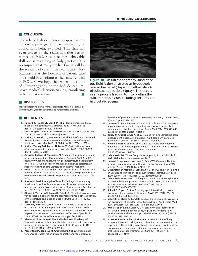
The major role of POCUS in evaluating skin and soft-tissue infection is to detect abscess formation in the soft tissue. It has been found to change management in more than half of patients presenting with a skin or soft-tissue infection.55 The cobblestone appearance of cellulitis (Figure 10) is nonspecifi c and can be seen in any cause of subcutaneous edema, while ultra-sonography is 98% sensitive and 88% specifi c for abscess detection (PLR 8.17, NLR 0.02).56
The appearance of abscesses ranges from an-echoic to hyperechoic and may demonstrate posterior acoustic enhancement. Color Doppler is highly informative, as abscess cavities do not have internal Doppler fl ow. The presence of fl ow within the cav-ity may suggest a vascular structure such as a pseudoaneurysm. Air within the cavity, sug-gestive of high-grade infection, can be easily detected with ultrasonography. Ultrasonography may also help diagnose necrotizing fasciitis by detecting fascial and subcutaneous thickening, abnormal fl uid accumulation in the deep fascia layer, and subcutaneous air. However, ultrasonography should not be used to rule out the diagnosis of necrotizing fasciitis.
■ ULTRASONOGRAPHY FOR PROCEDURAL GUIDANCE
Numerous procedures common to hospital medicine practice may be performed more safely and effectively with ultrasonography. The Society of Hospital Medicine has pub-lished recommendations for the use of ultra-sonography in common hospital medicine procedures, including abdominal paracen-tesis,42 thoracentesis,57 lumbar puncture,58 and
The major role of POCUS in evaluating skin and soft-tissue infection is to detect abscess formationin soft tissue
With applied compression of probe Without applied compression of probe
Figure 9. Right common femoral vein deep vein thrombosis. The left image shows lack of compression of the vein with applied compression of the probe. The right image shows vein without compression.
on December 2, 2021. For personal use only. All other uses require permission.www.ccjm.orgDownloaded from

356 CLEVELAND CLINIC JOURNAL OF MEDICINE VOLUME 88 • NUMBER 6 JUNE 2021
POCUS FOR THE HOSPITALIST
venous access,59 as well as for procedural cre-dentialing.60
Procedures may be “ultrasound-assisted” or “static” (ie, ultrasonography is used for site selec-tion, then the procedure is performed without ultrasonography) vs “ultrasound-guided” or “dy-namic” (ie, the procedure is performed with live ultrasonographic guidance, with the ultrasound probe in one hand and a needle in the other).
Central venous catheter insertionFor central venous catheter insertion, ultraso-nography reduces time to completion and de-crease failed attempts, with fewer complications like pneumothorax and arterial punctures. It also aids in preprocedural detection of stenosis and thrombosis of the target vein, and it is currently the standard of care for upper-extremity central venous catheter insertion.61 Nonetheless, this procedure remains highly user-dependent, and adequate training is critical.62
Peripheral intravenous catheter insertionUltrasonography is increasingly used to guide peripheral intravenous catheter insertion. In ad-dition to increasing patient satisfaction, it has demonstrated a higher success rate, particularly in patients with diffi cult access, reducing the need for a central venous catheter. Ultrasonog-raphy can also be used to confi rm the correct placement by visualizing the catheter in the vein or detecting bubbles with saline fl ush.63
Abdominal paracentesisUltrasonographic guidance of paracentesis has been found to have a 95% success rate com-pared with 61% using the traditional landmark-based method.64 Unsurprisingly, paracentesis was successfully completed with ultrasonogra-
phy in 87% of the patients for whom the land-mark method failed. In a large observational database study of 70,000 patients undergoing paracentesis, ultrasonographic guidance sig-nifi cantly reduced bleeding complications.65,66 In addition, a linear probe can help identi-fy underlying vasculature, including the infe-rior epigastric artery, further minimizing major bleeding risk.
ThoracentesisUltrasonography has also demonstrated a high-er rate of success and fewer complications for thoracentesis. In a meta-analysis of 24 studies with 6,605 thoracentesis procedures, ultraso-nography signifi cantly reduced pneumothorax compared with the landmark technique, even with inexperienced operators.67 The procedure can be performed using static or dynamic ul-trasonographic guidance. If static technique is used, the patient position needs to be main-tained after marking the spot. Evaluation of normal lung sliding preproce-dure and postprocedure obviates the need for chest radiographs to rule out pneumothorax.57
Common pitfallsUltrasound gel can prevent effective preproce-dural aseptic skin preparation and postproce-dural dressing adherence. Gel should dry before cleaning the skin or applying a dressing. In addition, use of ultrasonography may sometimes lead to failure to look at anatomi-cal landmarks, leading to performing a pro-cedure at a nonideal site. Users should be mindful of anatomic landmarks in addition to sonographic features.
TABLE 6
Meta-analyses evaluating point-of-care ultrasonographyfor diagnosing deep vein thrombosis
Meta-analysisNo. of studies
No. of patients
Pooled sensitiv-ity
95% confi dence interval
Pooled specifi city
95% confi dence interval
Positive likelihood ratio
Negative likelihood ratio
Burnside et al,50 2008
6 936 95% 87%–99% 96% 87%– 99% 23.75 0.05
Pomero et al,51 2013
16 2,379 96.1% 90.6%–98.5% 96.8% 94.6%–98.1% 30.03 0.04
West et al,52 2015 13 1,806 96.5% 90.1%–98.8% 96.8% 94.7% –98.0% 30.16 0.04
Many predict that POCUSwill bethe standardof care in the near future
on December 2, 2021. For personal use only. All other uses require permission.www.ccjm.orgDownloaded from
CLEVELAND CLINIC JOURNAL OF MEDICINE VOLUME 88 • NUMBER 6 JUNE 2021 357
THIND AND COLLEAGUES
■ CONCLUSION
The role of bedside ultrasonography has un-dergone a paradigm shift, with a variety of applications being explored. This shift has been driven by the realization that perfor-mance of POCUS is a readily achievable skill and is rewarding in daily practice. It is no surprise that many predict that it will be the standard of care in the near future. Hos-pitalists are at the forefront of patient care and should be cognizant of the many benefi ts of POCUS. We hope that wider utilization of ultrasonography at the bedside can im-prove medical decision-making, translating to better patient care. ■
■ DISCLOSURESThe authors report no relevant fi nancial relationships which, in the context of their contributions, could be perceived as a potential confl ict of interest.
Figure 10. On ultrasonography, subcutane-ous fl uid is demonstrated as hypoechoic or anechoic (dark) layering within islands of subcutaneous tissue (gray). This occurs in any process leading to fl uid within the subcutaneous tissue, including cellulitis and hydrostatic edema.
■ REFERENCES 1. Howard ZD, Noble VE, Marill KA, et al. Bedside ultrasound maxi-
mizes patient satisfaction. J Emerg Med 2014; 46(1):46–53. doi:10.1016/j.jemermed.2013.05.044
2. Fox S, Dugar S. Point-of-care ultrasound and COVID-19. Cleve Clin J Med 2020 May 14. doi:10.3949/ccjm.87a.ccc019
3. Soni NJ, Schnobrich D, Mathews BK, et al. Point-of-care ultrasound for hospitalists: a position statement of the Society of Hospital Medicine. J Hosp Med 2019; 14:E1–E6. doi:10.12788/jhm.3079
4. Soni NJ, Tierney DM, Jensen TP, Lucas BP. Certifi cation of point-of-care ultrasound competency. J Hosp Med 2017; 12(9):775–776. doi:10.12788/jhm.2812
5. American College of Physicians. ACP statement in support of point-of-care ultrasound in internal medicine. Accessed April 20, 2021. https://www.acponline.org/meetings-courses/focused-topics/point-of-care-ultrasound-pocus-for-internal-medicine/acp-statement-in-support-of-point-of-care-ultrasound-in-internal-medicine.
6. PSNet. “The ultrasound looked fi ne”: point-of-care ultrasound and patient safety. Accessed April 20, 2021. https://www.psnet.ahrq.gov/web-mm/ultrasound-looked-fi ne-point-care-ultrasound-and-patient-safety.
7. Blaivas M, Pawl R. Analysis of lawsuits fi led against emergency physicians for point-of-care emergency ultrasound examination performance and interpretation over a 20-year period. Am J Emerg Med 2012; 30(2):338–341. doi:10.1016/j.ajem.2010.12.016
8. Alrajab S, Youssef AM, Akkus NI, Caldito G. Pleural ultrasonography versus chest radiography for the diagnosis of pneumothorax: review of the literature and meta-analysis. Crit Care 2013; 17(5):R208. doi:10.1186/cc13016
9. Maw AM, Hassanin A, Ho PM, et al. Diagnostic accuracy of point-of-care lung ultrasonography and chest radiography in adults with symptoms suggestive of acute decompensated heart failure: a systematic review and meta-analysis. JAMA Netw Open 2019; 2(3):e190703. doi:10.1001/jamanetworkopen.2019.0703
10. Alzahrani SA, Al-Salamah MA, Al-Madani WH, Elbarbary MA. Systematic review and meta-analysis for the use of ultrasound versus radiology in diagnosing of pneumonia. Crit Ultrasound J 2017; 9(1):6. doi:10.1186/s13089-017-0059-y
11. Yousefi fard M, Baikpour M, Ghelichkhani P, et al. Screening per-formance characteristic of ultrasonography and radiography in
detection of pleural effusion: a meta-analysis. Emerg (Tehran) 2016; 4(1):1–10. pmid:26862542
12. Laursen CB, Sloth E, Lassen AT, et al. Point-of-care ultrasonography in patients admitted with respiratory symptoms: a single-blind, randomised controlled trial. Lancet Respir Med 2014; 2(8):638–646. doi:10.1016/S2213-2600(14)70135-3
13. Rouby JJ, Arbelot C, Gao Y, et al. Training for lung ultrasound score measurement in critically ill patients. Am J Respir Crit Care Med 2018; 198(3):398–401. doi:10.1164/rccm.201802-0227LE
14. Pivetta E, Goffi A, Lupia E, et al. Lung ultrasound-implemented diagnosis of acute decompensated heart failure in the ED: a SIMEU multicenter study. Chest 2015; 148(1):202–210. doi:10.1378/chest.14-2608
15. Lichtenstein DA. Whole Body Ultrasonography in the Critically Ill. Berlin Heidelberg: Springer-Verlag; 2010.
16. Husain LF, Hagopian L, Wayman D, Baker WE, Carmody KA. Sono-graphic diagnosis of pneumothorax. J Emerg Trauma Shock 2012; 5(1):76–81. doi:10.4103/0974-2700.93116
17. Lichtenstein D, Mezière G, Biderman P, Gepner A. The “lung point”: an ultrasound sign specifi c to pneumothorax. Intensive Care Med 2000; 26(10):1434–1440. doi:10.1007/s001340000627
18. Lichtenstein D, Mezière G. A lung ultrasound sign allowing bedside distinction between pulmonary edema and COPD: the comet-tail artifact. Intensive Care Med 1998; 24(12):1331–1334. doi:10.1007/s001340050771
19. Soldati G, Copetti R, Sher S. Sonographic interstitial syndrome: the sound of lung water. J Ultrasound Med 2009; 28(2):163–174. doi:10.7863/jum.2009.28.2.163
20. Volpicelli G, Mussa A, Garofalo G, et al. Bedside lung ultrasound in the assessment of alveolar-interstitial syndrome. Am J Emerg Med 2006; 24(6):689–696. doi:10.1016/j.ajem.2006.02.013
21. Wang Y, Shen Z, Lu X, Zhen Y, Li H. Sensitivity and specifi city of ultrasound for the diagnosis of acute pulmonary edema: a sys-tematic review and meta-analysis. Med Ultrason 2018; 1(1):32–36. doi:10.11152/mu-1223
22. Prosen G, Klemen P, Štrnad M, Grmec S. Combination of lung ultrasound (a comet-tail sign) and N-terminal pro-brain natriuretic peptide in differentiating acute heart failure from chronic obstruc-tive pulmonary disease and asthma as cause of acute dyspnea in prehospital emergency setting. Crit Care 2011; 15(2):R114. doi:10.1186/cc10140
on December 2, 2021. For personal use only. All other uses require permission.www.ccjm.orgDownloaded from

358 CLEVELAND CLINIC JOURNAL OF MEDICINE VOLUME 88 • NUMBER 6 JUNE 2021
POCUS FOR THE HOSPITALIST
23. Xie HQ, Zhang WW, Sun S, et al. A simplifi ed lung ultrasound for the diagnosis of interstitial lung disease in connective tissue disease: a meta-analysis. Arthritis Res Ther 2019; 21(1):93. doi:10.1186/s13075-019-1888-9
24. Lo Giudice V, Bruni A, Corcioni E, Corcioni B. Ultrasound in the evaluation of interstitial pneumonia. J Ultrasound 2008; 11(1):30–38. doi:10.1016/j.jus.2007.10.002
25. Dietrich CF, Mathis G, Blaivas M, et al. Lung B-line artefacts and their use. J Thorac Dis 2016; 8(6):1356–1365. doi:10.21037/jtd.2016.04.55
26. Lichtenstein DA, Lascols N, Mezière G, Gepner A. Ultrasound diag-nosis of alveolar consolidation in the critically ill. Intensive Care Med 2004; 30(2):276–281. doi:10.1007/s00134-003-2075-6
27. Kitazono MT, Lau CT, Parada AN, Renjen P, Miller WT Jr. Differentia-tion of pleural effusions from parenchymal opacities: accuracy of bedside chest radiography. AJR Am J Roentgenol 2010; 194(2):407–412. doi:10.2214/AJR.09.2950
28. Ibitoye BO, Idowu BM, Ogunrombi AB, Afolabi BI. Ultrasonographic quantifi cation of pleural effusion: comparison of four formulae. Ultrasonography 2018; 37(3):254–260. doi:10.14366/usg.17050
29. Lucas BP, Candotti C, Margeta B, et al. Diagnostic accuracy of hospitalist-performed hand-carried ultrasound echocardiography after a brief training program. J Hosp Med 2009; 4(6):340–349. doi:10.1002/jhm.438
30. Gudmundsson P, Rydberg E, Winter R, Willenheimer R. Visually estimated left ventricular ejection fraction by echocardiography is closely correlated with formal quantitative methods. Int J Cardiol 2005; 101(2):209–212. doi:10.1016/j.ijcard.2004.03.027
31. Melamed R, Sprenkle MD, Ulstad VK, Herzog CA, Leatherman JW. Assessment of left ventricular function by intensivists using hand-held echocardiography. Chest 2009; 135(6):1416–1420. doi:10.1378/chest.08-2440
32. Breitkreutz R, Price S, Steiger HV, et al. Focused echocardiographic evaluation in life support and periresuscitation of emergency patients: a prospective trial. Resuscitation 2010; 81(11):1527–1533. doi:10.1016/j.resuscitation.2010.07.013
33. Casazza F, Bongarzoni A, Capozi A, Agostoni O. Regional right ventricular dysfunction in acute pulmonary embolism and right ventricular infarction. Eur J Echocardiogr 2005; 6(1):11–14. doi:10.1016/j.euje.2004.06.002
34. Chamsi-Pasha MA, Sengupta PP, Zoghbi WA. Handheld echocar-diography: current state and future perspectives. Circulation 2017; 136(22):2178–2188. doi:10.1161/CIRCULATIONAHA.117.026622
35. Rudski LG, Lai WW, Afi lalo J, et al. Guidelines for the echocardio-graphic assessment of the right heart in adults: a report from the American Society of Echocardiography endorsed by the European Association of Echocardiography, a registered branch of the Euro-pean Society of Cardiology, and the Canadian Society of Echocar-diography. J Am Soc Echocardiogr 2010; 23(7):685–788. doi:10.1016/j.echo.2010.05.010
36. Schmidt GA. POINT: should acute fl uid resuscitation be guided primarily by inferior vena cava ultrasound for patients in shock? Yes. Chest 2017; 151(3):531–532. doi:10.1016/j.chest.2016.11.021
37. Mandavia DP, Hoffner RJ, Mahaney K, Henderson SO. Bedside echocardiography by emergency physicians. Ann Emerg Med 2001; 38(4):377–382. doi:10.1067/mem.2001.118224
38. Klein AL, Abbara S, Agler DA, et al. American Society of Echocar-diography clinical recommendations for multimodality cardiovas-cular imaging of patients with pericardial disease: endorsed by the Society for Cardiovascular Magnetic Resonance and Society of Cardiovascular Computed Tomography. J Am Soc Echocardiogr 2013; 26(9):965–1012.e15. doi:10.1016/j.echo.2013.06.023
39. Alpert EA, Amit U, Guranda L, Mahagna R, Grossman SA, Bentancur A. Emergency department point-of-care ultrasonography improves time to pericardiocentesis for clinically signifi cant effusions. Clin Exp Emerg Med 2017; 4(3):128–132. doi:10.15441/ceem.16.169
40. Savatmongkorngul S, Wongwaisayawan S, Kaewlai R. Focused assessment with sonography for trauma: current perspectives. Open Access Emerg Med 2017; 9:57–62. doi:10.2147/OAEM.S120145
41. Bhoi S, Sinha TP, Ramchandani R, Kurrey L, Galwankar S. To de-termine the accuracy of focused assessment with sonography for trauma done by nonradiologists and its comparative analysis with radiologists in emergency department of a level 1 trauma center of India. J Emerg Trauma Shock 2013; 6(1):42–46. doi:10.4103/0974-2700.106324
42. Cho J, Jensen TP, Reierson K, et al. Recommendations on the use of ultrasound guidance for adult abdominal paracentesis: a position statement of the Society of Hospital Medicine. J Hosp Med 2019; 14:E7–E15. doi:10.12788/jhm.3095
43. Wong C, Teitge B, Ross M, Young P, Robertson HL, Lang E. The accuracy and prognostic value of point-of-care ultrasound for neph-rolithiasis in the emergency department: a systematic review and meta-analysis. Acad Emerg Med 2018; 25(6):684–698. doi:10.1111/acem.13388
44. Caronia J, Panagopoulos G, Devita M, et al. Focused renal sonogra-phy performed and interpreted by internal medicine residents. J Ul-trasound Med 2013; 32(11):2007–2012. doi:10.7863/ultra.32.11.2007
45. Ross M, Brown M, McLaughlin K, et al. Emergency physician-per-formed ultrasound to diagnose cholelithiasis: a systematic review. Acad Emerg Med 2011; 18(3):227–235. doi:10.1111/j.1553-2712.2011.01012.x
46. Hilsden R, Leeper R, Koichopolos J, et al. Point-of-care biliary ultra-sound in the emergency department (BUSED): implications for surgi-cal referral and emergency department wait times. Trauma Surg Acute Care Open 2018; 3(1):e000164. doi:10.1136/tsaco-2018-000164
47. Lahham S, Becker BA, Gari A, et al. Utility of common bile duct mea-surement in ED point of care ultrasound: a prospective study. Am J Emerg Med 2018; 36(6):962–966. doi:10.1016/j.ajem.2017.10.064
48. Kory PD, Pellecchia CM, Shiloh AL, Mayo PH, DiBello C, Koenig S. Accuracy of ultrasonography performed by critical care physicians for the diagnosis of DVT. Chest 2011; 139(3):538–542. doi:10.1378/chest.10-1479
49. Fischer EA, Kinnear B, Sall D, et al. Hospitalist-operated compression ultrasonography: a point-of-care ultrasound study (HOCUS-POCUS). J Gen Intern Med 2019; 34(10):2062–2067. doi:10.1007/s11606-019-05120-5
50. Burnside PR, Brown MD, Kline JA. Systematic review of emergency physician-performed ultrasonography for lower-extremity deep vein thrombosis. Acad Emerg Med 2008; 15(6):493–498. doi:10.1111/j.1553-2712.2008.00101.x
51. Pomero F, Dentali F, Borretta V, et al. Accuracy of emergency physician-performed ultrasonography in the diagnosis of deep-vein thrombosis: a systematic review and meta-analysis. Thromb Hae-most 2013; 109(1):137–145. doi:10.1160/TH12-07-0473
52. West JR, Shannon AW, Chilstrom ML. What is the accuracy of emergency physician-performed ultrasonography for deep venous thrombosis? Ann Emerg Med 2015; 65(6):699–701. doi:10.1016/j.annemergmed.2014.06.025
53. Adhikari S, Zeger W, Thom C, Fields JM. Isolated deep venous thrombosis: implications for 2-point compression ultrasonogra-phy of the lower extremity. Ann Emerg Med 2015; 66(3):262–266. doi:10.1016/j.annemergmed.2014.10.032
54. Zitek T, Baydoun J, Yepez S, Forred W, Slattery DE. Mistakes and pitfalls associated with two-point compression ultrasound for deep vein thrombosis. West J Emerg Med 2016; 17(2):201–208. doi:10.5811/westjem.2016.1.29335
55. Tayal VS, Hasan N, Norton HJ, Tomaszewski CA. The effect of soft-tissue ultrasound on the management of cellulitis in the emergency department. Acad Emerg Med 2006; 13(4):384–388. doi:10.1197/j.aem.2005.11.074
56. Squire BT, Fox JC, Anderson C. ABSCESS: applied bedside sonogra-phy for convenient evaluation of superfi cial soft tissue infections. Acad Emerg Med 2005; 12(7):601–606. doi:10.1197/j.aem.2005.01.016
57. Dancel R, Schnobrich D, Puri N, et al. Recommendations on the use of ultrasound guidance for adult thoracentesis: a position statement of the Society of Hospital Medicine. J Hosp Med 2018; 13(2):126–135. doi:10.12788/jhm.2940
on December 2, 2021. For personal use only. All other uses require permission.www.ccjm.orgDownloaded from

CLEVELAND CLINIC JOURNAL OF MEDICINE VOLUME 88 • NUMBER 6 JUNE 2021 359
THIND AND COLLEAGUES
58. Soni NJ, Franco-Sadud R, Kobaidze K, et al. Recommendations on the use of ultrasound guidance for adult lumbar puncture: a position statement of the Society of Hospital Medicine. J Hosp Med 2019; 14(10):591–601. doi:10.12788/jhm.3197
59. Franco-Sadud R, Schnobrich D, Mathews BK, et al. Recommenda-tions on the use of ultrasound guidance for central and peripheral vascular access in adults: a position statement of the Society of Hos-pital Medicine. J Hosp Med 2019; 14:E1–E22. doi:10.12788/jhm.3287
60. Lucas BP, Tierney DM, Jensen TP, et al. Credentialing of hospitalists in ultrasound-guided bedside procedures: a position statement of the Society of Hospital Medicine. J Hosp Med 2018; 13(2):117–125. doi:10.12788/jhm.2917
61. Lalu MM, Fayad A, Ahmed O, et al. Ultrasound-guided subclavian vein catheterization: a systematic review and meta-analysis. Crit Care Med 2015; 43(7):1498–1507. doi:10.1097/CCM.0000000000000973
62. Blaivas M, Adhikari S. An unseen danger: frequency of posterior vessel wall penetration by needles during attempts to place internal jugular vein central catheters using ultrasound guidance. Crit Care Med 2009; 37(8):2345–2359. doi:10.1097/CCM.0b013e3181a067d4
63. Gottlieb M, Sundaram T, Holladay D, Nakitende D. Ultrasound-guided peripheral intravenous line placement: a narrative review of
evidence-based best practices. West J Emerg Med 2017; 18(6):1047–1054. doi:10.5811/westjem.2017.7.34610
64. Nazeer SR, Dewbre H, Miller AH. Ultrasound-assisted paracentesis performed by emergency physicians vs the traditional technique: a prospective, randomized study. Am J Emerg Med 2005; 23(3):363–367. doi:10.1016/j.ajem.2004.11.001
65. Patel PA, Ernst FR, Gunnarsson CL. Evaluation of hospital complica-tions and costs associated with using ultrasound guidance during abdominal paracentesis procedures. J Med Econ 2012; 15(1):1–7. doi:10.3111/13696998.2011.628723
66. Mercaldi CJ, Lanes SF. Ultrasound guidance decreases complications and improves the cost of care among patients undergoing thoracen-tesis and paracentesis. Chest 2013; 143(2):532–538. doi:10.1378/chest.12-0447
67. Gordon CE, Feller-Kopman D, Balk EM, Smetana GW. Pneumothorax following thoracentesis: a systematic review and meta-analysis. Arch Intern Med 2010; 170(4):332–339. doi:10.1001/archinternmed.2009.548
Address: Guramrinder Singh Thind, MD, Department of Critical Care Medicine, A90, Respiratory Institute, Cleveland Clinic, 9500 EuclidAvenue, Cleveland, OH 44195; [email protected]
on December 2, 2021. For personal use only. All other uses require permission.www.ccjm.orgDownloaded from