Dental Operating Microscope
74
Submitted by: - Dinesh solanki OPERATING MICROSCOPE IN DENTISTRY
-
Upload
drdineshsolanki -
Category
Documents
-
view
200 -
download
11
Transcript of Dental Operating Microscope
Submitted by: -
Dinesh solanki
OPERATING MICROSCOPE IN DENTISTRY
• Introduction
• Loupes
• Operating microscope
• Magnification
• Illumination
• Ergonomics
• Imaging
• Parts of operating microscope
• Comparison between different companies
• Companies

REMEMBER THIS ONE IMPORTANT POINT
• The better, the dentist can see when he or she is doing treatment, the greater the probability of the treatment being successful.
THE LIMIT OF HUMAN EYES
• Resolution???
• Resolving power of human aided eyes – 0.2mm
• Dentist can increase their resolving………
• presbyopia
WHY ENHANCED VISION IS NECESSARY IN DENTISTRY
• Operating microscope - .006mm
• Small space, large instruments…..
LOUPES
• Its classified by optical methods in to
• 1- A diopter
• 2- A surgical telescope with a Galilean system configuration
• 3- A surgical telescope with a Keplerian system configuration
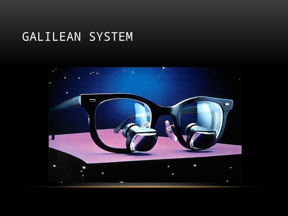
GALILEAN SYSTEM
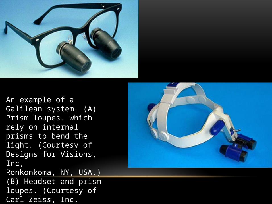
An example of a Galilean system. (A) Prism loupes. which rely on internal prisms to bend the light. (Courtesy of Designs for Visions, Inc,Ronkonkoma, NY, USA.) (B) Headset and prism loupes. (Courtesy of Carl Zeiss, Inc, Germany.)
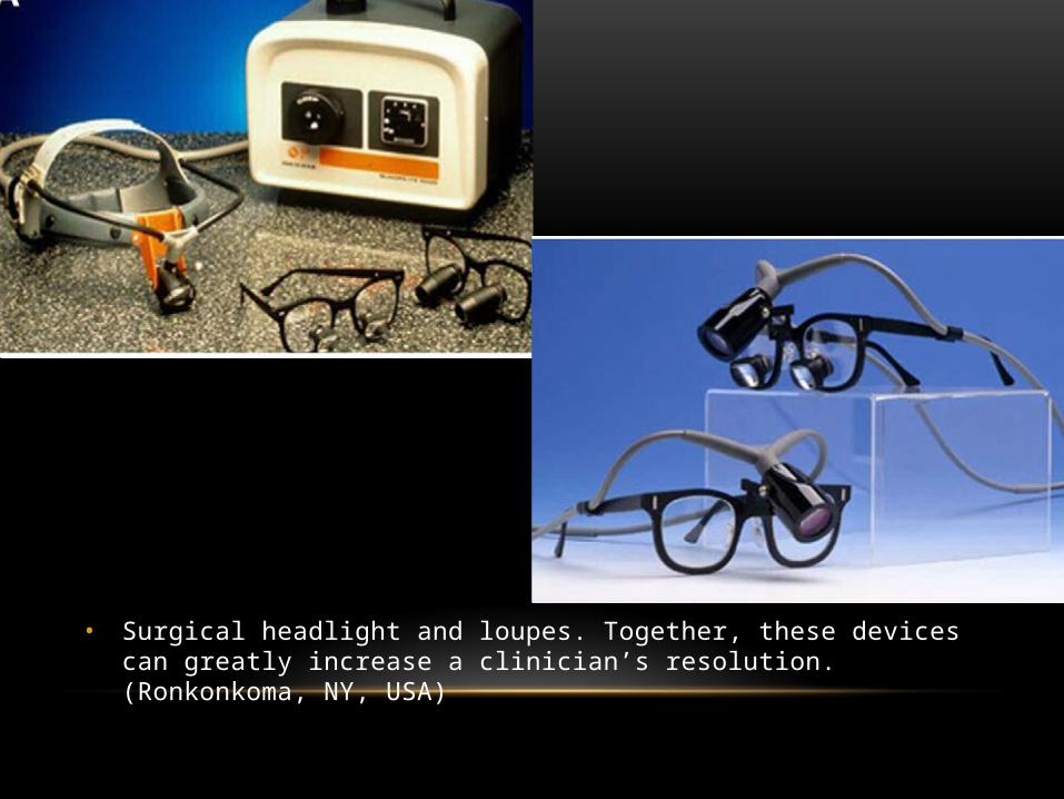
• Surgical headlight and loupes. Together, these devices can greatly increase a clinician’s resolution. (Ronkonkoma, NY, USA)
• operating microscope possess the additional benefit of Galilean optics.
• As opposed to loupes, which have convergent optics, it focus at infinity and send parallel beams of light to each eye. With parallel light, the operator’s eye are at rest, as though looking off into the distance, permitting performance of time-consuming procedures without inducing eye fatigue

OPERATING MICROSCOPE
• Apotheker in 1981
• Howard selden
• Gary carr in 1999
• Magnification is essential for performing not only endodontic procedures, but almost any other dental procedure one can imagine, except perhaps Orthodontics. A microscope is absolutely the best modality for consistent accurate visualization for dental procedures.
MAGNIFICATION
• Microscopes provide multiple telescopic magnifications with a twist of a dial, from 3x to 24x. The higher magnification provided by microscopes allow for a wider field of view and a deeper depth of field than loupes.
• Although loupes are available with magnification above 3x, there are drawbacks. Loupes have only a single magnification factor. Additionally, loupes with any factor above 3x have increasingly longer and heavier lenses, with a progressively smaller field of view. The additional weight makes holding one’s head steady a challenge, and the use of higher magnifications becomes difficult at best .
ILLUMINATION
• Whenever the degree of magnification is increased, the available light is spread out. For this reason anything under higher magnification appears darker. Additional light is required to compensate for this phenomenon. In order to achieve adequate illumination with loupes, one should purchase the optional headband light source which provides the spot illumination needed.
• Microscopes on the other-hand have an integrated, through the lens, bright fiber-optic light source. This light source completely eliminates shadows because it incorporates a coaxial (line of sight) light path that is always directed where we are looking. Some microscopes even have a built-in curing light filter that prevents light activated composites from polymerizing while using the scope for general dental procedures

.
ERGONOMICS
• Microscopes really deliver on the promise of comfortable sit-down-dentistry. With a scope, it is no longer necessary to bend your body to obtain good visualization. With proper microscope training, one can sit at the 12 o’clock position even while working with mandibular molars by learning to bring the patient to you, instead of you bringing yourself to the patient. Under a microscope, subtle patient head movements are all that is required for proper patient positioning to affect a more comfortable operating posture.
IMAGING
• One may add an integrated video camera and a monitor to the scope for image capture. A scope with a camera will fulfill the same function as does an intra-oral camera, but with the advantage of imaging done in “real time”. While working with the scope.
• So we don’t have to stop in the middle of the procedure to get the wand, find the tooth, focus, and capture the image. What we see is what we get in "live action video". The image can be saved to the computer, emailed, printed on a video or computer printer.
• Justifying a proposed treatment plan to an insurance company is facilitated when a printed picture accompanies the plan .

PATIENT ACCEPTANCE
• Viewing their own teeth as seen through a scope is an impressive patient education tool that portrays the dentist as state of the art. They become participants in their treatment planning process and actually help suggest work they want to have completed.
OPERATING MICROSCOPE
• It consist of three important parts: -
1- the supporting structure
2- body of the microscope and
3- light source.
SUPPORTING STRUCTURE
• It stable the microscope during procedure, so yet remain maneuverable with ease and exceptional precision, particularly when used at high power.
• It can be mounted on the floor, ceiling, or wall.
• As the distance between the fixation and the body of the microscope is decreased, the stability of the setup is increased.
• In clinical sitting with high ceilings or distal walls, the floor mount is preferable.
BODY OF THE MICROSCOPE
• Eyepiece are used in the overall magnification. They are available in various power, ranging from 6.3x to 20x; the two most commonly used are 10x and 12.5x.
• The end of the each eyepiece has a rubber cup that can be lowered for clinicians who wear glasses. Eyepiece also have adjustable diopter settings.
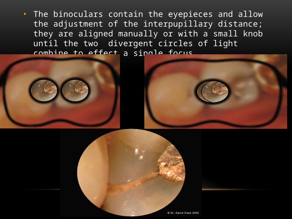
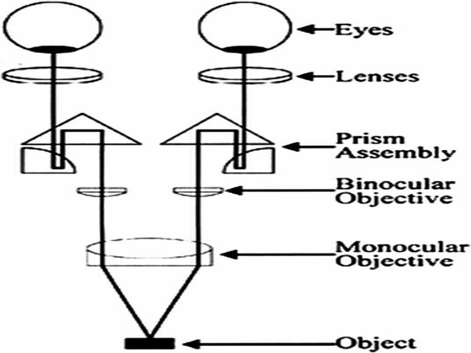
• The binoculars contain the eyepieces and allow the adjustment of the interpupillary distance; they are aligned manually or with a small knob until the two divergent circles of light combine to effect a single focus.
• Binocular are available with straight, inclined or inclinable tubes.
• Straight tubes are not well suited for dentistry.
• Inclined or inclinable tubes are preferred to allow the clinician to establish a comfortable position.
• Inclined tubes are fixed at 45 degree angle to the line of the sight of the microscope.
• Magnification changers are available as 3, 5, or 6-step manual changers, or a power-zoom changers. They consist of lenses mounted that is connected to a dial located on the side of the microscope. The magnification is altered by rotating the dial.
• The objective lens is the final optical elements, and its focal length determines the working distance between the microscope and the surgical field. The range of the focal length varies from 100mm to 400mm.
• A 200mm focal length allows approximately 20cm of working distance, which is generally adequate for utilization in intraoral procedure.
• The total magnification of microscope is represented by the following formula:
TM=(FLT/FLOL) X EP X MV
• TM: Total magnification
• FLT: Focal length of tube
• FLOL: Focal length of objective lengths
• EP: Eyepiece power
• MV: Magnification value
LIGHT SOURCE
• Its one of the most important feature of operating microscope.
• The illumination is coaxial with the line of sight, which eliminates the presence of any shadow. The light source is generally powered by a 100 to 150-walt halogen light bulb that is connected to the microscope with a high efficiency fiberoptic cable.
• The light passes through a condensing lens, a series of prisms, and then through the objective lens to the surgical site.
• The intensity of light is controlled by a rheostat.
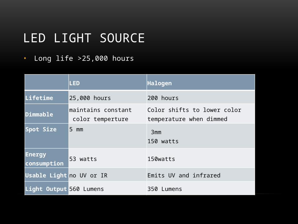
LED LIGHT SOURCE• Long life >25,000 hours
LED Halogen
Lifetime 25,000 hours 200 hours
Dimmable maintains constant color temperture
Color shifts to lower color temperature when dimmed
Spot Size 5 mm 3mm150 watts
Energy consumption 53 watts 150watts
Usable Light no UV or IR Emits UV and infrared
Light Output 560 Lumens 350 Lumens
Advantages vs. halogen and metal halide lamps
• Long life• Energy efficient• Immediately stable output - no warm-up time• Consistent color temperature over lifetime and when dimmed• No high voltage power supply
XENON LIGHT SOURCE
• Xenon light cold light source, When it used in the Clinical, the product can supply good illumination, imaging clearly for various procedure; the other hand, to provide good lighting to the dentist when examination, treatment, surgery and photography and video during treatment procedure.
PARTS AND ACCESSORIES OF THE MICROSCOPE
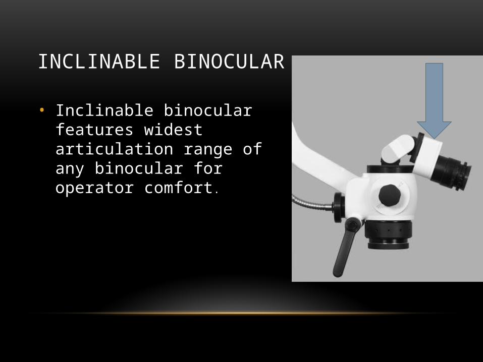
INCLINABLE BINOCULAR
• Inclinable binocular features widest articulation range of any binocular for operator comfort.
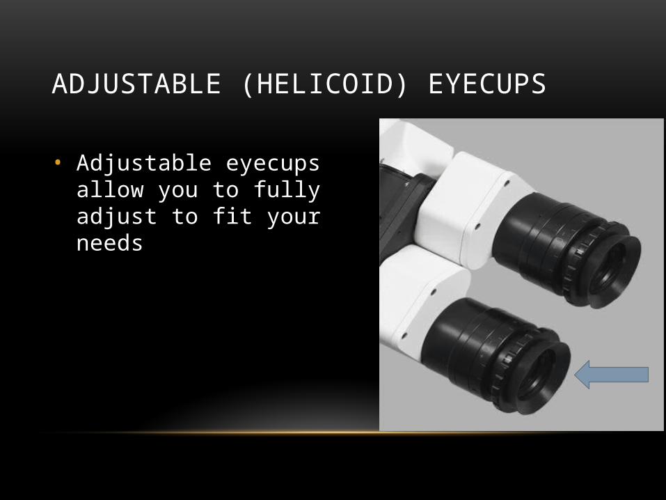
ADJUSTABLE (HELICOID) EYECUPS
• Adjustable eyecups allow you to fully adjust to fit your needs

• The widefield eyepieces of the Pico OPMI. The upper rotating button, which regulates the interpupillar distance
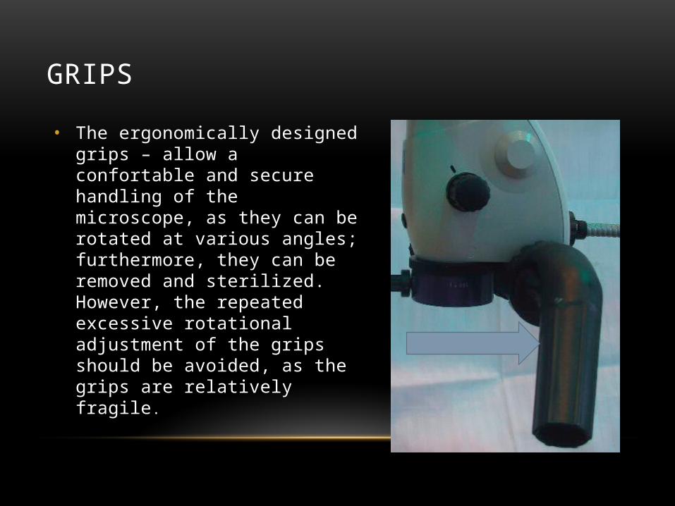
GRIPS
• The ergonomically designed grips – allow a confortable and secure handling of the microscope, as they can be rotated at various angles; furthermore, they can be removed and sterilized. However, the repeated excessive rotational adjustment of the grips should be avoided, as the grips are relatively fragile.
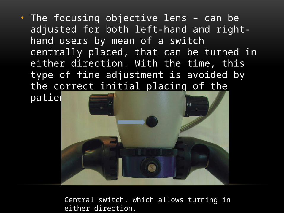
• The focusing objective lens – can be adjusted for both left-hand and right-hand users by mean of a switch centrally placed, that can be turned in either direction. With the time, this type of fine adjustment is avoided by the correct initial placing of the patient’s head .
Central switch, which allows turning in either direction.
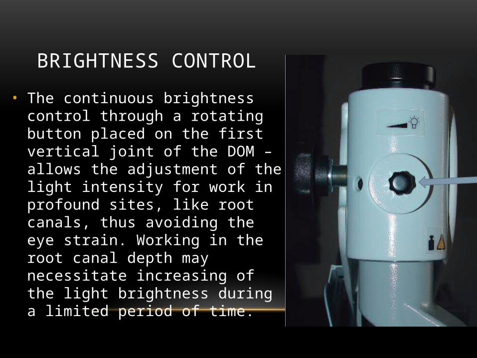
BRIGHTNESS CONTROL
• The continuous brightness control through a rotating button placed on the first vertical joint of the DOM – allows the adjustment of the light intensity for work in profound sites, like root canals, thus avoiding the eye strain. Working in the root canal depth may necessitate increasing of the light brightness during a limited period of time.
MORA
• Designed by Dr.Assad Mora, the MORA interface raises the mobility of microscopes.
• Basically, the MORA interface allows the vertical tilting Thus, dentists continue looking straight into the microscope, while sitting in an upright position in each phase of the treatment. The MORA interface also enables DOM body in a panning motion to the left and to the right sides of the mouth (25° in each direction);

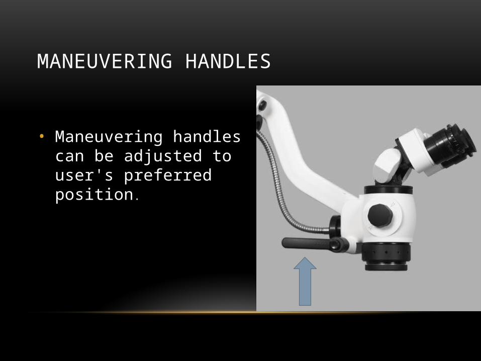
MANEUVERING HANDLES
• Maneuvering handles can be adjusted to user's preferred position.
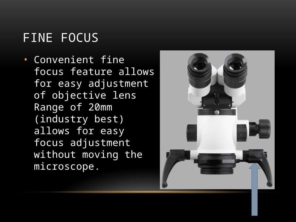
FINE FOCUS
• Convenient fine focus feature allows for easy adjustment of objective lens Range of 20mm (industry best) allows for easy focus adjustment without moving the microscope.
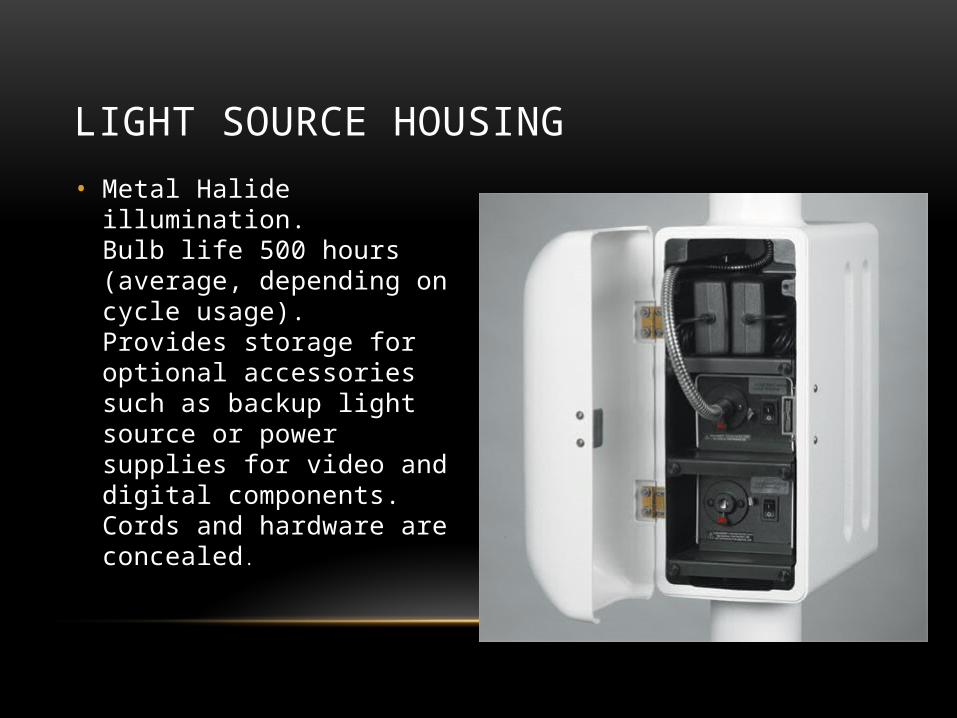
LIGHT SOURCE HOUSING
• Metal Halide illumination.Bulb life 500 hours (average, depending on cycle usage).Provides storage for optional accessories such as backup light source or power supplies for video and digital components. Cords and hardware are concealed.
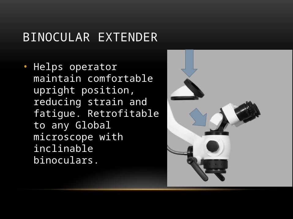
BINOCULAR EXTENDER
• Helps operator maintain comfortable upright position, reducing strain and fatigue. Retrofitable to any Global microscope with inclinable binoculars.
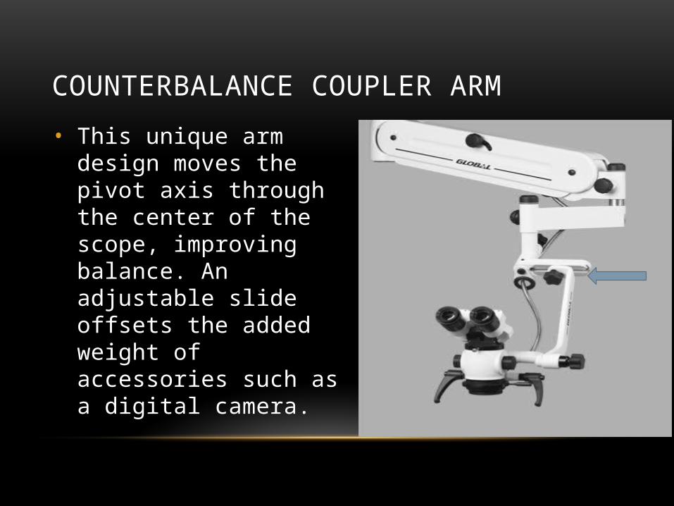
COUNTERBALANCE COUPLER ARM
• This unique arm design moves the pivot axis through the center of the scope, improving balance. An adjustable slide offsets the added weight of accessories such as a digital camera.

EXTENSION ARM
• Provides additional maneuverabilityProvides extra reach
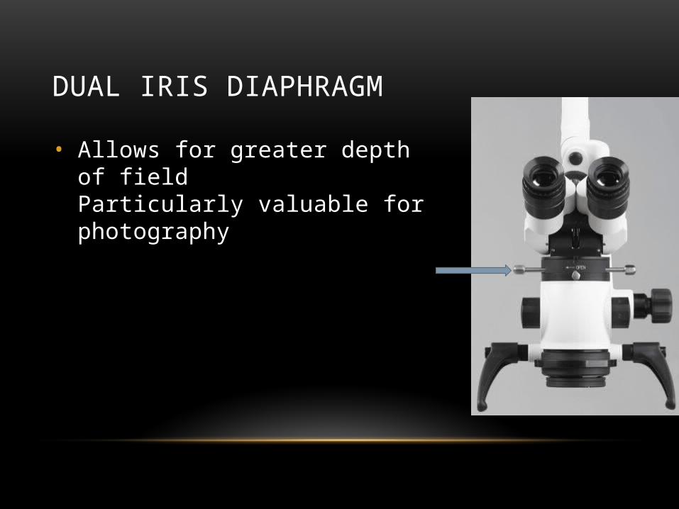
DUAL IRIS DIAPHRAGM
• Allows for greater depth of fieldParticularly valuable for photography
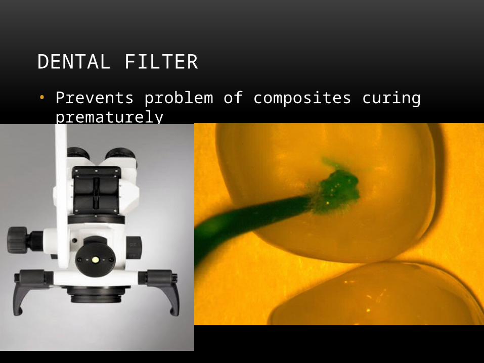
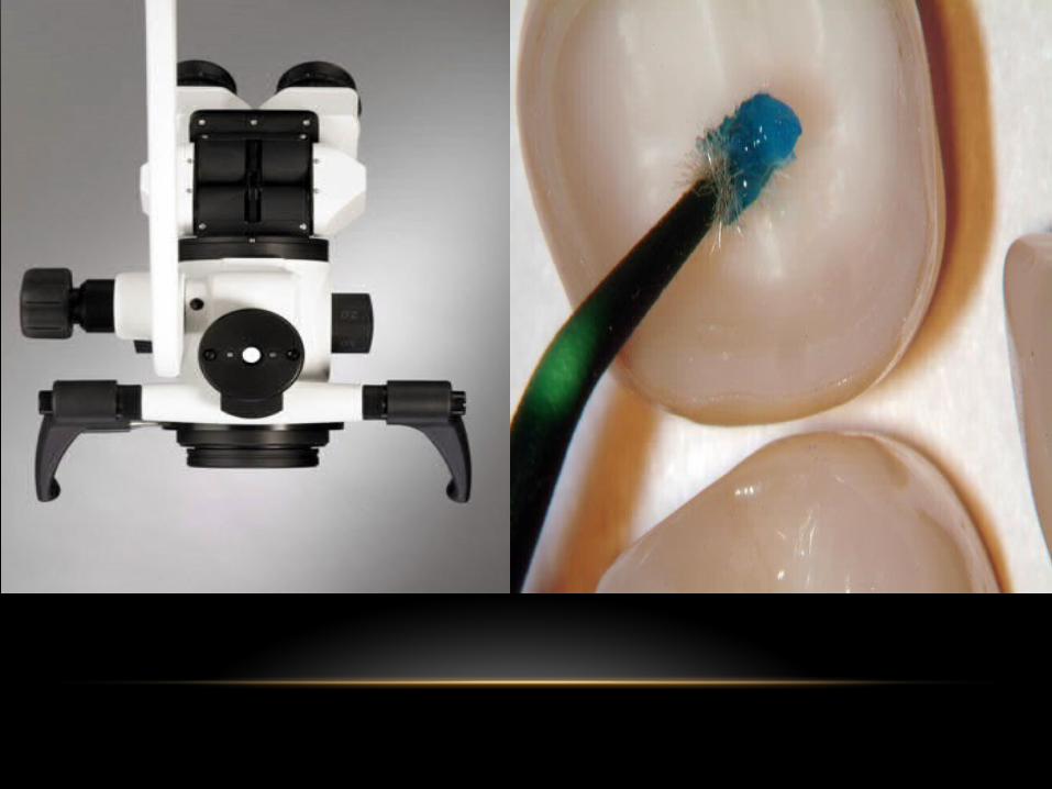
DENTAL FILTER
• Prevents problem of composites curing prematurely

• G3 Microscope
• The G3 from Global is ideal for general restorative dentistry. The microscope has three turret settings with a magnification ranging from x3.2 up to x12.8.
• G4 Microscope
• The G4 from Global is ideal for more involved restorative dentistry as it provides an additional magnification setting for added flexibility and coverage between turret steps. The microscope has four turret settings with a magnification ranging from x3.2 up to x12.8.
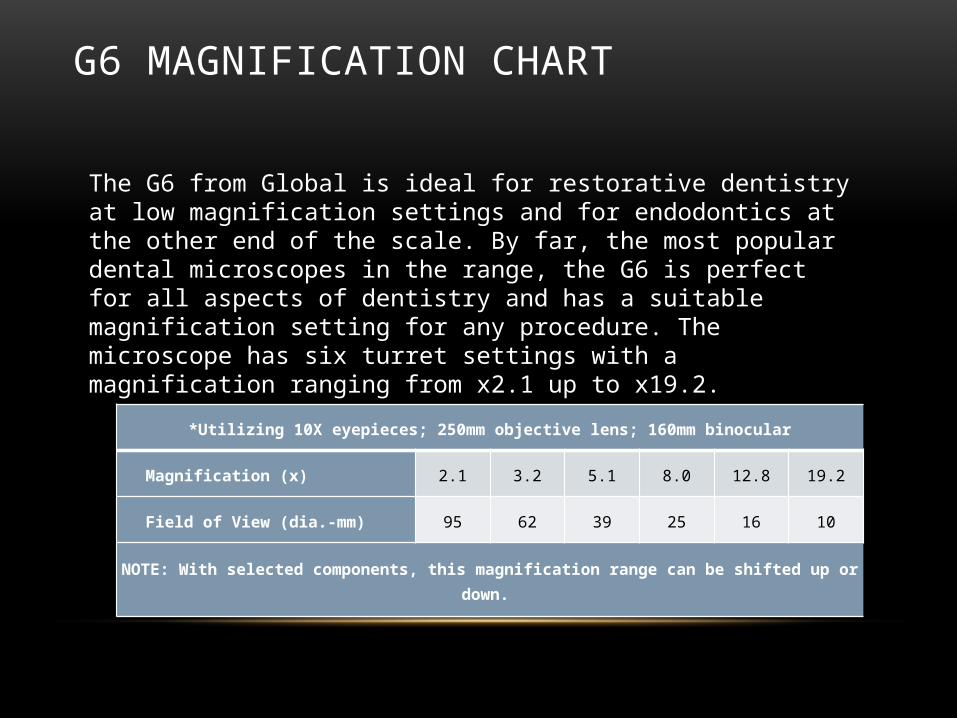
G6 MAGNIFICATION CHART
*Utilizing 10X eyepieces; 250mm objective lens; 160mm binocular
Magnification (x) 2.1 3.2 5.1 8.0 12.8 19.2
Field of View (dia.-mm) 95 62 39 25 16 10
NOTE: With selected components, this magnification range can be shifted up or down.
The G6 from Global is ideal for restorative dentistry at low magnification settings and for endodontics at the other end of the scale. By far, the most popular dental microscopes in the range, the G6 is perfect for all aspects of dentistry and has a suitable magnification setting for any procedure. The microscope has six turret settings with a magnification ranging from x2.1 up to x19.2.
COMPARISON BETWEEN DIFFERENT COMPANIES OF DENTAL MICROSCOPES
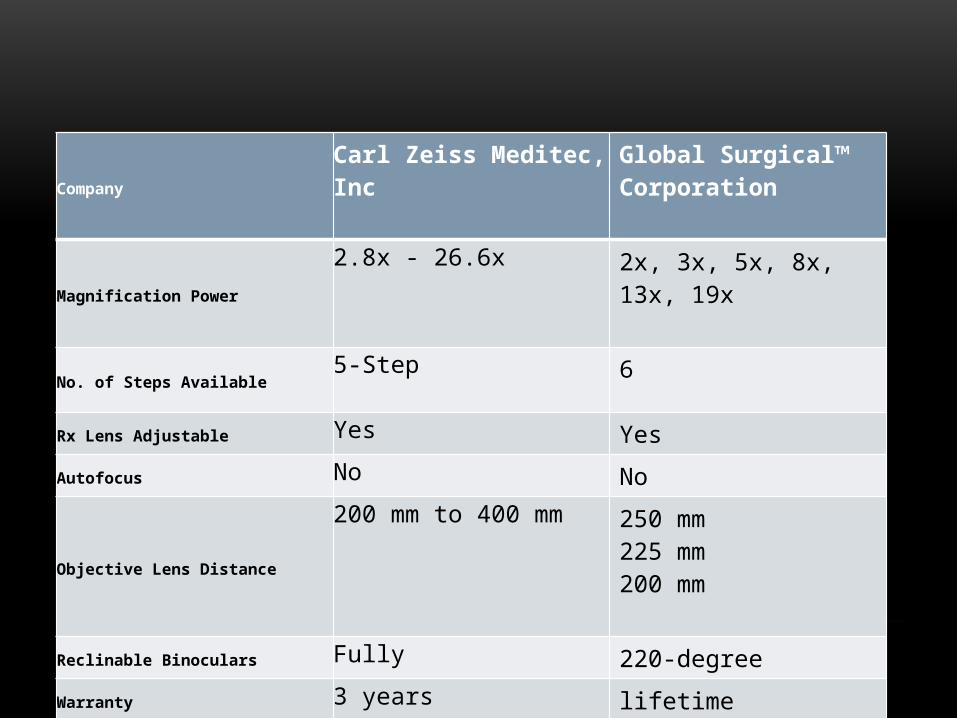
CompanyCarl Zeiss Meditec, Inc
Global Surgical™ Corporation
Magnification Power2.8x - 26.6x 2x, 3x, 5x, 8x, 13x, 19x
No. of Steps Available5-Step 6
Rx Lens Adjustable Yes Yes
Autofocus No No
Objective Lens Distance
200 mm to 400 mm 250 mm 225 mm 200 mm
Reclinable Binoculars Fully 220-degree
Warranty 3 years lifetime
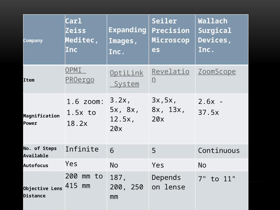
Company
Carl Zeiss Meditec, Inc
Expanding Images, Inc.
Seiler Precision Microscopes
Wallach Surgical Devices, Inc.
Item
OPMI PROergo
OptiLink System
Revelation ZoomScope
Magnification Power1.6 zoom: 1.5x to 18.2x
3.2x, 5x, 8x, 12.5x, 20x
3x,5x, 8x, 13x, 20x
2.6x - 37.5x
No. of Steps Available
Infinite 6 5 Continuous
Autofocus Yes No Yes No
Objective Lens Distance
200 mm to 415 mm
187, 200, 250 mm
Depends on lense
7" to 11"
Reclinable Binoculars
Fully Partially Fully No
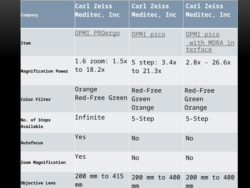
Company
Carl Zeiss Meditec, Inc
Carl Zeiss Meditec, Inc
Carl Zeiss Meditec, Inc
Item
OPMI PROergo OPMI pico OPMI pico with MORA interface
Magnification Power
1.6 zoom: 1.5x to 18.2x
5 step: 3.4x to 21.3x
2.8x - 26.6x
Color Filter
Orange Red-Free Green
Red-Free GreenOrange
Red-Free GreenOrange
No. of Steps AvailableInfinite 5-Step 5-Step
AutofocusYes No No
Zoom MagnificationYes No No
Objective Lens Distance
200 mm to 415 mm 200 mm to 400 mm
200 mm to 400 mm
Reclinable Binoculars Fully Fully Fully
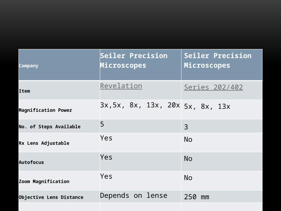
Company
Seiler Precision Microscopes
Seiler Precision Microscopes
ItemRevelation Series 202/402
Magnification Power3x,5x, 8x, 13x, 20x 5x, 8x, 13x
No. of Steps Available 5 3
Rx Lens AdjustableYes No
AutofocusYes No
Zoom MagnificationYes No
Objective Lens Distance Depends on lense 250 mm

COMPANIES
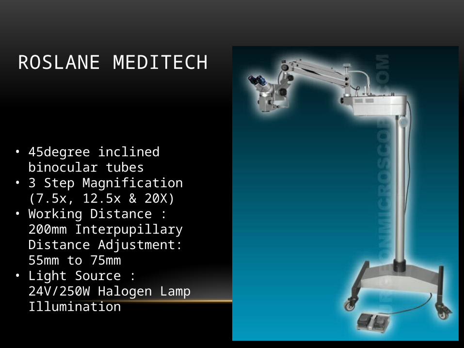
ROSLANE MEDITECH
• 45degree inclined binocular tubes
• 3 Step Magnification (7.5x, 12.5x & 20X)
• Working Distance : 200mm Interpupillary Distance Adjustment: 55mm to 75mm
• Light Source : 24V/250W Halogen Lamp Illumination
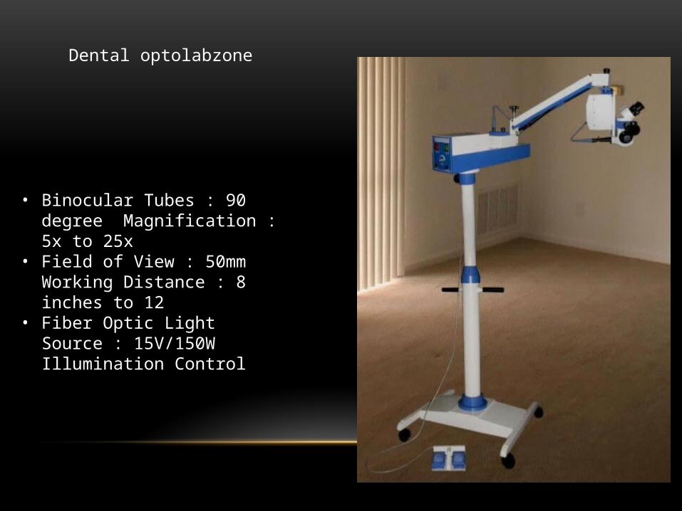
• Binocular Tubes : 90 degree Magnification : 5x to 25x
• Field of View : 50mm Working Distance : 8 inches to 12
• Fiber Optic Light Source : 15V/150W Illumination Control
Dental optolabzone
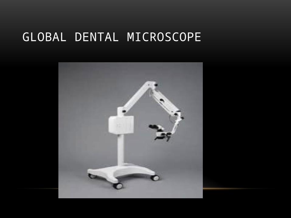
GLOBAL DENTAL MICROSCOPE
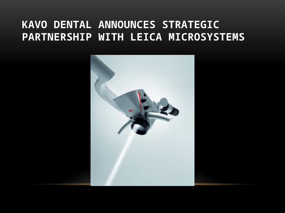
KAVO DENTAL ANNOUNCES STRATEGIC PARTNERSHIP WITH LEICA MICROSYSTEMS
• Leica M320 F12 – Designed for Dentists
It is the only dental microscope with LED illumination and High-
Definition image technology. LED illumination yields crisp, bright
and natural images with tremendous depth of field. With an
operational lifespan of 60,000 hours, the cost of operation is
appreciably lower than that of conventional light sources.
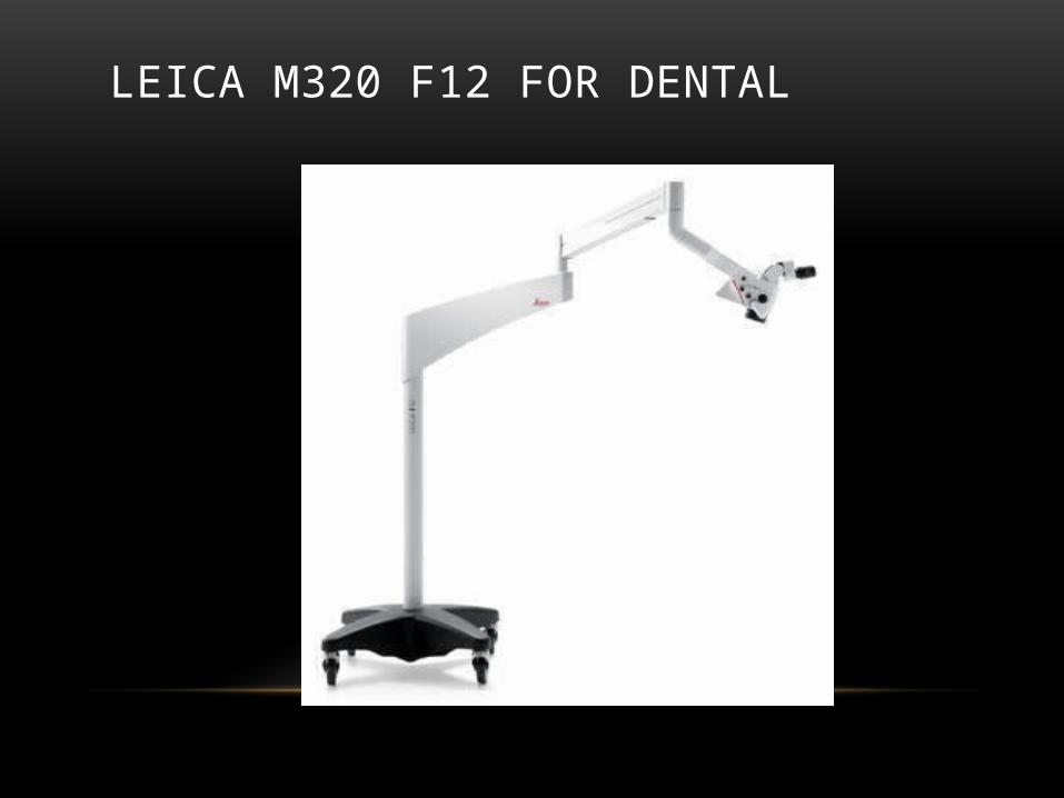
LEICA M320 F12 FOR DENTAL
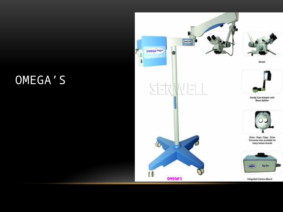
OMEGA’S

DENTAL MICROSCOPE - OPTOFILO
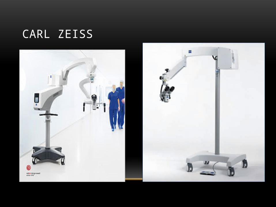
CARL ZEISS

WE HAVE 3 DIFFERENT OPTIONS TO CHOOSE FROM.
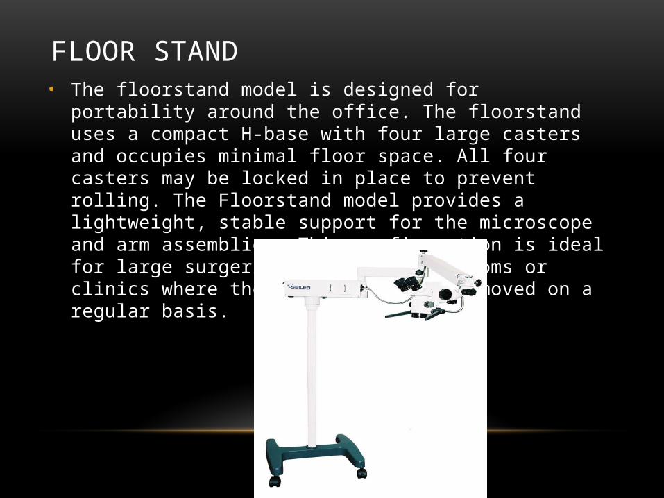
FLOOR STAND• The floorstand model is designed for portability around the office.
The floorstand uses a compact H-base with four large casters and occupies minimal floor space. All four casters may be locked in place to prevent rolling. The Floorstand model provides a lightweight, stable support for the microscope and arm assemblies. This configuration is ideal for large surgeries with multiple rooms or clinics where the scope needs to be moved on a regular basis.
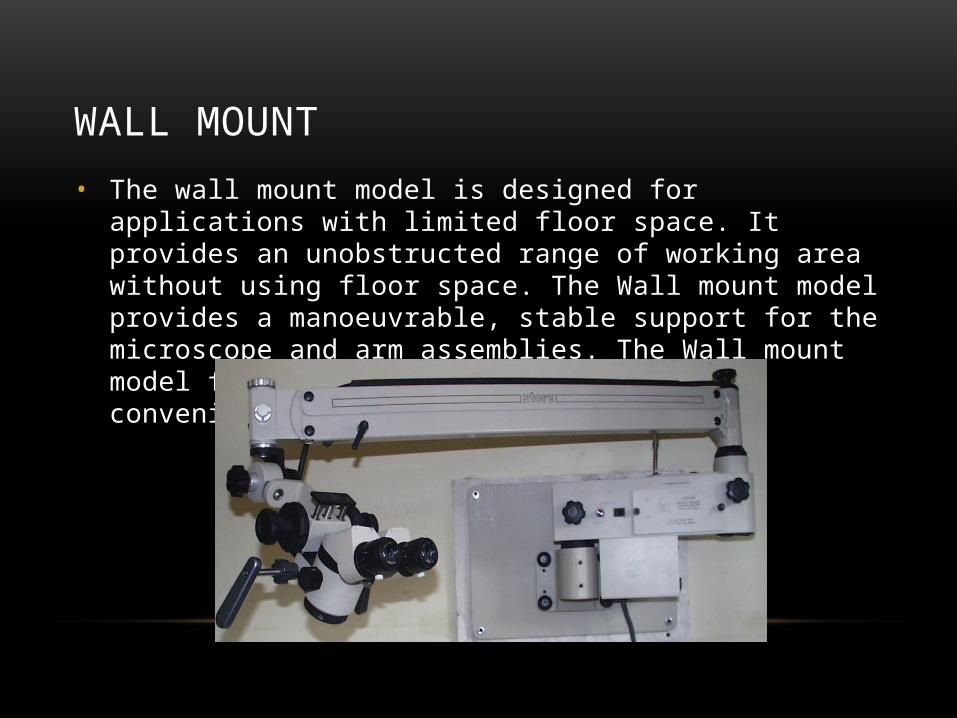
WALL MOUNT
• The wall mount model is designed for applications with limited floor space. It provides an unobstructed range of working area without using floor space. The Wall mount model provides a manoeuvrable, stable support for the microscope and arm assemblies. The Wall mount model folds flat against the wall for convenient storage.
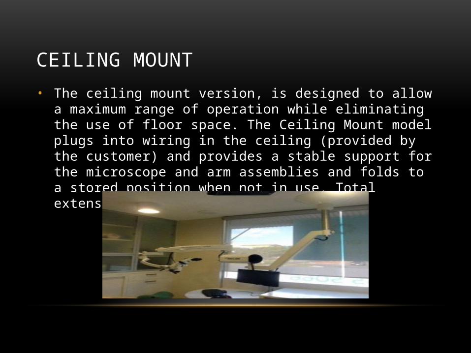
CEILING MOUNT
• The ceiling mount version, is designed to allow a maximum range of operation while eliminating the use of floor space. The Ceiling Mount model plugs into wiring in the ceiling (provided by the customer) and provides a stable support for the microscope and arm assemblies and folds to a stored position when not in use. Total extension reaches over 1.8 meters
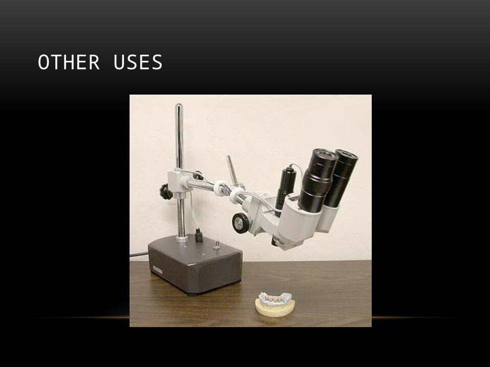
OTHER USES

THANK YOU