
Dental implants with versus without peri-implant bone ... · Dental implants with versus without...
8
J Clin Exp Dent. 2015;7(3):e361-8. Implants with versus without defects treated GBR e361 Journal section: Oral Surgery Publication Types: Research Dental implants with versus without peri-implant bone defects treated with guided bone regeneration Amparo Aloy-Prósper 1 , David Peñarrocha-Oltra 2 , Maria Peñarrocha-Diago 3 , Miguel Peñarrocha-Diago 4 1 DDS, MSc, Collaborating Professor of the Master in Oral Surgery and Implant Dentistry, Stomatology Department, Faculty of Medicine and Dentistry, University of Valencia, Spain 2 DDS, MSc, PhD, Junior Researcher, Collaborating Professor of the Master in Oral Surgery and Implant Dentistry, Stomatology Department, Faculty of Medicine and Dentistry, University of Valencia, Spain 3 DDS, PhD, Full Professor of Oral Surgery, Stomatology Department, Faculty of Medicine and Dentistry, University of Valencia, Spain 4 MD, PhD, Chairman of Oral Surgery, Stomatology Department, Faculty of Medicine and Dentistry, University of Valencia, Spain Correspondence: Clínicas odontológicas Gascó Oliag 1 46021- Valencia, Spain [email protected] Received: 07/01/2015 Accepted: 26/03/2015 Abstract Background: The guided bone regeneration (GBR) technique is highly successful for the treatment of peri-implant bone defects. The aim was to determine whether or not implants associated with GBR due to peri-implant defects show the same survival and success rates as implants placed in native bone without defects. Material and Methods: Patients with a minimum of two submerged dental implants: one suffering a dehiscence or fenestration defect during placement and undergoing simultaneous guided bone regeneration (test group), versus the other entirely surrounded by bone (control group) were treated and monitored annually for three years. Com- plications with the healing procedure, implant survival, implant success and peri-implant marginal bone loss were assessed. Statistical analysis was performed with non-parametric tests setting an alpha value of 0.05. Results: Seventy-two patients and 326 implants were included (142 test, 184 control). One hundred and twenty-five dehiscences (average height 1.92±1.11) and 18 fenestrations (average height 3.34±2.16) were treated. At 3 years post-loading, implant survival rates were 95.7% (test) and 97.3% (control) and implant success rates were 93.6% and 96.2%, respectively. Mean marginal bone loss was 0.54 (SD 0.26 mm) for the test group and 0.43 (SD 0.22 mm) for the control group. No statistically significant differences between both groups were found. Conclusions: Within the limits of this study, implants with peri-implant defects treated with guided bone regene- ration exhibited similar survival and success rates and peri-implant marginal bone loss to implants without those defects. Large-scale randomized controlled studies with longer follow-ups involving the assessment of esthetic parameters and hard and soft peri-implant tissue stability are needed. Key words: Guided bone regeneration, peri-implant defects, dental implants, marginal bone level, success rate, survival rate. doi:10.4317/jced.52292 http://dx.doi.org/10.4317/jced.52292 Article Number: 52292 http://www.medicinaoral.com/odo/indice.htm © Medicina Oral S. L. C.I.F. B 96689336 - eISSN: 1989-5488 eMail: [email protected] Indexed in: Pubmed Pubmed Central® (PMC) Scopus DOI® System Aloy-Prósper A, Peñarrocha-Oltra D, Peñarrocha-Diago MA, Peña- rrocha-Diago M. Dental implants with versus without peri-implant bone defects treated with guided bone regeneration. J Clin Exp Dent. 2015;7(3):e361-8. http://www.medicinaoral.com/odo/volumenes/v7i3/jcedv7i3p361.pdf
Transcript of Dental implants with versus without peri-implant bone ... · Dental implants with versus without...
J Clin Exp Dent. 2015;7(3):e361-8. Implants with versus without defects treated GBR
e361
Journal section: Oral Surgery Publication Types: Research
Dental implants with versus without peri-implant bone defects treated with guided bone regeneration
Amparo Aloy-Prósper 1, David Peñarrocha-Oltra 2, Maria Peñarrocha-Diago 3, Miguel Peñarrocha-Diago 4
1 DDS, MSc, Collaborating Professor of the Master in Oral Surgery and Implant Dentistry, Stomatology Department, Faculty of Medicine and Dentistry, University of Valencia, Spain2 DDS, MSc, PhD, Junior Researcher, Collaborating Professor of the Master in Oral Surgery and Implant Dentistry, Stomatology Department, Faculty of Medicine and Dentistry, University of Valencia, Spain3 DDS, PhD, Full Professor of Oral Surgery, Stomatology Department, Faculty of Medicine and Dentistry, University of Valencia, Spain4 MD, PhD, Chairman of Oral Surgery, Stomatology Department, Faculty of Medicine and Dentistry, University of Valencia, Spain
Correspondence:Clínicas odontológicasGascó Oliag 146021- Valencia, [email protected]
Received: 07/01/2015Accepted: 26/03/2015
Abstract Background: The guided bone regeneration (GBR) technique is highly successful for the treatment of peri-implant bone defects. The aim was to determine whether or not implants associated with GBR due to peri-implant defects show the same survival and success rates as implants placed in native bone without defects.Material and Methods: Patients with a minimum of two submerged dental implants: one suffering a dehiscence or fenestration defect during placement and undergoing simultaneous guided bone regeneration (test group), versus the other entirely surrounded by bone (control group) were treated and monitored annually for three years. Com-plications with the healing procedure, implant survival, implant success and peri-implant marginal bone loss were assessed. Statistical analysis was performed with non-parametric tests setting an alpha value of 0.05.Results: Seventy-two patients and 326 implants were included (142 test, 184 control). One hundred and twenty-five dehiscences (average height 1.92±1.11) and 18 fenestrations (average height 3.34±2.16) were treated. At 3 years post-loading, implant survival rates were 95.7% (test) and 97.3% (control) and implant success rates were 93.6% and 96.2%, respectively. Mean marginal bone loss was 0.54 (SD 0.26 mm) for the test group and 0.43 (SD 0.22 mm) for the control group. No statistically significant differences between both groups were found.Conclusions: Within the limits of this study, implants with peri-implant defects treated with guided bone regene-ration exhibited similar survival and success rates and peri-implant marginal bone loss to implants without those defects. Large-scale randomized controlled studies with longer follow-ups involving the assessment of esthetic parameters and hard and soft peri-implant tissue stability are needed.
Key words: Guided bone regeneration, peri-implant defects, dental implants, marginal bone level, success rate, survival rate.
doi:10.4317/jced.52292http://dx.doi.org/10.4317/jced.52292
Article Number: 52292 http://www.medicinaoral.com/odo/indice.htm© Medicina Oral S. L. C.I.F. B 96689336 - eISSN: 1989-5488eMail: [email protected] in:
PubmedPubmed Central® (PMC)ScopusDOI® System
Aloy-Prósper A, Peñarrocha-Oltra D, Peñarrocha-Diago MA, Peña-rrocha-Diago M. Dental implants with versus without peri-implant bone defects treated with guided bone regeneration. J Clin Exp Dent. 2015;7(3):e361-8.http://www.medicinaoral.com/odo/volumenes/v7i3/jcedv7i3p361.pdf

J Clin Exp Dent. 2015;7(3):e361-8. Implants with versus without defects treated GBR
e362
IntroductionHorizontal alveolar bone defects often result in a dehis-cence or a fenestration defect exposing part of the im-plant surface (1,2). Several clinical studies have shown that at least 1mm of bone width buccal and lingual to the implant surface is needed to ensure long-term bone coverage and therefore implant success (3,4). When this is lacking at the moment of implant placement, guided bone regeneration has been proposed to augment the bone width in a single simultaneous surgical interven-tion (5,6).An important issue is whether or not implants placed in sites associated with bone regeneration provide survival and success rates similar to those of implants placed in sites with sufficient native bone (7). Although there are a variety of case series studies on bone regeneration, three systematic reviews (1,8,9) on implant survival in sites regenerated with GBR identified only three studies (10-12) that compared implants with peri-implant de-fects that required bone grafts versus implants entirely surrounded by pristine bone as control implants in their analysis.The purpose of the present study was to retrospectively evaluate whether or not implants associated with bone regeneration due to peri-implant defects show the same survival and success rates as implants placed in native bone without such defects, and to evaluate long-term outcomes of implants with dehiscences and fenestrations treated with guided bone regeneration with a minimum follow-up of three years post-loading.
Subject and site inclusion criteria:A minimum of two dental implants, one with a dehiscence or fenestration bony defect during implant place-- ment treated with particulate bone graft and resorbable membranes (Test Group), and another implant entirely surrounded by bone (Control Group).Submerged implants- Tooth/teeth at implant site extracted >6 months previously- Rehabilitation with fixed or removable implant-supported prosthesis - Age > 18 years- No relevant medical conditions- Non-smoking or smoking - ≤ 20 cigarettes/day (all pipe or cigar smokers were excluded)Follow-up for at least three years after prosthetic loading -
Subject and site exclusion criteria:Patient’s with systemic or local conditions contraindicating implant therapy (previous chemotherapy, previous - irradiation of the head and neck region, active progressive periodontitis and/or immunosuppression).Pregnant and lactating patients- Sites with acute infection- Poor oral hygiene- Implants with sinus augmentation- Immediate implants or placed in a bone with a recent extraction (less than six months)- Re-implants- Implants placed in bone previously regenerated with bone grafts.- Patients failing to attend follow-up visits-
Table 1. Inclusion and exclusion criteria.
Material and Methods -Patient selectionA retrospective controlled clinical study was made of patients with a minimum of two dental implants, one implant demonstrating a dehiscence or fenestration bony defect with exposed implant surface during implant placement and so undergoing simultaneous particulate bone grafting with resorbable membranes (test group), and the other implant site entirely surrounded by bone (control group). All implants had to be left submerged. Patients were treated between January 2005 and Dec-ember 2009 at the Oral Surgery Unit of the University of Valencia in Spain and were monitored annually for a minimum of 3-years post-loading. Patients were given full information about the surgical procedures and duly signed informed consent forms. Preoperative analysis included registering complete medical histories and per-forming clinical and radiographic examinations. Inclu-sion and exclusion criteria are detailed in table 1. The present study is reported in accordance with the STRO-BE (Strengthening the Reporting of Observational Stu-dies in Epidemiology) statement (13).-Pre-Operative EvaluationThorough medical histories, clinical examinations and panoramic radiographs were performed in all cases. Cone-beam computed tomographic scans were obtained to assess the availability of bone whenever the surgeon considered this necessary. Periodontal treatment was provided whenever necessary to control inflammation prior implant placement surgery. Within 10 days of the

J Clin Exp Dent. 2015;7(3):e361-8. Implants with versus without defects treated GBR
e363
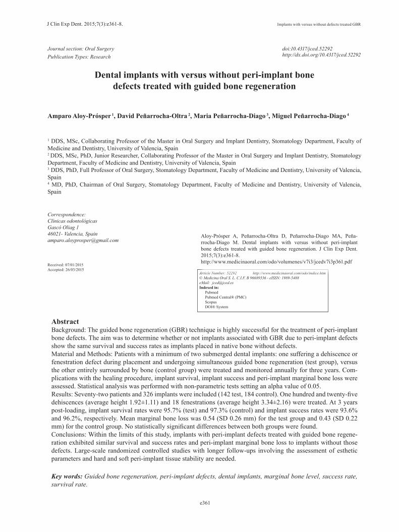
implant placement surgery, a full mouth professional prophylaxis appointment was scheduled.-Surgical proceduresSurgical procedures were performed by the same sur-geon with an extensive experience in regenerative pro-cedures. All procedures were performed under local anesthesia using 4% articaine 1:100,000 adrenalin (Ini-bsa, Lliça de Vall, Spain) and intravenous conscious se-dation with 1% propofol solution, administered by an anesthesiologist. All patients received antibiotic pro-phylaxis (amoxicillin and clavulanic acid, 1g every 8 hours starting 1 day preoperatively (14)). An initial in-cision was made slightly palatal/lingual of the alveolar crest. One or two releasing incisions were made and a mucoperiosteal flap was raised. The exposed alveolar bone was curetted to remove all soft tissues. To enhance primary stability, drills and osteotomes were combined to prepare implant beds. TSATM implants with Avant-blast surface (Phibo Dental Solutions S.L., Sentmenat, Barcelona, Spain) were inserted using standard proce-dures following the manufacturer’s guidelines. These implants have a polished surface portion of 1.5 mm. All implants were placed with adequate primary stability (≥35 Ncm). In implants that did not need bone regene-ration, bone width from the implant head to the facial plate was over 1.5 mm. In implants that needed bone regeneration, autologous bone grafts harvested from the conformation of implant beds during drilling and was adjusted to the bone contour. When the autologous bone obtained was of insufficient quantity to cover the peri-implant defects, synthetic bone (Kera-OsTM, Keramat, Coruña, Spain) was added. Grafted bone was protected with a textured collagen membrane (Lyopstic, B Braun, Aesculap, Germany). Periosteal incisions were made to allow flap mobilization and tension free primary wound closure. Implants were left submerged. Flaps were clo-sed with horizontal sutures using Polisoft® 4/0 sutures (Sweden & Martina, Due Carrare, Italy) (Figs. 1-10).
Fig. 1. Occlusal preoperative view.
Fig. 2. Dental implants placed in 4.5, 4.6 and 4.7 position.
Fig. 3. Autogenous bone graft over dehiscence.
Fig. 4. Resorbable membrane over bone graft.
Patients were prescribed amoxicillin and clavulanic acid 1g (GlaxoSmithKline, Madrid, Spain) twice daily for six days, 600 mg ibuprofen (Bexistar, Laboratorio Bacino, Barcelona, Spain) three times per day for five days and mouthrinse with chlorhexidine 0.12% (GUM, John O Butler/Sunstar, Chicago, IL, U.S.A.) twice daily, com-mencing three days prior to surgery and for two weeks
J Clin Exp Dent. 2015;7(3):e361-8. Implants with versus without defects treated GBR
e364
Fig. 5. Suture.
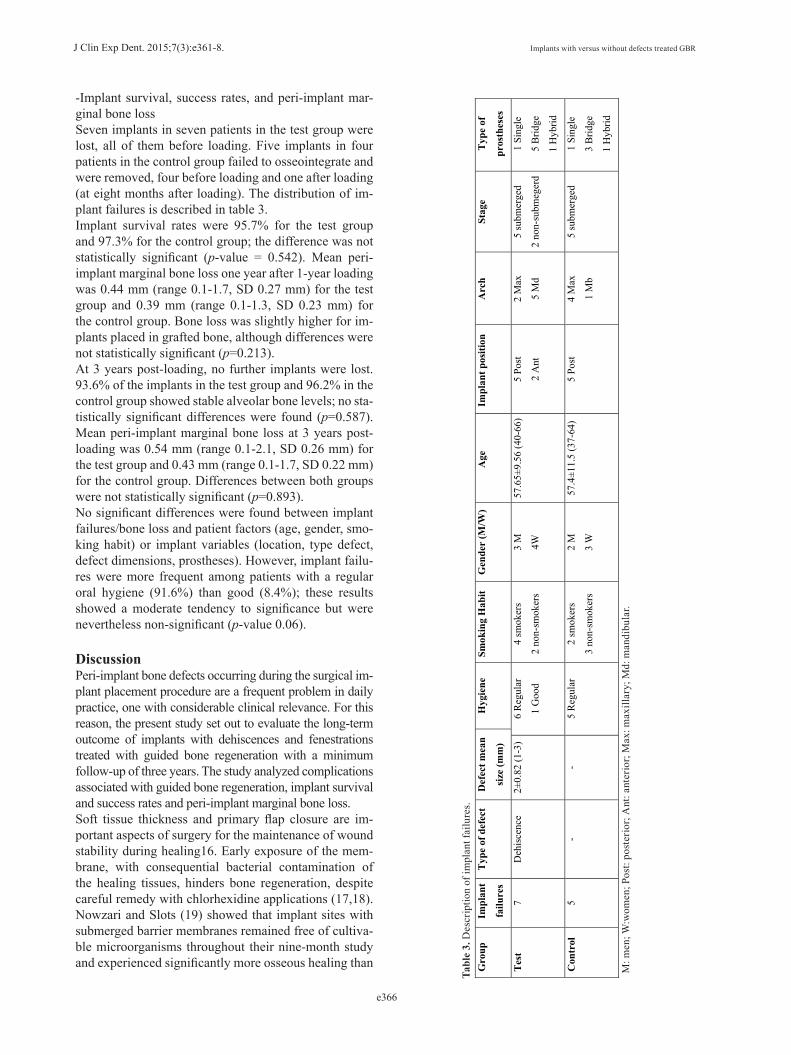
Fig. 6. Panoramic radiography after implant placement.
Fig. 7. Healed soft tissues. Fig. 8. Final prosthesis place-ment.
Fig. 9. Frontal view after final prosthesis placement.
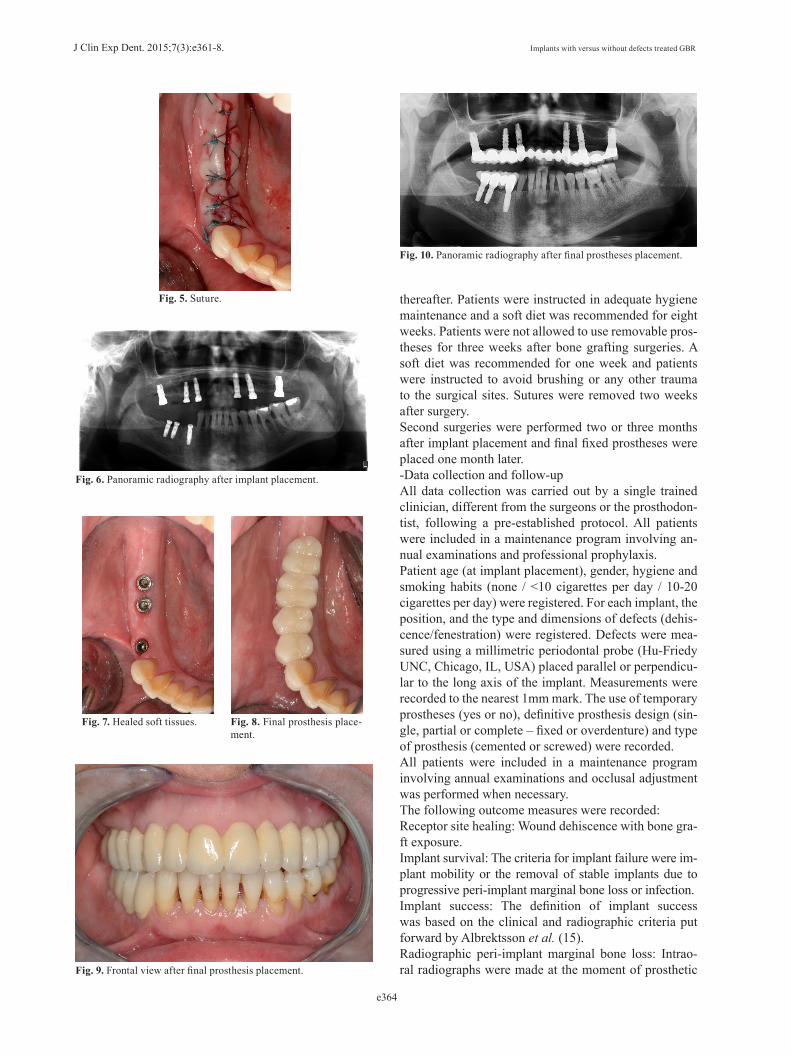
Fig. 10. Panoramic radiography after final prostheses placement.
thereafter. Patients were instructed in adequate hygiene maintenance and a soft diet was recommended for eight weeks. Patients were not allowed to use removable pros-theses for three weeks after bone grafting surgeries. A soft diet was recommended for one week and patients were instructed to avoid brushing or any other trauma to the surgical sites. Sutures were removed two weeks after surgery.Second surgeries were performed two or three months after implant placement and final fixed prostheses were placed one month later.-Data collection and follow-upAll data collection was carried out by a single trained clinician, different from the surgeons or the prosthodon-tist, following a pre-established protocol. All patients were included in a maintenance program involving an-nual examinations and professional prophylaxis.Patient age (at implant placement), gender, hygiene and smoking habits (none / <10 cigarettes per day / 10-20 cigarettes per day) were registered. For each implant, the position, and the type and dimensions of defects (dehis-cence/fenestration) were registered. Defects were mea-sured using a millimetric periodontal probe (Hu-Friedy UNC, Chicago, IL, USA) placed parallel or perpendicu-lar to the long axis of the implant. Measurements were recorded to the nearest 1mm mark. The use of temporary prostheses (yes or no), definitive prosthesis design (sin-gle, partial or complete – fixed or overdenture) and type of prosthesis (cemented or screwed) were recorded.All patients were included in a maintenance program involving annual examinations and occlusal adjustment was performed when necessary.The following outcome measures were recorded:Receptor site healing: Wound dehiscence with bone gra-ft exposure.Implant survival: The criteria for implant failure were im-plant mobility or the removal of stable implants due to progressive peri-implant marginal bone loss or infection. Implant success: The definition of implant success was based on the clinical and radiographic criteria put forward by Albrektsson et al. (15).Radiographic peri-implant marginal bone loss: Intrao-ral radiographs were made at the moment of prosthetic

J Clin Exp Dent. 2015;7(3):e361-8. Implants with versus without defects treated GBR
e365
loading (baseline), one-year post-loading and at 3 year control radiograph, using the X-MindTM intraoral sys-tem (Satelec-Pierre Rolland Group, Merignac, France) and an RVGTM intraoral digital receptor (Dürr Dental, Bietigheim-Bissingen, Germany) with the aid of a Rinn XCPTM (Dentsply Rinn, Elgin, IL, U.S.A.) to achieve parallelism. Evaluation of the marginal bone level around implants was performed using image analysis software (Autocad 2006, version Z 54.10, Autodesk, USA), which is designed to compensate for radiographic distortion. Each image was calibrated using the known length of the implants. The vertical distance from the outer edge of the implant shoulder (reference point) to the most co-ronal point of bone-to-implant contact was evaluated at the mesial and distal aspect of each implant. Peri-im-plant marginal bone resorption at 3-year post-loading was calculated from the change in bone level between the 1-year post-loading and the 3-year control radiogra-ph; for each pair of measurements (mesial and distal) the largest value was used. Intra-examiner calibration was analyzed before evaluating the entire implant sample by reassessing bone loss at a total of 30 randomly selec-ted sites (using the random function of Microsoft Excel 2010) on duplicate measurements performed on different days. An intraclass correlation coefficient of 0.833 was obtained, showing a high concordance between the two sets of data. According to Dahlberg’s d-value, a 0.046 mm error was estimated for the measurement method.-Statistical Analysis Statistical analysis was performed using non-parametric tests for implant success, as this was a non-continuous variable, and marginal bone loss, as this was an asym-metric distribution. The Chi2 test and the Mann-Whitney test (MW) were used to evaluate homogeneity within the two groups in relation to the demographic factors, clini-cal parameters, implant survival and success rates. The MW test was used to compare bone loss between groups. The relationship between the implant failure and bone loss with respect to age, gender, smoking and hygiene habit, position and location of the implants, type defect
and defect dimensions or type of graft was studied with non-parametric tests. The statistical power for this test was 99.5% to detect an effect of 0.27 with a confidence of 95% and alpha set at 0.05. Statistical analysis was performed using SPSS 17.0 software (SPSS Inc. Chica-go, IL, U.S.A.).
ResultsA total of 129 patients with submerged dental implants placed with dehiscences and fenestrations and treated with particulate bone graft and implants without these bony defects in the same patient were included. Eleven patients were excluded as a result of failing to attend the scheduled recall visits and 20 because control implants were not submerged.The final study sample included 72 patients (37 women and 35 men) with a mean age of 55.4 ± 11.7 (25-87). Hygiene maintenance was good in 47 patients and re-gular in 25. Forty-eight patients were non-smokers, 11 smoked less than 10 cigarettes per day, 7 between 10 and 20 cigarettes and 6 were ex-smokers.A total of 326 dental implants (142 test group, 184 control group) were placed. Out of 183 implants in test group, 162 had dehiscences and 21 with fenestrations. The mean dimensions of the resulting dehiscence defects were: 1.92±1.11 mm (range 1-6) length and 3.29 mm (range 3 to 5.5 mm) width. The mean dimensions of the resulting fenestration defects were: 3.34±2.16 mm (range 1-8) and 2.1 mm (range 1.5 to 3 mm) width. The distribution of patient or implant variables is described in table 2. No sta-tistically significant differences were found between the two study groups regarding implant variables.-Receptor site healingWound dehiscence with membrane exposure during the early postoperative period occurred in twelve grafted si-tes in twelve patients (8.4% membrane exposure rate). These exposures did not exceed 3mm in diameter. In these cases, 0.2% chlorhexidine gel was prescribed three times daily over the exposed membrane for 6 weeks af-ter surgery. All sites re-epithelialized uneventfully.
Table 2. Description of implants distribution sample.
N implants Group 1 (142) Group 2 (184) Test pArch Maxillary 69 88 Chi2 0.18
Mandibular 83 86 Position Anterior 47 32 Chi2 <0.01
Posterior 95 152 Temporary prostheses Yes 1 5 Chi2 0.236
No 182 230 Prostheses design Single 28 37 Chi2 0.21
Partial 89 87 Hybrid 13 32
Full arch 5 17 Overdenture 7 11
Type prostheses Cemented 64 63 Chi2 0.06 Screwed 42 67
J Clin Exp Dent. 2015;7(3):e361-8. Implants with versus without defects treated GBR
e366
Gro
up
Impl
ant
failu
res
Typ
e of
def
ect
Def
ect m
ean
size
(mm
)
Hyg
iene
Sm
okin
g H
abit
Gen
der
(M/W
) A
ge
Impl
ant p
ositi
on
Arc
h St
age
Typ
e of
pros
thes
es
Tes
t7
Deh
isce
nce
2±0.
82 (1
-3)
6 R
egul
ar
1 G
ood
4 sm
oker
s
2 no
n-sm
oker
s
3 M
4W
57.6
5±9.
56 (4
0-66
)5
Post
2 A
nt
2 M
ax
5 M
d
5 su
bmer
ged
2 no
n-su
bmeg
erd
1 Si
ngle
5 B
ridge
1 H
ybrid
Con
trol
5 -
- 5
Reg
ular
2
smok
ers
3 no
n-sm
oker
s
2 M
3 W
57.4
±11.
5 (3
7-64
) 5
Post
4
Max
1 M
b
5 su
bmer
ged
1 Si
ngle
3 B
ridge
1 H
ybrid
Tabl
e 3.
Des
crip
tion
of im
plan
t fai
lure
s.
M: m
en; W
:wom
en; P
ost:
post
erio
r; A
nt: a
nter
ior;
Max
: max
illar
y; M
d: m
andi
bula
r.
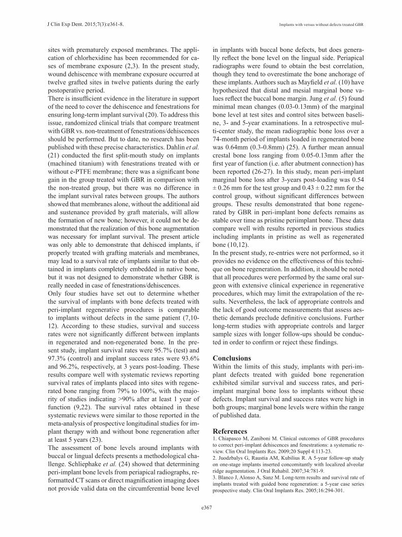
-Implant survival, success rates, and peri-implant mar-ginal bone lossSeven implants in seven patients in the test group were lost, all of them before loading. Five implants in four patients in the control group failed to osseointegrate and were removed, four before loading and one after loading (at eight months after loading). The distribution of im-plant failures is described in table 3.Implant survival rates were 95.7% for the test group and 97.3% for the control group; the difference was not statistically significant (p-value = 0.542). Mean peri-implant marginal bone loss one year after 1-year loading was 0.44 mm (range 0.1-1.7, SD 0.27 mm) for the test group and 0.39 mm (range 0.1-1.3, SD 0.23 mm) for the control group. Bone loss was slightly higher for im-plants placed in grafted bone, although differences were not statistically significant (p=0.213).At 3 years post-loading, no further implants were lost. 93.6% of the implants in the test group and 96.2% in the control group showed stable alveolar bone levels; no sta-tistically significant differences were found (p=0.587). Mean peri-implant marginal bone loss at 3 years post-loading was 0.54 mm (range 0.1-2.1, SD 0.26 mm) for the test group and 0.43 mm (range 0.1-1.7, SD 0.22 mm) for the control group. Differences between both groups were not statistically significant (p=0.893).No significant differences were found between implant failures/bone loss and patient factors (age, gender, smo-king habit) or implant variables (location, type defect, defect dimensions, prostheses). However, implant failu-res were more frequent among patients with a regular oral hygiene (91.6%) than good (8.4%); these results showed a moderate tendency to significance but were nevertheless non-significant (p-value 0.06).
DiscussionPeri-implant bone defects occurring during the surgical im-plant placement procedure are a frequent problem in daily practice, one with considerable clinical relevance. For this reason, the present study set out to evaluate the long-term outcome of implants with dehiscences and fenestrations treated with guided bone regeneration with a minimum follow-up of three years. The study analyzed complications associated with guided bone regeneration, implant survival and success rates and peri-implant marginal bone loss.Soft tissue thickness and primary flap closure are im-portant aspects of surgery for the maintenance of wound stability during healing16. Early exposure of the mem-brane, with consequential bacterial contamination of the healing tissues, hinders bone regeneration, despite careful remedy with chlorhexidine applications (17,18). Nowzari and Slots (19) showed that implant sites with submerged barrier membranes remained free of cultiva-ble microorganisms throughout their nine-month study and experienced significantly more osseous healing than
J Clin Exp Dent. 2015;7(3):e361-8. Implants with versus without defects treated GBR
e367
sites with prematurely exposed membranes. The appli-cation of chlorhexidine has been recommended for ca-ses of membrane exposure (2,3). In the present study, wound dehiscence with membrane exposure occurred at twelve grafted sites in twelve patients during the early postoperative period.There is insufficient evidence in the literature in support of the need to cover the dehiscence and fenestrations for ensuring long-term implant survival (20). To address this issue, randomized clinical trials that compare treatment with GBR vs. non-treatment of fenestrations/dehiscences should be performed. But to date, no research has been published with these precise characteristics. Dahlin et al. (21) conducted the first split-mouth study on implants (machined titanium) with fenestrations treated with or without e-PTFE membrane; there was a significant bone gain in the group treated with GBR in comparison with the non-treated group, but there was no difference in the implant survival rates between groups. The authors showed that membranes alone, without the additional aid and sustenance provided by graft materials, will allow the formation of new bone; however, it could not be de-monstrated that the realization of this bone augmentation was necessary for implant survival. The present article was only able to demonstrate that dehisced implants, if properly treated with grafting materials and membranes, may lead to a survival rate of implants similar to that ob-tained in implants completely embedded in native bone, but it was not designed to demonstrate whether GBR is really needed in case of fenestrations/dehiscences.Only four studies have set out to determine whether the survival of implants with bone defects treated with peri-implant regenerative procedures is comparable to implants without defects in the same patient (7,10-12). According to these studies, survival and success rates were not significantly different between implants in regenerated and non-regenerated bone. In the pre-sent study, implant survival rates were 95.7% (test) and 97.3% (control) and implant success rates were 93.6% and 96.2%, respectively, at 3 years post-loading. These results compare well with systematic reviews reporting survival rates of implants placed into sites with regene-rated bone ranging from 79% to 100%, with the majo-rity of studies indicating >90% after at least 1 year of function (9,22). The survival rates obtained in these systematic reviews were similar to those reported in the meta-analysis of prospective longitudinal studies for im-plant therapy with and without bone regeneration after at least 5 years (23).The assessment of bone levels around implants with buccal or lingual defects presents a methodological cha-llenge. Schliephake et al. (24) showed that determining peri-implant bone levels from periapical radiographs, re-formatted CT scans or direct magnification imaging does not provide valid data on the circumferential bone level
in implants with buccal bone defects, but does genera-lly reflect the bone level on the lingual side. Periapical radiographs were found to obtain the best correlation, though they tend to overestimate the bone anchorage of these implants. Authors such as Mayfield et al. (10) have hypothesized that distal and mesial marginal bone va-lues reflect the buccal bone margin. Jung et al. (5) found minimal mean changes (0.03-0.13mm) of the marginal bone level at test sites and control sites between baseli-ne, 3- and 5-year examinations. In a retrospective mul-ti-center study, the mean radiographic bone loss over a 74-month period of implants loaded in regenerated bone was 0.64mm (0.3-0.8mm) (25). A further mean annual crestal bone loss ranging from 0.05-0.13mm after the first year of function (i.e. after abutment connection) has been reported (26-27). In this study, mean peri-implant marginal bone loss after 3-years post-loading was 0.54 ± 0.26 mm for the test group and 0.43 ± 0.22 mm for the control group, without significant differences between groups. These results demonstrated that bone regene-rated by GBR in peri-implant bone defects remains as stable over time as pristine periimplant bone. These data compare well with results reported in previous studies including implants in pristine as well as regenerated bone (10,12). In the present study, re-entries were not performed, so it provides no evidence on the effectiveness of this techni-que on bone regeneration. In addition, it should be noted that all procedures were performed by the same oral sur-geon with extensive clinical experience in regenerative procedures, which may limit the extrapolation of the re-sults. Nevertheless, the lack of appropriate controls and the lack of good outcome measurements that assess aes-thetic demands preclude definitive conclusions. Further long-term studies with appropriate controls and larger sample sizes with longer follow-ups should be conduc-ted in order to confirm or reject these findings.
ConclusionsWithin the limits of this study, implants with peri-im-plant defects treated with guided bone regeneration exhibited similar survival and success rates, and peri-implant marginal bone loss to implants without these defects. Implant survival and success rates were high in both groups; marginal bone levels were within the range of published data.
References1. Chiapasco M, Zaniboni M. Clinical outcomes of GBR procedures to correct peri-implant dehiscences and fenestrations: a systematic re-view. Clin Oral Implants Res. 2009;20 Suppl 4:113-23.2. Juodzbalys G, Raustia AM, Kubilius R. A 5-year follow-up study on one-stage implants inserted concomitantly with localized alveolar ridge augmentation. J Oral Rehabil. 2007;34:781-9.3. Blanco J, Alonso A, Sanz M. Long-term results and survival rate of implants treated with guided bone regeneration: a 5-year case series prospective study. Clin Oral Implants Res. 2005;16:294-301.

J Clin Exp Dent. 2015;7(3):e361-8. Implants with versus without defects treated GBR
e368
4. Lekholm U, Sennerby L, Ross J, Becker W. Soft tissue and mar-ginal bone condition at osseointegrated implants that have exposed threads: A 5-year retrospective study. Int J Oral Maxillofac Implants. 1996;11:599-604.5. Jung RE, Hälg GA, Thoma DS, Hämmerle CH. A randomized, controlled clinical trial to evaluate a new membrane for guided bone regeneration around dental implants. Clin Oral Implants Res. 2009;20:162-8.6. Schwarz F, Mihatovic I, Golubovic V, Hegewald A, Becker J. In-fluence of two barrier membranes on staged guided bone regeneration and osseointegration of titanium implants in dogs: part 1. Augmenta-tion using bone graft substitutes and autogenous bone. Clin Oral Im-plants Res. 2012;23:83-9.7. Benić GI, Jung RE, Siegenthaler DW, Hämmerle CH. Clinical and radiographic comparison of implants in regenerated or native bone: 5-year results. Clin Oral Implants Res. 2009;20:507-13.8. Hämmerle CH, Jung RE, Feloutzis A. A systematic review of the survival of implants in bone sites augmented with barrier membranes (guided bone regeneration) in partially edentulous patients. J Clin Pe-riodontol. 2002;29 Suppl 3:226-31; discussion 232-3.9. Fiorellini JP, Nevins ML. Localized ridge augmentation/preserva-tion. A systematic review. Ann Periodontol. 2003;8:321-7.10. Mayfield L, Skoglund A, Nobréus N, Attström R. Clinical and ra-diographic evaluation, following delivery of fixed reconstructions, at GBR treated titanium fixtures. Clin Oral Implants Res. 1998;9:292-302.11. Corrente G, Abundo R, Cardaropoli D, Cardaropoli G, Martus-celli G. Long-term evaluation of osseointegrated implants in regene-rated and nonregenerated bone. Int J Periodontics Restorative Dent. 2000;20:390-7.12. Zitzmann NU, Schärer P, Marinello CP, Schüpbach P, Berglundh T. Alveolar ridge augmentation with Bio-Oss: a histologic study in hu-mans. Int J Periodontics Restorative Dent. 2001;21:288-95.13. Kuller LH, Goldstein BD. Suggestions for STROBE recommenda-tions. Epidemiology. 2007;18:792-3.14. Esposito M, Grusovin MG, Loli V, Coulthard P, Worthington HV. Does antibiotic prophylaxis at implant placement decrease early im-plant failures? A Cochrane systematic review. Eur J Oral Implantol. 2010;3:101-110.15. Albrektsson T, Zarb G, Worthington P, Eriksson AR. The long-term efficacy of currently used dental implants: a review and proposed crite-ria of success. Int J Oral Maxillofac Implants. 1986;1:11-25.16. Hwey-Chin, Kuang-Wei. Guided bone regeneration for fenestra-tion defects in dental implants. Ghang Gung Med J. 2003;26:684-9.17. Zitzmann NU, Naef R, Schärer P. Resorbable versus nonresorbable membranes in combination with Bio-Oss for guided bone regenera-tion. Int J Oral Maxillofac Implants. 1997;12:844-52. Erratum in: Int J Oral Maxillofac Implants. 1998;13:576.18. Simion M, Baldoni M, Rossi P, Zaffe D. A comparative study of the effectiveness of e-PTFE membranes with and without early ex-posure during thehealing period. Int J Periodontics Restorative Dent. 1994;14:166-80.19. Nowzari H, Slots J. Microorganism in polytetrafluoroethyle-ne membranes for guided tissue regeneration. J Clin Periodontol. 1994;21:203-10.20. Chiapasco M, Zaniboni M, Boisco M. Augm.entation procedures for the rehabilitation of deficient edentulous ridges with oral implants. Clin Oral Implants Res. 2006;17 Suppl 2:136-59.21. Dahlin C, Lekholm U, Becker W, Becker B, Higuchi K, Callens A, van Steenberghe D. Treatment of fenestration and dehiscence bone defects around oral implants using the guided tissue regeneration tech-nique: a prospective multicenter study. Int J Oral Maxillofac Implants. 1995;10:312-8.22. Hämmerle C, Feloutzis A. A systematic review of the survival of implants in bone sites augmented with barrier membranes (guided bone regeneration) in partially edentulous patients. J Clin Periodontol. 2002;29:226-31.23. Berglundh T, Persson L. Klinge B. A systematic review of the in-cidence of biological and technical complications in implant dentistry
reported in prospective longitudinal studies of at least 5 years. Journal of Clinical Periodontology. 2002; 29:197-212.24. Schliephake H, Wichmann M, Donnerstag F, Vogt S. Imaging of peri-implant bone levels of implants with buccal bone defects. Clin Oral Implants Res. 2003;14:193-200.25. Nevins M, Mellonig JT, Clem DS, Reiser GM, Buser DA. Implants in regenerated bone: long-term survival. Int J Periodont Rest Dent. 1998;18:34–45.26. Ahlqvist J, Borg K, Gunne J, Nilson H, Olsson M, Astrand P. Os-seointegrated implants in edentulous jaws: a 2-year longitudinal study. Int J Oral Maxillofac Impl. 1990;5:155-63.27. Adell R, Lekholm U, Gröndahl K, Bränemark PI, Lindström J, Ja-cobsson M. Reconstruction of severely resorbed edentulous maxillae using osseointegrated fixtures in immediate autogenous bone grafts. Int J Oral MAxillofac Impl. 1990;5:233-46.
Conflict of InterestNone of the authors have a conflict of interest.


