dental emengency Ch4
17
Chapter 4 Acute Oral Medical and Surgical Conditions P.J. Thomson Introduction Oral medicine and oral surgery are specialised branches of dentistry that deal with a wide range of disorders affecting the mouth, face and jaws and which normally require medical or surgical intervention. There is considerable over- lap in the range of conditions that present to medical and surgical specialists, but a number of very important clinical conditions can present acutely to dental practitioners. Whilst the detailed management of these disorders is outside the scope of this book, it is important that the dental emergency clinic practitioner is aware not only of the type and medical significance of such presentations, but also which patients should be referred for specialist assessment and treatment. As most dental emergency clinics function as integral parts of dental hospitals, close working relationships exist with oral and maxillofacial surgery units and oral medicine departments that facilitate such care. An acute condition may be defined as a suddenly presenting disorder, usu- ally with only a short history of symptoms, but with a degree of severity that causes significant disruption to the patient. They include traumatic injuries (described in Chapter 7), facial pain (which is discussed in detail in Chapter 6), swellings arising both intra-orally and around the face, jaws and neck, blister- ing and ulcerative disorders of the oral mucosa, disturbed oro-facial sensory or motor function and haemorrhage. This chapter will summarise a wide range of clinical disorders that practitioners should be aware of and which may present as acute conditions in the emergency clinic. Oro-facial swelling A swelling is defined as a transient enlargement or protuberance of part of the body and may arise intra-orally or externally around the face, jaws Dental Emergencies, First Edition. Edited by Mark Greenwood and Ian Corbett. C 2012 Blackwell Publishing Ltd. Published 2012 by Blackwell Publishing Ltd. 39
-
Upload
eugene-njebarikanuye -
Category
Health & Medicine
-
view
33 -
download
0
Transcript of dental emengency Ch4

P1: SFK/UKS P2: SFK
BLBK407-c04 BLBK407-Greenwood January 24, 2012 11:7 Trim: 244mm×172mm
Chapter 4
Acute Oral Medical and SurgicalConditionsP.J. Thomson
Introduction
Oral medicine and oral surgery are specialised branches of dentistry that deal
with a wide range of disorders affecting the mouth, face and jaws and which
normally require medical or surgical intervention. There is considerable over-
lap in the range of conditions that present to medical and surgical specialists,
but a number of very important clinical conditions can present acutely to
dental practitioners.
Whilst the detailed management of these disorders is outside the scope of
this book, it is important that the dental emergency clinic practitioner is aware
not only of the type and medical significance of such presentations, but also
which patients should be referred for specialist assessment and treatment. As
most dental emergency clinics function as integral parts of dental hospitals,
close working relationships exist with oral and maxillofacial surgery units and
oral medicine departments that facilitate such care.
An acute condition may be defined as a suddenly presenting disorder, usu-
ally with only a short history of symptoms, but with a degree of severity that
causes significant disruption to the patient. They include traumatic injuries
(described in Chapter 7), facial pain (which is discussed in detail in Chapter 6),
swellings arising both intra-orally and around the face, jaws and neck, blister-
ing and ulcerative disorders of the oralmucosa, disturbed oro-facial sensory or
motor function and haemorrhage. This chapter will summarise a wide range of
clinical disorders that practitioners should be aware of and which may present
as acute conditions in the emergency clinic.
Oro-facial swelling
A swelling is defined as a transient enlargement or protuberance of part
of the body and may arise intra-orally or externally around the face, jaws
Dental Emergencies, First Edition. Edited by Mark Greenwood and Ian Corbett.C© 2012 Blackwell Publishing Ltd. Published 2012 by Blackwell Publishing Ltd.
39

P1: SFK/UKS P2: SFK
BLBK407-c04 BLBK407-Greenwood January 24, 2012 11:7 Trim: 244mm×172mm
40 Dental Emergencies
and neck. Acute swellings are usually caused by trauma, or infection and
inflammation.
Traumatic swellings include haematoma, facial or dento-alveolar fractures
and temporomandibular joint effusions or dislocations. Signs and symptoms
that help the clinician to recognise facial fractures are summarised in Table 4.1.
Fractures inevitably display localised swelling, bruising and deformity and
the patient will experience both pain and some loss of function. The precise
Table 4.1 Signs and symptoms of facial fractures.
Type of facial fracture Sign and symptom
Unilateral condyle Fracture side:� Pain, swelling, bruising� Joint immobility� Deviation of mandible on mouth opening� Premature tooth contact
Opposite side:� Lateral open bite� Inability to move mandible laterally
Bilateral condyle � Pain, swelling, bruising and immobility ofboth joints
� Anterior open bite� No lateral mandibular movement possible
Mandible � Pain, swelling, bruising at site of fracture� Trismus� Abnormal mobility of fractured segments� Step deformity at lower border of mandible� Deranged occlusion� Numbness of lip and chin
Zygomatico-orbitalcomplex
� Pain, swelling, bruising over cheek andaround eye
� Subconjunctival haemorrhage� Later, flattening and depression of cheekprominence
� Step deformity at infra-orbital margin� Numbness of upper lip, lateral nose andcheek
� Intra-oral bruising in upper buccal sulcus� Trismus if coronoid process impacts ondisplaced zygoma
Middle third fractures � Can be gross facial swelling and tenderness� Bilateral circumorbital bruising� Mobility at infra-orbital margins orzygomatico-frontal sutures
� Widespread facial numbness� Deranged occlusion with anterior open bite� Mobile maxillary alveolus

P1: SFK/UKS P2: SFK
BLBK407-c04 BLBK407-Greenwood January 24, 2012 11:7 Trim: 244mm×172mm
Acute Oral Medical and Surgical Conditions 41
Figure 4.1 Minor salivary gland tumour arising in palatal tissue.
history of the preceding injury, examination of the anatomical site involved
and standard radiographic assessmentwill help clarify the diagnosis, and these
cases should be referred to specialist oral and maxillofacial surgery opinion.
Localised intra-oral swellings are very common, and although sometimes of
long standing may present as ‘acute’ problems due to sudden patient aware-
ness. Gingival swellingsmay represent localised acute periodontal problems or
rarer disorders such as giant cell granulomas, whilst palatal swellings can arise
fromacute dental abscesses or infected odontogenic cysts, particularly involv-
ing the lateral incisor or palatal roots of molar teeth, but may be due to other
more sinister causes such as minor salivary gland tumours. Tumours arising
from minor salivary glands are often malignant and the practitioner should
have a high index of suspicion, especially for diffuse, firm palatal swellings in
the absence of obvious dental disease. Figure 4.1 illustrates a salivary gland
tumour arising in the palate.
Similarly, patientsmay presentwith lip swellings thatmay be diffuse ormore
localised. Acute, diffuse lip swelling usually represents an allergic response but
localised lesions may arise from inflammation or obstruction of minor salivary
gland lesions. Whilst mucus extravasation cysts, due to local trauma, account
for the majority of lower lip lesions, upper lip swellings are more commonly
the result of minor salivary gland tumours.
Acute inflammatory and infective swellings may arise in a number of
anatomical sites around the mouth and face, presenting intra-orally as lo-
calised dental abscesses or cervico-facial swellings due to spreading tissue
space infections. Figure 4.2 shows facial swelling characteristic of a buccal
space infection following an intra-oral dental abscess. The various types of
clinical presentation, the anatomical sites where infections can localise and
their relevant diagnostic features are summarised in Table 4.2.
The rapid spread of infection through connective tissue spaces, often re-
ferred to as cellulitis, can give rise to airway obstruction and life-threatening
conditions, such as Ludwig’s angina, which is a large infective swelling

P1: SFK/UKS P2: SFK
BLBK407-c04 BLBK407-Greenwood January 24, 2012 11:7 Trim: 244mm×172mm
42 Dental Emergencies
Figure 4.2 Buccal space infection giving rise to facial swelling.
involving bilateral submandibular, sublingual and submental spaces. Swelling
may extend down the anterior neck, with massive distension of the floor of
the mouth and elevation and protrusion of the tongue. Such presentations areacute surgical emergencies and require immediate referral.
The cardinal signs of acute inflammation comprise swelling (often with re-
sultant suppuration and abscess formation), pain, redness, heat and loss of
function. Sometimes, patients may present with cutaneous swellings or si-
nuses as a result of discharge of infected material from dental abscesses.
There may also be systemic signs such as pyrexia, malaise, sweating, dehy-
dration and rapid pulse.
Clearly, clinical management is dependent upon the precise cause of infec-
tion and the general state of the patient. Localised dento-alveolar abscesses
may be appropriately treated by intra-oral drainage via tooth extraction, open-
ing of root canals and/or intra-oral incision and drainage. Wherever there are
signs of spreading cervico-facial infection or significant systemic disturbance,
however, patients should be referred urgently to the maxillofacial unit for
admission and further management.
One of the commonest acute oral surgery conditions to present to the emer-
gency clinic is pericoronitis, inflammation around the crown of a partly erupted
or impacted tooth usually the mandibular third molar. In acute cases, the pa-
tient experiences pain and swelling around the tooth, together with a foul taste
and often halitosis. There may also be trismus, dysphagia, facial swelling and
pyrexia. Diagnosis follows radiographic examination and confirmation that the
infection has not arisen from infected adjacent teeth. Extraction of traumatic
upper third molars, localised antibacterial treatment and antibiotic prescrip-
tion may be required to ease the acute condition before referral for specialist
advice on surgical removal. As in all infective conditions, rapidly spreadingfacial or neck swelling requires urgent referral and hospital admission.
A number of acute oral mucosal infections, such as pseudomembranous
candidiasis or herpetic gingivostomatitis may give rise not only to classical

P1: SFK/UKS P2: SFK
BLBK407-c04 BLBK407-Greenwood January 24, 2012 11:7 Trim: 244mm×172mm
Acute Oral Medical and Surgical Conditions 43
Table 4.2 Oro-facial tissue space infections.
Anatomical site Location Clinical signs
Lower jaw tissue spaces
Submental Between mylohyoid above andskin below
Firm swelling beneath chin
Submandibular Between anterior andposterior digastric muscles
Submandibular swelling
Sublingual Lingual mandible betweenmylohyoid and mucosa
Floor of mouth swelling andraised tongue
Buccal Between buccinator andmasseter
Swelling behind angle ofmouth to lower mandibularborder
Submasseteric Between masseter and lateralramus
Trismus, swelling confined tomasseter
Pterygomandibular Between medial pterygoid andmedial ramus
Severe trismus, dysphagia,limited buccal andsubmandibular swelling
Lateralpharyngeal
Skull base to hyoid, mediallypharyngeal muscle, laterallyfascia
Pyrexia, malaise, dysphagia,trismus, swollen fauces
Peritonsillar Between pharyngeal muscleand tonsil
Dysphagia, ‘hot potato’speech, swollen fauces andsoft palate
Upper jaw tissue spaces
Lip Between orbicularis oris andmucosa
Diffuse labial swelling
Canine fossa Between levator muscles andfacial skin
Diffuse swelling lip, cheek andlower eyelid
Palatal Subperiosteal space Circumscribed fluctuantpalatal swelling
Infratemporalfossa
Below greater wing ofsphenoid, ramus lateral andpterygoid plate medial
Trismus, pyrexia
Subtemporalis Between temporal bone andtemporalis muscle
Temporal swelling, trismus
intra-oral discomfort or ulceration but also to labial and facial swelling and
cervical lymphadenopathy.
Acute swelling of the major salivary glands may also present to the dental
emergency clinic. Whilst an acute bacterial parotitis, usually a consequence
of dehydration or obstructed salivary flow, is rarely seen these days, viral
parotitis due to mumps infection is becoming more common. In the UK, in
particular, this may be related to a lack of uptake of the MMR vaccine due to
adverse publicity in the late 1990s. Whilst the classic description of mumps

P1: SFK/UKS P2: SFK
BLBK407-c04 BLBK407-Greenwood January 24, 2012 11:7 Trim: 244mm×172mm
44 Dental Emergencies
Table 4.3 Differential diagnosis of neck lumps.
Type of lump in next Cause
Skin and superficialfascial lesions
Infective skin lesionsEpidermoid cystLipoma
Cystic lesions Sublingual dermoidThyroglossal cystBranchial cystLymphangioma
Cervicallymphadenopathy
Acute lymphadenitisChronic infectionsMetastatic malignant diseaseLeukaemia or lymphoma
Others Thyroid gland diseaseAneurysmSternomastoid tumourCervical ribCarotid body tumour
emphasises bilateral parotid swellingwith everted earlobes and accompanying
submandibular swelling, patients often present initiallywith an acute unilateral
parotid swelling that may pose a diagnostic dilemma.
Isolated acute submandibular salivary gland swellings are less common,
more usually presenting as intermittent obstructive swellings due to calcu-
lus formation in the ductal system. The history and clinical examination is
usually sufficient to establish the diagnosis. Salivary gland disorders are best
referred to oral and maxillofacial surgery specialists for further investigation
and management.
Swellings may also present as more discrete neck lumps. Whilst there is a
wide range of causes for a lump in the neck, summarised in Table 4.3, many
of these may be long-standing, chronic disorders that have only just been
noticed by the patient. Most patients are well aware of the potential sinister
nature of lumps and will usually be anxious. The salient features to ascertain
on examination of neck lumps are summarised in Box 4.1.
Acute cervical lymphadenopathy is not a common presentation in dental
emergency clinics but can arise as a result of bacterial infection anywhere in
the head and neck, viral infections such as infectious mononucleosis, rubella
or cat scratch fever, unusual infections such as toxoplasmosis or as a result
of haematological malignancies such as acute leukaemia or lymphoma. In the
absence of an obvious dental cause, investigation and management of neck
lumps is a surgical problem and appropriate referral to a maxillofacial unit is
essential.
Angioedema is a rare, immune-mediated disorder that gives rise to a
rapid, oedematous swelling of the face, particularly involving the lips or
tongue. Figure 4.3 demonstrates the acute, transient diffuse swelling of the

P1: SFK/UKS P2: SFK
BLBK407-c04 BLBK407-Greenwood January 24, 2012 11:7 Trim: 244mm×172mm
Acute Oral Medical and Surgical Conditions 45
Figure 4.3 Angioedema producing characteristic acute swelling of the upperlip and face.
Box 4.1 Examination of a neck lump� Anatomical site, tissue of origin and depth� Single or multiple� Associated cervical lymphadenopathy� Size (measure accurately)� Shape� Surface – smooth/lobulated/irregular� Edge – defined/diffuse� Colour� Consistency – soft/firm/rubbery/hard� Tender or warm on palpation� Fluctuation� Transillumination� Pulsation� General condition of the patient – well/pyrexial/cachexic
upper lip so characteristic of angioedema. It may be allergic in origin due to
a type I hypersensitivity reaction or hereditary as a result of complement
C1 esterase inhibitor deficiency. Although often transient and self-limiting,
resolving spontaneously over a few hours, oedema may spread to the neck
and threaten the airway. Whilst mild cases may be managed in the emergency
clinic with reassurance and/or the use of oral antihistamines, more severe
cases should be treated as acute anaphylactic reactions and referred to oral
and maxillofacial surgery.
Blistering disorders of the oral mucosa
Blisters are localised pockets of fluid that form within or beneath the oral mu-
cosa. Although blistering disorders are rare, symptoms can be very alarming

P1: SFK/UKS P2: SFK
BLBK407-c04 BLBK407-Greenwood January 24, 2012 11:7 Trim: 244mm×172mm
46 Dental Emergencies
Table 4.4 Blistering disorders of the oral mucosa.
Type Blister/disorder
Infective Herpetic gingivostomatitisRecurrent herpes simplex infectionVaricella zoster infectionHand, foot and mouth diseaseHerpangina
Autoimmune Mucous membrane pemphigoidPemphigusLinear IgA diseaseDermatitis herpetiformis
Others Erythema multiformeAngina bullosa haemorrhagica
IgA, immunoglobulin A.
for patients and when severe with systemic involvement may require general
medical care. The range of conditions that can present as blistering disorders
is summarised in Table 4.4.
Although it is primarily a chronicmucocutaneous disorder, lichen planus can
also present in an acute erosive form especially involving the buccal mucosa
as illustrated in Figure 4.4. Mucous membrane pemphigoid primarily affects
elderly females and gives rise to subepithelial bullae, painful erosions and mu-
cosal scarring, which involving the eye may risk blindness. Pemphigus vulgaris
is a rare condition affecting middle-aged females in which intra-epithelial vesi-
cles and bullae form in the oral mucosa together with widespread skin involve-
ment, which can be fatal without systemic treatment. In erythemamultiforme,
which often affects young adult males and may be drug-induced, systemic
illness and fever is associated with swollen, bleeding and crusted lips together
with erythema, ulceration and sloughing of the anterior oral mucosa.
Figure 4.4 Extensive erosive lichenoid lesion affecting buccal mucosa.

P1: SFK/UKS P2: SFK
BLBK407-c04 BLBK407-Greenwood January 24, 2012 11:7 Trim: 244mm×172mm
Acute Oral Medical and Surgical Conditions 47
There is a range of other much rarer mucosal conditions such as epidermol-
ysis bullosa, pyostomatitis vegetans, and linear IgA diseases together with oral
hypersensitivity reactions to drugs all of which may give rise to vesicles, intra-
epithelial abscesses and bulla formation, but their diagnosis and management
lie outwith the remit of an emergency clinic. All suspected vesiculobullous
disease patients should be referred for specialist oral medicine advice.
Desquamative gingivitis
A particular clinical presentation of red and raw attached gingivae is termed
desquamative gingivitis, and may be caused by erosive lichen planus, pem-
phigoid or pemphigus. This is usually easily distinguished from either an acute
necrotising ulcerative gingivitis, in which anaerobic bacterial infection causes-
crater shaped ulcers arising in interdental papillae, or primary herpetic gin-
givostomatitis in which there is diffuse erythema and oedema of the gingiva
associated with widespread intra-oral vesicles and ulcers.
Angina bullosa haemorrhagica describes a specific condition, often seen in
older patients, in which blood-filled blisters develop suddenly, usually on the
soft palate or pharyngealmucosa, giving rise to discomfort and dysphagia. The
blisters breakdown and rupture after a day or two leaving ragged ulcerations,
which eventually heal. There is no clear cause, although mucosal fragility with
age and occasionally use of inhalers has been thought to be contributory. No
treatment is particularly helpful and reassurance is usually all that is required.
Repeatedor persistent bouts of blistering should prompt referral to a specialist
oral medicine clinic to rule out an underlying vesiculobullous disorder.
Oral ulceration
Ulcers are full-thickness breaks in the continuity of oralmucosa, usually caused
by death of epithelial cells, whilst erosions refer to areas of superficial epithe-
lial tissue loss. A variety of mucosal disorders may present acutely as ulcera-
tions and these are listed in Table 4.5. Definitive diagnosis and management
Table 4.5 Causes of acute mucosal ulceration.
Trauma: Mechanical/thermal/chemical
Drug induced: Cytotoxics/nicorandil/NSAIDs
Recurrent aphthous stomatitis
Mucocutaneous disorders: Lichen planus/pemphigoid/pemphigus/erythema multiforme
Haematological disease: Anaemia/leukaemia
Malignancy: Squamous cell carcinoma/minorsalivary gland carcinoma
NSAID, non-steroidal anti-inflammatory drug.

P1: SFK/UKS P2: SFK
BLBK407-c04 BLBK407-Greenwood January 24, 2012 11:7 Trim: 244mm×172mm
48 Dental Emergencies
usually requires referral to oral medicine services but there are a series of
important clinical observations every clinician should make when examining
an ulcer (Box 4.2).
Box 4.2 Examination of an ulcer� Define anatomical site� Single or multiple� Size (measure accurately)� Shape – round/oval/irregular� Base – soft/indurated/fixed� Floor – smooth/granulating/sloughing/fungating� Edge – distinct/raised/everted
Whilst multiple persistent ulcerations may represent mucocutaneous dis-
eases such as pemphigoid or lichen planus, the most significant clinical pre-
sentation is, of course, the solitary non-healing ulcer present for longer than 2
weeks and which persists in the absence of an obvious traumatic cause. Here,
one must consider oral squamous cell carcinoma as a possible cause. Classic
signs of malignancy include irregular ulcer margins, raised or everted edges,
induration (hardness) of the ulcer base and fixity to the surrounding tissues.
Figure 4.5 illustrates the classical appearance of an invasive oral squamous
cell carcinoma.
Oral cancer may present in a number of ways, however, and these are sum-
marised in Box 4.3. It is also worth remembering that non-healing ulcerated or
nodular lesions on the face, representing skin cancers such as basal cell car-
cinoma or squamous carcinoma may be noticed, often as incidental findings,
Figure 4.5 Invasive oral squamous cell carcinoma.

P1: SFK/UKS P2: SFK
BLBK407-c04 BLBK407-Greenwood January 24, 2012 11:7 Trim: 244mm×172mm
Acute Oral Medical and Surgical Conditions 49
during oro-facial examination and practitioners should be alert to recognising
and documenting such lesions during patient examination.
Box 4.3 Clinical presentation of oral cancer� Oral ulceration (non-healing)� Red or white patches� Abnormal swellings� Loss of tongue mobility� Cauliflower-like growths� Abnormal, localised tooth mobility� Non-healing tooth sockets� Colour changes in mucosa (brown/blue)� Erosions in mucosa� Reduced or altered sensation
An interesting, rare acute benign ulceration particularly affecting the
tongue is the eosinophilic granuloma. Clinically, this tumour, like ulceration,
may be mistaken for a squamous cell carcinoma but is usually a much softer
lesion on palpation composed of eosinophils and histiocytes beneath the ul-
cerated mucosal surface. Whilst referral and biopsy for histopathological con-
firmation is mandatory, these lesions usually heal spontaneously within a few
weeks, especially after an incisional biopsy that appears to stimulate the heal-
ing process.
Urgent referral to the local maxillofacial service is required for further
assessment andmanagement of all persistent or clinically suspicious ulcerated
lesions.
Disturbed oro-facial sensory or motor function
Sensory changes
Acute onset of sensory disturbance in the trigeminal nerve distribution may
be the result of traumatic injury, such as mental nerve anaesthesia follow-
ing mandibular fracture or infra-orbital nerve paraesthesia after zygomatic
fracture, but can also arise in infective conditions such as osteomyelitis.
Whilst many of these diagnoses can be confirmed on standard radiographic
examination, the detailed assessment and management of sensory distur-
bance is better carried out in specialist oral medicine or maxillofacial clinics
and patients with unexplained, persistent sensory loss should be referred for
further investigation.

P1: SFK/UKS P2: SFK
BLBK407-c04 BLBK407-Greenwood January 24, 2012 11:7 Trim: 244mm×172mm
50 Dental Emergencies
Trismus
An acute onset of trismus, inability or limited ability to open the mouth, may
represent an acute exacerbation of an underlying temporomandibular disor-
der, a submasseteric or pterygoid infection or, particularly in older patients,
herald the presence of a previously occult squamous cell carcinoma arising in
the retromolar and pterygoid regions.
In the event of suspicion, it is wise to refer such cases to an oral and max-
illofacial surgeon for formal examination (often, under general anaesthesia)
and head and neck imaging by computed tomography or magnetic resonance
scanning.
Facial palsy
Acute onset of facial paralysis is not uncommon inmedical or surgical practice,
but is less likely to present as a dental emergency. Nonethless, it is important
that the dental practitioner has a clear understanding of the nature and pre-
sentation of this condition.
Facial nerve weakness may arise due to upper motor neurone lesions, such
as a cerebrovascular accident, head injury or intra-cranial tumour, and gives
rise to disturbed function in the lower part of the face only. These lesions pre-
serve motor function to the forehead, which receives a bilateral innervation,
and allows reactive facial expression with emotional response.
In contrast, lower motor neurone lesions arising from a Bell’s palsy (nerve
oedema in the facial nerve canal probably due to herpes virus infection),
a malignant parotid gland or skull base tumour, or localised nerve trauma,
produce total, ipsilateral facial weakness. Occasionally, a temporary facial
palsymay follow administration of an inferior alveolar local analgesic injection
during which diffusion of anaesthetic solution posteriorly through the parotid
gland affects the main trunk of the facial nerve.
Patients sometimes present to dental clinics with a combination of trigem-
inal sensory nerve deficits together with facial nerve and other cranial nerve
defects, and this usually suggests a more sinister underlying pathology such
as a demyelinating disorder (multiple sclerosis) or a neoplastic process.
Patients presenting with persistent neurological symptoms should be re-
ferred to theirmedical practitioner or oral andmaxillofacial surgery for further
investigation and diagnosis.
Haemorrhage
Whilst haemorrhage from the oro-facial region may present spontaneously,
particularly from gingival tissue as a result of a bleeding diathesis or a haema-
tological abnormality such as leukaemia, the most common cause is in re-
sponse to traumaor a post-operative haemorrhage following dental extraction
(see Chapter 11).

P1: SFK/UKS P2: SFK
BLBK407-c04 BLBK407-Greenwood January 24, 2012 11:7 Trim: 244mm×172mm
Acute Oral Medical and Surgical Conditions 51
The history should help to determine the precise cause of a presenting
haemorrhage, but a thorough and detailed clinical examination should be ex-
pedited to assess the patient’s general condition, and pulse and blood pressure
measurements should be taken to determine the risk of hypovolaemic shock.
The management of an intra-oral haemorrhage is summarised in Box 4.4.
Box 4.4 Management of Intra-oral haemorrhage� Review medical history and any recent surgery.� Assess patient’s general condition and measure pulse and blood
pressure.� Reassure patient and clean away excess blood.� Careful oral examination in good light with adequate suction.� Identify the precise source of bleeding.� Administer local anaesthesia and apply pressure to wound for 10
minutes.� Suture with or without packing of wound.� Re-examine to confirm haemostasis.� If persistent bleeding, refer to maxillofacial unit for specialist
surgical management.
Any patient in whom haemorrhage fails to respond to appropriate local
control measures should be referred urgently to oral andmaxillofacial surgery
for further investigation and management.
Other acute conditions
Immunodeficiency
Patients may be immunocompromised due to a number of factors, most com-
monly due to the long term use of immunosuppressive drugs following organ
transplant but both congenital and acquired immunodeficiency states exist.
Patients with HIV disease or AIDS may present with acute infective disorders
such as florid candidosis, necrotising gingivitis and accelerated periodonti-
tis, hairy leukoplakia (due to Epstein–Barr virus) or neoplastic disorders such
as lymphoma or Kaposi’s sarcoma. Once again, the patient’s medical history
and clinical examination should alert the clinician to the underlying cause and
liaison with medical or infectious disease practitioners is mandatory.
Facial soft tissue problems
Patients may present with acute infective conditions involving the facial skin
such as erysipelas, impetigo or localised non-specific cellulitis. Similarly, acute
sinus infections may give rise to facial and orbital swellings. Once a dental
P1: SFK/UKS P2: SFK
BLBK407-c04 BLBK407-Greenwood January 24, 2012 11:7 Trim: 244mm×172mm
52 Dental Emergencies
cause has been excluded, such patients are best referred to the maxillofa-
cial unit for definitive management. This can be especially urgent for or-
bital swellings where untreated intra-orbital infection may endanger eyesight.
Marked eyelid oedema and congestion, conjunctival redness and oedema, ex-
ophthalmos and pain are all signs requiring urgent surgical intervention.
Soft tissue abrasions and lacerations associated with facial or dento-
alveolar fractures are commonly seen, as are the identification of foreign
bodies, including tooth fragments, in the facial soft tissues. Whilst simple in-
juries may be treated in the emergency clinic, more extensive or complex soft
tissue injuries should be referred for specialist maxillofacial surgery.
Bony pathology
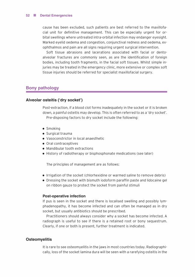
Alveolar osteitis (‘dry socket’)
Post-extraction, if a blood clot forms inadequately in the socket or it is broken
down, a painful osteitis may develop. This is often referred to as a ‘dry socket’.
Pre-disposing factors to dry socket include the following:
� Smoking� Surgical trauma� Vasoconstrictor in local anaesthetic� Oral contraceptives� Mandibular tooth extractions� History of radiotherapy or bisphosphonate medications (see later)
The principles of management are as follows:
� Irrigation of the socket (chlorhexidine or warmed saline to remove debris)� Dressing the socket with bismuth iodoform paraffin paste and lidocaine gel
on ribbon gauze to protect the socket from painful stimuli
Post-operative infectionIf pus is seen in the socket and there is localised swelling and possibly lym-
phadenopathy, it has become infected and can often be managed as in dry
socket, but usually antibiotics should be prescribed.
Practitioners should always consider why a socket has become infected. A
radiograph is useful to see if there is a retained root or bony sequestrum.
Clearly, if one or both is present, further treatment is indicated.
Osteomyelitis
It is rare to see osteomyelitis in the jaws in most countries today. Radiographi-
cally, loss of the socket lamina dura will be seen with a rarefying osteitis in the

P1: SFK/UKS P2: SFK
BLBK407-c04 BLBK407-Greenwood January 24, 2012 11:7 Trim: 244mm×172mm
Acute Oral Medical and Surgical Conditions 53
bone. Sequestra may be seen. If caught early, osteomyelitis may be treated
by antimicrobial therapy alone but often sequestrectomy is needed.
Osteoradionecrosis arises due to the death of irradiated and lethally dam-
aged bone cells stimulated to divide following traumatic stimuli such as dental
extractions or localised infection. Diminished vascularity of the periosteum
also exists as a result of late radiation effects on endothelial lining cells, which
is particularly pertinent for the dense and less vascular mandibular bone. The
radionecrotic process usually starts as ulceration of the alveolar mucosa with
brownish dead bone exposed at the base. Pathological fractures may occur in
weakened bone and secondary infection leads to severe discomfort, trismus,
foetor oris and general malaise. Radiographically, the earliest changes are
a ‘moth-eaten’ appearance of the bone, followed by sequestration. All these
potential complications should be considered in patients who have undergone
radiotherapy of the head and neck, presenting at a dental emergency clinic.
Treatment should be predominately conservative, with long-term antibiotic
and topical antiseptic therapy and careful local removal of sequestra when
necessary. Hyperbaric oxygen and ultrasound therapy to increase tissue blood
flow and oxygenation have also been recommended and are used as a treat-
ment modality in the United Kingdom. It is best treated by a specialist in oral
and maxillofacial surgery and prompt referral is important.
Osteonecrosis is a recently recognised complication of bisphosphonate
treatment (Figure 4.6). This condition is defined as exposed bone in the max-
illofacial region for longer than 8 weeks in the absence of radiotherapy but in
a patient using bisphosphonates. It is diagnosed clinically but local malignancy
must be excluded. Bisphosphonates are a group of drugs, including alendronic
acid, disodium etidronate and risedronate sodium, which are adsorbed onto
hydroxyapatite crystals, thus slowing their rate of growth and dissolution.
They have been used in treatment of bony metastases, the hypercalcaemia of
malignancy and the management of osteoporosis in post-menopausal women.
Figure 4.6 Osteonecrosis following dental extractions.

P1: SFK/UKS P2: SFK
BLBK407-c04 BLBK407-Greenwood January 24, 2012 11:7 Trim: 244mm×172mm
54 Dental Emergencies
Dental extractions should be avoided wherever possible while patients are
on bisphosphonate therapy to reduce the risk of necrosis. Established cases
require analgesia, long-term antibiotic and topical antiseptic therapy, together
with careful local debridement to remove limited bony sequestra similar to
the management of osteoradionecrosis. Risk factors that will increase the
possibility of osteonecrosis developing include local infection, steroid use,
trauma, chemotherapy and periodontal disease.
The mechanism by which bisphosphonates increase the risk of osteonecro-
sis is not fully understood. Trauma caused by a dental extraction in the
presence of impaired osteoclast function may cause inadequate clearance
of necrotic debris. Local osteonecrosis may also occur due to secondary in-
fection. It is also thought that bisphosphonates might have toxic effects on
soft tissues around the extraction site and thereby impair the function of
vascular and epithelial cells.
Chemotherapy agents are inevitably highly toxic and risk important sys-
temic effects such as infections and bleeding due to bone marrow involve-
ment and resultant neutropaenia and thrombocytopaenia. It is important to
liaise with an individual patient’s oncologist to ensure dental or oral surgical
treatments are timed to avoid periods of maximum bone marrow depression.
Management of established mucositis includes systemic analgesia, the use
of intra-oral ice and topical analgesics such as benzydamine hydrochloride or
2% lidocaine lollipops or mouthwash.
Subsequent to radiotherapy and chemotherapy, meticulous oral hygiene is
essential, especially during treatment when the mouth is inflamed and sore.
Dilute chlorhexidine mouthwashes, topical fluoride applications, saliva substi-
tutes and active restorative care may all be needed to preserve the remaining
dentition. Should teeth have to be extracted, this is best carried out in a
specialist oral and maxillofacial surgery unit and it is essential that atrau-
matic techniques are used, with primary closure of oral mucosa together with
antibiotic therapy until healing is complete. Similar considerations apply to
patients taking bisphosphonates. The timing of extractions in patients under-
going chemotherapy is critical. This should be coordinated with the treating
oncologist so that the ideal ‘window of opportunity’ is used.
Effects of drugs used in patients with oral malignancy on patientmanagement in the dental emergency clinic
As mentioned previously, many drugs used in the management of malignant
disease will affect white cell and platelet numbers. This means that bleed-
ing and infection are risks of surgical dentistry such as extractions. A full
blood count is needed to ensure that any extractions can be performed safely.
Elective extractions should be carried out when the blood picture is normal,
however, emergency extractions may need to be performed. If the platelet
count is less than 50 × 109/L, then intra-oral surgery is contraindicated un-
less a platelet transfusion is provided; if less than 100 × 109/L, then sockets
should be packed with a haemostatic agent such as SurgicelR©and sutured. If

P1: SFK/UKS P2: SFK
BLBK407-c04 BLBK407-Greenwood January 24, 2012 11:7 Trim: 244mm×172mm
Acute Oral Medical and Surgical Conditions 55
the white cell count is less than 2.5 × 10⁹/L, then prophylactic antibiotics are
recommended.
It was mentioned previously that xerostomia and stomatitis are side effects
of radiotherapy. These can also be unwanted effects of some drugs used to
treatmalignancy. Thus, excellent oral hygiene and caries preventionmeasures
such as the use of fluoride are recommended. If dentures are ill-fitting, these
should be removed as they may worsen drug-induced mucositis.
Some of the drugs used to treatmalignancies will interfere withmedications
dentists might prescribe. Examples include paracetamol and metronidazole,
both of which increase the toxicity of busulphan by inhibiting metabolism and
increasing plasma concentration of the cytotoxic drug. Similarly, erythromycin
increases the toxicity of the chemotherapeutic drug vinblastine. The toxicity of
methotrexate is increased with concomitant administration of non-steroidal
anti-inflammatory drugs, penicillins and tetracyclines. These are just some
examples of pertinent drug interactions. The dental surgeon should consult a
publication such as the British National Formulary or discuss with the patient’s
oncologist.
Summary
It is clear that many oral diseases may present in the dental emergency clinic
presenting as amyriad of fascinating oral surgery and oral medical conditions.
The emergency clinic practitioner’s role, however, is not one of specialist
diagnosis and management but rather the recognition of those important
and/or life-threatening disorders that require referral. Indeed, a close and
harmonious working relationship between the dental emergency clinic and
their local departments of oral and maxillofacial surgery and oral medicine is
imperative for appropriate, comprehensive and efficacious patient care.
Further reading
Meechan JG, Greenwood M, Moore UJ, Thomson PJ, Brook IM, Smith KG (2006)Minor Oral Surgery in Dental Practice. Quintessence Publishing Co Ltd, London.
Moore UJ (ed.) (2011) Principles of Oral and Maxillofacial Surgery (6th edition).Blackwell Science, Oxford.
Scully C (2004) Oral and Maxillofacial Medicine. Wright, Edinburgh.