Dendrimer-Based Postnatal Therapy for Neuroinflammation ... · Dendrimer-Based Postnatal Therapy...
12
DOI: 10.1126/scitranslmed.3003162 , 130ra46 (2012); 4 Sci Transl Med et al. Sujatha Kannan Palsy in a Rabbit Model Dendrimer-Based Postnatal Therapy for Neuroinflammation and Cerebral Editor's Summary suspected of having CP. in preclinical testing in rabbits, this dendrimer-drug conjugate shows promise for postnatal treatment of babies promoted greater uptake by activated microglia and astrocytes, with no toxicity to surrounding neurons. Although still comparison, NAC alone or saline had no effect. The authors believe that conjugating NAC to the dendrimers successfully treated kits also had neuron counts and low inflammation similar to healthy control animals. By postnatal ''rescue'' with D-NAC, given on day 1 of life, allowed CP kits to develop normally, able to walk and hop. The -acetyl-l-cysteine), or a dendrimer-NAC (D-NAC) conjugate. This N either a saline solution, a free drug known as NAC ( toxin into the rabbit mother's uterus at about 90% term gestation. When the kits were born, they were administered Escherichia coli as well as the motor deficits in children. To generate this model, Kannan and colleagues injected The authors chose to use the rabbit model of CP, which replicates the neuroinflammation seen in human brains rabbits (kits), opening the door to new treatment options in humans. have designed a dendrimer-based therapeutic for treating this developmental disorder in baby et al. Now, Kannan There is no cure for CP, and the best option for affected children is intensive physical therapy to improve motor skills. either in the womb or during the early months of life, but is often not diagnosed until children are 2 to 3 years of age. Cerebral palsy (CP) is a developmental disorder caused by injury to a baby's brain while it is still developing, One Hop at a Time http://stm.sciencemag.org/content/4/130/130ra46.full.html can be found at: and other services, including high-resolution figures, A complete electronic version of this article http://stm.sciencemag.org/content/suppl/2012/04/16/4.130.130ra46.DC1.html can be found in the online version of this article at: Supplementary Material http://www.sciencemag.org/content/sci/336/6079/286.full.html http://stm.sciencemag.org/content/scitransmed/4/130/130fs8.full.html can be found online at: Related Resources for this article http://www.sciencemag.org/about/permissions.dtl in whole or in part can be found at: article permission to reproduce this of this article or about obtaining reprints Information about obtaining is a registered trademark of AAAS. Science Translational Medicine rights reserved. The title NW, Washington, DC 20005. Copyright 2012 by the American Association for the Advancement of Science; all last week in December, by the American Association for the Advancement of Science, 1200 New York Avenue (print ISSN 1946-6234; online ISSN 1946-6242) is published weekly, except the Science Translational Medicine on May 22, 2013 stm.sciencemag.org Downloaded from
Transcript of Dendrimer-Based Postnatal Therapy for Neuroinflammation ... · Dendrimer-Based Postnatal Therapy...
DOI: 10.1126/scitranslmed.3003162, 130ra46 (2012);4 Sci Transl Med
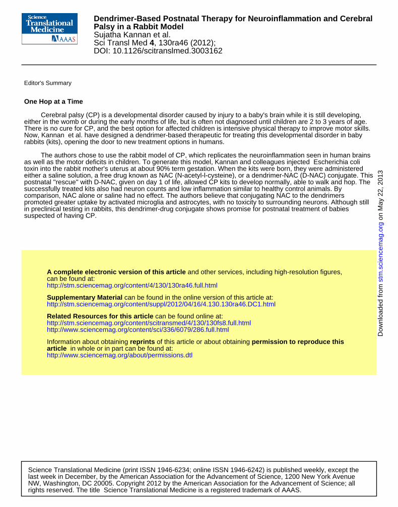
et al.Sujatha KannanPalsy in a Rabbit ModelDendrimer-Based Postnatal Therapy for Neuroinflammation and Cerebral
Editor's Summary
suspected of having CP.in preclinical testing in rabbits, this dendrimer-drug conjugate shows promise for postnatal treatment of babies promoted greater uptake by activated microglia and astrocytes, with no toxicity to surrounding neurons. Although stillcomparison, NAC alone or saline had no effect. The authors believe that conjugating NAC to the dendrimers successfully treated kits also had neuron counts and low inflammation similar to healthy control animals. Bypostnatal ''rescue'' with D-NAC, given on day 1 of life, allowed CP kits to develop normally, able to walk and hop. The
-acetyl-l-cysteine), or a dendrimer-NAC (D-NAC) conjugate. ThisNeither a saline solution, a free drug known as NAC (toxin into the rabbit mother's uterus at about 90% term gestation. When the kits were born, they were administered
Escherichia colias well as the motor deficits in children. To generate this model, Kannan and colleagues injected The authors chose to use the rabbit model of CP, which replicates the neuroinflammation seen in human brains
rabbits (kits), opening the door to new treatment options in humans. have designed a dendrimer-based therapeutic for treating this developmental disorder in babyet al.Now, Kannan
There is no cure for CP, and the best option for affected children is intensive physical therapy to improve motor skills.either in the womb or during the early months of life, but is often not diagnosed until children are 2 to 3 years of age.
Cerebral palsy (CP) is a developmental disorder caused by injury to a baby's brain while it is still developing,
One Hop at a Time
http://stm.sciencemag.org/content/4/130/130ra46.full.htmlcan be found at:
and other services, including high-resolution figures,A complete electronic version of this article
http://stm.sciencemag.org/content/suppl/2012/04/16/4.130.130ra46.DC1.html can be found in the online version of this article at: Supplementary Material
http://www.sciencemag.org/content/sci/336/6079/286.full.html http://stm.sciencemag.org/content/scitransmed/4/130/130fs8.full.html
can be found online at:Related Resources for this article
http://www.sciencemag.org/about/permissions.dtl in whole or in part can be found at: article
permission to reproduce this of this article or about obtaining reprintsInformation about obtaining
is a registered trademark of AAAS. Science Translational Medicinerights reserved. The title NW, Washington, DC 20005. Copyright 2012 by the American Association for the Advancement of Science; alllast week in December, by the American Association for the Advancement of Science, 1200 New York Avenue
(print ISSN 1946-6234; online ISSN 1946-6242) is published weekly, except theScience Translational Medicine
on
May
22,
201
3st
m.s
cien
cem
ag.o
rgD
ownl
oade
d fr
om
R E S EARCH ART I C L E
CEREBRAL PALSY
Dendrimer-Based Postnatal Therapy forNeuroinflammation and Cerebral Palsy in aRabbit ModelSujatha Kannan,1,2*† Hui Dai,1,2 Raghavendra S. Navath,1,3 Bindu Balakrishnan,1,2* Amar Jyoti,1,2*James Janisse,4 Roberto Romero,1† Rangaramanujam M. Kannan1,3†‡
y 22
, 201
3
Cerebral palsy (CP) is a chronic childhood disorder with no effective cure. Neuroinflammation, caused by activatedmicroglia and astrocytes, plays a key role in the pathogenesis of CP and disorders such as Alzheimer’s disease andmultiple sclerosis. Targeting neuroinflammation can be a potent therapeutic strategy. However, delivering drugsacross the blood-brain barrier to the target cells for treating diffuse brain injury is a major challenge. We show thatsystemically administered polyamidoamine dendrimers localize in activated microglia and astrocytes in the brain ofnewborn rabbits with CP, but not healthy controls. We further demonstrate that dendrimer-based N-acetyl-L-cysteine (NAC) therapy for brain injury suppresses neuroinflammation and leads to a marked improvement in mo-tor function in the CP kits. The well-known and safe clinical profile for NAC, when combined with dendrimer-basedtargeting, provides opportunities for clinical translation in the treatment of neuroinflammatory disorders in humans.The effectiveness of the dendrimer-NAC treatment, administered in the postnatal period for a prenatal insult, sug-gests a window of opportunity for treatment of CP in humans after birth.
n M
a
ostm
.sci
ence
mag
.org
Dow
nloa
ded
from
INTRODUCTION
Cerebral palsy (CP) is a broad term encompassing a group of disor-ders involving variable degrees of motor, sensory, and cognitive im-pairment that occur as a result of an injury/insult to the developingfetal or infant brain (1). This chronic childhood disability may resultfrom diverse etiologies, with a prevalence of 3.3 per 1000 children (2),and is associated with substantial social, personal, and financial bur-dens (3). Intrauterine infection and inflammation are risk factors forthe development of periventricular leukomalacia (PVL) and CP in theneonate (4–7). PVL, the pathophysiological mechanism proposed forthe development of CP in humans, is characterized by focal necrosisaround the ventricles and diffuse microglial and astrocyte activation inthe immature white matter (8). Microglia, immune cells in the brain,play an important role in remodeling and growth in the fetal and post-natal period (9). Activation of these cells can result in an exaggeratedinflammatory response with formation of free radicals, excitotoxic me-tabolites, and proinflammatory cytokines, leading to brain injury (10, 11).In severe inflammation, astrocytes that normally participate in theprotection of neurons and in preventing oxidative injury are unableto maintain their neuroprotective role (12).
Treatment of disorders such as CP is challenging for several reasons.Inflammation and injury are often diffuse in the white matter, preclud-
1Perinatology Research Branch, Eunice Kennedy Shriver National Institute of ChildHealth and Human Development, National Institutes of Health, Detroit, MI 48201,USA. 2Department of Pediatrics, Children’s Hospital of Michigan, Detroit MedicalCenter, Wayne State University, Detroit, MI 48201, USA. 3Department of Chemical En-gineering and Material Science, Wayne State University, Detroit, MI 48201, USA. 4De-partment of Family Medicine and Public Health Sciences, Wayne State University, Detroit,MI 48201, USA.*Present address: Department of Anesthesiology and Critical Care Medicine, JohnsHopkins University School of Medicine, Baltimore, MD 21287, USA.†To whom correspondence should be addressed. E-mail: [email protected] (R.M.K.);[email protected] (S.K.); [email protected] (R.R.)‡Present address: Center for Nanomedicine/Wilmer Eye Institute, Ophthalmology,Johns Hopkins University School of Medicine, Baltimore, MD 21287, USA.
www.Scie
ing local brain delivery. Furthermore, clinical diagnosis of CP is madewell after birth, so postnatal treatment of a prenatal injury to the brain isnot expected to result in improvement in motor function. Finally, trans-port of drugs across the blood-brain barrier (BBB) is difficult to achieve.We hypothesized that a postnatal therapeutic strategy targeting activatedmicroglia and astrocytes for sustained attenuation of ongoing neuro-inflammation would improve outcomes in an animal model of CP.
Taking advantage of the differences between cellular localizationof nanomaterials in healthy and diseased tissues may help addressthese treatment challenges. Here, we used polyamidoamine (PAMAM)dendrimers as vehicles for delivery of N-acetyl-L-cysteine (NAC). NAC,an antioxidant and anti-inflammatory agent, has a long history of clin-ical use as an antidote for acetaminophen poisoning (doses from 50to 150 mg/kg) (13) and is being explored in several ongoing clinicaltrials for potential neuroprotective effects in autism spectrum dis-orders (ClinicalTrials.gov IDs: NCT00453180 and NCT00627705),in pregnant women for the treatment of maternal and fetal inflam-mation (NCT00397735 and NCT00724594), in Alzheimer’s disease(NCT01320527), and in animal models of perinatal brain injury (14, 15).Dendrimers are viewed as synthetic biomimics of globular proteins,with versatile tailorable surface functionalities. They are being ex-plored in preclinical studies for cancer therapy, inflammation, and tar-geted delivery applications (16–19). We have previously shown thathydroxyl-terminated PAMAM dendrimers localize in activated mi-croglia and astrocytes when injected into the subarachnoid space ofneonatal rabbit brains with a CP phenotype, but not in age-matchedhealthy controls (20). Building upon this work, we investigated wheth-er these dendrimers can localize in activated microglia and astrocyteseven when administered systemically to newborn rabbits with neuro-inflammation and motor deficits. In addition, we asked whether deliv-ering NAC to activated microglia and astrocytes using these dendrimerswould lead to improvements in motor function. To this end, we showthat NAC conjugated to the PAMAM dendrimer, administered intra-venously on day 1 of life to rabbit kits with CP, resulted in significant
nceTranslationalMedicine.org 18 April 2012 Vol 4 Issue 130 130ra46 1

R E S EARCH ART I C L E
improvement in motor function along with a decrease in markers ofneuroinflammation and oxidative injury by day 5 of life. This studysuggests that targeted therapy of drugs using dendrimers may be ef-fective in the treatment of neuroinflammatory disorders.
on
May
22,
201
3st
m.s
cien
cem
ag.o
rgD
ownl
oade
d fr
om
RESULTS
Preparation, characterization, and biodistributionof dendrimer-NAC conjugatesNAC was conjugated to generation-4, hydroxyl-terminated PAMAMdendrimers (“D”), [D-(OH)64], using a disulfide linker, through a four-step reaction (Fig. 1 and fig. S1) (Supplementary Methods). In the firsttwo steps, a bifunctional dendrimer [(HO)39-D-(GABA-NH2)25, 4] with25 reactive amines was synthesized using our previously publishedprotocol (21). The intermediate 4 was reacted with the heterobifunc-tional cross-linkerN-succinimidyl-3-(2-pyridyldithio)-propionate (SPDP)to yield amide-linked 2-pyridyldithiopropanoyl (PDP)–functionalizeddendrimer, [(HO)39-D-(GABA-PDP)25, 5]. The appearance of aro-matic thiopyridine protons in 1H NMR (nuclear magnetic resonance)confirmed the formation of PDP-functionalized dendrimer 5. In thefinal step, 5 was treated with water-soluble NAC in phosphate-bufferedsaline (PBS) solution (pH 7.4) to obtain the desired conjugate witha disulfide linkage between the drug and the dendrimer (D-NAC, 1).The characteristic peaks in 1H NMR spectrum of 1 corresponding to thedendrimer, NAC, and the linker confirm the formation of the productand the presence of disulfide bond between the dendrimer and NAC(fig. S1A) (22, 23). The drug payload on the dendrimer was estimatedby NMR andMALDI-TOF (matrix-assisted laser desorption/ionization–time-of-flight) mass spectrometry to be 19% (suggestive of 20 moleculesof NAC) (table S1 and fig. S1B). The reverse-phase high-performanceliquid chromatography (HPLC) chromatogram of D-NAC 1 at 210 nmshows a relatively narrow peak, different from that of the starting mate-rials and intermediates, suggesting a relatively pure conjugate nano-device (fig. S1C). The z potential changed from −2.1 mV for the startingdendrimer to−10.6mV for theD-NAC 1 conjugate, due to surfacemod-ification with NAC, resulting in some carboxylic acid end functional-ities. Particle size analysis by dynamic light scattering (DLS) showedthat D-NAC is larger in size (5.4 nm) than D-OH 2 as expected, owingto higher molecular weight of the conjugate.
www.Scie
The hydroxyl-terminated PAMAM dendrimers used in this studyare nontoxic, nonimmunogenic, and are cleared intact through thekidneys (16, 24). Biosafety of the dendrimer after systemic administra-tion (550 mg/kg) on day 1 was evaluated in healthy newborn kits atdays 5 and 15 of age. There was no change in renal and hepatic func-tions or neurobehavior noted at both time points when compared tohealthy kits administered PBS (table S2 and fig. S2). Liver enzymesremained normal, indicating that there was no hepatocellular injurywith dendrimer administration. Ten percent of this amount was usedas the maximum dendrimer dose in the present study.
In vitro NAC release from the D-NAC conjugateGlutathione (GSH)–cleavable disulfide linkers were used between thedrug and the dendrimer to enable intracellular release (22, 23). Atphysiological conditions, in the absence of GSH, the conjugate wasstable without releasing NAC over a 72-hour period. In vitro releaseof NAC was investigated at seven different GSH concentrations start-ing from 2 mM (plasma level) to 10 mM (intracellular levels) (Supple-mentary Methods). At extracellular and plasma GSH levels (2 mM),the conjugate did not release measurable NAC. As the GSH level wasincreased, proportionately more of NAC was released (fig. S3). At in-tracellular GSH concentrations (2 and 10 mM), the conjugate readilyreleased the drug (>80% in 100 min), which could be detected mostlyas free NAC, and as NAC-GSH to a smaller extent within 40 min.This indicated that the use of a disulfide linker enabled rapid releaseof NAC from the conjugate, but only when it was exposed to an intra-cellular GSH-rich environment (22, 23). The mechanism of GSH-based release from dendrimer conjugates has been described previously(23). Reported GSH levels in microglia (~25 mM) and astrocytes(~4 to 20 mM) are well within the range shown for drug release fromthe conjugate (25). Even if the intracellular GSH levels were reducedsignificantly due to inflammation, there will still be sufficient GSH torelease the drug.
In vivo brain biodistribution of dendrimersAll in vivo studies were performed with a previously described rabbitmodel, where CP is induced by maternal intrauterine endotoxin ad-ministration (26, 27). Fluorescein-labeled dendrimer (D-FITC) wasadministered intravenously to newborn kits, and brains were ex-amined after 24 hours. D-FITC was found to colocalize in activated
Fig. 1. Synthesis and characterization of the D-NAC conjugate. Reaction schematic forthe synthesis of the dendrimer-NAC (D-NAC) conjugate 1, starting from the free dendrimerand free NAC.
nceTranslationalMedicine.org 18 April 2012 Vol 4 Issue 130 130ra46 2

R E S EARCH ART I C L E
microglia and astrocytes in the periventricular region (PVR), in kits withCP, but not in healthy age-matched controls (Fig. 2). This increasedbrain uptake in CP kits was also consistent with results from positronemission tomography (PET) imaging of 64Cu-labeled dendrimer. Anincrease in tracer activity was seen in CP kits injected with 64Cu-dendrimer but not in controls (fig. S4). Very little uptake was seen in
on
May
22,
201
3st
m.s
cien
cem
ag.o
rgD
ownl
oade
d fr
om
the control and CP kits injected with64CuCl2 alone. This selective localizationin activated microglia and astrocytes wassimilar to that noted upon subarachnoidadministration in CP kits and upon in-travitreal administration in a rat modelof neuroinflammation-induced retinal de-generation (19, 20).
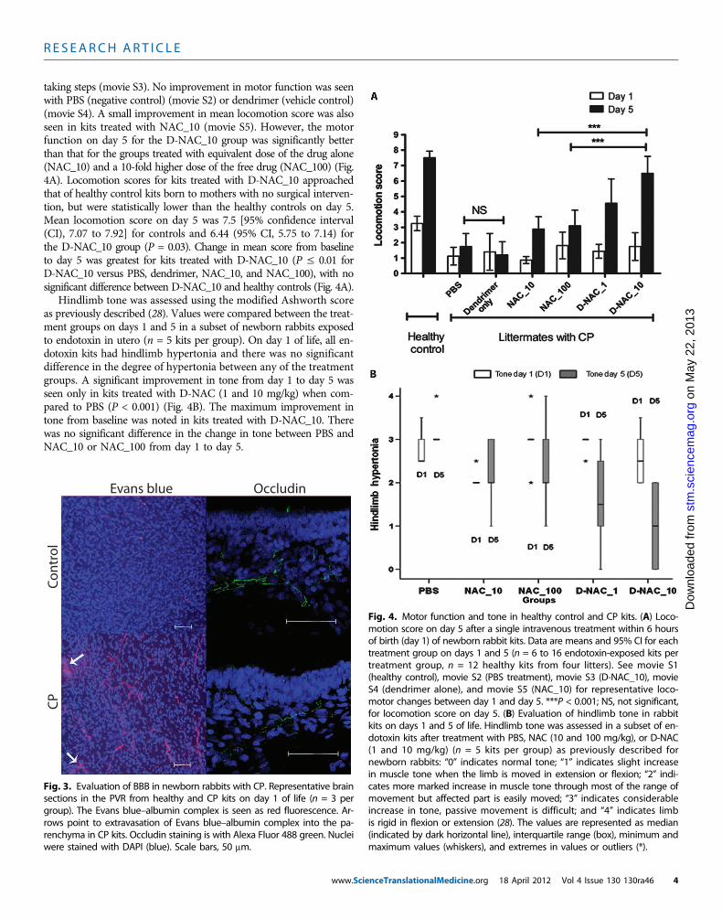
Impairment in BBB integrity was ob-served in CP kits on day 1 of life. This wasdemonstrated by decreased occludin ex-pression, indicating loss of tight junctionproteins and increased permeability evi-denced by extravasation of Evans blue–albumin complex (Fig. 3).
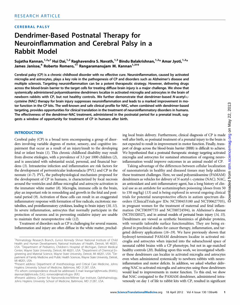
D-NAC therapy improves motorfunction in CP kitsTo determine whether targeting NAC toareas of neuroinflammation results in im-provedmotor function, we randomly treatedlittermates with CP with NAC at 10 mg/kg(NAC_10), NAC at 100 mg/kg (NAC_100),D-NAC with NAC at 1 mg/kg (D-NAC_1),D-NAC with NAC at 10 mg/kg (D-NAC_10), dendrimer alone (delivery ve-hicle control), or PBS (negative control).A total of 69 kits from 14 dams thatunderwent laparotomy and intrauterineendotoxin administration were used forthe study. To minimize the variation be-tween treated animals, we administeredkits from the same litter with differenttherapies. The healthy control group con-sisted of kits born to dams that had noendotoxin intervention (healthy control).All treatments were administered intra-venously as a single dose within 6 hoursafter birth (day 1). Using modifications ofobservational motor function scores forrabbits, we evaluated the kits in a blindedmanner for change in motor function onday 5 (during the peak myelination periodin rabbits), as described in Materials andMethods (28, 29).
Endotoxin-exposed kits were bornwith impaired motor function involvinginability to take steps, decreased coordi-nation, impaired balance, and hypertoniaof the hindlimbs suggestive of a pheno-type of CP as described previously in thismodel (27, 30). The endotoxin-exposed
www.Scie
kits had similar motor function scores on day 1 of life, whereas healthycontrol kits that had no intervention were normal and had signif-icantly better motor function (Fig. 4A and movies S1 and S2). Themost favorable response in motor function from day 1 to day 5 wasseen in kits treated with D-NAC_10 (Fig. 4A), manifested by markedimprovement in coordination and motor control while hopping and
Fig. 2. Cellular localization of FITC-labeled dendrimer in the brains of 1-day-old newborn healthy(control) and CP rabbits upon intravenous administration. Representative images of the PVR from
healthy control and endotoxin-exposed newborn rabbits (CP group). Microglia were identified bystaining with tomato lectin (red in microglia panels). Astrocytes were stained with an anti–glial fibril-lary acidic protein (GFAP) antibody (red in astrocyte panels). Images were merged to observe coloca-lization of dendrimer-FITC (D-FITC) with microglia and astrocyte cell types. Nuclei were stained with4′,6-diamidino-2-phenylindole (DAPI) (blue). Scale bars, 20 mm. Inset shows microglia and astrocytes athigher magnification (scale bar, 5 mm).nceTranslationalMedicine.org 18 April 2012 Vol 4 Issue 130 130ra46 3
R E S EARCH ART I C L E
on
May
22,
201
3em
ag.o
rg
taking steps (movie S3). No improvement in motor function was seenwith PBS (negative control) (movie S2) or dendrimer (vehicle control)(movie S4). A small improvement in mean locomotion score was alsoseen in kits treated with NAC_10 (movie S5). However, the motorfunction on day 5 for the D-NAC_10 group was significantly betterthan that for the groups treated with equivalent dose of the drug alone(NAC_10) and a 10-fold higher dose of the free drug (NAC_100) (Fig.4A). Locomotion scores for kits treated with D-NAC_10 approachedthat of healthy control kits born to mothers with no surgical interven-tion, but were statistically lower than the healthy controls on day 5.Mean locomotion score on day 5 was 7.5 [95% confidence interval(CI), 7.07 to 7.92] for controls and 6.44 (95% CI, 5.75 to 7.14) forthe D-NAC_10 group (P = 0.03). Change in mean score from baselineto day 5 was greatest for kits treated with D-NAC_10 (P ≤ 0.01 forD-NAC_10 versus PBS, dendrimer, NAC_10, and NAC_100), with nosignificant difference between D-NAC_10 and healthy controls (Fig. 4A).
Hindlimb tone was assessed using the modified Ashworth scoreas previously described (28). Values were compared between the treat-ment groups on days 1 and 5 in a subset of newborn rabbits exposedto endotoxin in utero (n = 5 kits per group). On day 1 of life, all en-dotoxin kits had hindlimb hypertonia and there was no significantdifference in the degree of hypertonia between any of the treatmentgroups. A significant improvement in tone from day 1 to day 5 wasseen only in kits treated with D-NAC (1 and 10 mg/kg) when com-pared to PBS (P < 0.001) (Fig. 4B). The maximum improvement intone from baseline was noted in kits treated with D-NAC_10. Therewas no significant difference in the change in tone between PBS andNAC_10 or NAC_100 from day 1 to day 5.
stm
.sci
enc
Dow
nloa
ded
from
Evans blue Occludin
Co
ntr
ol
CP
Fig. 3. Evaluation of BBB in newborn rabbits with CP. Representative brainsections in the PVR from healthy and CP kits on day 1 of life (n = 3 per
group). The Evans blue–albumin complex is seen as red fluorescence. Ar-rows point to extravasation of Evans blue–albumin complex into the pa-renchyma in CP kits. Occludin staining is with Alexa Fluor 488 green. Nucleiwere stained with DAPI (blue). Scale bars, 50 mm.www.Scie
Fig. 4. Motor function and tone in healthy control and CP kits. (A) Loco-motion score on day 5 after a single intravenous treatment within 6 hours
of birth (day 1) of newborn rabbit kits. Data are means and 95% CI for eachtreatment group on days 1 and 5 (n = 6 to 16 endotoxin-exposed kits pertreatment group, n = 12 healthy kits from four litters). See movie S1(healthy control), movie S2 (PBS treatment), movie S3 (D-NAC_10), movieS4 (dendrimer alone), and movie S5 (NAC_10) for representative loco-motor changes between day 1 and day 5. ***P < 0.001; NS, not significant,for locomotion score on day 5. (B) Evaluation of hindlimb tone in rabbitkits on days 1 and 5 of life. Hindlimb tone was assessed in a subset of en-dotoxin kits after treatment with PBS, NAC (10 and 100 mg/kg), or D-NAC(1 and 10 mg/kg) (n = 5 kits per group) as previously described fornewborn rabbits: “0” indicates normal tone; “1” indicates slight increasein muscle tone when the limb is moved in extension or flexion; “2” indi-cates more marked increase in muscle tone through most of the range ofmovement but affected part is easily moved; “3” indicates considerableincrease in tone, passive movement is difficult; and “4” indicates limbis rigid in flexion or extension (28). The values are represented as median(indicated by dark horizontal line), interquartile range (box), minimum andmaximum values (whiskers), and extremes in values or outliers (*).nceTranslationalMedicine.org 18 April 2012 Vol 4 Issue 130 130ra46 4
R E S EARCH ART I C L E
Kit weight gain and survivalBecause the inflammatory stimulus occurs 3 days before birth, all en-dotoxin kits have intrauterine growth restriction with lower birthweights compared to healthy control kits on day 1 of life (P < 0.001).
on
May
22,
201
3st
m.s
cien
cem
ag.o
rgD
ownl
oade
d fr
om
There was no significant difference in day1 weights between the different endotoxin-exposed treatment groups (table S3). Therewas no significant difference in weight gainfrom day 1 to day 5 between healthy controlsand NAC- or D-NAC–treated CP animals,but PBS- and dendrimer-treated animalsgained less weight than the kits treated withNAC or D-NAC (P < 0.01) (table S3). Anincreased catabolic rate due to ongoing in-flammation may account for the lowerweight gain in kits treated with PBS anddendrimer alone. Survival up to day 5 wassimilar between all endotoxin (CP) groupsand ranged from 77 to 85%.
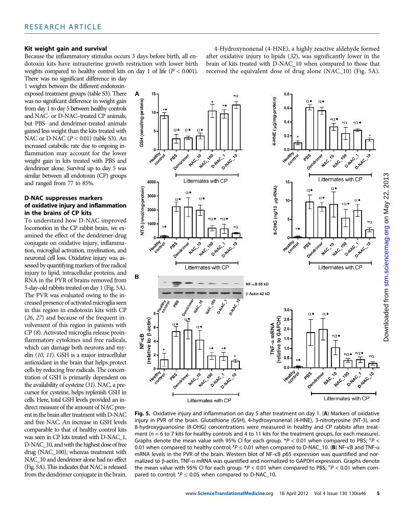
D-NAC suppresses markersof oxidative injury and inflammationin the brains of CP kitsTo understand how D-NAC improvedlocomotion in the CP rabbit brain, we ex-amined the effect of the dendrimer-drugconjugate on oxidative injury, inflamma-tion, microglial activation, myelination, andneuronal cell loss. Oxidative injury was as-sessed by quantifyingmarkers of free radicalinjury to lipid, intracellular proteins, andRNA in the PVR of brains removed from5-day-old rabbits treated on day 1 (Fig. 5A).The PVR was evaluated owing to the in-creased presence of activatedmicroglia seenin this region in endotoxin kits with CP(26, 27) and because of the frequent in-volvement of this region in patients withCP (8). Activated microglia release proin-flammatory cytokines and free radicals,which can damage both neurons and my-elin (10, 11). GSH is a major intracellularantioxidant in the brain that helps protectcells by reducing free radicals. The concen-tration of GSH is primarily dependent onthe availability of cysteine (31). NAC, a pre-cursor for cysteine, helps replenish GSH incells. Here, total GSH levels provided an in-directmeasure of the amount of NACpres-ent in the brain after treatmentwithD-NACand free NAC. An increase in GSH levelscomparable to that of healthy control kitswas seen in CP kits treated with D-NAC_1,D-NAC_10, andwith thehighestdoseof freedrug (NAC_100), whereas treatment withNAC_10 and dendrimer alone had no effect(Fig. 5A). This indicates thatNAC is releasedfrom the dendrimer conjugate in the brain.
www.Scie
4-Hydroxynonenal (4-HNE), a highly reactive aldehyde formedafter oxidative injury to lipids (32), was significantly lower in thebrain of kits treated with D-NAC_10 when compared to those thatreceived the equivalent dose of drug alone (NAC_10) (Fig. 5A).
Fig. 5. Oxidative injury and inflammation on day 5 after treatment on day 1. (A) Markers of oxidativeinjury in PVR of the brain. Glutathione (GSH), 4-hydroxynonenal (4-HNE), 3-nitrotyrosine (NT-3), and8-hydroxyguanosine (8-OHG) concentrations were measured in healthy and CP rabbits after treat-ment (n = 6 to 7 kits for healthy controls and 4 to 11 kits for the treatment groups, for each measure).Graphs denote the mean value with 95% CI for each group. *P < 0.01 when compared to PBS; aP <0.01 when compared to healthy control; •P ≤ 0.01 when compared to D-NAC_10. (B) NF-kB and TNF-amRNA levels in the PVR of the brain. Western blot of NF-kB p65 expression was quantified and nor-malized to b-actin. TNF-a mRNA was quantified and normalized to GAPDH expression. Graphs denotethe mean value with 95% CI for each group. *P < 0.01 when compared to PBS; aP < 0.01 when com-pared to control; •P ≤ 0.05 when compared to D-NAC_10.
nceTranslationalMedicine.org 18 April 2012 Vol 4 Issue 130 130ra46 5
R E S EARCH ART I C L E
13
Oxidative injury to proteins by ONOO− (peroxynitrate), one of themost potent free radicals, was measured by evaluating 3-nitrotyrosine(NT-3), which is produced by nitration of tyrosine residues on pro-teins (33). NT-3 levels decreased upon treatment with D-NAC_10when compared to PBS- and free NAC–treated kits (Fig. 5A), indicat-ing an improvement in oxidative injury with D-NAC_10 therapy. Fi-nally, levels of 8-hydroxyguanosine (8-OHG), an early and sensitivemarker for RNA oxidation in various neurodegenerative disorders(34), were significantly reduced with D-NAC_10 in CP littermateswhen compared to free NAC alone even at the highest dose (Fig. 5A).The greatest decrease in oxidative injury was seen in kits treated withD-NAC_10, which was two- to sixfold better than the equivalent doseof drug alone. D-NAC_10 was significantly better than 10 times thedose of the drug alone in suppressing NT-3 and 8-OHG.
NAC is known to suppress activation of nuclear factor kB (NF-kB),which induces transcription of proinflammatory genes, such as tumornecrosis factor–a (TNF-a) (35). TNF-a is responsible for microglialproliferation and activation perpetuating the inflammatory process
on
May
22,
20
stm
.sci
ence
mag
.org
Dow
nloa
ded
from
in the brain (8, 11). A single dose ofD-NAC_10 led to a 3.5-fold decrease inNF-kB expression when compared toequivalent dose of the free drug (NAC_10)(Fig. 5B). A significant decrease in mRNAexpression of TNF-awas also noted in thebrain of kits treated with D-NAC (1 and10 mg/kg) when compared to kits treatedwith the free drug alone (Fig. 5B).
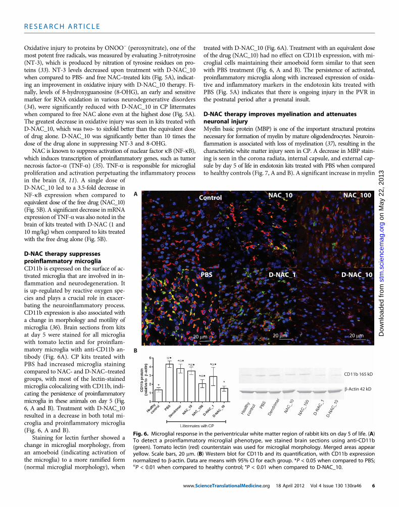
D-NAC therapy suppressesproinflammatory microgliaCD11b is expressed on the surface of ac-tivated microglia that are involved in in-flammation and neurodegeneration. Itis up-regulated by reactive oxygen spe-cies and plays a crucial role in exacer-bating the neuroinflammatory process.CD11b expression is also associated witha change in morphology and motility ofmicroglia (36). Brain sections from kitsat day 5 were stained for all microgliawith tomato lectin and for proinflam-matory microglia with anti-CD11b an-tibody (Fig. 6A). CP kits treated withPBS had increased microglia stainingcompared to NAC- and D-NAC–treatedgroups, with most of the lectin-stainedmicroglia colocalizing with CD11b, indi-cating the persistence of proinflammatorymicroglia in these animals on day 5 (Fig.6, A and B). Treatment with D-NAC_10resulted in a decrease in both total mi-croglia and proinflammatory microglia(Fig. 6, A and B).
Staining for lectin further showed achange in microglial morphology, froman amoeboid (indicating activation ofthe microglia) to a more ramified form(normal microglial morphology), when
www.Scie
treated with D-NAC_10 (Fig. 6A). Treatment with an equivalent doseof the drug (NAC_10) had no effect on CD11b expression, with mi-croglial cells maintaining their amoeboid form similar to that seenwith PBS treatment (Fig. 6, A and B). The persistence of activated,proinflammatory microglia along with increased expression of oxida-tive and inflammatory markers in the endotoxin kits treated withPBS (Fig. 5A) indicates that there is ongoing injury in the PVR inthe postnatal period after a prenatal insult.
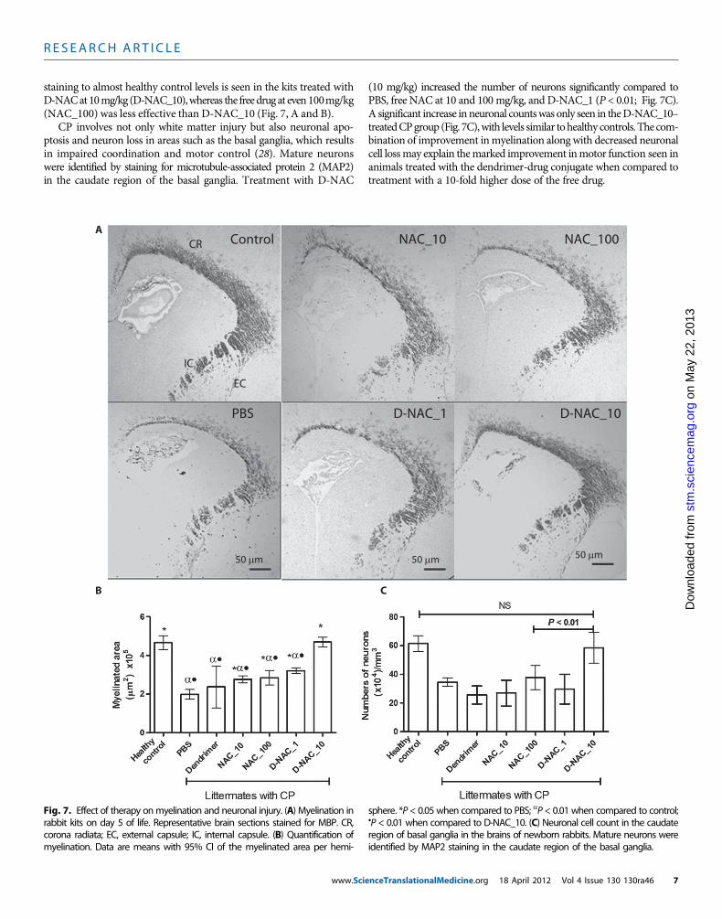
D-NAC therapy improves myelination and attenuatesneuronal injuryMyelin basic protein (MBP) is one of the important structural proteinsnecessary for formation of myelin by mature oligodendrocytes. Neuroin-flammation is associated with loss of myelination (37), resulting in thecharacteristic white matter injury seen in CP. A decrease in MBP stain-ing is seen in the corona radiata, internal capsule, and external cap-sule by day 5 of life in endotoxin kits treated with PBS when comparedto healthy controls (Fig. 7, A and B). A significant increase in myelin
A
B
Fig. 6. Microglial response in the periventricular white matter region of rabbit kits on day 5 of life. (A)To detect a proinflammatory microglial phenotype, we stained brain sections using anti-CD11b
(green). Tomato lectin (red) counterstain was used for microglial morphology. Merged areas appearyellow. Scale bars, 20 mm. (B) Western blot for CD11b and its quantification, with CD11b expressionnormalized to b-actin. Data are means with 95% CI for each group. *P < 0.05 when compared to PBS;aP < 0.01 when compared to healthy control; •P < 0.01 when compared to D-NAC_10.nceTranslationalMedicine.org 18 April 2012 Vol 4 Issue 130 130ra46 6
R E S EARCH ART I C L E
staining to almost healthy control levels is seen in the kits treated withD-NACat10mg/kg (D-NAC_10),whereas the freedrugat even100mg/kg(NAC_100) was less effective than D-NAC_10 (Fig. 7, A and B).
CP involves not only white matter injury but also neuronal apo-ptosis and neuron loss in areas such as the basal ganglia, which resultsin impaired coordination and motor control (28). Mature neuronswere identified by staining for microtubule-associated protein 2 (MAP2)in the caudate region of the basal ganglia. Treatment with D-NAC
www.Scie
(10 mg/kg) increased the number of neurons significantly compared toPBS, free NAC at 10 and 100 mg/kg, and D-NAC_1 (P < 0.01; Fig. 7C).A significant increase inneuronal countswas only seen in theD-NAC_10–treatedCPgroup (Fig. 7C),with levels similar tohealthy controls.The com-bination of improvement inmyelination along with decreased neuronalcell lossmay explain themarked improvement inmotor function seen inanimals treated with the dendrimer-drug conjugate when compared totreatment with a 10-fold higher dose of the free drug.
on
May
22,
201
3st
m.s
cien
cem
ag.o
rgD
ownl
oade
d fr
om
A
B C
Fig. 7. Effect of therapy onmyelination and neuronal injury. (A) Myelination inrabbit kits on day 5 of life. Representative brain sections stained for MBP. CR,
sphere. *P < 0.05 when compared to PBS; aP < 0.01 when compared to control;•P < 0.01 when compared to D-NAC_10. (C) Neuronal cell count in the caudate
corona radiata; EC, external capsule; IC, internal capsule. (B) Quantification ofmyelination. Data are means with 95% CI of the myelinated area per hemi-
region of basal ganglia in the brains of newborn rabbits. Mature neurons wereidentified by MAP2 staining in the caudate region of the basal ganglia.
nceTranslationalMedicine.org 18 April 2012 Vol 4 Issue 130 130ra46 7
R E S EARCH ART I C L E
on
May
22,
201
3st
m.s
cien
cem
ag.o
rgD
ownl
oade
d fr
om
DISCUSSION
Current management for CP primarily focuses on rehabilitation andimproving quality of life. Therapeutic approaches being explored in-clude hypothermia for perinatal asphyxia and stem cell infusion forCP (38) (ClinicalTrials.gov IDs NCT01192776, NCT01147653, andNCT01072370). A key challenge in evaluating therapies for CP hasbeen the paucity in animal models demonstrating the phenotype asseen in humans. The parallels in the timing of white matter develop-ment, along with microglial presence in the perinatal human andrabbit brain, make rabbit models of fetal brain injury more represent-ative of CP in humans (27–29). The presence of activated microgliain the periventricular white matter, oxidative injury, impaired my-elination, and neuronal loss seen in this model are consistent with his-tological findings seen in postmortem brain of patients with PVL (8). Inaddition, we find a predominance of hindlimb involvement in theseanimals, which is similar to the increased incidence of diparetic CP (in-volving lower extremities) in children born preterm to mothers withplacental inflammation or infection (6).
We showed that systemic administration of hydroxyl-terminatedPAMAM dendrimer resulted in their selective accumulation in acti-vated microglia and astrocytes only in kits with CP. We attribute thisincreased brain and cell uptake in CP kits, to impairment of the BBBin the PVR, presumably leading to the increased permeability to den-drimers. This is consistent with previous reports of PAMAM dendrimerscrossing the blood-brain-tumor barrier in models of malignant gliomas(39). An increase in the number of activated microglia and astrocytes,with enhanced phagocytic abilities under pathological conditions,may further facilitate the selective cellular localization of dendrimersin kits with CP (40). Technical and ethical considerations make directevaluation of the BBB difficult in patients. However, studies in newbornanimal models of white matter injury have shown increased perme-ability of the BBB in the presence of inflammation (41). Impairment ofthe BBB, in the presence of neuroinflammation, has also been reportedin stroke, multiple sclerosis, and Alzheimer’s disease (42). Passive target-ing with dendrimers may facilitate delivery of therapeutics to neuro-inflammation in these indications.
Intravenous administration of a single dose of D-NAC (10 mg/kg)resulted in a significant improvement in neuronal injury and motorfunction in CP kits (movie S3), whereas free NAC at 100 mg/kg didnot, suggesting the importance of targeted drug delivery in the treat-ment of ongoing neuroinflammation. Although free NAC_100 showedsome efficacy in attenuating inflammation and oxidative injury in thebrain, the improvement did not translate to improved myelination, neu-ronal counts, or motor function. Moreover, the improvements seenwith NAC_100 were similar to that seen with D-NAC at 1% of thedose (D-NAC_1). We speculate that this could be due to several fac-tors, including poor bioavailability of free NAC (43), improved uptakeand efficacy of D-NAC when compared to free NAC in activated mi-croglia, as shown previously in vitro (23), delivery of a higher drugpayload to the target cells (activated microglia and astrocytes) bythe dendrimer in vivo, and decreased toxicity of the drug to neuronswhen conjugated with the dendrimer.
In the presence of inflammation and oxidative stress, depletion ofGSH is one of the mechanisms by which the neuroprotective functionof astrocytes is compromised (12). Regulated neuroglial transport ofGSH and cysteine from astrocytes to neurons may play a role in neu-roprotection (44). Hence, replenishing GSH specifically in astrocytes
www.Scie
by D-NAC may help improve neuronal survival. In addition, excessextracellular L-cysteine concentrations have been shown to result inneuronal degeneration by NMDA (N-methyl-D-aspartate)–mediatedexcitotoxicity both in vitro and in vivo (45). Therefore, targeted deliv-ery of the drug to activated microglia and astrocytes can not only helpattenuate inflammation but may also prevent excess extracellular levelsof L-cysteine produced from NAC that may be toxic to neurons andoligodendrocytes in the immature brain.
A therapeutic response was not seen upon treatment with dendrimeralone, which indicates that the dendrimer acts as a drug delivery ve-hicle. Although PAMAM dendrimers are not yet approved for clinicaluse, they are the subject of several preclinical studies (16, 19). We usehydroxyl-terminated PAMAM dendrimers with a good safety profilein newborn kits, which may enable translation. In humans, because theexact time of the perinatal brain injury may vary, multiple injections ofD-NAC or sustained release formulations may be needed for effectivetherapy. Future longitudinal studies focusing on long-term efficacy ofthis therapy up to adulthood will facilitate clinical translation. Theplatform described herein to target activated microglia and astrocyteshas broad implications for the treatment of neurological diseases giventhe growing body of evidence that neuroinflammation plays a key rolein the pathogenesis of disorders such as multiple sclerosis, Alzheimer’sdisease, and stroke. A similar therapeutic response with dendrimer-based targeting is also seen in models of retinal degeneration (19).
This work demonstrates that targeted attenuation of ongoing neuro-inflammation can have significant implications for the treatment ofmaternal intrauterine infection and inflammation-induced brain inju-ry, which leads to disorders such as CP. The effectiveness of the D-NACtreatment, administered in the postnatal period for a prenatal insult,suggests a new window of opportunity for the treatment of CP afterbirth in humans. Early detection of neuroinflammation using non-invasive, in vivo imaging techniques, such as PET and MRI (magneticresonance imaging), can help in identifying patients at high risk fordeveloping motor deficits in the newborn period (26, 30). Targetedtherapy for attenuation of neuroinflammation in at-risk patients, deliv-ered at an early stage after birth, can potentially arrest or prevent thedevelopment of motor and cognitive deficits associated with perinatalbrain injury and CP. Using dendrimers to deliver drugs to activatedmicroglia and astrocytes may eventually provide a versatile platformfor the treatment of other neuroinflammatory disorders.
MATERIALS AND METHODS
Rabbit model of CPAll animal procedures were approved by the Institutional Animal Careand Use Committee of Wayne State University and are as described pre-viously (26, 27, 30). Timed pregnant New Zealand white rabbits wereobtained from Covance Research Products Inc. Briefly, pregnant rabbitsin the endotoxin group (n = 14 dams) underwent laparotomy at gesta-tional day 28 (term pregnancy 31 days) and were injected with 1 ml ofsaline containing Escherichia coli endotoxin (20 mg/kg) (serotype O127:B8, Sigma Aldrich) along the length of the uterus (26, 27). At this dose,the newborn kits have been shown to have uniform microglial activationin the PVR and display a phenotype of CP with predominantly hindlimbhypertonia (26, 27, 30). The healthy control group (n = 4 dams) includedpregnant rabbits that had no surgery or intervention. All kits were bornspontaneously on gestational day 31 and were used for the experiments.
nceTranslationalMedicine.org 18 April 2012 Vol 4 Issue 130 130ra46 8
R E S EARCH ART I C L E
on
May
22,
201
3st
m.s
cien
cem
ag.o
rgD
ownl
oade
d fr
om
Biodistribution of dendrimers in the brainNewborn rabbit kits with CP (CP group, neuroinflammation, n = 3from three different litters), and kits born to healthy rabbits that hadno intervention (healthy control group, no neuroinflammation, n = 3from three different litters), were intravenously administered D-FITC(10 mg/kg) on day 1 of birth and euthanized 24 hours later (25). ForPET studies, newborn kits (controls, n = 2; CP kits, n = 3) wereadministered 64Cu-dendrimer (or 64CuCl2 control) intravenously onday 1 and imaged by PET after 24 hours.
Postnatal NAC therapyA total of 69 kits from 14 dams in the endotoxin group were used forthe different intravenous therapies. The kits were randomly distributedsuch that littermates (kits from the mother) were treated with either200 ml of PBS (positive control; n = 18 kits from 11 mothers) or the samevolume of PBS containing NAC_10 (n = 10 kits from six mothers),NAC_100 (n = 11 kits from eight mothers), D-NAC_1 (n = 11 kits fromseven mothers), D-NAC_10 (n = 12 kits from six mothers), or dendrimeralone at a dose equivalent to that in D-NAC_10 (n = 7 kits from threemothers). To minimize potential variability among animals, we allo-cated kits from the same litter to different therapeutic interventions.The treatment groups were compared to control kits born to mothersthat had no intervention (negative control, n = 13 kits from fourmothers). These age-matched healthy controls (no intervention) wereused for the comparison to demonstrate the extent of deviation fromnormal for the endotoxin kits and to compare response to treatmentto see if it recovers to the level of the healthy control kits. This wouldsimulate clinical studies where typical comparisons would be withcompletely healthy age-matched controls.
Behavioral testingNewborn rabbits underwent neurobehavioral testing on days 1 and 5 oflife with a modified scoring system, based on those described for rabbits(28). Because abnormalities in posture and movement are commonmanifestations in CP, the number of steps and hops taken was evalu-ated as an objective measure of motor function. Newborn kits werevideotaped for 5 to 10 min and scored on the basis of the maximumnumber of steps and hops taken without falls during 1 min of contin-uous activity with the scoring system described below, by an operatormasked to the treatment. The number of steps was scored from 0 to 4,with 0 for “drags or no steps, uses whole body to move, or not able tomove”; 1 for “1 step or falls with almost every step”; 2 for “2 to 5 stepswithout falling”; 3 for “6 to 9 steps”; and 4 for “≥10 steps.” The numberof hops taken was scored similarly as 0 for no hops, 1 for “attempts tohop but falls,” 2 for “one hop,” 3 for “two to three hops,” and 4 for “fouror more hops.” A “hop” was defined as lifting both hindlimbs off theground to make a leap. Because normal, healthy rabbits do not hop onday 1 of life, the maximum possible score on day 1 was 4 and the max-imum possible score on day 5 was 8. A detailed method for assessmentof tone is provided in the Supplementary Methods.
Tissue isolation and preparation for analysis of oxidativeinjury and inflammationFor all tissue analysis, the region around the ventricles, where maxi-mal neuroinflammation is seen in CP kits, was evaluated. The brainwas sectioned in the coronal plane into 1-mm blocks. Then, the areaaround the ventricle, including parts of the corpus callosum, coronaradiata, internal capsule, caudate, and dorsal hippocampus, was dissected
www.Scie
from the level of the beginning of the lateral ventricle to the beginningof the dorsal hippocampus (denoted as the PVR), homogenized, andused for evaluation of oxidative injury, reverse transcription–polymerasechain reaction (RT-PCR), and Western blots. For all these measures,n = 6 to 7 kits from four litters (healthy controls), n = 5 to 11 kits fromfive to nine litters (endotoxin/PBS treatment), n = 5 kits from four litters(endotoxin/NAC_10), n = 6 kits from six litters (endotoxin/NAC_100),n = 6 kits from five litters (endotoxin/D-NAC_1), n = 6 kits fromfour litters (endotoxin/D-NAC_10), and n = 4 kits from three litters(endotoxin/dendrimer control).
GSH levels and oxidative injuryCommercially available immunoassays for GSH, 4-HNE, and NT-3 (CellBiolabs Inc.) were validated and performed as per the manufacturer’s in-structions, after quantification of protein (Bradford) with the Coomassieprotein assay kit. For the measurement of 8-OHG, a biomarker of oxi-dative injury to RNA, the OxiSelect Oxidative RNA Damage Kit (CellBiolabs Inc.) was used. RNA samples extracted and purified with AllPrepDNA/RNA/Protein Mini Kit (Qiagen) were evaluated as per the manu-facturer’s instructions. All samples were run in duplicates.
Reverse transcription–PCRTotal RNA from brain tissue was purified (AllPrep DNA/RNA/ProteinMini Kit; Qiagen), quantified (NanoDrop ND-1000 Spectrophotometer;Thermo Scientific), and integrity-verified (Agilent 2100 Bioanalyzerwith Eukaryote Total RNA Nanoassay). Single-stranded complemen-tary DNA (cDNA) was reverse-transcribed from total RNA samples withthe High-Capacity cDNA Reverse Transcription Kit with RNase inhibitor(Applied Biosystems), followed by PCR amplification with the TaqManUniversal Master Mix (Applied Biosystems). Primer sequences usedwere as follows: 5′-cttctgtctactgaacttcggggt-3′ (forward), 5′-tggaactgat-gagagggagcc-3′ (reverse), and TGGAGTTCCGGATGTAT (probe) forTNF-a; 5′-cctacccccaatgtatccgttgtg-3′ (forward), 5′-ggaggaatgggagtt-gctgttgaa-3′ (reverse), and CACCCACTCCTCTACC (probe) for GAPDH(glyceraldehyde-3-phosphate dehydrogenase). Amplification conditionswere the following: 30 min at 48°C, 10 min at 95°C, 40 cycles at 95°Cfor 15 s, and 60°C for 1 min. Samples were quantified with the DCt
(threshold cycle, amount of target = 2−DDCt) method, normalized to theinternal control gene GAPDH.
Western blot analysisFor analysis of NF-kB p65 and CD11b expression, nuclear and cyto-plasmic extracts were prepared from brain tissue lysates with a nuclearextraction kit (Millipore) and separated on 4 to 12% NuPage NovexBis-Tris MiniGels (Invitrogen). Proteins were transferred onto a poly-vinylidene difluoride (PVDF) membrane and were probed with the fol-lowing primary antibodies and dilutions: mouse anti-CD11b (1:100;AbD Serotec) and mouse anti–NF-kB p65 antibody (1:200; Abcam).Horseradish peroxidase (HRP)–conjugated goat anti-mouse secondaryantibodies (1:500; Abcam) were used for detection. Expression of NF-kBand CD11b was developed with the electrochemiluminescence system(WesternBreeze Immunodetection Kit, Invitrogen) and x-ray photo-graphic film (Eastman Kodak). The same blots were developed withWesternBreeze Chromogenic Kit (Invitrogen) for b-actin. The proteinsize was confirmed by molecular weight standards (Invitrogen). Theintegrated intensity for a fixed area of the bands for NF-kB, CD11b,and b-actin was obtained after background subtraction with ImageJ(National Institutes of Health). The values obtained for NF-kB and
nceTranslationalMedicine.org 18 April 2012 Vol 4 Issue 130 130ra46 9
R E S EARCH ART I C L E
on
May
22,
201
3st
m.s
cien
cem
ag.o
rgD
ownl
oade
d fr
om
CD11b were normalized to the b-actin from the same gel and expressedas ratios relative to the b-actin expression.
ImmunohistochemistryStaining protocols for colocalization of D-FITC with microglia andastrocytes have been previously described (20) (n = 10 to 12 sectionsper brain per kit; three kits per group).
Staining for CD11b, MBP, and MAP2All protocols have been described previously (26, 27, 30, 46). Sections wereincubated with primary antibodies, mouse anti-rabbit CD11b (1:200;AbD Serotec), MBP (1:450; Covance), and MAP2 (1:500; Covance),followed by corresponding secondary antibodies, goat anti-mouse AlexaFluor 488 (1:400; Invitrogen) or biotinylated goat anti-mouse (1:200;Vector Laboratories). For MAP2 and MBP, DAB staining was per-formed with ABC kit and developed by DAB peroxidase substratekit (Vector Laboratories). For each of these markers, 4 to 7 brain sec-tions per rabbit kit were stained and analyzed (n = 3 to 4 kits per group;15 to 21 sections per group for each marker). Images were obtainedwith a Leica TCS SP-5 confocal microscope. Detailed methods for eval-uation of BBB impairment with Evans blue dye and staining for oc-cludin are provided in the Supplementary Methods.
Quantification of myelination and neuronal countsFor myelin quantification, 30-mm sections (five to seven sections, 120 mmapart; three to four kits per group) were evaluated at the level of bregma(1-mm anterior and 1-mm posterior). All images were captured with thesame settings at ×10 magnification (Olympus) to cover the whole hem-isphere. These images were processed and analyzed with Volocitysoftware (Perkin Elmer), and myelinated areas were identified with thesame threshold and object size limits for all images. Average area stainedfor myelin per hemisphere was obtained for each group. The number ofneurons in the caudate nucleus was evaluated in every fifth section (fourto five total sections per kit and three to four kits per group) from thebeginning of the lateral ventricle to the end of dorsal hippocampus withan optical fractionator probe (Stereo Investigator). After the boundary ofthe caudate nucleus was defined by drawing a contour, MAP2-stainedneurons were counted as previously described (46).
Statistical analysisBecause of the nesting of kits within litters and the repeated measure-ments for each kit, generalized estimating equations (GEE) was used tocompare the outcomes between the groups (47). GEE accounts for the lackof independence that arises due to nesting of kits within a litter. GEE willalso handle outcomes that are not normally distributed (for example, di-chotomous data and count data) and data that are repeated across time.For assessment of neurobehavior on days 1 and 5, time was entered as awithin-subjects variable as well as a predictor in the analyses. ModifiedBonferroni corrections were applied to post hoc comparisons. All dataare expressed as means and 95% CI obtained from the GEE analysis.
SUPPLEMENTARY MATERIALSwww.sciencetranslationalmedicine.org/cgi/content/full/4/130/130ra46/DC1Materials and MethodsFig. S1. Characterization of the D-NAC conjugate.Fig. S2. Motor function evaluation of healthy rabbit kits at days 5 and 15.Fig. S3. In vitro NAC release from D-NAC in PBS at various glutathione levels.Fig. S4. PET images of 64Cu-labeled dendrimer in control and CP kits 24 hours after tracerinjection.
www.Scien
Table S1. Molecular weight, payload, and purity of D-NAC and its intermediates.Table S2. Evaluation of liver and kidney function in healthy rabbit kits at days 5 and 15.Table S3. Weight gain from day 1 to day 5 of life of treated newborn rabbits.Movie S1. Healthy control kit on days 1 and 5 of life.Movie S2. CP kit treated with PBS on day 1, videotaped on days 1 and 5.Movie S3. CP kit treated with D-NAC_10 on day 1, videotaped on days 1 and 5.Movie S4. CP kit treated with dendrimer (55 mg/kg) (vehicle control) on day 1, videotaped ondays 1 and 5.Movie S5. CP kit treated with NAC_10 on day 1, videotaped on days 1 and 5.
REFERENCES AND NOTES1. P. Rosenbaum, N. Paneth, A. Leviton, M. Goldstein, M. Bax, D. Damiano, B. Dan, B. Jacobsson,
A report: The definition and classification of cerebral palsy April 2006. Dev. Med. Child Neurol.Suppl. 109, 8–14 (2007).
2. R. S. Kirby, M. S. Wingate, K. Van Naarden Braun, N. S. Doernberg, C. L. Arneson, R. E. Benedict,B. Mulvihill, M. S. Durkin, R. T. Fitzgerald, M. J. Maenner, J. A. Patz, M. Yeargin-Allsopp, Preva-lence and functioning of children with cerebral palsy in four areas of the United Statesin 2006: A report from the Autism and Developmental Disabilities Monitoring Network.Res. Dev. Disabil. 32, 462–469 (2011).
3. Centers for Disease Control and Prevention (CDC), Economic costs associated with mentalretardation, cerebral palsy, hearing loss, and vision impairment—United States, 2003. MMWRMorb. Mortal. Wkly. Rep. 53, 57–59 (2004).
4. A. Leviton, F. Gilles, Maternal urinary-tract infections and fetal leukoencephalopathy. N. Engl.J. Med. 301, 661 (1979).
5. B. H. Yoon, R. Romero, J. S. Park, C. J. Kim, S. H. Kim, J. H. Choi, T. R. Han, Fetal exposure toan intra-amniotic inflammation and the development of cerebral palsy at the age of threeyears. Am. J. Obstet. Gynecol. 182, 675–681 (2000).
6. A. Leviton, E. N. Allred, K. C. Kuban, J. L. Hecht, A. B. Onderdonk, T. M. O’Shea, N. Paneth,Microbiologic and histologic characteristics of the extremely preterm infant’s placenta predictwhite matter damage and later cerebral palsy. The ELGAN study. Pediatr. Res. 67, 95–101(2010).
7. Y. W. Wu, G. J. Escobar, J. K. Grether, L. A. Croen, J. D. Greene, T. B. Newman, Chorioamnionitisand cerebral palsy in term and near-term infants. JAMA 290, 2677–2684 (2003).
8. R. L. Haynes, R. D. Folkerth, R. J. Keefe, I. Sung, L. I. Swzeda, P. A. Rosenberg, J. J. Volpe,H. C. Kinney, Nitrosative and oxidative injury to premyelinating oligodendrocytes in peri-ventricular leukomalacia. J. Neuropathol. Exp. Neurol. 62, 441–450 (2003).
9. A. Monier, H. Adle-Biassette, A. L. Delezoide, P. Evrard, P. Gressens, C. Verney, Entry and dis-tribution of microglial cells in human embryonic and fetal cerebral cortex. J. Neuropathol.Exp. Neurol. 66, 372–382 (2007).
10. M. A. Dommergues, F. Plaisant, C. Verney, P. Gressens, Early microglial activation following neo-natal excitotoxic brain damage in mice: A potential target for neuroprotection. Neuroscience121, 619–628 (2003).
11. J. Li, E. R. Ramenaden, J. Peng, H. Koito, J. J. Volpe, P. A. Rosenberg, Tumor necrosis factor amediates lipopolysaccharide-induced microglial toxicity to developing oligodendrocyteswhen astrocytes are present. J. Neurosci. 28, 5321–5330 (2008).
12. N. J. Maragakis, J. D. Rothstein, Mechanisms of disease: Astrocytes in neurodegenerativedisease. Nat. Clin. Pract. Neurol. 2, 679–689 (2006).
13. J. Brok, N. Buckley, C. Gluud, Interventions for paracetamol (acetaminophen) overdose.Cochrane Database Syst. Rev. 2, CD003328 (2006).
14. X. Wang, P. Svedin, C. Nie, R. Lapatto, C. Zhu, M. Gustavsson, M. Sandberg, J. O. Karlsson,R. Romero, H. Hagberg, C. Mallard, N-Acetylcysteine reduces lipopolysaccharide-sensitizedhypoxic-ischemic brain injury. Ann. Neurol. 61, 263–271 (2007).
15. M. K. Paintlia, A. S. Paintlia, E. Barbosa, I. Singh, A. K. Singh, N-Acetylcysteine preventsendotoxin-induced degeneration of oligodendrocyte progenitors and hypomyelinationin developing rat brain. J. Neurosci. Res. 78, 347–361 (2004).
16. A. R. Menjoge, R. M. Kannan, D. A. Tomalia, Dendrimer-based drug and imaging conjugates:Design considerations for nanomedical applications. Drug Discov. Today 15, 171–185 (2010).
17. M. Hayder, M. Poupot, M. Baron, D. Nigon, C. O. Turrin, A. M. Caminade, J. P. Majoral,R. A. Eisenberg, J. J. Fournié, A. Cantagrel, R. Poupot, J.-L. Davignon, A phosphorus-based dendrimer targets inflammation and osteoclastogenesis in experimental arthritis.Sci. Transl. Med. 3, 81ra35 (2011).
18. C. C. Lee, J. A. MacKay, J. M. Fréchet, F. C. Szoka, Designing dendrimers for biological ap-plications. Nat. Biotechnol. 23, 1517–1526 (2005).
19. R. Iezzi, B. R. Guru, I. V. Glybina, M. K. Mishra, A. Kennedy, R. M. Kannan, Dendrimer-basedtargeted intravitreal therapy for sustained attenuation of neuroinflammation in retinal de-generation. Biomaterials 33, 979–988 (2012).
20. H. Dai, R. S. Navath, B. Balakrishnan, B. R. Guru, M. K. Mishra, R. Romero, R. M. Kannan,S. Kannan, Intrinsic targeting of inflammatory cells in the brain by polyamidoaminedendrimers upon subarachnoid administration. Nanomedicine 5, 1317–1329 (2010).
ceTranslationalMedicine.org 18 April 2012 Vol 4 Issue 130 130ra46 10
R E S EARCH ART I C L E
on
May
22,
201
3st
m.s
cien
cem
ag.o
rgD
ownl
oade
d fr
om
21. A. R. Menjoge, R. S. Navath, A. Asad, S. Kannan, C. J. Kim, R. Romero, R. M. Kannan, Transportand biodistribution of dendrimers across human fetal membranes: Implications for intra-vaginal administration of dendrimer-drug conjugates. Biomaterials 31, 5007–5021 (2010).
22. R. S. Navath, Y. E. Kurtoglu, B. Wang, S. Kannan, R. Romero, R. M. Kannan, Dendrimer–drugconjugates for tailored intracellular drug release based on glutathione levels. Bioconjug. Chem.19, 2446–2455 (2008).
23. Y. E. Kurtoglu, R. S. Navath, B. Wang, S. Kannan, R. Romero, R. M. Kannan, Poly(amidoamine)dendrimer–drug conjugates with disulfide linkages for intracellular drug delivery. Biomaterials30, 2112–2121 (2009).
24. H. Kobayashi, M. W. Brechbiel, Nano-sized MRI contrast agents with dendrimer cores. Adv. DrugDeliv. Rev. 57, 2271–2286 (2005).
25. S. Chatterjee, H. Noack, H. Possel, G. Keilhoff, G. Wolf, Glutathione levels in primary glialcultures: Monochlorobimane provides evidence of cell type-specific distribution. Glia 27,152–161 (1999).
26. S. Kannan, F. Saadani-Makki, O. Muzik, P. Chakraborty, T. J. Mangner, J. Janisse, R. Romero,D. C. Chugani, Microglial activation in perinatal rabbit brain induced by intrauterine in-flammation: Detection with 11C-(R)-PK11195 and small-animal PET. J. Nucl. Med. 48,946–954 (2007).
27. F. Saadani-Makki, S. Kannan, X. Lu, J. Janisse, E. Dawe, S. Edwin, R. Romero, D. Chugani,Intrauterine administration of endotoxin leads to motor deficits in a rabbit model: A linkbetween prenatal infection and cerebral palsy. Am. J. Obstet. Gynecol. 199, 651.e1–651.e7(2008).
28. M. Derrick, N. L. Luo, J. C. Bregman, T. Jilling, X. Ji, K. Fisher, C. L. Gladson, D. J. Beardsley,G. Murdoch, S. A. Back, S. Tan, Preterm fetal hypoxia-ischemia causes hypertonia andmotor deficits in the neonatal rabbit: A model for human cerebral palsy? J. Neurosci.24, 24–34 (2004).
29. G. Vinukonda, A. Csiszar, F. Hu, K. Dummula, N. K. Pandey, M. T. Zia, N. R. Ferreri, Z. Ungvari,E. F. LaGamma, P. Ballabh, Neuroprotection in a rabbit model of intraventricular haemorrhageby cyclooxygenase-2, prostanoid receptor-1 or tumour necrosis factor-alpha inhibition. Brain133, 2264–2280 (2010).
30. S. Kannan, F. Saadani-Makki, B. Balakrishnan, P. Chakraborty, J. Janisse, X. Lu, O. Muzik,R. Romero, D. C. Chugani, Magnitude of [11C]PK11195 binding is related to severity of motordeficits in a rabbit model of cerebral palsy induced by intrauterine endotoxin exposure.Dev. Neurosci. 33, 231–240 (2011).
31. K. Aoyama, M. Watabe, T. Nakaki, Regulation of neuronal glutathione synthesis. J. Pharmacol.Sci. 108, 227–238 (2008).
32. H. Esterbauer, R. J. Schaur, H. Zollner, Chemistry and biochemistry of 4-hydroxynonenal,malonaldehyde and related aldehydes. Free Radic. Biol. Med. 11, 81–128 (1991).
33. F. Groenendaal, H. Lammers, D. Smit, P. G. J. Nikkels, Nitrotyrosine in brain tissue of neo-nates after perinatal asphyxia. Arch. Dis. Child. Fetal Neonatal Ed. 91, F429–F433 (2006).
34. X. Shan, Y. Chang, C. L. Lin, Messenger RNA oxidation is an early event preceding celldeath and causes reduced protein expression. FASEB J. 21, 2753–2764 (2007).
35. S. Oka, H. Kamata, K. Kamata, H. Yagisawa, H. Hirata, N-Acetylcysteine suppresses TNF-inducedNF-kB activation through inhibition of IkB kinases. FEBS Lett. 472, 196–202 (2000).
36. F. González-Scarano, G. Baltuch, Microglia as mediators of inflammatory and degenerativediseases. Annu. Rev. Neurosci. 22, 219–240 (1999).
37. S. C. Zhang, B. D. Goetz, J. L. Carré, I. D. Duncan, Reactive microglia in dysmyelination anddemyelination. Glia 34, 101–109 (2001).
38. A. D. Edwards, D. V. Azzopardi, Therapeutic hypothermia following perinatal asphyxia.Arch. Dis. Child. Fetal Neonatal Ed. 91, F127–F131 (2006).
39. H. Sarin, A. S. Kanevsky, H. Wu, K. R. Brimacombe, S. H. Fung, A. A. Sousa, S. Auh, C. M. Wilson,K. Sharma, M. A. Aronova, R. D. Leapman, G. L. Griffiths, M. D. Hall, Effective transvasculardelivery of nanoparticles across the blood-brain tumor barrier into malignant glioma cells.J. Transl. Med. 6, 80 (2008).
40. N. Choucair, V. Laporte, R. Levy, A. S. Arnold, J. P. Gies, P. Poindron, Y. Lombard, Phagocyticfunctions of microglial cells in the central nervous system and their importance in two neuro-degenerative diseases: Multiple sclerosis and Alzheimer’s disease. CEJB 1, 463–493 (2006).
www.Scien
41. H. B. Stolp, K. M. Dziegielewska, C. J. Ek, M. D. Habgood, M. A. Lane, A. M. Potter, N. R. Saunders,Breakdown of the blood–brain barrier to proteins in white matter of the developing brainfollowing systemic inflammation. Cell Tissue Res. 320, 369–378 (2005).
42. H. E. de Vries, J. Kuiper, A. G. de Boer, T. J. Van Berkel, D. D. Breimer, The blood-brain barrierin neuroinflammatory diseases. Pharmacol. Rev. 49, 143–155 (1997).
43. B. Olsson, M. Johansson, J. Gabrielsson, P. Bolme, Pharmacokinetics and bioavailability ofreduced and oxidized N-acetylcysteine. Eur. J. Clin. Pharmacol. 34, 77–82 (1988).
44. X. F. Wang, M. S. Cynader, Astrocytes provide cysteine to neurons by releasing glutathione.J. Neurochem. 74, 1434–1442 (2000).
45. R. Janáky, V. Varga, A. Hermann, P. Saransaari, S. S. Oja, Mechanisms of L-cysteine neuro-toxicity. Neurochem. Res. 25, 1397–1405 (2000).
46. S. Kannan, F. Saadani-Makki, B. Balakrishnan, H. Dai, P. K. Chakraborty, J. Janisse, O. Muzik,R. Romero, D. C. Chugani, Decreased cortical serotonin in neonatal rabbits exposed toendotoxin in utero. J. Cereb. Blood Flow Metab. 31, 738–749 (2011).
47. K. Y. Liang, S. L. Zeger, Longitudinal data analysis using generalized linear models. Biometrika73, 13–22 (1986).
48. R. S. Navath, A. R. Menjoge, B. Wang, R. Romero, S. Kannan, R. M. Kannan, Amino acid-functionalized dendrimers with heterobifunctional chemoselective peripheral groups fordrug delivery applications. Biomacromolecules 11, 1544–1563 (2010).
49. S. Kannan, B. Balakrishnan, O. Muzik, R. Romero, D. Chugani, Positron emission tomographyimaging of neuroinflammation. J. Child Neurol. 24, 1190–1199 (2009).
50. M. Kaya, S. Gulturk, I. Elmas, R. Kalayci, N. Arican, Z. C. Kocyildiz, M. Kucuk, H. Yorulmaz, A. Sivas,The effects of magnesium sulfate on blood-brain barrier disruption caused by intracarotidinjection of hyperosmolar mannitol in rats. Life Sci. 76, 201–212 (2004).
51. X. Chen, J. W. Gawryluk, J. F. Wagener, O. Ghribi, J. D. Geiger, Caffeine blocks disruption ofblood brain barrier in a rabbit model of Alzheimer’s disease. J. Neuroinflammation 5, 12(2008).
52. L. J. Gage, Hand-Rearing Wild and Domestic Mammals (Wiley-Blackwell, New York,2002).
Acknowledgments: We thank M. Mishra (manuscript preparation), S. Kambhampati (drugrelease studies), and the PET Center at Wayne State University. Funding: Supported in partby the Perinatology Research Branch, Division of Intramural Research, Eunice Kennedy ShriverNational Institute of Child Health and Human Development (NICHD), NIH, and by NICHD5K08HD050652 (S.K.). Author contributions: S.K. was responsible for all animal studies, im-aging, and efficacy assessment and provided the expertise in animal model developmentand cerebral palsy. H.D. was involved in animal surgeries and evaluation of inflammationand oxidative injury. R.S.N. prepared and characterized the dendrimer nanodevices. B.B.was involved in animal experiments and was responsible for neurobehavioral evaluationand neuronal injury assessment. A.J. was involved in evaluation of immunohistochemistrymyelination. J.J. provided the statistical expertise. R.R. participated in the conception, researchplanning, data analysis, and manuscript writing. He also provided the expertise in perinatalmedicine and cerebral palsy. R.M.K. was responsible for all aspects of dendrimer chemistry,characterization, formulation, and assessment of efficacy. S.K. and R.M.K. conceived the ideaand were principal investigators responsible for directing and conducting the work, analysisand interpretation of data, and manuscript writing. All authors contributed to the writing of themanuscript. Competing interests: R.M.K., S.K., R.R., R.S.N., and H.D. have filed a patent (pending)(U.S. 12/797,657 and PCT/US10/38068).
Submitted 2 September 2011Accepted 22 February 2012Published 18 April 201210.1126/scitranslmed.3003162
Citation: S. Kannan, H. Dai, R. S. Navath, B. Balakrishnan, A. Jyoti, J. Janisse, R. Romero,R. M. Kannan, Dendrimer-based postnatal therapy for neuroinflammation and cerebral palsyin a rabbit model. Sci. Transl. Med. 4, 130ra46 (2012).
ceTranslationalMedicine.org 18 April 2012 Vol 4 Issue 130 130ra46 11