Dementias See Box 16-3 for classification. Alzheimer’s Disease Degenerative, progressive, –#1...
61
Dementias See Box 16-3 for classification
-
Upload
leon-booker -
Category
Documents
-
view
216 -
download
0
Transcript of Dementias See Box 16-3 for classification. Alzheimer’s Disease Degenerative, progressive, –#1...

DementiasSee Box 16-3 for classification

Alzheimer’s Disease
Degenerative, progressive, – #1 cause of dementia (60-80% all cases)– #6 cause of death in US– > 5 million Americans affected
Disruption of neuron communication, metabolism, repair

Average life expectancy 8 years after diagnosis
No specific antemortem test—dx by exclusion early, then post mortem– MRI, CT scans will reveal shrinking of
cortices as disease progresses– EEG traces slow in activity as progresses
50% of population over 85 probably affected– Exercise helps delay onset and progression

Histopathology of Alzheimers
Neurofibrillary tangles of tau protein and collapse of neuron skeleton inside neurons
Senile plaques of beta amyloid in interstitial fluid around neurons– byproduct of normal amyloid precursor
protein (membrane protein)– Believe that plaques cause tau protein to form

Beta amyloid plaques– accumulation disrupts inter-neuronal
communication make neurons more susceptible to ischemia
– inflammation and microglial invasion might complicate matters
Loss of cholinergic neurons Loss of choline acetyl transferase and
somatostatin (50%) Disruption of neuron communication,
metabolism, and repair, leading to cell death

Alzheimer’s Disease
Early onset AD appears before 65 – autosomal dominant trait (3 genes)– 5-10% of all cases
Late onset AD – one copy of apolipoprotein E epsilon 4 in
genome predisposes to late AD– May be better predictor for Caucasian than
Hispanic or African Americans

Stages of AD
I 1-3 years II 2-10 years III 8-12 years

Signs and Symptoms
Initial damage to memory: first hippocampus, then cerebral cortex– Loss of language skill; impaired judgment; personality
changes– Emotional outbursts; wandering (sundowners);
agitation– Progressively more apathetic
Bedridden; incontinent by Stage 3– Rigid, flexed limbs, severe mental deterioration

Diagnostic tests
Diagnose by exclusion or on PM– CBC/CS (include electrolytes), thyroid tests,
Vitamin B 12– Review all medications being taken
CT scans—cortical atrophy, ventricular expansion, no masses (Stage 2 & 3)
MRI, PET scans CSF protein analysis

Potential complicating factors in development of pathology
Inflammation--Use of NSAIDS to decrease incidence of AD
Oxidative stress—free radicals from metabolic reactions damage DNA, cell membranes
Long term damage from subclinical interruptions of blood flow
Mental stagnation---use it or lose it

Other dementias characterized by tangles of amyloid fibrils
– Prion diseases—BSE– Parkinson’s Disease– Huntington’s Disease

Nutritional dementias
B vitamin deficiency (esp B1, B6, B12, niacin, pantothenic acid
Alcoholism– Alcoholics often malnourished, have chronic
illnesses and untreated infections

Central Motor Disorders

Parkinson’s Disease Most common disorder of extrapyramidal
system, 2nd most common CNS problem in US
Most cases are idiopathic, onset usually 60+, no known cure
Characterized by loss of dopamine production and neurons in basal ganglia and substantia nigra
Diagnosed by response to L-dopa

Signs and Symptoms
Rigidity, tremor at rest, loss of postural reflexes, akinesia or bradykinesia– Muscle rigidity may be unilateral or bilateral
“lead pipe rigidity”“cogwheel rigidity”
Increased tonus of both extensors and flexors– Tremor at rest, worse if stressed or tired– Cramped, small handwriting
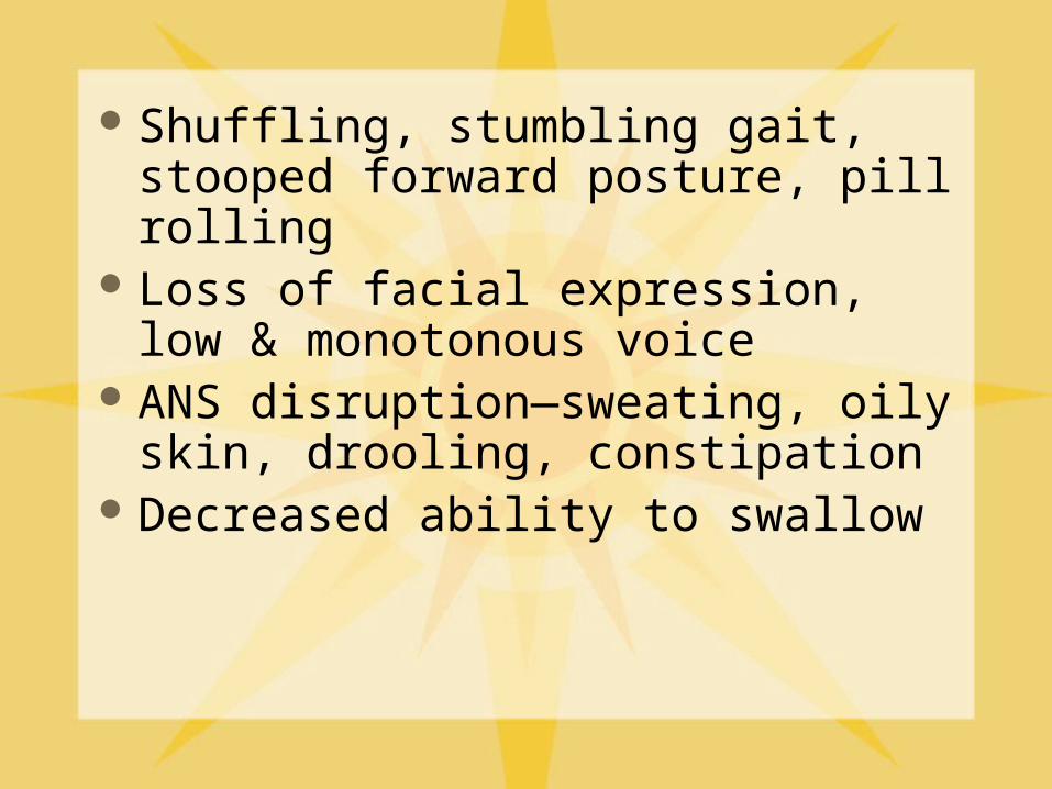
Shuffling, stumbling gait, stooped forward posture, pill rolling
Loss of facial expression, low & monotonous voice
ANS disruption—sweating, oily skin, drooling, constipation
Decreased ability to swallow
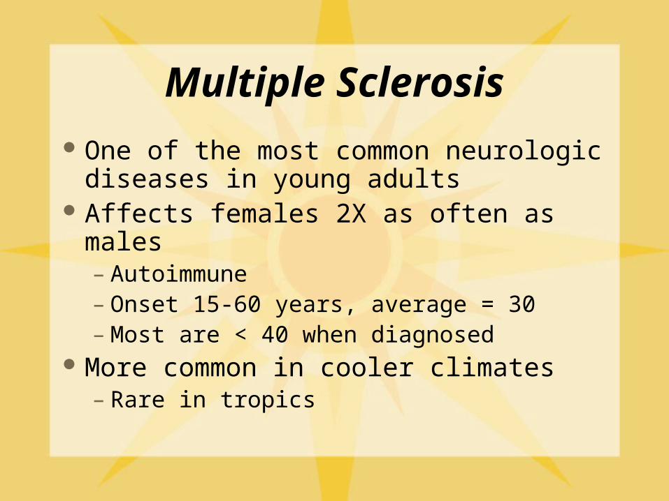
Multiple Sclerosis
One of the most common neurologic diseases in young adults
Affects females 2X as often as males – Autoimmune – Onset 15-60 years, average = 30– Most are < 40 when diagnosed
More common in cooler climates– Rare in tropics

MS
Idiopathic but probably follows viral illness—worse after gamma interferon– Precipitating factors include PG, infection,
injury, emotional stress– Probably multiple causes– Familial patterns suggests common exposure
or genetic predisposition History of attacks followed by periods of
remission with progressive damage

Underlying pathology
Widespread demyelination in CNS, with hard, yellow plaques in white matter– Pyramidal tracts, dorsal columns most often
affected– Cerebellar peduncles, brainstem, optic nerve
and tract– As myelin degenerates, macrophages enter and
remove debris

Established Syndromes
Mixed/generalized (50%)– Visual system attacked
Spinal (30-40%)– Weakness or numbness in one or more limbs– UMN signs are unilateral, spinal signs are
bilateral, legs more often than arms Cerebellar (5%)
– Symmetrical deficits, nystagmus, ataxia Amaurotic (5%)
– blindness

Signs and symptoms (any combination)
Visual problems most often initial symptoms Sensory disorders—dorsal column problems Spastic weakness of limbs—one or more Nystagmus, uncoordinated movement
– Cerebellar dysfunction Bladder dysfunction—corticospinal tracts Euphoria or dementia —frontal lobes

MS
No single diagnostic test—MRI might show plaques
CSF—elevated WBC, IgG, and myelin basic protein
Progression is variable—disability in 10-20 years in most cases
Relapsing-remitting form is most common

Chronic, progressive MS Primary progressive--steady, gradual
decline– Fairly rare
Secondary progressive--eventually affects 2/3 of pts– Relapsing/remission form– Start to experience decline between attacks
Progressive relapsing--rarest, no remission– Occasional bouts of increased severity

ALS—Amyotrophic lateral sclerosis
AKA Lou Gehrig’s Disease Progressive, idiopathic neurologic deterioration of
40-70 year olds– 5-10% have an inherited form
Destruction of upper and lower neurons in motor tracts– Ventral horns of spinal cord, lower brainstem, cerebral
cortex are destroyed– No inflammation
Results in muscular atrophy– Pt presents with hand or leg weakness, incoordination,
or difficulty speaking (stammer, stutter)

ALS NO sensory loss, no memory loss, patient
remains aware of everything Fast glycolytic fibers go first, fast oxidative next
– Slow twitch fibers go last Cranial often goes before caudal
– Difficulty chewing, swallowing, speaking, breathing– Fatigue in arms or legs, tripping, drop objects– Control of eye and bladder are lost last
Usually die 2-6 years after diagnosis, of respiratory failure

Pathophysiology of ALS Evidence that damage to SOD1 (superoxide
dismutase) gene allows damage to neuron by free radicals
First signs of degeneration begin at distal axon near synapse– accumulation of xs neurofilaments and disruption
of microtubules blocks nutrient transport inside neuron
– Later dysfunction of proteosomes in cell body allow buildup of degenerative products
– Breakdown of surrounding glial cells

Lower Motor Neuron diseases:Myasthenia gravis
Only neuromuscular disease with rapid fatigue and prolonged recovery
Younger patients: Women 3X as likely as men to be affected
Patients > 50 are more often males Usually die from respiratory insufficiency Histology—autoimmune destruction of
ACh receptor at myoneural junction

Generalized autoimmune myasthenia
May have periodic relapses with prolonged remission
May be slowly progressive with no remission
May be rapidly fulminating and fatal Often graded I (ocular disease only)
through IV (crisis)

Signs and symptoms Progressive weakness and fatigability—
eye and face often first– Double vision (diplopia) and drooping
lids (ptosis)– Hanging jaw sign, inability to swallow
Weakness increases with activity, strength improves with rest
Strength improves with ACh esterase inhibitors

Signs are aggravated by:
Hormonal imbalance (PG, phases of menstrual cycle, thyroid)
Concurrent illness or emotional stress Alcohol, especially Gin &Tonic

Diagnosis
EMG of muscle in repetitive action Serum antibodies to ACh receptors (80%
of patients) Tensilon test—ACh esterase inhibitor
– immediate improvement 70-80% have abnormal thymus (males) Increased risk of other AI diseases

Crisis—unable to swallow, clear respiratory secretions, or breathe adequately – Myasthenic crisis
Usually occurs 3-4 hours post meds
– Cholinergic crisisDrug OD, occurs within 1 hours of medsSee other signs of increased smooth muscle
activity
Death from respiratory arrest in either case

Infections of CNS
Meningitis
Encephalitis
Reye syndrome

Meningitis Etiology
– Viral meningitis—usually self limitingEnteroviruses, Herpes viruses, MyxovirusesAlso called aseptic meningitis, non-purulent
mengitis, lymphocytic meningitis– Bacterial meningitis
Meningococcus and PneumococcusUsually begins in another part of the bodyMay spread to ventricles and CSF

Pathophysiology Bacteria enter bloodstream, break through
choroid plexus into subarachnoid space– Inflammatory reaction in meningeal vessels– Purulent exudate may obstruct villi and produce
interstitial edema
Abrupt onset of severe, throbbing headache, fever, stiff neck– Photophobia, decreased LOC if spread to brain– May/may not have nausea, vomiting, abdominal pain,
malaise

Encephalitis Acute, febrile, usually viral origin Signs of meningitis plus decreased level of
consciousness – Delirium, confusion, seizures– Increased ICP– Herpes associated with hallucinations, abnormal
behavior Much poorer prognosis than meningitis Differentiate from brain abscesses, tumors,
parasites– Brain abscess may follow any case of encephalitis

Reye syndrome
Associated with giving aspirin to children with influenza or other viral infections– 2 phase illness--viral infection then Reyes
Syndrome Appear to recover from viral illness, then
vomiting, convulsions, delirium– Acute, rare, multi-organ (liver and brain
typically) in apparently healthy child– NO FEVER at this time

Dysfunction of hepatic mitochondria underlies pathology
Fatty infiltration of liver, heart and kidneys but no inflammation or necrosis– Profound hypoglycemia, hyperammonemia– Increase in short chain FA in serum
Electrolyte disturbances (decreased Na and K, high ammonia)– Cerebral edema, increased ICP, swelling of
mitochondria in neurons Mortality 25-50%, survivors may have
permanent CNS damage--retardation, seizures, paralysis

Spinal Cord Trauma
Most often in young, single males Etiology—car accidents, falls, sports
injuries– hyperextension, hyperflexion, vertical
compression, rotational forces Quadriplegia—injuries to cervical spine Paraplegia—injuries to thoracic, lumbar,
and sacral spine
Mechanism of Injury
Most damage occurs at time of initial injury
Area above damage usually survives– 2º damage from continued movement, rubbing
on damaged vertebrae– ischemia after trauma major damage
(methylprednisolone) XS glutamate damages surviving cells

Common complications
Chronic pain and muscle spasms Bed sores from constant pressure Deep vein thrombosis from inactivity CHF, pulmonary edema from compromised
circulation Pneumonia from accumulation of mucous in
upper respiratory tract

Life expectancy has improved
Infection is #1 cause of death– Quadriplegics usually die within 5 years
Renal failure #2 cause of death

Quality of life depends on level of damage to cord
C1 rare but usually fatal C2- 4 life threatening—phrenic nerve C5 retain head, neck, shoulder,
respiratory control C6 retain control through wrist C7 retain some finger control C7-T-1 retain hand control

Paraplegia
T2-12 retain upper body and some trunk control
L1-5 usually retain full trunk control and some leg control
S1-5 some bowel and bladder dysfunction

Spinal shock--SNS is in T-L spine
temporary loss of cord function initial loss of reflex activity below level of
injury—flaccid paralysis loss of T control, loss of vasomotor tone,
atonic bowel and bladder recovery in hours to weeks (30 days) must maintain BP and urine flow with
fluids; keep bowel emptied

Autonomic dysreflexia (hyperreflexia)
Occurs after recovery from spinal shock– The higher the cord lesion, the more likely this
will develop Loss of higher level control on SNS
outflow– In effect, an Upper Motor Neuron syndrome

Irritation stimulates massive SNS activity—arterial spasm, increased BP
Heart slows because of PNS response to increased BP
Severe pounding headache, flushed /pale skin, goose bumps
Must lower BP or potential stroke

Pain
Assessment is always subjective—no test to measure or confirm
If the patient says she hurts, she hurts
Often accompanied by increased SNS activity and stress response

Classification
Underlying cause– Nocioceptive– Neuropathic (non-nocioceptive)
Duration– Acute – Chronic
Etiology Regional

Physiologic events in nocioceptive (acute) pain
Transduction—noxious stimuli activate nocioceptors– Histamine, bradykinin, TNF and other
inflammatory chemicals Nocioceptors release Substance
Pvasodilation, edema, more bradykinin and histamine– Small myelinated (A-delta—fast) and non-
myelinated (C-slow) fibers transmit AP’s

Transmission along A-delta and C fibers
Synapse with second order neurons, cross spinal cord and rise in anterolateral spinothalamic tract– A-delta fibers are direct with no side
branches—sharp, localized pain– C fibers send information to reticular
formation—burning, aching, diffuseMay be most important tract in chronic pain
Perception Hypothalamus and limbic system modulate
perception Endogenous opioid peptides and receptors
throughout sensory system Pain threshold: point at which stimulus is
perceived as painfulLittle variation between individuals
Pain tolerance: length of time pain is endured without complaint

Pain tolerance
Influenced by: personality, culture, past experiences, mental/emotional state– Decreased by repeated exposures,
fatigue, anxiety, sleep deprivation, fear– Increased by distraction, cold, warmth,
alcohol, hypnosis, cultureFrontal cortex determines response

Acute pain Table 15-2
Sudden onset, specific cause, lasts < 6 months
Resolves after healing or successful drug therapy
See increased SNS response and stress reaction

Visceral acute pain Viscera respond to stretch, ischemia, and
inflammation Visceral pain often stimulates the autonomic
nervous system and induces changes in blood pressure, sweating, vomiting/diarrhea
May see contraction of local muscles– Surgical abdomen, guarding
Typically projects to superficial areas along same dermatome--referred pain– Gall bladder to area between scapulae– Myocardial ischemia to left arm and jaw

Chronic pain Table 15-2 Continuous or intermittent, lasts longer than 6
months– Low back pain is #1 example
Cause often unknown, or not responsive to analgesics
May come on suddenly or over a period of years– May afflict as much as 25% of population– Little or no SNS stimulation at this point– Severe emotional distress, depression, insomnia– Become hypersensitive to any touch—allodynia
Neuropathic pain
Trauma to or disease of nerves– Burning/tingling, shooting, stabbing, gnawing– Not responsive to NSAIDS or analgesics– Intensifies with physical/emotional stress– May be result of CNS or PNS damage
Neurons in dorsal root ganglia become more active with repetitive stimuli– May be result of loss of central inhibition

Peripheral hypersensitivity
Nocioceptors on C fiber neurons are sensitized or directly stimulated by inflammatory mediators– Decreased threshold for excitation– Activation of normally silent receptors
Response is longer and more intense Allodynia: perceive low intensity stimuli as
painful Secondary hyperalgesia of surrounding
tissues may develop

Central Sensitization
Repetitive and high frequency stimulation of C fibers
Release of glutamate and Substance P– Activation of AMPA and NMDA receptors
in CNS May also lose loss of pain inhibition by
GABA or other neurotransmitters

Wind up pain
Chronic NMDA receptor activation causes changes in protein synthesis, density of receptors, and enhanced glutamate release– May develop long term changes in sp9inal
cord (central sensitization) Increased frequency of firing of
neurons Allodynia develops

Phantom limb pain
Chronic pain, tingling, burning, itching affecting 50-85% of amputees– More common if limb was painful before
amputation– Most cases decrease significantly over 6 months
May also occur in spinal trauma patients
Not well understood, but probably involves peripheral and central activation