Delayed Tension Pneumocephalus due to Rupture of the ... · resolution of the tension...
3
online © ML Comm 0CASE REPORT0 106 Copyright ⓒ 2009 Journal of Korean Neurotraumatology Society J Kor Neurotraumatol Soc 2009;5:106-108 ISSN 1738-8708 Delayed Tension Pneumocephalus due to Rupture of the Pneumocyst Won-Chul Lee, MD, Dae-Hee Seo, MD, Il-Seung Choe, MD, PhD and Sung-Choon Park, MD, PhD Department of Neurosurgery, Myongji Hospital, Kwandong University College of Medicine, Goyang, Korea Tension pneumocephalus is an unusual delayed presentation of trauma. This case is presentation of a young man who suffered sudden deterioration of mental status from the rupture of the preexisting pneumocyst. He had a history of basal skull fracture, had a ventriculoperitoneal shunt placed due to posttraumatic hydrocephalus and had also undergone the repairing operation of basal skull defect at another hospital. The successful treatment of tension pneumocephalus and persistent pneumocyst at our hospital is described. The neurosurgeon must be aware the possibility of delayed pneumocyst formation and tension pneumocephalus in patients who experienced basal skull fracture quite a long time ago. ( J Kor Neurotraumatol Soc 2009;5: 106-108 ) KEY WORDS: Tension pneumocephalus·Pneumocyst·Basal skull fracture. Introduction Tension pneumocephalus is a serious condition requir- ing prompt treatment. 1) It is usually occurred after trauma and associated with basal skull fracture. Some cases were reported after neurosurgical manipulation. 3,4,6,8,9) We de- scribe a case of delayed tension pneumocephalus that pre- sumably developed from rupture of the incompletely treated pneumocyst of the right anterior cranial fossa. Case Report This 35-year-old man visited outpatient clinic of our hos- pital complaining headache, insomnia and memory distur- bance. His medical record showed that he had a diffuse ax- onal injury by traffic accident 15 years ago and was treated at another hospital. A ventriculoperitoneal (VP) shunt was placed at that time due to hydrocephalus. The patient said that the second operation was performed due to a right fron- tal pneumocyst at the same hospital three years later. The initial study and follow up data were not obtainable and we were not certain whether the pneumocyst was completely treated or not. On the initial computed tomography (CT) scan at our hospital, a round pneumocyst was found on the right anterior cranial fossa (Figure 1). He refused further study and operation for removal of pneumocyst and repair of the defect. Conservative treatment including headache regimen was done and the patient was followed up for two years. He had no history of cerebrospinal fluid (CSF) leak- age. On the day of emergent admission, the patient’s mother reported that he had complained severe headache for two hours before deterioration. There was no known recent head trauma history and signs of CSF infection. On the neurological examination, the patient was stupor- ous and hemiplegic on the left side. Axial CT scans reveal- ed frontotemporal air collection with multiple cisternal air densities and compression of the parenchyma of brain bi- laterally. The characteristic separation of the frontal lobe instead of peaking sign only was consistent with the diag- nosis of tension pneumocephalus. 7) The size of the previous air pocket seemed slightly decreased than the previous CT image (Figure 2). In consideration of the rapid deterioration, a twist drill trephination instead of a craniotomy with repair of the de- fect was performed on the right frontal bone and an external- ized drain was connected to the closed drainage system. Two days later, the patient improved markedly. The CT scan Received: July 1, 2009 / Revised: July 13, 2009 Accepted: September 11, 2009 Address for correspondence: Sung-Choon Park, MD, PhD Department of Neurosurgery, Myongji Hospital, Kwandong Universit y College of Medicine, 697-24 Hwajeong- dong, Deogyang- gu, Goyang 412- 270, Korea Tel: +82-31-810-6630, Fax: +82-31-969-0500 E-mail: [email protected]
Transcript of Delayed Tension Pneumocephalus due to Rupture of the ... · resolution of the tension...
online © ML Comm
0CASE REPORT0
106 Copyright ⓒ 2009 Journal of Korean Neurotraumatology Society
J Kor Neurotraumatol Soc 2009;5:106-108 ISSN 1738-8708
Delayed Tension Pneumocephalus due to Rupture of the Pneumocyst
Won-Chul Lee, MD, Dae-Hee Seo, MD, Il-Seung Choe, MD, PhD and Sung-Choon Park, MD, PhD
Department of Neurosurgery, Myongji Hospital, Kwandong University College of Medicine, Goyang, Korea
Tension pneumocephalus is an unusual delayed presentation of trauma. This case is presentation of a young man who suffered sudden deterioration of mental status from the rupture of the preexisting pneumocyst. He had a history of basal skull fracture, had a ventriculoperitoneal shunt placed due to posttraumatic hydrocephalus and had also undergone the repairing operation of basal skull defect at another hospital. The successful treatment of tension pneumocephalus and persistent pneumocyst at our hospital is described. The neurosurgeon must be aware the possibility of delayed pneumocyst formation and tension pneumocephalus in patients who experienced basal skull fracture quite a long time ago. (J Kor Neurotraumatol Soc 2009;5: 106-108) KEY WORDS: Tension pneumocephalus·Pneumocyst·Basal skull fracture.
Introduction
Tension pneumocephalus is a serious condition requir-ing prompt treatment.1) It is usually occurred after trauma and associated with basal skull fracture. Some cases were reported after neurosurgical manipulation.3,4,6,8,9) We de-scribe a case of delayed tension pneumocephalus that pre-sumably developed from rupture of the incompletely treated pneumocyst of the right anterior cranial fossa.
Case Report
This 35-year-old man visited outpatient clinic of our hos-pital complaining headache, insomnia and memory distur-bance. His medical record showed that he had a diffuse ax-onal injury by traffic accident 15 years ago and was treated at another hospital. A ventriculoperitoneal (VP) shunt was placed at that time due to hydrocephalus. The patient said that the second operation was performed due to a right fron-tal pneumocyst at the same hospital three years later. The
initial study and follow up data were not obtainable and we were not certain whether the pneumocyst was completely treated or not. On the initial computed tomography (CT) scan at our hospital, a round pneumocyst was found on the right anterior cranial fossa (Figure 1). He refused further study and operation for removal of pneumocyst and repair of the defect. Conservative treatment including headache regimen was done and the patient was followed up for two years. He had no history of cerebrospinal fluid (CSF) leak-age. On the day of emergent admission, the patient’s mother reported that he had complained severe headache for two hours before deterioration. There was no known recent head trauma history and signs of CSF infection.
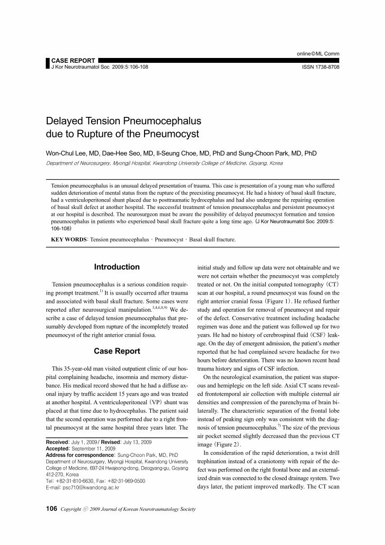
On the neurological examination, the patient was stupor-ous and hemiplegic on the left side. Axial CT scans reveal-ed frontotemporal air collection with multiple cisternal air densities and compression of the parenchyma of brain bi-laterally. The characteristic separation of the frontal lobe instead of peaking sign only was consistent with the diag-nosis of tension pneumocephalus.7) The size of the previous air pocket seemed slightly decreased than the previous CT image (Figure 2).
In consideration of the rapid deterioration, a twist drill trephination instead of a craniotomy with repair of the de-fect was performed on the right frontal bone and an external- ized drain was connected to the closed drainage system. Two days later, the patient improved markedly. The CT scan
Received: July 1, 2009 / Revised: July 13, 2009 Accepted: September 11, 2009 Address for correspondence: Sung-Choon Park, MD, PhD Department of Neurosurgery, Myongji Hospital, Kwandong UniversityCollege of Medicine, 697-24 Hwajeong-dong, Deogyang-gu, Goyang412-270, Korea Tel: +82-31-810-6630, Fax: +82-31-969-0500 E-mail: [email protected]
Won-Chul Lee, et al.
www.neurotrauma.or.kr 107
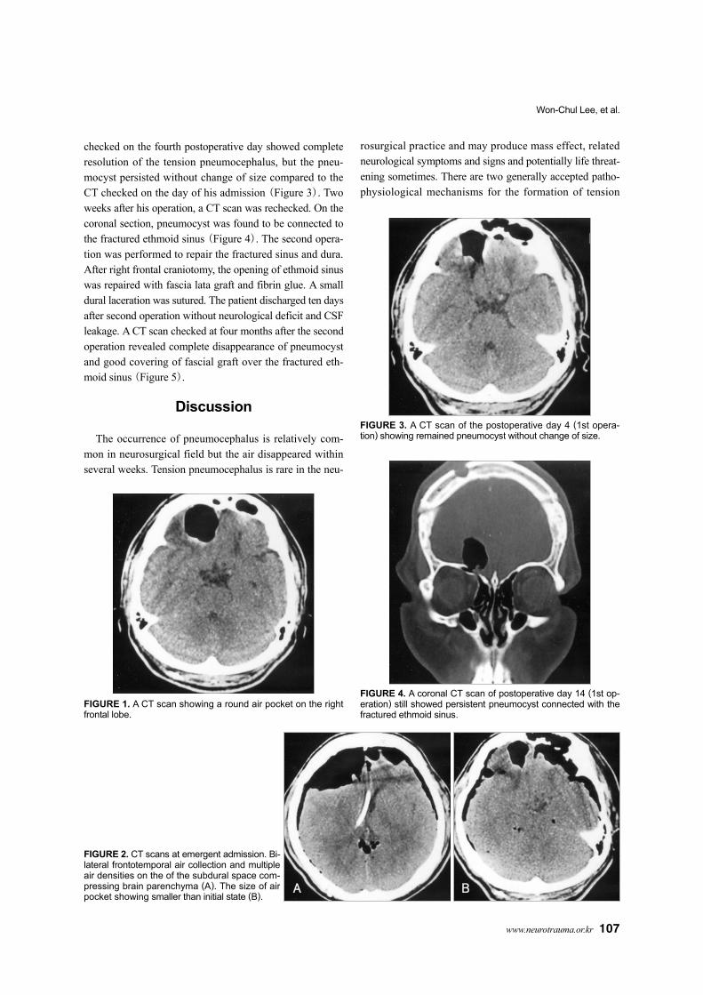
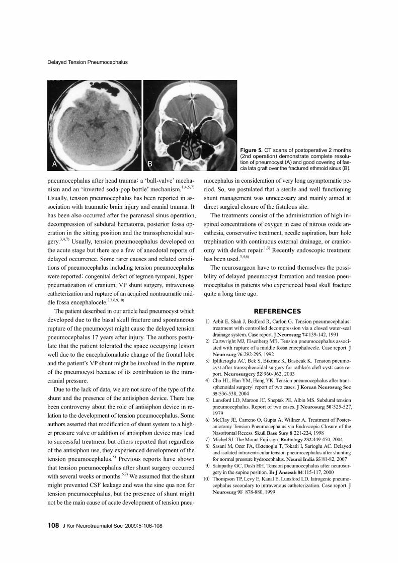
checked on the fourth postoperative day showed complete resolution of the tension pneumocephalus, but the pneu-mocyst persisted without change of size compared to the CT checked on the day of his admission (Figure 3). Two weeks after his operation, a CT scan was rechecked. On the coronal section, pneumocyst was found to be connected to the fractured ethmoid sinus (Figure 4). The second opera-tion was performed to repair the fractured sinus and dura. After right frontal craniotomy, the opening of ethmoid sinus was repaired with fascia lata graft and fibrin glue. A small dural laceration was sutured. The patient discharged ten days after second operation without neurological deficit and CSF leakage. A CT scan checked at four months after the second operation revealed complete disappearance of pneumocyst and good covering of fascial graft over the fractured eth-moid sinus (Figure 5).
Discussion
The occurrence of pneumocephalus is relatively com-mon in neurosurgical field but the air disappeared within several weeks. Tension pneumocephalus is rare in the neu-
rosurgical practice and may produce mass effect, related neurological symptoms and signs and potentially life threat-ening sometimes. There are two generally accepted patho-physiological mechanisms for the formation of tension
FIGURE 2. CT scans at emergent admission. Bi-lateral frontotemporal air collection and multipleair densities on the of the subdural space com-pressing brain parenchyma (A). The size of airpocket showing smaller than initial state (B).
A B
FIGURE 1. A CT scan showing a round air pocket on the rightfrontal lobe.
FIGURE 3. A CT scan of the postoperative day 4 (1st opera-tion) showing remained pneumocyst without change of size.
FIGURE 4. A coronal CT scan of postoperative day 14 (1st op-eration) still showed persistent pneumocyst connected with thefractured ethmoid sinus.
Delayed Tension Pneumocephalus
108 J Kor Neurotraumatol Soc 2009;5:106-108
pneumocephalus after head trauma: a ‘ball-valve’ mecha-nism and an ‘inverted soda-pop bottle’ mechanism.1,4,5,7) Usually, tension pneumocephalus has been reported in as-sociation with traumatic brain injury and cranial trauma. It has been also occurred after the paranasal sinus operation, decompression of subdural hematoma, posterior fossa op-eration in the sitting position and the transsphenoidal sur-gery.3,4,7) Usually, tension pneumocephalus developed on the acute stage but there are a few of anecdotal reports of delayed occurrence. Some rarer causes and related condi-tions of pneumocephalus including tension pneumocephalus were reported: congenital defect of tegmen tympani, hyper-pneumatization of cranium, VP shunt surgery, intravenous catheterization and rupture of an acquired nontraumatic mid-dle fossa encephalocele.2,3,6,9,10)
The patient described in our article had pneumocyst which developed due to the basal skull fracture and spontaneous rupture of the pneumocyst might cause the delayed tension pneumocephalus 17 years after injury. The authors postu-late that the patient tolerated the space occupying lesion well due to the encephalomalatic change of the frontal lobe and the patient’s VP shunt might be involved in the rupture of the pneumocyst because of its contribution to the intra-cranial pressure.
Due to the lack of data, we are not sure of the type of the shunt and the presence of the antisiphon device. There has been controversy about the role of antisiphon device in re-lation to the development of tension pneumocephalus. Some authors asserted that modification of shunt system to a high-er pressure valve or addition of antisiphon device may lead to successful treatment but others reported that regardless of the antisiphon use, they experienced development of the tension pneumocephalus.8) Previous reports have shown that tension pneumocephalus after shunt surgery occurred with several weeks or months.6,9) We assumed that the shunt might prevented CSF leakage and was the sine qua non for tension pneumocephalus, but the presence of shunt might not be the main cause of acute development of tension pneu-
mocephalus in consideration of very long asymptomatic pe-riod. So, we postulated that a sterile and well functioning shunt management was unnecessary and mainly aimed at direct surgical closure of the fistulous site.
The treatments consist of the administration of high in-spired concentrations of oxygen in case of nitrous oxide an-esthesia, conservative treatment, needle aspiration, burr hole trephination with continuous external drainage, or craniot-omy with defect repair.1,3) Recently endoscopic treatment has been used.3,4,6)
The neurosurgeon have to remind themselves the possi-bility of delayed pneumocyst formation and tension pneu-mocephalus in patients who experienced basal skull fracture quite a long time ago.
REFERENCES 1) Arbit E, Shah J, Bedford R, Carlon G. Tension pneumocephalus:
treatment with controlled decompression via a closed water-seal drainage system. Case report. J Neurosurg 74:139-142, 1991
2) Cartwright MJ, Eisenberg MB. Tension pneumocephalus associ-ated with rupture of a middle fossa encephalocele. Case report. J Neurosurg 76:292-295, 1992
3) Iplikcioglu AC, Bek S, Bikmaz K, Basocak K. Tension pneumo-cyst after transsphenoidal surgery for rathke’s cleft cyst: case re-port. Neurosurgery 52:960-962, 2003
4) Cho HL, Han YM, Hong YK. Tension pneumocephalus after trans-sphenoidal surgery: report of two cases. J Korean Neurosurg Soc 35:536-538, 2004
5) Lunsford LD, Maroon JC, Sheptak PE, Albin MS. Subdural tension pneumocephalus. Report of two cases. J Neurosurg 50:525-527, 1979
6) McClay JE, Carreno O, Gupta A, Willner A. Treatment of Postcr-aniotomy Tension Pneumocephalus via Endoscopic Closure of the Nasofrontal Recess. Skull Base Surg 8:221-224, 1998
7) Michel SJ. The Mount Fuji sign. Radiology 232:449-450, 2004 8) Sasani M, Ozer FA, Oktenoglu T, Tokatli I, Sarioglu AC. Delayed
and isolated intraventricular tension pneumocephalus after shunting for normal pressure hydrocephalus. Neurol India 55:81-82, 2007
9) Satapathy GC, Dash HH. Tension pneumocephalus after neurosur-gery in the supine position. Br J Anaesth 84:115-117, 2000
10) Thompson TP, Levy E, Kanal E, Lunsford LD. Iatrogenic pneumo-cephalus secondary to intravenous catheterization. Case report. J Neurosurg 91: 878-880, 1999
Figure 5. CT scans of postoperative 2 months (2nd operation) demonstrate complete resolu-tion of pneumocyst (A) and good covering of fas-cia lata graft over the fractured ethmoid sinus (B).
A B