Deg nala disease
24
A CHALLENGE OF VETERINARY PRACTITIONER FROM DEGNALA DISEASE Dr.Jibachha Sah M.V.SC (T.U.) VHRTC(P)LTD
-
Upload
jibachha-shah -
Category
Education
-
view
160 -
download
1
Transcript of Deg nala disease
A CHALLENGE OF VETERINARY PRACTITIONER FROM DEGNALADISEASE
Dr.Jibachha SahM.V.SC (T.U.)VHRTC(P)LTD
Introduction
Degnala disease, which is believed to be amycotoxicosis, has clinical syndromesimilar to chronic ergotism and ischaracterised by development of oedema,necrosis and gangrene of the legs, tail,ears, etc.
Etiology
The most frequently found fungi species recorded from rice straw were: Aspergillus, Aspergillus flavus and Penicillium notatum.
Species affected
Buffaloes are more frequently affected than cattle and younger animals appear to be more susceptible.
Epizootical
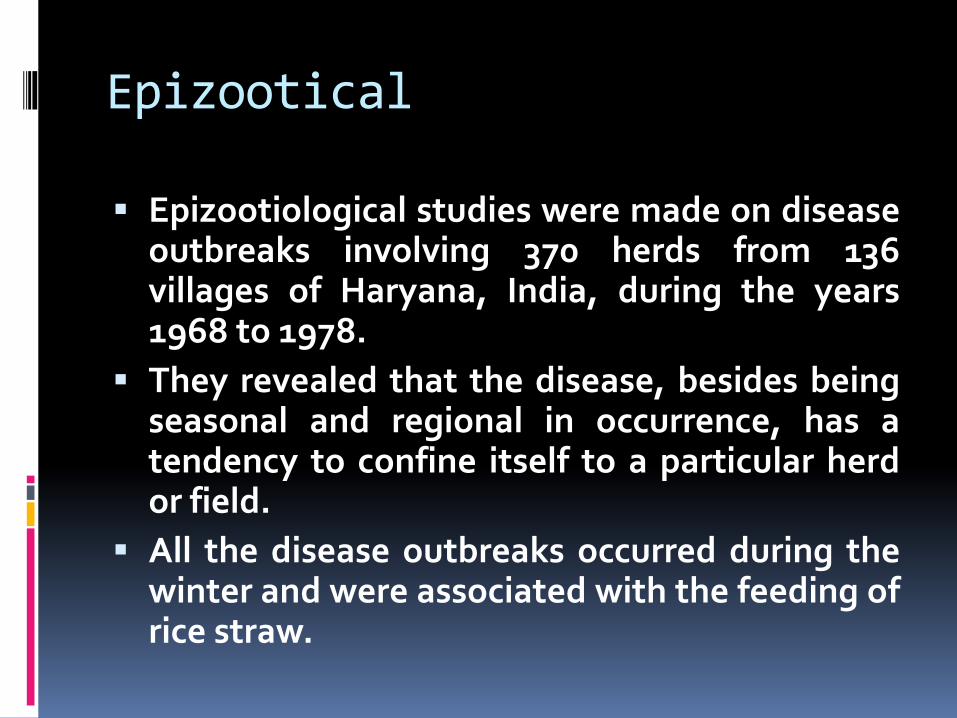
Epizootiological studies were made on diseaseoutbreaks involving 370 herds from 136villages of Haryana, India, during the years1968 to 1978.
They revealed that the disease, besides beingseasonal and regional in occurrence, has atendency to confine itself to a particular herdor field.
All the disease outbreaks occurred during thewinter and were associated with the feeding ofrice straw.
Prevalence in Nepal
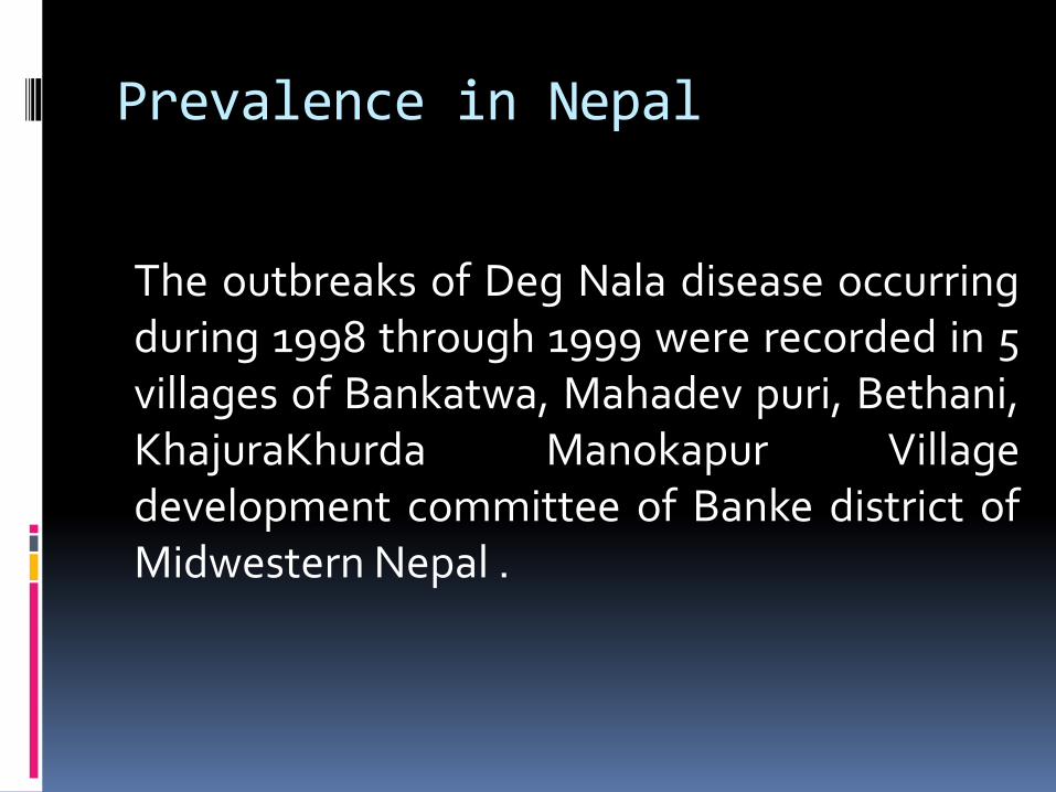
The outbreaks of Deg Nala disease occurringduring 1998 through 1999 were recorded in 5villages of Bankatwa, Mahadev puri, Bethani,KhajuraKhurda Manokapur Villagedevelopment committee of Banke district ofMidwestern Nepal .
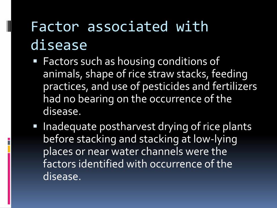
Factor associated with disease Factors such as housing conditions of
animals, shape of rice straw stacks, feeding practices, and use of pesticides and fertilizers had no bearing on the occurrence of the disease.
Inadequate postharvest drying of rice plants before stacking and stacking at low-lying places or near water channels were the factors identified with occurrence of the disease.

Morbidity and mortality
The morbidity and mortality rates were61.61% and 13.93%, respectively, inbuffaloes and 13.49% and 2.41% in cattle,with no sex and age differences.
Symptoms
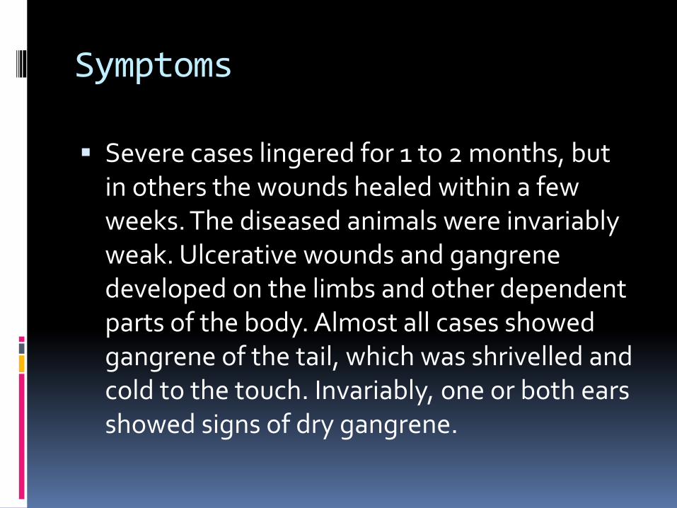
Severe cases lingered for 1 to 2 months, but in others the wounds healed within a few weeks. The diseased animals were invariably weak. Ulcerative wounds and gangrene developed on the limbs and other dependent parts of the body. Almost all cases showed gangrene of the tail, which was shrivelled and cold to the touch. Invariably, one or both ears showed signs of dry gangrene.
In some cases the muzzle and even the tip of the tonguebecame gangrenous and was shed. One or more hoovesshowed lesions in varying stages of development. In somecases the affected feet and legs were swollen up to the knee;hair was denuded and inflammatory changes set in. Later,wounds appeared on the coronet, fetlock, pastern, knee and inthe hock region.
In very advanced cases the lower regions of the feet becomegangrenous. In some cases the hooves were shed and boneswere exposed. The gangrenous portions of the tail, tips of theears, tongue and other affected parts of the body, dropped off,although wounds healed in the course of time.
Secondary bacterial infections of the lesions were at least partially responsible for the severity of the disease. To address this complicating factor, long-acting Terramycinwas injected parenterally.
Moreover simultaneous use of antifungal(Diethylamine acetarsol, Acetylarson,Antidegnala liquior) induced development ofimmunity and was proven to be effectiveagainst infection.
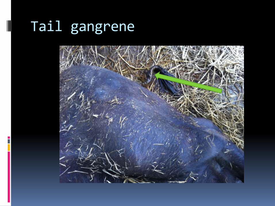
Tail gangrene
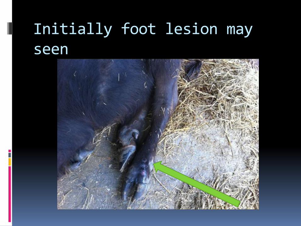
Initially foot lesion may seen

Foot lesion
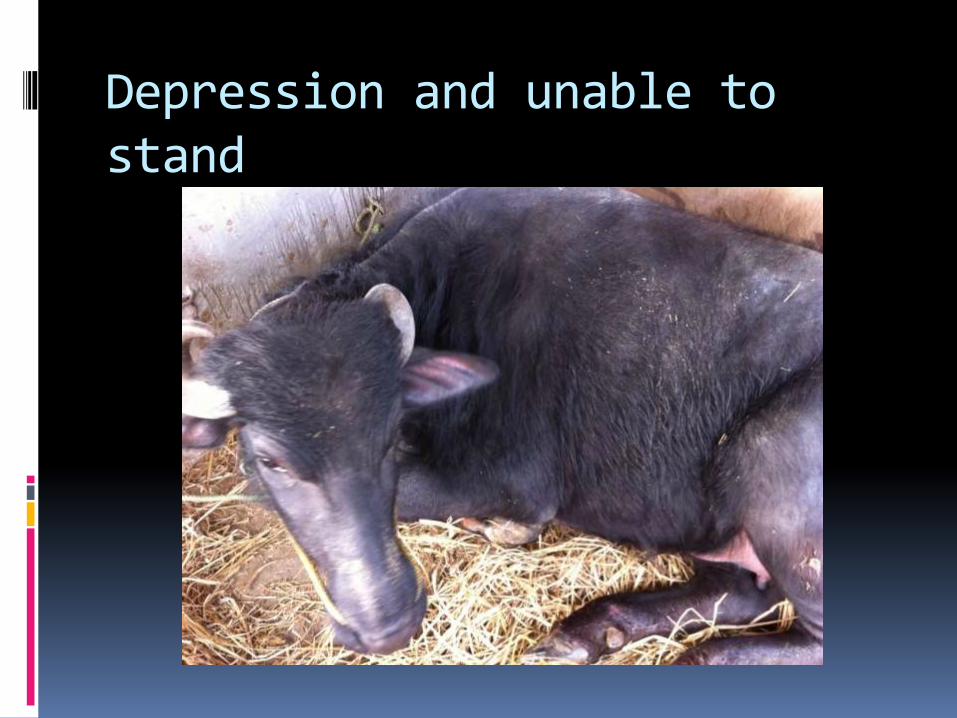
Depression and unable to stand
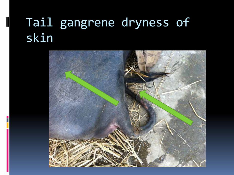
Tail gangrene dryness of skin
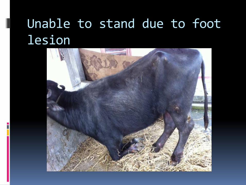
Unable to stand due to foot lesion

After treatment improvement in tail gangrene
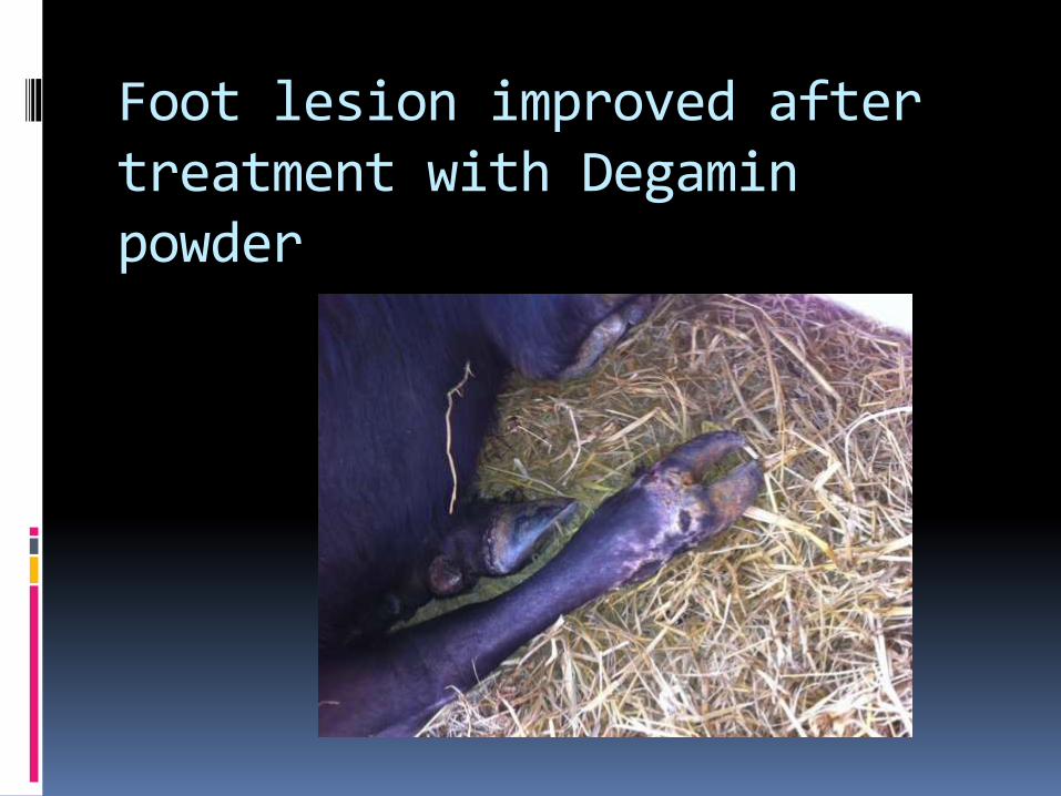
Foot lesion improved after treatment with Degaminpowder
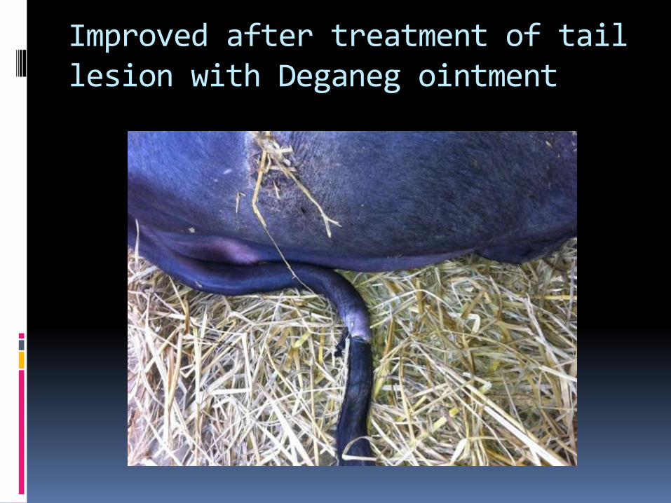
Improved after treatment of tail lesion with Deganeg ointment
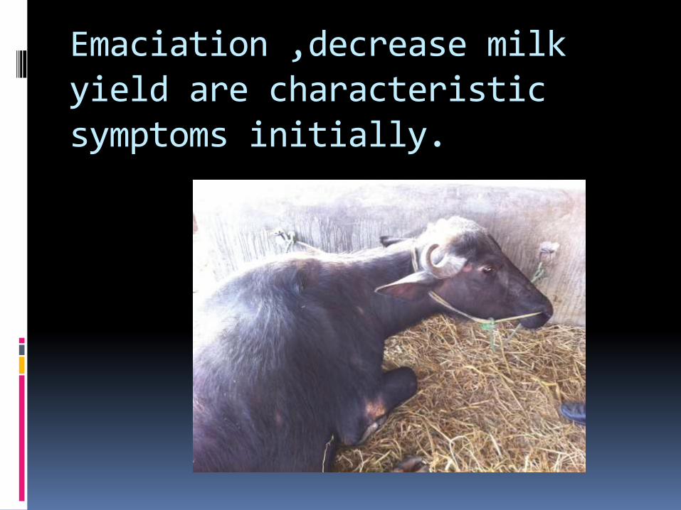
Emaciation ,decrease milk yield are characteristic symptoms initially.
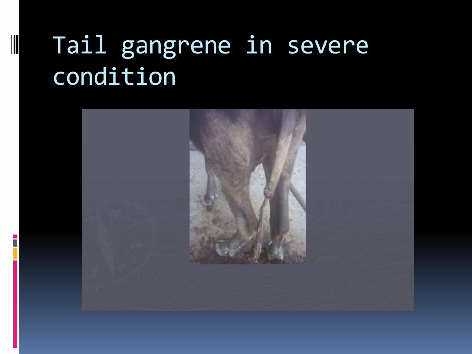
Tail gangrene in severe condition
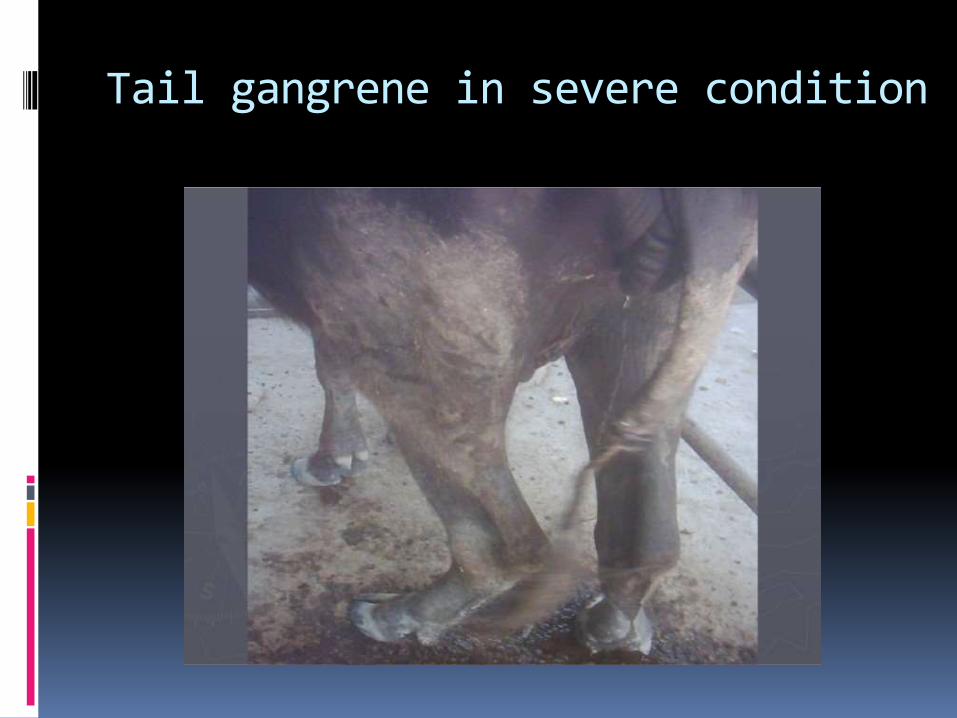
Tail gangrene in severe condition
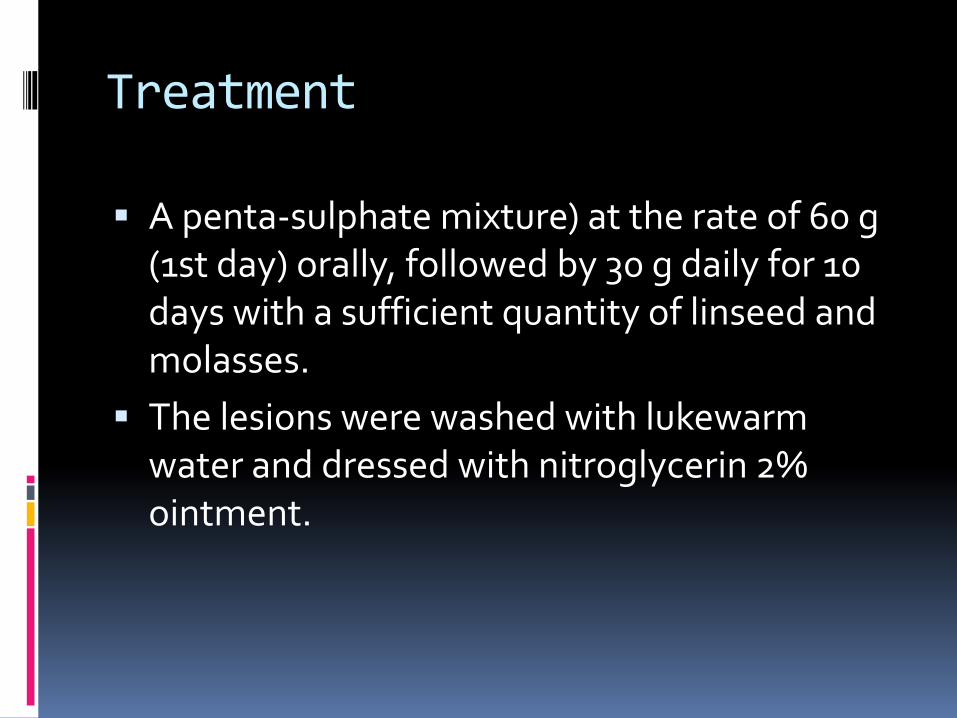
Treatment
A penta-sulphate mixture) at the rate of 60 g (1st day) orally, followed by 30 g daily for 10 days with a sufficient quantity of linseed and molasses.
The lesions were washed with lukewarm water and dressed with nitroglycerin 2% ointment.