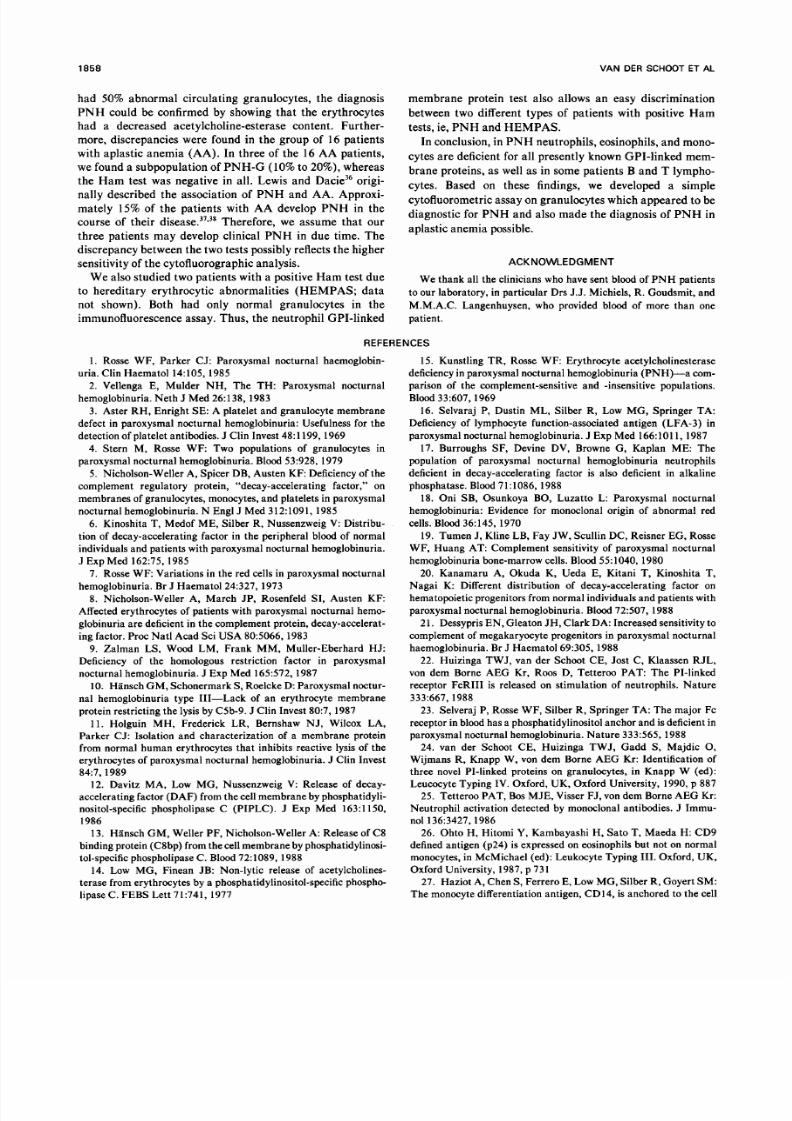
Deficiency of GPI of Leukocytes in Paroxysmal Nocturnal Hematuria
7
Deficiency of Glycosyl-Phosphatidylinositol-Linked Membrane Glycoproteins of Leukocytes in Paroxysmal Nocturnal Hemoglobinuria, Description of a New Diagnostic Cytofluorometric Assay By C. Ellen van der Schoot, Tom W.J. Huizinga, Elisabeth T. van ‘t Veer-Korth of, Rosemarie Wijmans, Jantina Pinkster, and Albert E.G. Kr. von dem Borne Paroxysmal nocturnal hemoglobinuria (PNH) is a disease tha t affects not only red cel ls, but other blood cel ls as well. The common defect is supposed to be an acquired defi- ciency of glycosyl-p hosphatidy linositol GPII -anchored mem- brane proteins, which may be present already at the hematopoietic stem cell level. Recently, a panel of mono- clonal antibodies (MoAbs) has become available directed against various GPI-linked membrane proteins. This makes it possible to study various cell lineages for the deficiency of such proteins in PNH in more detail. Using cytofluorogra- 20 PNH patients miss not only GPI-linked FcRlll (CD16 anti- gen). but also three other GPI-linked proteins, ie , CD24 antigen, CD67 antigen and a granulocyte-specific 50 t o 80 Kd antigen. The affected granulocytes were not only neutrophils but also eosinophils, as was found in a more detailed analysis of thr ee patients. Moreover , in all 10 PNH patients tested, the monocytes were found to be defici ent for the GPI-l inked CD14 anti gen, and we found with CD24 AROX YSMAL nocturnal hemoglob inur ia (PNH ) is an P cquired hematologic disorder characterized by inter- mittent intravascular PN H patients have abnor- mal erythrocytes, platelets, and granulocyte^^-^ that have an increased sensitivity to complement. Two types of PNH erythrocytes (PNH-E) have been recognized, based on differences in sensitivity to complement-mediated lysis.7 PNH-E type I1 are three to five times more sensitive to complement than normal red cells, PNH- E type I11 15 to 25 times mo re. 7 PNH-E with normal sensitivity are called P NH type-I cells. The increased sensitivity of the erythrocytes to complemen t (and pro bably of the other blo od cells as we ll) is due to the deficiency of cell-membrane-associated comple- ment regulatory proteins.’-” Decay-accelerating factor (DAF = CD55 antigen) is one o f th e missing proteins in PNH.’ In addi tion to DAF, PNH -E type 111 are deficient in the C8-binding protein (C8bp)9.’0 nd the recently described 18-Kd protein termed membrane inhibito r o f reactive lysis (MIRL = CD59 antigen).” Because DAF ,12 C8bp,I3 and MIRL as well as red-cell acetyl-ch~linesterase ’~ nother protein deficient in PN H, ” belong to the class of glycosyl- phosphatidylinositol (GP1)-linked membrane proteins, Davitz et a 1 suggested that the molecular defect i n P NH may lie in the posttranslational attachment of the GPI anchor.12 The demonstration of deficiency of two other GPI-linked cellular membrane proteins in PN H, LFA-3I6 and alkaline phosphat ase,” is in agreemen t with this hypothesis. Ho w- ever, it is not clear whether in PNH all or only a subset of GPI-linked membrane proteins is deficient. That PNH is a clonal disorder has been shown by glucose-6-phosphate dehydrogenase enzyme polymorphism studies.” The fact that abnormalities exist in red cells, granulocytes, monocytes, and platelets as well as in their precursor^'^-^' suggests tha t the lesi on specific for PN H is, at least in some patients, present in hematopoietic stem cells and CD55 (DAF) antibodies that lymphocytes may be involved as well. However, abnormal 6 and T lymphocytes were detected only in a subset of patients (2 of 1 0 tested). The uniform deficiency of GPI-linked proteins of granulo- cytes allows the introduc tion of a ne w diagnostic cytofluo- rometric assay for PNH with MoAbs against GPI-linked granulocytic antigens. This test was positive in all PNH patients studied and not in a group of 4 ontrol patients or 5 normal donors, with the exception of three of 16 aplastic anemi a AA) patients. In the three AA patients, (10 20 ) detected, whereas these patients had a negative acidified serum (Ham) test. This indicat es that t he n ew tes t is more sensitive than the Ham tes t and allows the early diagnosis of PNH in AA. An advantage of th e ne utrophil assay is that, in contrast t o th e Ham test, it is not influenced by recent red-cell transfusions. Moreover, it is possible to quantify th e number of affected cells by single cell analysis. 1990 by The American Society of Hematology. from which all these cell lines derive. However, it is not known whether all these blood-cell lineages in PNH are affected as well always. Recently, we as well as others demonstrated that the FcRIII of neutrophils (GD16) is GPI-linked.22.23y screen- ing a panel of MoAbs submitted to the Fourth Workshop o f Leucocyte Differenti ation Antigens, we identified three other GPI-linked proteins on granulocytes: the CD24 antigen, the CD67 antigen, and the antigen recognized by cLBgra11/5.~~ In this report, we demonstrate tha t all these GPI-anchored proteins are deficient on PNH granulocytes. In contrast to the supposed heterogeneity in red cell types in PN H, we did not find indications for t he ex istence of diffe rent typ es of affected granulocytes . By using MoAbs against GPI-linked antigens present not only on neutrophils (FcRIII and CLB- gran/5 antigens) but also on eosinophils (CD24 and CD67 From the Department of Immunological Hematology, Central Laboratory o f the Netherlands Red Cross Blood Transfusion Service, Laboratory for Experimental and Clinical Immunology. University o f Amsterdam; Department o f Haematology. Academic Medical Centre, Amsterdam: and Department of Pediatrics. Univer- sity Hospital Leiden. Leiden, The Netherlands. Submitted March 6,19 90; accepted July 9.1990. Supported by a grant from the Queen Wilhelmina Fund/ Netherlands Fou ndation fo r the Fight Against Cancer. Address reprint requests to C. Ellen van der Schoot. MD, c/o Publication Secretariat, Central Laboratory o f the Netherlands Red Cross Blood Transfusion Service, PO Box 9406, 1006 AK Amsterdam, The Netherlands. The publication costs o f this article were defrayed in par t by page charge paym ent. This article must therefore be hereby marked “advertisement” in accordance with 18 U.S.C. ection 1734 solely to indicate this fact. 990 by The American Society o f Hematology. 0006-4971/90/7609-0028 3.00/0 Blood, Vol76, No 9 (November 1 1 , 1990: pp 1853-1859 1853
-
Upload
katherine-bajada -
Category
Documents
-
view
221 -
download
0
Transcript of Deficiency of GPI of Leukocytes in Paroxysmal Nocturnal Hematuria
8/12/2019 Deficiency of GPI of Leukocytes in Paroxysmal Nocturnal Hematuria
http://slidepdf.com/reader/full/deficiency-of-gpi-of-leukocytes-in-paroxysmal-nocturnal-hematuria 1/7
Deficiency of Glycosyl-Phosphatidylinositol-Linked Membrane Glycoproteins of
Leukocytes in Paroxysm al Nocturnal Hem oglobinuria, Description of a NewDiag nostic C ytofluorometric Assay
By C.Ellen van der Schoot, Tom W.J. Huizinga, ElisabethT. van ‘t Veer-Korthof, Rosemarie Wijmans, Jantina Pinkster,
and Albert E.G. Kr. von dem Borne
Paroxysmal nocturnal hemoglobinuria (PNH) is a disease
that affects not only red cells, but other blood cells as well.
The common defect is supposed to be an acquired defi-
ciency of glycosyl-phosphatidylinositol GPII-anchored mem-
brane proteins, which may be present already at the
hematopoietic stem cell level. Recently, a panel of mono-
clonal antibodies (MoAbs) has become available directed
against various GPI-linked membrane proteins. This makes
it possible to study various cell lineages for the deficiency
of such proteins in PNH in more detail. Using cytofluorogra-
phy, we could show tha t the granulocytes of 20 different
PNH patients miss not only GPI-linked FcRlll (CD16 anti-
gen). but also three other GPI-linked proteins, ie, CD24
antigen, CD67 antigen and a granulocyte-specific 50 to 80
Kd antigen. The affected granulocytes were not only
neutrophils but also eosinophils, as was found in a moredetailed analysis of three patients. Moreover, in all 10 PNH
patients tested, the monocytes were found to be deficient
for the GPI-linkedCD14 antigen, and we found with CD24
AROXYSMAL nocturnal hemoglobinuria (PNH) is anP cquired hematologic disorder characterized by inter-
mittent intravascular PNH patients have abnor-
mal erythrocytes, platelets, and granulocyte^^-^ that have an
increased sensitivity to complement. Two types of PNH
erythrocytes (PNH-E) have been recognized, based on
differences in sensitivity to complement-mediated lysis.7
PNH-E type I1 are three to five times more sensitive to
complement than normal red cells, PNH-E type I1115 to 25times more.7 PNH-E with normal sensitivity are called PNH
type-I cells. The increased sensitivity of the erythrocytes to
complement (and probably of the other blood cells as well) is
due to the deficiency of cell-membrane-associated comple-
ment regulatory proteins.’-” Decay-accelerating factor
(DAF = CD55 antigen) is one of the missing proteins in
PNH.’ In addition to DAF, PNH-E type111are deficient in
the C8-binding protein (C8bp)9.’0 nd the recently described
18-Kd protein termed membrane inhibitor of reactive lysis
(MIRL = CD59 antigen).” Because DAF,12 C8bp,I3 and
MIRL as well as red-cell acetyl-ch~linesterase ’~nother
protein deficient in PNH,” belong to the class of glycosyl-
phosphatidylinositol (GP1)-linked membrane proteins,Davitz et a1 suggested that the molecular defect in PNH may
lie in the posttranslational attachment of the GPI anchor.12
The demonstration of deficiency of two other GPI-linked
cellular membrane proteins in PNH, LFA-3I6 and alkaline
phosphatase,” is in agreement with this hypothesis. How-
ever, it is not clear whether in PNH all or only a subset of
GPI-linked membrane proteins is deficient.
That PNH is a clonal disorder has been shown by
glucose-6-phosphate dehydrogenase enzyme polymorphism
studies.” The fact that abnormalities exist in red cells,
granulocytes, monocytes, and platelets as well as in their
precursor^'^-^' suggests that the lesion specific for PNH is, at
least in some patients, present in hematopoietic stem cells
and CD55 (DAF) antibodies that lymphocytes may be
involved as well. However, abnormal 6 and T lymphocytes
were detected only in a subset of patients (2 of 10 tested).
The uniform deficiency of GPI-linked proteins of granulo-
cytes allows the introduction of a new diagnostic cytofluo-
rometric assay for PNH with MoAbs against GPI-linked
granulocytic antigens. This test was positive in all PNH
patients studied and not ina group of 4 ontrol patients or
5 normal donors, with the exception of three of 16
aplastic anemia A A ) patients. In the three AA patients,
subpopulations(10 to 20 ) of PNH granulocytes could be
detected, whereas these patients had a negative acidified
serum (Ham) test. This indicates that the new tes t is more
sensitive than the Ham tes t and allows the early diagnosis
of PNH in AA. An advantage of the neutrophil assay is that,
in contrast to the Ham test, it is not influenced by recentred-cell transfusions. Moreover, it is possible to quantify
the number of affected cells by single cell analysis.
1990by The American Society of Hematology.
from which all these cell lines derive. However, it is not
known whether all these blood-cell lineages in PNH are
affected as well always.
Recently, we as well as others demonstrated that the
FcRIII of neutrophils (GD16) is GPI-linked.22.23 y screen-
ing a panel of MoAbs submitted to the Fourth Workshop of
Leucocyte Differentiation Antigens, we identified three other
GPI-linked proteins on granulocytes: the CD24 antigen, the
CD67 antigen, and the antigen recognized by cLBgra11/5.~~In this report, we demonstrate that all these GPI-anchored
proteins are deficient on PNH granulocytes. In contrast to
the supposed heterogeneity in red cell types in PNH, we did
not find indications for the existence of different types of
affected granulocytes. By using MoAbs against GPI-linked
antigens present not only on neutrophils (FcRIII and CLB-
gran/5 antigens) but also on eosinophils (CD24 and CD67
From the Department of Immunological Hematology, Central
Laboratory of the Netherlands Red Cross Blood Transfusion
Service, Laboratory for Experimental and Clinical Immunology.
University of Amsterdam; Department of Haematology. Academic
Medical Centre, Amsterdam: and Department of Pediatrics. Univer-
sity Hospital Leiden. Leiden, The Netherlands.
Submi t ted March 6,19 90; accepted July 9.19 90.
Supported by a grant from the Queen Wilhelmina Fund/
Netherlands Fou ndation fo r the Fight Against Cancer.
Address reprint requests to C. Ellen van der Schoot. MD, c/o
Publication Secretariat, Central Laboratory of the Netherlands
Red Cross Blood Transfusion Service, PO Box 9406, 1006 AK
Amsterdam, The Netherlands.
The publication costs of this article were defrayed in par t by pag e
charge paym ent. This article must therefore be hereby marked
“advertisement” in accordance with 18 U.S.C. ection 1734 solely to
indicate this fac t.
990 by The American Society ofHematology.
0006-4971/90/7609-0028 3.00/0
Blood, Vol76, No 9 (November 11, 1990: pp 1853-1859 1853
8/12/2019 Deficiency of GPI of Leukocytes in Paroxysmal Nocturnal Hematuria
http://slidepdf.com/reader/full/deficiency-of-gpi-of-leukocytes-in-paroxysmal-nocturnal-hematuria 2/7
1854 VAN DER SCHOOT ET AL
antigen), B lymphocytes (CD24 antigen and DAF), Tlymphocytes (DAF), and monocytes (CD14 antigen), we
found that the m embrane lesion specific for P N H could be
demonstrated on these cells as well. Because neutrophils,
eosinophils, an d monocytes deficient for GPI-lin ked proteins
could be demonstrated in all PN H patients tested, but only in
some patients affected lymphocytes, we assume tha t in P N Hthe abnormal cells are always derived at least from com-
monly affected progenitors (CF U-G EM M) . Therefore, it is
possible to introduce a new diagnostic test for P N H based on
th e cytofluorographic analysi s of the reactivity of Mo Abs
against G PI-linked proteins o n leukocytes and specifically on
the easily testable granulocytes. In contrast to t he standard
diagnostic red cell assays for PNH, the reliability of this
assay will not be influenced by previous red cell transfusions,
which a re often administered to P N H patients. Moreover, it
is possible to quantify the percentage of affected cells by
single cell analysis.
MATERIALS AND METHODSPeripheral blood was obtained from 12 patients with
previously diagnosed PNH and 8 patients with newly diagnosed
PNH. The latter 8 patients came from a series of 48 patients with
clinical suspicion for PN H, whose blood was sent to our laboratory
for routine screening in the acidified serum (Ham) test. In 17 of
these patients, th e diagnosis was confirmed by a positive H am test,
whereas in 2 patients the H am test (as well as the sucrose lysis test)
were only dubiously positive. In one patient, the Ham test and the
sucrose lysis test were negative. However, in this patient the
acetylcholinesterase content of the erythrocytes was lowered (0.23
pmol DT NB /min /L, normal values 0.25 to 0.40 pmol). Blood from
50 normal donors was analyzed as well. Furthermore, peripheral
blood was obtained from 16 patients (all children) with aplastic
anem ia with no clinical signs of increased hemolysis. None of these
patients had a positive Ham test, but in two patients the sucrose lysis
test was positive. T he cells of two p atients with th e ra re congenital
disorder here ditary eryth rocytic multinuclearity with positive acidi-
fied serum test ( HE M PA S) were also tested.
Cells were isolated from heparin-anticoagu-
lated peripheral blood. By Ficoll-Isopaque (6 = 1.077 g/cm3) centrif-
ugation the m ononuclear cells were separated from the granulocytes.
The red cells were removed by lysis in NH,CI. The granulocytes
were tested directly, th e mononuclear cells were tested either directly
or after cryopreservation in a medium containing 10 DM SO and
20 fetal calf serum (FCS). In the mononuclear cell fraction,
monocytes were recognized by their forward and side scatter in the
FACSCAN, T lymphocytes by CD3 antigen, B lymphocytes by
CD20 antigen, NK cells by C D56 antigen, and eosinophils by
CD9-antigen expression, respectively.Immunofluorescence. The cells were fixed in 1 (wt/vo l)
parafor malde hyde except for erythrocytes, which were tested un-
fixed. They were then incubated with the MoAbs in appropriatedilutions, washed with phosphate buffered saline (PBS) containing
0.2 (wt/vo l) bovine serum albumin (BSA) and subsequently
incubated with FITC-conjugated goat-anti-mouse Ig (GM17-01-FF, CLB) or phycoerythrin (PE)-co njugate d rat-anti-mouse kapp a-
chain (purchased from Becton Dickinson, Mountain View, CA ). In
case of double fluorescence experiments, the free antigen-binding
sites of the second antibody were blocked with irrelevant murine
IgG l and IgG 2a M oAbs. Thereafter, the cells were incubated with a
directly conjugated antibody. Th e fluorescence intensity (FI) was
quantified by flow cytom etry (FAC SC AN ). W hen cells were studied
Patients.
Cell separation.
in double-fluorescence procedures, at least 2,000 cells tha t were
reactive with the MoAb applied to recognize the leukocyte subset
were analyzed. The mean fluorescence intensity for the different
antibodies was expressed in arbitrary units on a linear scale after
subtra ction of t he mean fluorescence of cells incubated with a n
irrelevant isotype-matched antibody. The following MoAbs were
used: Leu4-PE (CD3, IgGl) , CLBthromb/l (CD9, IgGZa), CLB-
mon /l (CD14, IgGZa), CLB-B4.3 (CD15, IgM), CL BFcR gran/l
(CD16-FcRII1 , IgGZa) , Le ul lc -P E (CD 16-FcRII1 , Ig Gl ) ,
Leul6-PE (CD20, IgGl) , C LB-gran/Bly/l (CD24, IgGl) , ESIVC7
(CD36, IgGl) , BRIC llO (CD55-DAF, IgGl) , Leul9-PE (CD56,
IgG l), B13.9 (CD67, IgG l), CLBgran/lO (CD66, IgGl), and the
antibody CLB gran/5 (unclustered, IgG l), directed against a 50 to
80-Kd GPI-linked antigen of neutrophil̂ ^^^^^ The BRIC antibodies
were kindly donated by Dr D. Anstee (Oxford, UK), YTH53 .1 was a
gift of Dr H. Waldmann (Cam bridge, UK). Leu4-PE. L eul lc-PE,
Leul6-P E, and Leu l9-P E were purchased from Becton Dickinson.
The other antibodies were produced in our own laboratory and,
except for CLBgran/5, clustered in the International Workshops on
Leucocyte Differentiation Antigens. The monoclonal antibodies
agains t PI-linked antigens are listed in Table 1.
IgGl), BRIC5 (CD58-LFA-3, IgG), YTH53.1 (CD59-MIRL,
RESULTS
GPI-linked glycoprotein dejciencies of PNH granulo-
cytes neutrophils, eosinophils). The Ficoll-Isopaque gran-
ulocyte fraction from th e blood of all patients w as tested by
cytofluorography for the expression of the GPI-linked CD16
(FcRIII) , CD24, CD67, and CLBgran/5 antigens. This
fraction was not tested routinely with CD 55 (anti-DA F) or
CD58 (anti-LFA-3) antibodies. DA F and L FA-3 a re a lso
GPI-linked membrane proteins. However, their expression
on granulocytes is too low for reliable cytofluorographic
analysis (mean FI, 20 to 40, compared with a mean FI of 200
to 500 for the other antigens) . To make sure that theanalyzed cells (gate d on differences in light scatter ing) were
indeed granulocy tes, they were also tested for t he expression
of the granulocyte-specific nonGPI-linked CD 15 an d CD 66
antigens.
The granulocyte fraction consists mainly of neutrophils
an d contain s only a small num ber of eosinophils (1.5 to
5 ). Thu s, results of the overall cytofluorographic analysis of
this fraction reflect mainly but not entirely the neutrophil
antigen make-up. Fc RIII (C D16 antigen) and CLBg ran/5
antigens are expressed only on neutrophils and not on
eosinophils. Therefo re, also in normal donors a sm all popula-
tion of granulocytes negative for these an tigens exists.
Table 1. PI-Linked Proteins of Blood Cells
CD Antigen MoAb Expression
14 55Kd
16 FcRl l l
24 38/41 Kd
55 DAF
59 MlRL
67 100Kd
58 LFA-3
50-80 Kd
CLBmon/l Monocytes
CLBFcRgran/l Neutrophils
CLEgran/Bly/ 1 Neutrophils eosinophils
BR lC ll O Leukocytes erythrocytes
BRIC5 Leukocytes erythrocytes
YTH53.1 Leukocytes erythrocytes
8 13.9 Neutrophils eosinophils
CLBgran/5 Neutrophils
B lymphocytes
8/12/2019 Deficiency of GPI of Leukocytes in Paroxysmal Nocturnal Hematuria
http://slidepdf.com/reader/full/deficiency-of-gpi-of-leukocytes-in-paroxysmal-nocturnal-hematuria 3/7
A FLUORESCENCE TEST FOR THE DIA GNO SIS OF PNH 1855
100
80
60
40
20
0
affected granulocytes
8
8
CONTROLIn-40)
A An.16)
PNHI n - 2 0 )
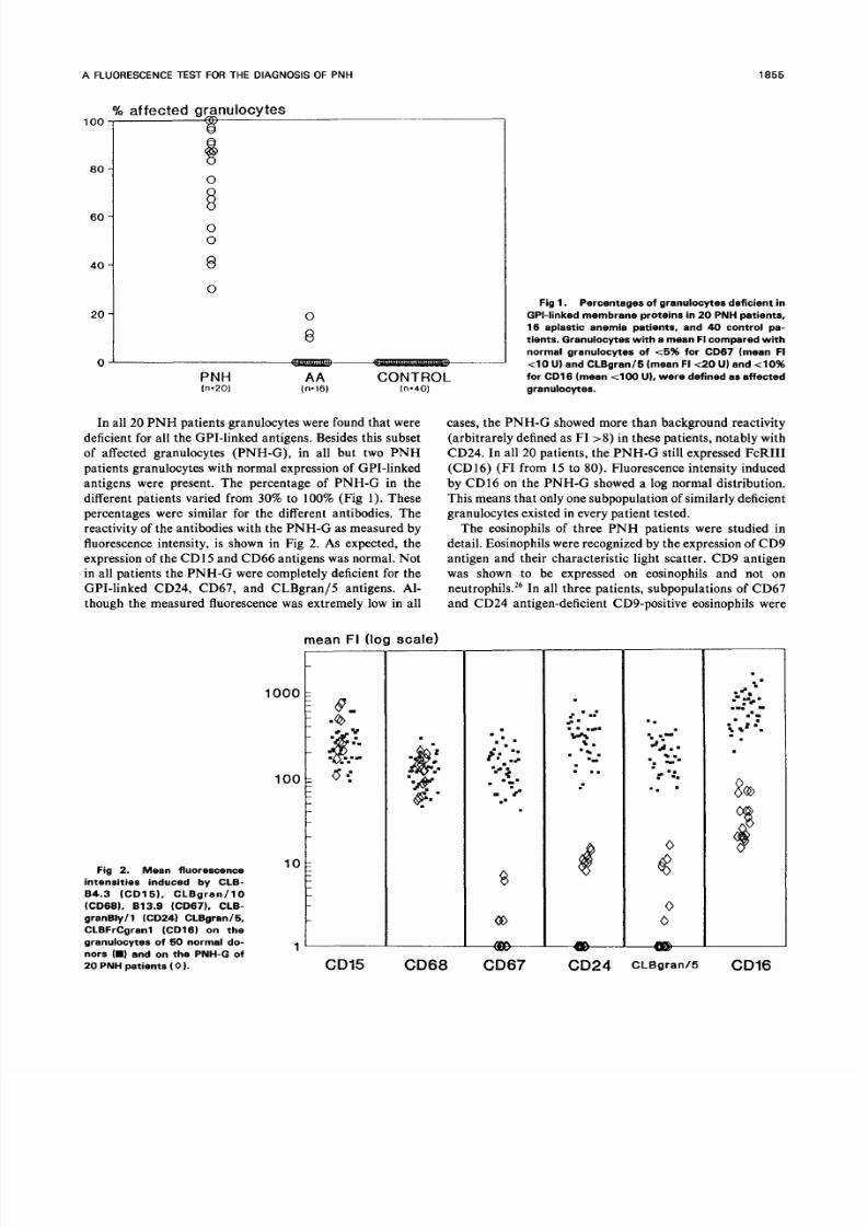
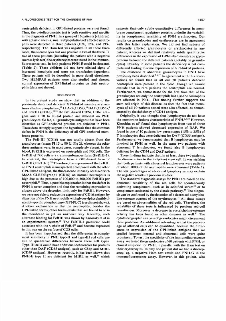
In all 20 P N H patients granulocytes were found that w ere
deficient for all the G PI-linked antigens. Besides this subset
of affected granulocytes (PNH-G), in all but two PNH
patients granulocytes with normal expression of GPI-linked
antigens were present. The percentage of PNH-G in the
different patients varied from 30 to 100 (Fig 1). These
percentages were similar for the different antibodies. Thereactivity of the antibodies with the PNH-G as measured by
fluorescence intensity, is shown in Fig 2. As expected, the
expression of the CD1 5 and C D66 antigens was normal. Notin all patients the P NH -G were completely deficient for the
GPI-linked CD24, CD67, and CLBgran/5 antigens. Al-though the measured fluorescence was extremely low in all
Fig 2. Mean fluorescence
intensities induced by CLB-
84.3 (CD15). CLBgran/ lO
(CD68). 813.9 (CD67). CLB-
granBly/l (CD24) CLBgran/5,
CLBFrCgranl (CD16I on the
granulocytes of 60 normal do-
nors ( and on the PNH-G of
20 PNH patients 0 ) .
mean FI log scale)
l o o k 8,
1 ° ~
CD15
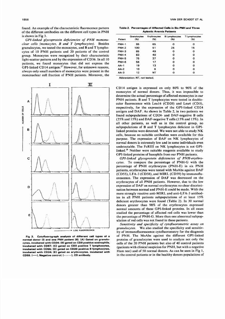
Fig 1. Percentages of granulocytes deficient in
GPI-linked membrane p rotei ns in 20 PNH patients,
16 aplastic anemia patients, and 4 control pa-
tients. Granulocytes with a mean FI compared with
normal granulocytes of t 5 for CD67 (mean FI
t 1 0 U) and CLBgran/5 (mean FIt2 U) and 4 0
for CD16 (mean 4 0 0 U), were defined as affected
granulocytes.
cases, the P NH -G showed more than background reactivity
(arbitrare ly defined as FI >8 n these patients, notably with
CD24. In all 20 patients, the PNH-G still expressed FcRIII
(CD16) (F I from 15 to 80). Fluorescence intensity induced
by CD16 on the PNH-G showed a log normal distribution.
This means tha t only one subpopulation of similarly deficient
granulocytes existed in every patient tested.
The eosinophils of three PNH patients were studied in
detail. Eosinophils were recognized by the expression of C D9
antigen and their charac teristic light scatter. C D9 antigen
was shown to be expressed on eosinophils and not on
neutrophils.26 In all thr ee patients , subpo pulations of CD 67and CD24 antigen-deficient CD9-positive eosinophils were
CD68
= -. C
?.. I
. ..
e
CD24
-.= =ad =. z
r -:.
..
go
CLBgranIC CD16
8/12/2019 Deficiency of GPI of Leukocytes in Paroxysmal Nocturnal Hematuria
http://slidepdf.com/reader/full/deficiency-of-gpi-of-leukocytes-in-paroxysmal-nocturnal-hematuria 4/7
8/12/2019 Deficiency of GPI of Leukocytes in Paroxysmal Nocturnal Hematuria
http://slidepdf.com/reader/full/deficiency-of-gpi-of-leukocytes-in-paroxysmal-nocturnal-hematuria 5/7
A FLUORESCENCE TEST FOR THE DIAGNOSIS OF PNH 1857
neutrophils deficient in GPI-linked proteins were not found.
Thus, the cytofluorometric test is both sensitive and specific
in the diagnosis of PNH. In a group of 16 patients (children)
with aplastic anemia, small subpopulations of affected neutro-
phils were detected in three patients (lo , 12 , and 19 ,
respectively). The Ham test was negative in all these three
cases, the sucrose lysis test was positive in two of the three. In
two of these patients (including the patient with a negative
sucrose lysis test) the erythrocytes were tested in the immuno-
fluorescence test. In both patients PNH-E could be detected
(Table 2). These children did not have clinical signs of
increased hemolysis and were not transfusion-dependent.
These patients will be described in more detail elsewhere.
Two HEMPAS patients were also studied and showed
normal expression of GPI-linked proteins on their neutro-
phils (data not shown).
DISCUSSION
In the present study we show that, in addition to the
previously described deficient GPI-linked membrane struc-
tures alkaline phosphatase,” LFA-3 (CD58),I6DAF (CD55),8
and FcRIII (CD16),22*23he GPI-linked CD24, CD67 anti-
gens and a 50 to 80-Kd protein are deficient on PNH
granulocytes.So far, all granulocyte antigens that have been
identified as GPI-anchored proteins are deficient in PNH.
These data strongly support the hypothesis that the common
defect in PNH is the deficiency of all GPI-anchored mem-
brane proteins.
The FcR-111 (CD16) was not totally absent from the
granulocytes (mean FI 15 to 80 U, Fig 2), whereas the other
three antigens were, in most cases, completely absent. In the
blood, FcRIII is expressed on neutrophils and NK cells. The
FcRIII of NK cells is a transmembranous form (FcRIII-2).
In contrast, the neutrophils have a GPI-linked form of
FcRIII (FcRIII-1).28Therefore, the expression of the FcRIII
on PNH neutrophils is unexpected. Compared with the other
GPI-linked antigens, the fluorescence ntensity obtained with
MoAb CLBFcRgran/ l (CD16) on normal neutrophils is
high due to the presence of 100,000 to 300,000 FcRIIIs per
ne~ t r oph i l . ~~hus, a possible explanation is that the defect in
PNH is never complete and that the remaining expression is
always above the detection limit only for FcRIII. However,
we were not able to reduce the expression of CD16 antigen by
digestion of the PNH neutrophils with glycosylphosphatidyli-
nositol-specific phospholipase (GPI-PLC) (results not shown).
Another explanation is that on neutrophils, besides theGPI-linked forms, other forms exists that are bound to or in
the membrane in yet an unknown way. Recently, such
alternate binding for FcRIII was shown by Kurosaki et a1 in
an experimental system.30 The FcRIII-1 precursor could
associate with the y-chain of FcRt13’ and become expressed
in this way on the surface of COS cells.
It has been hypothesized that the differences in comple-
ment sensitivity in PNH type-I1 and type-111 red cells are
due to qualitative differences between these cell types.
Type-I11 cells would have additional deficiencies for proteins
other than DAF (CD55 antigen), such as C8bp and MIRL
(CD59 antigen). However, recently, it has been shown that
PNH-E type I1 are deficient for MIRL as well,3’ which
suggests that only subtle quantitative differences in mem-
brane complement regulatory proteins underlie the variabil-
ity in complement sensitivity of PNH erythrocytes. Our
results on granulocytes and erythrocytes are in agreement
with this latter explanation. We did not find subsets of
differently affected granulocytes or erythrocytes in any
patient, whereas we did find relatively subtle quantitative
differences in the expression of GPI-linked membrane glyco-
proteins between the different patients (notably on granulo-
cytes). Possibly in some patients the deficiency is not com-
plete and leading to some expression of GPI-linked proteins.
The existence of abnormal granulocytes in PNH have
previously been de~cribed.~-~.”n agreement with this obser-
vations we found that in all our 20 patients deficient
neutrophils were present in the blood, though we cannot
exclude that in rare patients the neutrophils are normal.
Furthermore, we demonstrate for the first time that of the
granulocytes not only the neutrophils but also the eosinophils
are affected in PNH. This finding further supports the
stem-cell origin of this disease, as does the fact that mono-
cytes of all 10 patients tested were also affected, as demon-
strated by the deficiency of CD14 antigen.
Originally, it was thought that lymphocytes do not have
the membrane lesions characteristic of PNH.2.5.3Z owever,
Kinoshita et a16 found that lymphocytes from two of three
PNH patients showed decreased expression of DAF. We
found in two of 10patients low percentages (15 to 25 ) of
T lymphocytes that were deficient for DAF (CD55 antigen).
Furthermore, we demonstrated that B lymphocytes may be
involved in PNH as well. In the same two patients with
abnormal T lymphocytes, we found also B lymphocytes
deficient for the CD24 and DAF antigen.
These findings indicate that, in at least these two patients,
the disease arises in the totipotent stem cell. It was striking
that both patients with abnormal lymphocytes were patients
of whom 100 of the neutrophils were abnormal (Table 2).
The low percentages of abnormal lymphocytes may explain
the negative results in previous studies.
The standard diagnostic assays for PNH are based on the
abnormal sensitivity of the red cells for spontaneously
activating complement, such as in acidified serum33or to
complement activated by the classic pathway.34The diagno-
sis can be confirmed by the detection of a decreased acetylcho-
line-esterase content of the erythrocytes.” All these assays
are based on abnormalities of the red cells. Therefore, the
reliability of these tests is influenced by previous red-celltransfusions. Moreover, a decrease in acetylcholine-esterase
activity has been found in other diseases as well.35 The
cytofluorographic analysis of granulocytes might circumvent
these problems. An additional advantage is that the percent-
age of affected cells can be quantified, because the differ-
ences in expression of the GPI-linked antigens that we
studied between normal and abnormal cells were quite
prominent. To test the specificity of the immunofluorescence
assay, we tested the granulocytes of 60 patients with PNH,orclinical suspicion for PNH, in parallel with the Ham test on
their erythrocytes. In only one patient did we find a discrep-
ancy, eg, a negative Ham test result and PNH-G in the
immunofluorescence assay. However, in this patient, who
8/12/2019 Deficiency of GPI of Leukocytes in Paroxysmal Nocturnal Hematuria
http://slidepdf.com/reader/full/deficiency-of-gpi-of-leukocytes-in-paroxysmal-nocturnal-hematuria 6/7
1858 VAN DER SCHOOT ET AL
had 50 abnorm al circulating granulocytes, the diagnosis
P N H could be confirmed by showing that the erythrocytes
had a decreased acetylcholine-esterase content. Further-
more, discrepancies were found in the group of 16 patients
with aplastic anemia (AA). In three of the 16 AA patients,
we found a subpopulation of PN H- G (10 to 20 ), whereas
t he H a m t es t w as n eg ativ e in al l. L ew is an d D a ~ i e ~ ~rigi-nally described t he association of P N H an d AA. Approxi-
mately 15 of the patients with AA develop PNH in the
course of their di~ease.~’ .~’herefore, we assume that our
three patients may develop clinical P N H in due time. The
discrep ancy between th e two tests possibly reflects the higher
sensitivity of th e cytofluorographic analysis.
We also studied two patients with a positive H am test d ue
to hereditary erythrocytic abnormalit ies (HE MP AS ; data
not shown). Both h ad only normal g ranulocytes in the
immunofluorescence assay. Th us, th e neutrophil GP I-linked
membrane protein test also allows an easy discrimination
between two different types of patients with positive Ham
tests, ie, PN H and HE MPA S.
In conclusion, in P N H neutrophils, eosinophils, an d mono-
cytes are deficient for all presently known GPI-linked mem-
brane proteins, as well as in some patients B a nd T lympho-
cytes. Based on these findings, we developed a simple
cytofluorometric assay on granulocytes which appeared to be
diagnostic for PN H an d also made the diagnosis of P N H in
aplastic anem ia possible.
ACKNOWLEDGMENT
We tha nk all the clinicians who have sent blood of P N H patients
to our laboratory , in particular D rs J.J. Michiels, R. G oudsmit, and
M.M.A.C. Langenhuysen, who provided blood of more than one
patient.
REFERENCES
1. Rosse WF, Parker CJ: Paroxysmal nocturnal haemoglobin-uria. Clin Hae matol 14:105, 1985
2. Vellenga E, Mulder NH, The TH: Paroxysmal nocturnal
hemoglobinuria. Neth J Med 26:138, 1983
3. Aster R H, Enright SE: A platelet and granulocyte membrane
defect in paroxysmal no cturnal hem oglobinuria: Usefulness for the
detection of platelet antibodies. J Clin Invest 48:1199, 1969
4. Stern M , Rosse W F Two populations of granulocytes in
paroxysmal noctu rnal hemoglobinuria. Blood 53:928, 1979
5. Nicholson-Weller A, Spice r DB, Austen K F Deficiency of the
complement regulatory protein, “decay-accelerating factor,” on
mem branes of granulocytes, monocy tes, and platelets in paroxysmal
nocturn al hemoglobinuria. N Engl J Med 312:1091, 1985
6. Kinoshita T, Medof M E, Silber R, Nussenzweig V: Distribu-
tion of decay-accelerating factor in the peripheral blood of normalindividuals and patients with paroxysmal nocturnal hemog lobinuria.
J Exp Med 162:75,1985
7. Rosse WF: Variations in the red cells in paroxysmal nocturn al
hemoglobinuria. Br J Haematol24:327, 1973
8. Nicholson-Weller A March JP, Rosenfeld SI , Austen K F
Affected erythrocytes of patients with paroxysmal nocturnal hemo-
globinuria ar e deficient in the com plement protein, decay-accelerat-
ing factor. Proc Natl Acad Sci USA 80 :5066,1983
9. Zalman LS, Wood LM, Frank MM , Muller-Eberhard HJ:
Deficiency of the homologous restriction factor in paroxysmal
nocturn al hemoglobinuria. J Exp Med 165:572,1987
IO Hansch GM , SchonermarkS,Roelcke D: Paroxysmal noctur-
nal hemoglobinuria type 111-Lack of an erythroc yte mem brane
protein rest ricting the lysis by C5b -9. J Clin Invest 80:7, 1987
11. Holguin MH, Frederick LR, Bernshaw NJ, Wilcox LA,Parker CJ: Isolation and characterization of a membrane protein
from normal human erythrocytes that inhibits reactive lysis of the
erythrocytes of paroxysmal nocturnal hemoglobinuria. J Clin Invest
84:7, 1989
12. Davitz MA, Low MG, Nussenzweig V: Release of decay-
accelerating factor (DA F) from the cell mem brane by phosphatidyli-
nositol-specific phospholipase C (PIPL C). J Exp Med 163:1150,
1986
13. HBnsch GM , Weller PF, Nicholson-Weller A: Release of C8
binding protein (C8bp ) from the cell membrane by phosphatidylinosi-
tol-specific phosph olipase C. Blood 72:1089, 1988
14. Low MG , Finean JB: No n-lytic release of acetylcholines-
teras e from erythrocytes by a pho sphatidylinositol-specific phosph o-
lipase C. FE BS Lett 71:741, 1977
15. Kunstling TR, Rosse WF: Erythrocyte acetylcholinesterasedeficiency n paroxysmal nocturnal hemoglobinuria (PNH)-a com-
parison of the complement-sensitive and -insensitive populations.
Blood 33:607,1969
16. Selvaraj P, Dustin ML, Silber R, Low MG, Springer TA:
Deficiency of lymphocyte function-associated antigen (LFA-3) in
paroxysmal nocturnal hemoglobinuria. J Exp M ed 166: 1011, 1987
17. Burroughs SF, Devine DV, Browne G, Kaplan ME: The
population of paroxysmal nocturnal hemoglobinuria neutrophils
deficient in decay-accelerating factor is also deficient in alkaline
phosphatase. Blood 71:1086, 1988
18. Oni SB, Osunkoya BO, Luzatto L Paroxysmal nocturnal
hemoglobinuria: Evidence for mono clonal origin of abn ormal red
cells. Blood 36:145, 1970
19. Tumen J, Kline LB, Fay JW , Scullin DC, Reisner EG, RosseWF, Huang AT: Complement sensitivity of paroxysmal nocturnal
hemo globinuria bone-ma rrow cells. Blood 55: 1040, 1980
20. Kanamaru A, Okuda K, Ueda E, Kitani T, Kinoshita T,
Nagai K Different distribution of decay-accelerating factor on
hematopoietic progenitors from normal individuals and patients with
paroxysmal nocturnal hemoglobinuria. Blood 725 07 , 1 988
21. Dessypris EN , Gleaton J H, C lark DA: Increased sensitivity to
complement of megakaryocyte progenitors in paroxysmal noc turnal
haemoglobinuria. Br J Haematol69:305, 1988
22. Huizinga TW J, van der Schoot CE, Jost C, Klaassen R JL,
von dem Borne AEG Kr, Roos D, Tetteroo PAT: The PI-linked
receptor FcRIII is released on stimulation of neutrophils. Nature
333:667, 1988
23. Selveraj P, Rosse WF, Silber R, Springer TA: The major Fc
receptor in blood has a ph osphatidylinositol anch or and is deficient inparoxysmal nocturnal hemoglobinuria. Na tur e 3335 65, 1988
24. van der Schoot CE, Huizinga TWJ, Gadd S, Majdic 0
Wijmans R, Knapp W, von dem Borne AEG Kr: Identification of
three novel PI-linked proteins on granulocytes, in Knapp W (ed):
Leucocyte Typing IV. Oxford, UK , O xford University, 1990, p 887
25. Tetteroo PAT, Bos MJ E, Visser FJ, von dem Borne AEG K r:
Neutrop hil activation detected by m onoclonal antibodies. J Imm u-
no1 136:3427, 1986
26. Ohto H, Hitomi Y, Kambayashi H, Sa to T , Maeda H: CD 9
defined antigen (p24) is expressed on eosinophils but not on normal
monocytes, in McM ichael ed):eukocyte Typing 111.Oxford, UK,
Oxford University, 1987, p 73 1
27. Haziot A, Chen S, Ferrero E, Low M G, Silber R, Goyert SM :
The monocyte differentiation antigen, CD 14, is anchored to the cell
8/12/2019 Deficiency of GPI of Leukocytes in Paroxysmal Nocturnal Hematuria
http://slidepdf.com/reader/full/deficiency-of-gpi-of-leukocytes-in-paroxysmal-nocturnal-hematuria 7/7
A FLUORESCENCE TEST FOR THE DIAGNOSIS OF PNH 1859
membrane by a phosphatidylinositol linkage. J Immunol 141547,
1988
28. Ravetch JV, Perussia B: Alternative membrane forms of
FcR III (C D16) on hu man na tural killer cells and neutrophils. J Exp
Med 170:481,1989
29. Huizinga TWJ, Kerst M, Nuyens JH, Vlug A, Von dem
Borne AEGKr, Roos D, Tetteroo P AT: Binding characteristics of
dimeric IgG subclass complexes to human neutrophils. J Immunol
142:2359,1989
30. Kurosaki T, Ravetch JV: A single amino acid in the glycosyl
phosphatidylinositol attachm ent domain determ ined the memb rane
topology of FcyR III. Na tu re 3422305, 1989
31. Holguin MH , Wilcox LA, Bernshaw NJ , Rosse W F, Parker
CJ: Relationship between the membrane inhibitor of reactive lysis
and the erythrocyte phenotypes of paroxysmal nocturnal hemoglo-
binuria. J Clin Invest 84:1387, 1989
32. Cooper MR, Currie MS , Rustagi PK, Logue GL: T lympho-
cytes escape membrane defect on paroxysmal nocturnal haemoglo-
binuria. Br J Haematol55:263, 1983
33. Ha m TH : Chronic hemolytic anemia with paroxysmal noctur-
nal hemoglobinuria: Stud y of the mechanism of hemolysis in relation
to acid-base equilibrium. N Engl J Med 217:915, 1937
34. Rosse WF, Dacie JV: Immune lysis of normal and paroxys-
mal nocturnal hemoglobinuria (PNH) red blood cells. I. The
sensitivity of P N H red cells to lysis by complement a nd specific
antibody. J Clin Invest 45:736, 196635. Lawson AA, B arr RD : Acetylcholinesterase in red blood cells.
Am J Hematol26:101, 1987
36. Lewis SM, Dacie JV: The aplastic anaemia-paroxysmal
nocturnal haemoglobinuria syndrome. Br J Haematol 13:236, 1967
37. de Planque MM, Bacigalupo A, Wursch A, Hows JM,
Devergie A, Frickhoven N , Brand A, Nissen C: Long-term follow-up
of severe aplastic anemia patients treated with antithymocyte
globulin. Br J Haematol73:121, 1989
38. Tichelli A, Gratwohl A, Wursch A, Nissen C, Speck B La te
haematological complications in severe aplastic anaemia. Br J
Haematol69:413, 1988