Deep conservation of wrist and digit enhancers in fish - PNAS · Deep conservation of wrist and...
6
Deep conservation of wrist and digit enhancers in fish Andrew R. Gehrke a , Igor Schneider b , Elisa de la Calle-Mustienes c , Juan J. Tena c , Carlos Gomez-Marin c , Mayuri Chandran a , Tetsuya Nakamura a , Ingo Braasch d , John H. Postlethwait d , José Luis Gómez-Skarmeta c , and Neil H. Shubin a,1 a Department of Organismal Biology and Anatomy, The University of Chicago, Chicago, IL 60637; b Instituto de Ciencias Biologicas, Universidade Federal do Para, 66075, Belem, Brazil; c Centro Andaluz de Biología del Desarrollo, Consejo Superior de Investigaciones Científicas/Universidad Pablo de Olavide, Sevilla 41013, Spain; and d Institute of Neuroscience, University of Oregon, Eugene, OR 97403-1254 Contributed by Neil H. Shubin, November 17, 2014 (sent for review October 21, 2014; reviewed by Gunter P. Wagner) There is no obvious morphological counterpart of the autopod (wrist/ankle and digits) in living fishes. Comparative molecular data may provide insight into understanding both the homology of elements and the evolutionary developmental mechanisms behind the fin to limb transition. In mouse limbs the autopod is built by a “late” phase of Hoxd and Hoxa gene expression, orches- trated by a set of enhancers located at the 5′ end of each cluster. Despite a detailed mechanistic understanding of mouse limb de- velopment, interpretation of Hox expression patterns and their regulation in fish has spawned multiple hypotheses as to the or- igin and function of “autopod” enhancers throughout evolution. Using phylogenetic footprinting, epigenetic profiling, and trans- genic reporters, we have identified and functionally characterized hoxD and hoxA enhancers in the genomes of zebrafish and the spotted gar, Lepisosteus oculatus, a fish lacking the whole genome duplication of teleosts. Gar and zebrafish “autopod” enhancers drive expression in the distal portion of developing zebrafish pec- toral fins, and respond to the same functional cues as their murine orthologs. Moreover, gar enhancers drive reporter gene expres- sion in both the wrist and digits of mouse embryos in patterns that are nearly indistinguishable from their murine counterparts. These functional genomic data support the hypothesis that the distal radials of bony fish are homologous to the wrist and/or digits of tetrapods. evolution | development | autopod | gene regulation | Hox T he origin of novel features is a key question in evolutionary biology, and the autopod—wrists, fingers, ankles, and toes— is a hallmark example (1). Although paleontological data, such as that from the Devonian lobe fin Tiktaalik roseae, reveal a se- quence of changes in the elaboration of the bony elements of fins into limbs (2), in living taxa there is a lack of obvious homology between the wrist and digits of tetrapod limbs and the pectoral fin skeleton of extant fish (3). Tetrapod forelimbs are generally composed of a series of long bones (upper arm and forearm), followed by small nodular bones (wrist), and ending in another group of long bones (digits). Ray-finned (Actinopterygian) pec- toral fins are diverse but are usually composed of a series of long proximal radials, followed by a set of smaller distal radials. The open question remains: do extant fish have the equivalent of wrists or digits? The molecular mechanisms governing the development of mammalian limbs have been approached in mouse models through multiple levels of analysis, from chromatin dynamics, to enhancer sequence, to gene expression patterns (4). Murine limbs display two successive phases of gene expression of the HoxD and HoxA gene clusters. The initial or “early” phase of expression begins with members at the 3′ end of the clusters being expressed broadly, and members at the 5′ end of the cluster being activated in an increasingly restricted number of cells (5). This “early” phase of Hox expression is associated with the development of the upper arm (stylopod) and forearm (zeugopod). The initial wave of Hox expression is followed by a temporally distinct second activation of Hox genes, this time beginning with members of the 5′ end of the cluster being expressed most broadly and in the presumptive digits. This sec- ond, “late” phase of expression is necessary for autopod for- mation, as evidenced by a loss of this domain resulting in deletion of the wrist and digits (5). The genomic regulatory elements and chromatin dynamics responsible for enacting these two phases have been studied in detail in the HoxD cluster, where the “early” and “late” phases are governed by enhancers that lie on opposite sides of the HoxD cluster—3′ and 5′, re- spectively—and activated in turn by shifting domains of open chromatin (6, 7). In addition, recent work has identified a series of enhancers that drive late phase expression of the HoxA cluster in the developing mouse autopod in a fashion similar to that of HoxD (8). To what extent are the regulatory mechanisms that drive autopodial development present in fish fins and, if they are present, what is their developmental role? Previous work has shown that at least one of the “autopod” enhancers (CsB) is present and active in the common ancestor of gnathostomes (9). Additionally, recent work in zebrafish has shown that the early and late topological chromatin domains are indeed observed in bony fish (10). However, teleost fish enhancer domains were unable to drive reporter gene expression in the developing digits of transgenic mice, suggesting that although bony fish do contain a version of the autopod regulatory apparatus, these enhancers Significance The fossil record shows that the wrist and digits have an aquatic origin, becoming recognizable in a group of (mostly extinct) fish that contained robust fins. Do the fins of living fishes have the equivalent of these structures? Because com- parisons of fin and limb morphology have been inconclusive, we sought to investigate this question using developmental and molecular data. By utilizing a nonmodel fish (the spotted gar), we find that the regulatory networks that control “wrist and digit”-building genes (Hox) are deeply conserved between fish and tetrapods. The genomic architecture described here defines Hox gene activity in fins and limbs as equivalent, in turn suggesting equivalence between the distal bones of fish fins and the wrist and/or digits of tetrapods. Author contributions: A.R.G., I.S., J.L.G.-S., and N.H.S. designed research; A.R.G., I.S., E.d.l.C.-M., J.J.T., C.G.-M., M.C., T.N., and J.L.G.-S. performed research; A.R.G., E.d.l.C.-M., J.J.T., C.G.-M., I.B., J.H.P., and J.L.G.-S. contributed new reagents/analytic tools; A.R.G., I.S., J.J.T., C.G.-M., M.C., T.N., J.L.G.-S., and N.H.S. analyzed data; and A.R.G., I.S., and N.H.S. wrote the paper. Reviewers included: G.P.W., Yale University. The authors declare no conflict of interest. Freely available online through the PNAS open access option. Data deposition: The data reported in this paper have been deposited in the Gene Ex- pression Omnibus (GEO) database, www.ncbi.nlm.nih.gov/geo (accession nos. GSE61063 and GSE61065). 1 To whom correspondence should be addressed. Email: [email protected]. This article contains supporting information online at www.pnas.org/lookup/suppl/doi:10. 1073/pnas.1420208112/-/DCSupplemental. www.pnas.org/cgi/doi/10.1073/pnas.1420208112 PNAS | January 20, 2015 | vol. 112 | no. 3 | 803–808 EVOLUTION
Transcript of Deep conservation of wrist and digit enhancers in fish - PNAS · Deep conservation of wrist and...

Deep conservation of wrist and digit enhancers in fishAndrew R. Gehrkea, Igor Schneiderb, Elisa de la Calle-Mustienesc, Juan J. Tenac, Carlos Gomez-Marinc,Mayuri Chandrana, Tetsuya Nakamuraa, Ingo Braaschd, John H. Postlethwaitd, José Luis Gómez-Skarmetac,and Neil H. Shubina,1
aDepartment of Organismal Biology and Anatomy, The University of Chicago, Chicago, IL 60637; bInstituto de Ciencias Biologicas, Universidade Federal doPara, 66075, Belem, Brazil; cCentro Andaluz de Biología del Desarrollo, Consejo Superior de Investigaciones Científicas/Universidad Pablo de Olavide, Sevilla41013, Spain; and dInstitute of Neuroscience, University of Oregon, Eugene, OR 97403-1254
Contributed by Neil H. Shubin, November 17, 2014 (sent for review October 21, 2014; reviewed by Gunter P. Wagner)
There is no obvious morphological counterpart of the autopod(wrist/ankle and digits) in living fishes. Comparative moleculardata may provide insight into understanding both the homologyof elements and the evolutionary developmental mechanismsbehind the fin to limb transition. In mouse limbs the autopod isbuilt by a “late” phase of Hoxd and Hoxa gene expression, orches-trated by a set of enhancers located at the 5′ end of each cluster.Despite a detailed mechanistic understanding of mouse limb de-velopment, interpretation of Hox expression patterns and theirregulation in fish has spawned multiple hypotheses as to the or-igin and function of “autopod” enhancers throughout evolution.Using phylogenetic footprinting, epigenetic profiling, and trans-genic reporters, we have identified and functionally characterizedhoxD and hoxA enhancers in the genomes of zebrafish and thespotted gar, Lepisosteus oculatus, a fish lacking the whole genomeduplication of teleosts. Gar and zebrafish “autopod” enhancersdrive expression in the distal portion of developing zebrafish pec-toral fins, and respond to the same functional cues as their murineorthologs. Moreover, gar enhancers drive reporter gene expres-sion in both the wrist and digits of mouse embryos in patternsthat are nearly indistinguishable from their murine counterparts.These functional genomic data support the hypothesis that thedistal radials of bony fish are homologous to the wrist and/ordigits of tetrapods.
evolution | development | autopod | gene regulation | Hox
The origin of novel features is a key question in evolutionarybiology, and the autopod—wrists, fingers, ankles, and toes—
is a hallmark example (1). Although paleontological data, suchas that from the Devonian lobe fin Tiktaalik roseae, reveal a se-quence of changes in the elaboration of the bony elements of finsinto limbs (2), in living taxa there is a lack of obvious homologybetween the wrist and digits of tetrapod limbs and the pectoralfin skeleton of extant fish (3). Tetrapod forelimbs are generallycomposed of a series of long bones (upper arm and forearm),followed by small nodular bones (wrist), and ending in anothergroup of long bones (digits). Ray-finned (Actinopterygian) pec-toral fins are diverse but are usually composed of a series of longproximal radials, followed by a set of smaller distal radials. Theopen question remains: do extant fish have the equivalent ofwrists or digits?The molecular mechanisms governing the development of
mammalian limbs have been approached in mouse modelsthrough multiple levels of analysis, from chromatin dynamics, toenhancer sequence, to gene expression patterns (4). Murinelimbs display two successive phases of gene expression of theHoxD and HoxA gene clusters. The initial or “early” phase ofexpression begins with members at the 3′ end of the clustersbeing expressed broadly, and members at the 5′ end of thecluster being activated in an increasingly restricted number ofcells (5). This “early” phase of Hox expression is associated withthe development of the upper arm (stylopod) and forearm(zeugopod). The initial wave of Hox expression is followed bya temporally distinct second activation of Hox genes, this time
beginning with members of the 5′ end of the cluster beingexpressed most broadly and in the presumptive digits. This sec-ond, “late” phase of expression is necessary for autopod for-mation, as evidenced by a loss of this domain resulting indeletion of the wrist and digits (5). The genomic regulatoryelements and chromatin dynamics responsible for enacting thesetwo phases have been studied in detail in the HoxD cluster,where the “early” and “late” phases are governed by enhancersthat lie on opposite sides of the HoxD cluster—3′ and 5′, re-spectively—and activated in turn by shifting domains of openchromatin (6, 7). In addition, recent work has identified a seriesof enhancers that drive late phase expression of the HoxA clusterin the developing mouse autopod in a fashion similar to that ofHoxD (8).To what extent are the regulatory mechanisms that drive
autopodial development present in fish fins and, if they arepresent, what is their developmental role? Previous work hasshown that at least one of the “autopod” enhancers (CsB) ispresent and active in the common ancestor of gnathostomes (9).Additionally, recent work in zebrafish has shown that the earlyand late topological chromatin domains are indeed observed inbony fish (10). However, teleost fish enhancer domains wereunable to drive reporter gene expression in the developing digitsof transgenic mice, suggesting that although bony fish do containa version of the autopod regulatory apparatus, these enhancers
Significance
The fossil record shows that the wrist and digits have anaquatic origin, becoming recognizable in a group of (mostlyextinct) fish that contained robust fins. Do the fins of livingfishes have the equivalent of these structures? Because com-parisons of fin and limb morphology have been inconclusive,we sought to investigate this question using developmentaland molecular data. By utilizing a nonmodel fish (the spottedgar), we find that the regulatory networks that control “wristand digit”-building genes (Hox) are deeply conserved betweenfish and tetrapods. The genomic architecture described heredefines Hox gene activity in fins and limbs as equivalent, inturn suggesting equivalence between the distal bones of fishfins and the wrist and/or digits of tetrapods.
Author contributions: A.R.G., I.S., J.L.G.-S., and N.H.S. designed research; A.R.G., I.S.,E.d.l.C.-M., J.J.T., C.G.-M., M.C., T.N., and J.L.G.-S. performed research; A.R.G., E.d.l.C.-M., J.J.T.,C.G.-M., I.B., J.H.P., and J.L.G.-S. contributed new reagents/analytic tools; A.R.G., I.S., J.J.T.,C.G.-M., M.C., T.N., J.L.G.-S., and N.H.S. analyzed data; and A.R.G., I.S., and N.H.S. wrotethe paper.
Reviewers included: G.P.W., Yale University.
The authors declare no conflict of interest.
Freely available online through the PNAS open access option.
Data deposition: The data reported in this paper have been deposited in the Gene Ex-pression Omnibus (GEO) database, www.ncbi.nlm.nih.gov/geo (accession nos. GSE61063and GSE61065).1To whom correspondence should be addressed. Email: [email protected].
This article contains supporting information online at www.pnas.org/lookup/suppl/doi:10.1073/pnas.1420208112/-/DCSupplemental.
www.pnas.org/cgi/doi/10.1073/pnas.1420208112 PNAS | January 20, 2015 | vol. 112 | no. 3 | 803–808
EVOLU
TION
are not responsive to the regulatory program present in murinedigits (10). Thus, the number, extent, and function of “limb”enhancers in fish remain to be fully explored, especially in fishspecies outside those of traditional model systems that mightresemble ancestral characters more closely than teleosts.To address these issues, we used a combination of epigenetic
profiling in zebrafish and phylogenetic footprinting using thegenome assembly of the recently sequenced spotted gar (Lep-isosteus oculatus) (11) to investigate the enhancers that controlhoxd and hoxa expression in bony fish. The phylogenetic posi-tion of gar is crucial to our investigation, in that gar represents
a lineage that diverged from teleost fishes before the teleostgenome duplication, an event that may cloud studies of regula-tory evolution (Fig. 1A) (12–14). The data presented here revealan unprecedented and previously undescribed level of deepconservation of the vertebrate autopod regulatory apparatus,suggesting homology between the distal radials of bony fish andthe autopod of tetrapods.
ResultsTo identify orthologs of murine Hox limb enhancers in bony fish,we performed a multiple sequence alignment of the genomic
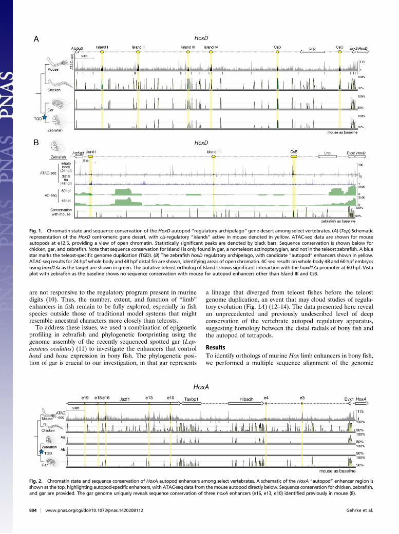
Fig. 1. Chromatin state and sequence conservation of the HoxD autopod “regulatory archipelago” gene desert among select vertebrates. (A) (Top) Schematicrepresentation of the HoxD centromeric gene desert, with cis-regulatory “islands” active in mouse denoted in yellow. ATAC-seq data are shown for mouseautopods at e12.5, providing a view of open chromatin. Statistically significant peaks are denoted by black bars. Sequence conservation is shown below forchicken, gar, and zebrafish. Note that sequence conservation for Island I is only found in gar, a nonteleost actinopterygian, and not in the teleost zebrafish. A bluestar marks the teleost-specific genome duplication (TGD). (B) The zebrafish hoxD regulatory archipelago, with candidate “autopod” enhancers shown in yellow.ATAC-seq results for 24 hpf whole-body and 48 hpf distal fin are shown, identifying areas of open chromatin. 4C-seq results on whole-body 48 and 60 hpf embryosusing hoxd13a as the target are shown in green. The putative teleost ortholog of Island I shows significant interaction with the hoxd13a promoter at 60 hpf. Vistaplot with zebrafish as the baseline shows no sequence conservation with mouse for autopod enhancers other than Island III and CsB.
Fig. 2. Chromatin state and sequence conservation of HoxA autopod enhancers among select vertebrates. A schematic of the HoxA “autopod” enhancer region isshown at the top, highlighting autopod-specific enhancers, with ATAC-seq data from themouse autopod directly below. Sequence conservation for chicken, zebrafish,and gar are provided. The gar genome uniquely reveals sequence conservation of three hoxA enhancers (e16, e13, e10) identified previously in mouse (8).
804 | www.pnas.org/cgi/doi/10.1073/pnas.1420208112 Gehrke et al.
region upstream of the HoxD and HoxA clusters from a numberof tetrapod and teleost species (Figs. 1A and 2). Our initialsurvey revealed a dearth of sequence conservation for late phaseenhancers in teleost species, aside from previously studied CsB,and an additional limb enhancer previously called Island III (6,9). As we broadened our taxonomic input by including the ge-nome of the spotted gar, we found peaks of conserved noncodingelements for the HoxD late phase digit enhancer Island I (Fig.1A) and the HoxA late phase enhancers e16, e13, and e10 (Fig. 2)(6, 8). We reasoned that the unduplicated nature of the gar ge-nome makes its Hox clusters a better representative of the com-mon ancestor of bony fish (Osteichthyes), revealing sequencesthat have diverged beyond recognition in derived, duplicatedteleost genomes (13).To identify potential orthologs in the zebrafish genome that
are not revealed by sequence conservation, we performed the“Assay for Transposable Accessible Chromatin” (ATAC-Seq) on24 hours postfertilization (hpf) zebrafish embryos, as well asembryonic day 12.5 (e12.5) mouse autopods as a control for theassay (15, 16). ATAC-seq on mouse autopods significantlyidentified the majority of validated HoxD and HoxA enhancerswith autopod activity (Figs. 1A and 2). Although not all pre-viously defined Hox autopod enhancers were identified (e.g.,Island I, CsB) in the mouse sample, we found substantial overlapof our ATAC-seq peaks with validated limb enhancers fromother studies (Table S1), making us confident that the assaywould be able to identify enhancers in zebrafish. We performed
ATAC-seq on whole-body zebrafish embryos and noticed anarea of open chromatin in the genome near the atp5g3a gene thatroughly matched the genomic coordinates of the late phase en-hancer Island I (Fig. 1B). To determine whether this regioninteracts with the zebrafish hoxd13a promoter, we performed“Circular Chromatin Conformation Capture” (4C-seq) to detectcontact with enhancers up to ∼1 Mb away, on 48 and 60 hpfwhole-body zebrafish embryos. We found that the area containingthe putative ortholog of Island I in zebrafish significantly interactswith the hoxd13a locus at 60 hpf (Fig. 1B). Because the assay wasperformed on whole embryos, it is possible that these interactionsmay not be specific to the developing fin. We did not observesignificant contact between Island I and hoxd13a before 60 hpf,possibly owing to limitations on the numbers of fin cells availablewhen using whole-body embryos for 4C-seq. Although the ATACand 4C-seq data revealed genomic areas in zebrafish that couldbe orthologous to HoxA enhancers (Fig. S1), these areas con-tained multiple candidates, and thus we sought to character-ize only the gar orthologs of HoxA where sequence orthologywas clear.Having identified potential enhancers according to chromatin
structure and sequence conservation, we sought to characterizetheir activity by cloning the sequences from gar and zebrafishinto reporter vectors and injecting them into zebrafish embryosto assay for domains of expression (17). We performed transientinjections of gar Island III, as well as the genomic “areas” fromthe gar genome that could potentially contain cryptic autopod
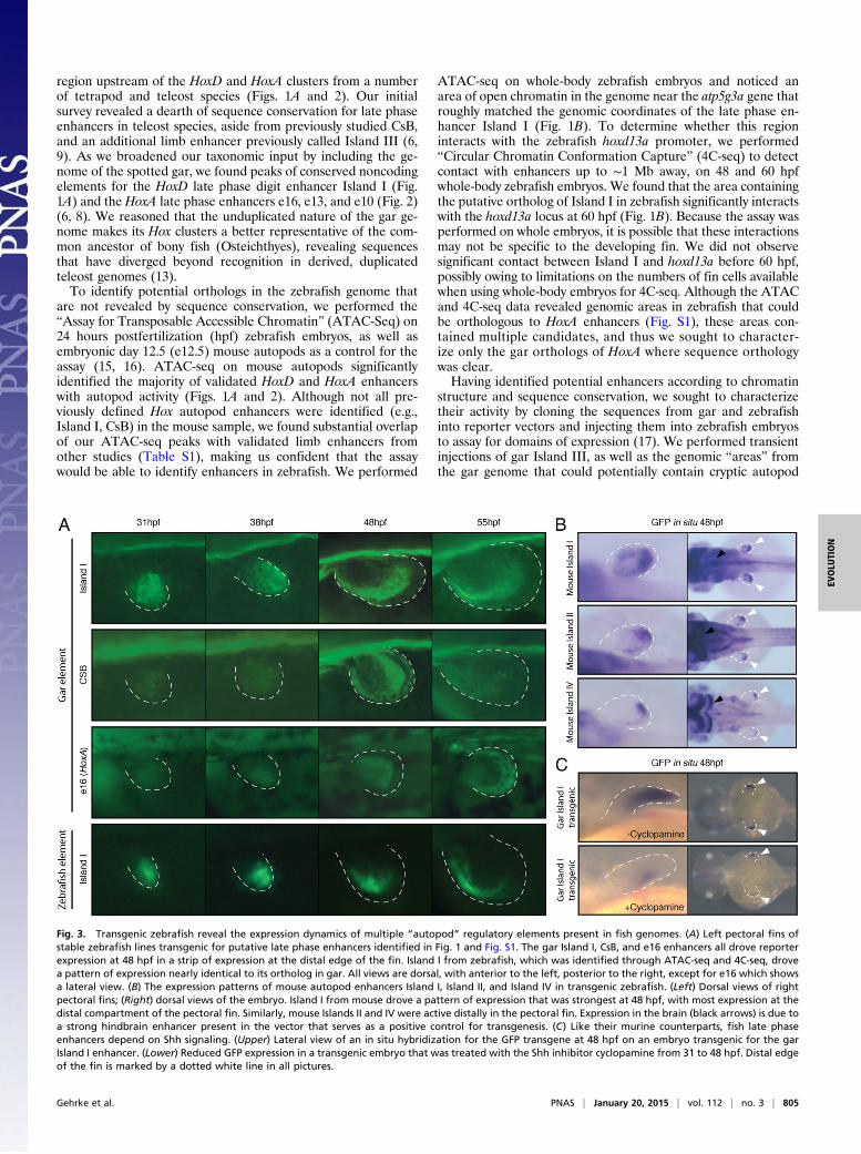
Fig. 3. Transgenic zebrafish reveal the expression dynamics of multiple “autopod” regulatory elements present in fish genomes. (A) Left pectoral fins ofstable zebrafish lines transgenic for putative late phase enhancers identified in Fig. 1 and Fig. S1. The gar Island I, CsB, and e16 enhancers all drove reporterexpression at 48 hpf in a strip of expression at the distal edge of the fin. Island I from zebrafish, which was identified through ATAC-seq and 4C-seq, drovea pattern of expression nearly identical to its ortholog in gar. All views are dorsal, with anterior to the left, posterior to the right, except for e16 which showsa lateral view. (B) The expression patterns of mouse autopod enhancers Island I, Island II, and Island IV in transgenic zebrafish. (Left) Dorsal views of rightpectoral fins; (Right) dorsal views of the embryo. Island I from mouse drove a pattern of expression that was strongest at 48 hpf, with most expression at thedistal compartment of the pectoral fin. Similarly, mouse Islands II and IV were active distally in the pectoral fin. Expression in the brain (black arrows) is due toa strong hindbrain enhancer present in the vector that serves as a positive control for transgenesis. (C) Like their murine counterparts, fish late phaseenhancers depend on Shh signaling. (Upper) Lateral view of an in situ hybridization for the GFP transgene at 48 hpf on an embryo transgenic for the garIsland I enhancer. (Lower) Reduced GFP expression in a transgenic embryo that was treated with the Shh inhibitor cyclopamine from 31 to 48 hpf. Distal edgeof the fin is marked by a dotted white line in all pictures.
Gehrke et al. PNAS | January 20, 2015 | vol. 112 | no. 3 | 805
EVOLU
TION
enhancers (potential orthologs of mouse Islands II and IV notfound by sequence alignment), and detected no fin signal forthese regions (Table S2). In contrast, we were able to detect GFPin the pectoral fin of zebrafish embryos injected with constructscontaining either gar Island I, CsB, or e16, which we raised tosexual maturity to obtain stable transgenic lines. The late phaseenhancer gar Island I exhibited little activity at 31 hpf but in-creased and became distally restricted by 38 hpf (Fig. 3A and Fig.S2). At 48 hpf, gar Island I drove a pattern of expression that wasrestricted to the anterior portion of the distal pectoral fin (Fig.3A and Fig. S2). Gar CsB drove a pattern of activity that wassimilar to that of Island I, with a peak of expression at 48 hpf butwith a posterior–distal restriction to its expression. Of the threehoxA autopod enhancers that were identified in the gar genomeby sequence conservation (Fig. 2), e16 from mouse has beenshown to drive the most robust native pattern of expressionthroughout the autopod of transgenic mice (8). As a result, wechose to make stable transgenic zebrafish lines of the gar e16regulatory element and found that it drove a distally restrictedpattern of gene expression at 48 hpf, much like the late phasehoxD enhancers from gar (Fig. 3A and Fig. S2).Using the data from ATAC-seq, 4C-seq, and gar conservation
as a guide, we cloned the putative region of Island I fromzebrafish and tested its potential activity by cloning it into a re-porter vector and producing stable zebrafish transgenic lines. Wefound that zebrafish Island I drives strong expression of GFP inthe distal pectoral fin at 48 hpf, in a pattern similar to that of itsgar ortholog (Fig. 3A and Fig. S2). To confirm that the activity ofzebrafish Island I was present in the distal fin (and not simplyactive elsewhere in the body), we grew multiple F1 fish from onefounder and collected a pool of distal fin cells from embryos at48 hpf using FACS. We subjected these distal fin cells to ATAC-seq and found that open chromatin signal for Island I wasenriched in this sample (Fig. 1B). Combined with ATAC-seq and4C-seq data, our transgenic zebrafish analysis indicates thatzebrafish Island I functions endogenously as a distal fin enhanceracting upon the hoxd13a gene.Because Island I from gar seemed to mark a distal compart-
ment of the zebrafish pectoral fin, we sought to investigatewhether Island I from mouse would elicit a similar pattern inzebrafish. To test this hypothesis, we cloned Island I from mouseinto a zebrafish enhancer detection vector and again createdindependent stable lines of transgenic zebrafish. Mouse Island Idrove a pattern of robust expression in the distal portion of theendochondral disk at 48 hpf, marking a distal segment of the finmuch like its gar ortholog (Fig. 3B). To further assess the activityof the other murine HoxD “autopod” enhancers when injectedinto zebrafish, we cloned mouse Islands II and IV and createdstable zebrafish lines (at least three independent lines/construct).Both of these mouse enhancers drove GFP in the distal com-partment of the zebrafish pectoral fin (Fig. 3B). These experi-ments demonstrate that the mouse autopod enhancer Island I,when transgenic in zebrafish, drives expression in the samepattern and area (the distal endochondral compartment of thefin) as the fish ortholog of this enhancer. Furthermore, mouseautopod enhancers that may not be present in fish genomes(Islands II and IV) are active in this same distal portion of thezebrafish pectoral fin.The late phase of Hox expression in the mouse autopod and
zebrafish distal pectoral fin depend on sonic hedgehog (Shh)(18–20). To test whether the expression driven by the gar latephase enhancers also depends on Shh signaling, we inhibited Shhfunction by adding its antagonist cyclopamine (20) to transgeniczebrafish embryos from 31 to 48 hpf. Embryos transgenic for thegar late phase enhancer Island I that were treated with cyclop-amine had markedly decreased expression of the reporter at48 hpf, whereas the overall fin morphology remained normal(Fig. 3C). We repeated these experiments on five independent
transgenic lines of gar Island I and found that nearly all embryosfor all lines exhibited a loss of late phase GFP expression whentreated with cyclopamine (n = 21 of 25, 5 embryos per line). Thesefindings show that both mouse and gar late phase enhancers de-pend on Shh signaling and suggest that Shh control of distal Hoxdexpression is an ancestral characteristic of bony fish.Finally, to test whether the fish enhancers could respond to
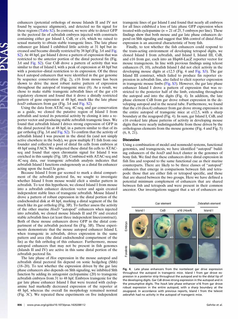
the trans-acting environment of developing tetrapod digits, wecloned Island I from zebrafish, and Island I, Island III, CsB,and e16 from gar, each into an Hsp68-LacZ reporter vector formouse transgenesis. In line with previous findings using teleostenhancers (9, 10), zebrafish Island I did not elicit activity in thedeveloping mouse digits at e12.5 (Fig. 4 and Fig. S3). The garIsland III construct, which failed to produce fin reporter ex-pression in zebrafish fins, also failed to elicit reporter expressionin transgenic mouse limbs (Fig. S3). However, the gar late phaseenhancer Island I drove a pattern of expression that was re-stricted to the posterior half of the limb, extending throughoutthe autopod and into the digits (Fig. 4). In addition, the latephase element CsB from gar drove robust expression in the de-veloping autopod and in the neural tube. Furthermore, we foundthat the e16 (hoxA) enhancer from gar drove strong expression inthe entire autopod of transgenic mice at e12.5, with a sharpboundary at the zeugopod (Fig. 4). In sum, gar Island I, CsB, ande16 evoked late phase patterns of activity in developing mousedigits that were nearly indistinguishable from those driven by theorthologous elements from the mouse genome (Fig. 4 and Fig. 5)(6, 8, 21).
DiscussionUsing a combination of model and nonmodel systems, functionalgenomics, and transgenesis, we have identified “autopod” build-ing enhancers of the hoxD and hoxA cluster in the genomes ofbony fish. We find that these enhancers drive distal expression infish fins and respond to the same functional cue as their murinecounterparts. There are likely to be three classes of “autopod”enhancers that emerge in comparisons between fish and tetra-pods: those that are either fish or tetrapod specific, and thosethat are shared between the two groups. Here we have defined apotential minimum complement of enhancers that are sharedbetween fish and tetrapods and were present in their commonancestor. Our investigations suggest that a set of enhancers are
Fig. 4. Late phase enhancers from the nonteleost gar drive expressionthroughout the autopod in transgenic mice. Island I from gar drove ex-pression in a posterior strip throughout the autopod and to the distal tip ofthe developing digits. Gar CsB drove strong expression in the autopod and inthe presumptive digits. The hoxA late phase enhancer e16 from gar droverobust expression in the entire autopod, with a sharp boundary at thezeugopod. In accordance with previous reports, Island I from the teleostzebrafish had no activity in the autopod of transgenic mice.
806 | www.pnas.org/cgi/doi/10.1073/pnas.1420208112 Gehrke et al.
tetrapod-specific (6, 8) and may have been assembled during thefin to limb transition to elaborate the autopod (22). However,a complete investigation of the total number of late phase Hoxenhancers in fish is necessary to determine the pattern of regu-latory elements gained or lost over evolutionary time. Overall, ourresults provide regulatory support for an ancient origin of the“late” phase of Hox expression that is responsible for buildingthe autopod.With a largely conserved regulatory system, the question
remains as to how (or whether) changes to HoxD and HoxAenhancers have contributed to appendage evolution. It has beensuggested that although late phase Hox expression is present infish pectoral fins, a type of genetic “rewiring” of the regulatorylandscape may have occurred in the transition from fins intolimbs to produce digits (10). Our data using zebrafish enhancers,together with previous reports, support the hypothesis that latephase enhancers are present in teleosts and are active during findevelopment but are unable to drive expression in the developingdigits of transgenic mice (Fig. 4) (9, 10). Our results show that incontrast to teleosts, gar late phase enhancers Island I, CsB, ande16 are able to drive expression throughout the mouse autopod(including digits) in a pattern that is markedly similar to thoseof the mouse enhancers (Figs. 4 and 5) (6, 8). Taken together,these data suggest that at least a subset of regulatory elementsthat drive late phase expression of Hox genes was present inthe last common ancestor of bony vertebrates (Osteichthyes)and have remained functionally conserved throughout evolu-tion. Teleosts, perhaps owing to genome duplication followed by
subfunctionalization (23), represent a derived state that haslost the ability in some taxa to respond to a distal program in thedeveloping limb.Regulatory data presented here define the late phases of Hoxd
and Hoxa expression in the pectoral appendages of ray-finnedfish and tetrapods as homologous domains. Thus, we suggest thatthe distal compartment marked by the late phase in fish, whichwill contribute to the distal radials, is equivalent to the autopodof tetrapods (Fig. 5). This relationship raises the intriguingpossibility that digits and/or mesopodials are an ancient featureof fish fins represented by distal radials, and makes a specificprediction testable by future genomic manipulations. Our datapoint to a generally conserved regulatory network governing Hoxgenes in fins, emphasizing the need to study subtle modificationsto Hox regulation as well as additional genomic regions that mayinfluence fin morphology and that emerged during the fin tolimb transition.
Materials and MethodsVISTA Alignments. Genomic segments of interest were downloaded from theEnsembl and University of California, Santa Cruz genome databases andaligned using the mVista LAGAN program. The following parameters wereused: calc window: 100 bps; Min Cons Width: 100 bps; Cons Identity: 70%.
Enhancer Cloning.Zebrafish and gar. Putative enhancers were amplified by PCR using primersfound in Table S3, purified genomic DNA, and Platinum Taq DNA polymeraseHigh Fidelity (Invitrogen). PCR fragments (∼3 kb in length to accommodateflanking sequence) were gel purified using a NucleoSpin Gel and PCR Clean-up Kit (Macherey-Nagel) and subcloned into PCR8GW/GW/TOPO vector(Invitrogen) according to the manufacturer’s protocols. Fragments were thenmoved via the Gateway system (Invitrogen) into either the pXIG-cFos-eGFPvector (17) for zebrafish transgenesis or to a Gateway-Hsp68-LacZ vector (kindgift of Marcelo Nobrega, The University of Chicago, Chicago) for mousetransgenesis. Final destination vectors were confirmed by restriction digestand sequencing.Mouse. Mouse islands were cloned in the PCR8GW/GW/TOPO vector as statedabove. These enhancers were shuttled via Gateway into an enhancer de-tection vector composed of a gata2 minimal promoter, an enhanced GFPreporter gene, and a strong midbrain enhancer (z48) that works as an in-ternal control for transgenesis. All these elements were previously reported(24) and were assembled with a tol2 transposon (25).Zebrafish transgenesis. All zebrafish andmouseworkwas performed according tostandard protocols approved by The University of Chicago. Zebrafish embryos(strain *AB) were collected from natural spawning. Transposase RNA was syn-thesized from the pCS2-zT2TP vector using the mMessage mMachine SP6 kit(Ambion) (26). Injections solutions were made following Fisher et al. (17), and ∼2nL were injected into the cytoplasm of embryos at the one- or two-cell stage.Injected embryos were raised to sexual maturity according to standard protocols(17, 26) and outcrossed to *AB fish to identify transgenic founders by visuali-zation of GFP. Transgenic embryos from founders were visualized using a LeicaM205FA microscope.Mouse transgenesis.Mouse transgenesis was performed by Cyagen Biosciences.Briefly, enhancer-Hsp68-LacZ vectors were linearized with SalI, gel purified,microinjected into fertilized mouse oocytes, and transferred to pseudopreg-nant females. Embryos were collected at e12.5, stained for β-galactosidaseactivity, and genotyped with LacZ primers using DNA extracted from yolksacs. A summary of primers used for mouse constructs as well as injectionresults appears in Fig. S2 and Table S2.Cyclopamine treatment and RNA in situ hybridization. Cyclopamine was resus-pended in 100% ethanol to make a stock of 10 mM and stored protectedfrom light. Zebrafish embryos were dechorionated manually and placed in-to embryo medium containing 100 μM cyclopamine (LC Laboratories) in thedark from 31 hpf until fixation at 48 hpf. RNA in situ hybridization experi-ments targeting the GFP transcript were performed after 2 d of fixation in4% PFA, according to standard protocols (27).
ATAC-Seq.Mouse. ATAC-seq experiments were performed as previously described (15).Mouse autopods at e12.5 were isolated by cutting the limb at the zeugopod/autopod boundary. Three autopods were placed in PBS supplemented with0.125% collagenase (Sigma) and left shaking at 300 rpms for 20 min. Thesolution was then pipetted to make a homogeneous solution and filtered
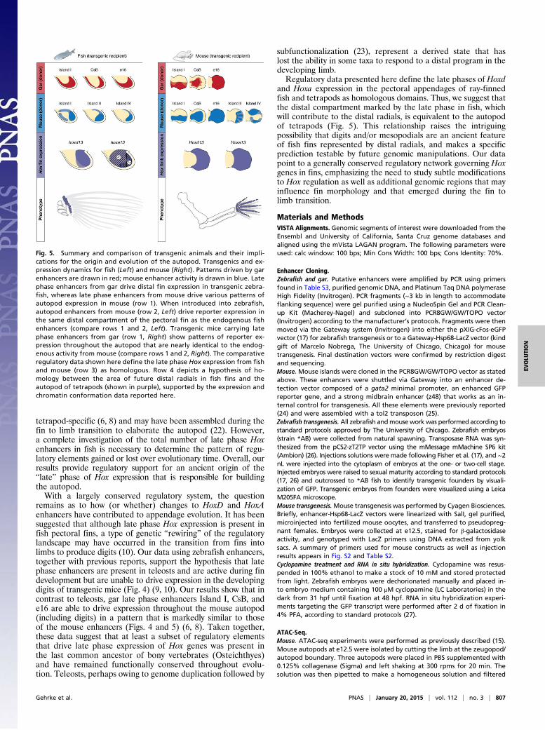
Fig. 5. Summary and comparison of transgenic animals and their impli-cations for the origin and evolution of the autopod. Transgenics and ex-pression dynamics for fish (Left) and mouse (Right). Patterns driven by garenhancers are drawn in red; mouse enhancer activity is drawn in blue. Latephase enhancers from gar drive distal fin expression in transgenic zebra-fish, whereas late phase enhancers from mouse drive various patterns ofautopod expression in mouse (row 1). When introduced into zebrafish,autopod enhancers from mouse (row 2, Left) drive reporter expression inthe same distal compartment of the pectoral fin as the endogenous fishenhancers (compare rows 1 and 2, Left). Transgenic mice carrying latephase enhancers from gar (row 1, Right) show patterns of reporter ex-pression throughout the autopod that are nearly identical to the endog-enous activity from mouse (compare rows 1 and 2, Right). The comparativeregulatory data shown here define the late phase Hox expression from fishand mouse (row 3) as homologous. Row 4 depicts a hypothesis of ho-mology between the area of future distal radials in fish fins and theautopod of tetrapods (shown in purple), supported by the expression andchromatin conformation data reported here.
Gehrke et al. PNAS | January 20, 2015 | vol. 112 | no. 3 | 807
EVOLU
TION
through a 0.45-μm cell strainer. A total of 50,000 cells were then sub-sequently used for transposition (15).Zebrafish. Briefly, 10 zebrafish embryos were manually dechorionated, anes-thetized with tricaine (Sigma), and disrupted in 500 μL of Ginzburg FishRingers (55 mM NaCl, 1.8 mM KCl, and 1.25 mM NaHCO3). The cells in theresulting solution were counted using a neubauer chamber, then pelleted at500 × g in a tabletop centrifuge and washed with cold PBS. A total of 75,000cells were lysed [lysis buffer: 10 μM Tris (pH 7.4), 10 μM NaCl, 3 μM MgCl2,and 0.1% IGEPAL (Sigma CA-630)] and incubated for 30 min at 37 °C with theTDE1 enzyme. The sample was then purified with a Qiagen Minelute kit, andPCR was performed with 13 cycles using Ad1F and Ad2.1R primers (15) withKAPA HiFi hotstart enzyme (Kapa Biosystems). The resulting library was se-quenced in a HisEq 2000 pair end lane producing 180 M of 49-bp reads perend. Reads were aligned using zebrafish July 2010 assembly (danRer7) as thereference genome. Duplicated pairs or those ones separated by more than 2kb were removed. The enzyme cleavage site was determined as the position−4 (minus strand) or +5 (plus strand) from each read start, and this positionwas extended 5 bp in both directions. ATAC-seq on FACS-sorted cells wereperformed and analyzed in the same manner with the following mod-ifications: ∼400 embryos from the gar Island I transgenic line were grown to48 hpf, anesthetized with tricaine (Sigma), and placed in PBS supplementedwith 0.125% collagenase (Sigma). Embryos were incubated at 37 °C for 1 hshaking at 300 rpm, with gentle pipetting every 20 min to disrupt embryos.The solution was filtered twice with a 0.45-μm cell strainer to obtain a sin-gle cell suspension, and FACS sorted for GFP using an Avalon 2-4 machine atthe University of Chicago. The resulting solution contained the 40,000brightest GFP+ cells and was processed immediately for ATAC-seq asdescribed above.
Statistically significant peaks were determined by extending ATAC-seqreads to 100 bp and using MACS software (28) with default parameters.Circular chromosome conformation capture. Circular chromosome conformationcapture (4C-seq) assays were performed as previously reported (29–32). Fivehundred zebrafish embryos at 48 or 60 hpf were dechorionated using pro-nase and disrupted in 1 mL of Ginzburg Fish Ringers (55 mM NaCl, 1.8 mM
KCl, and 1.25 mM NaHCO3). Isolated cells were treated with lysis buffer [lysisbuffer: 10 mM Tris·HCl (pH 8), 10 mM NaCl, 0.3% IGEPAL CA-630 (Sigma),and 1× protease inhibitor mixture (cOmplete, Roche)] and the DNA digestedwith DpnII (NEB) and Csp6I (Fermentas, Thermo Scientific) as primary andsecondary enzymes, respectively. T4 DNA ligase (Promega) was used for bothligation steps. Specific primers were designed at gene promoters, and Illu-mina adaptors were included in primer sequence. Eight PCRs were per-formed with the Expand Long Template PCR System (Roche) and pooledtogether. This library was purified with a High Pure PCR Product PurificationKit (Roche) and its concentration measured using the Quanti-iTTM Pico-Green dsDNA Assay Kit (Invitrogen, P11496) in a Qubit machine and sent fordeep sequencing in an Illumina HisEq 2000 machine multiplexing 10 samplesper lane. 4C-seq data were analyzed as previously described (31). Briefly, rawsequencing data were demultiplexed using the primer sequences as barc-odes and aligned using zebrafish July 2010 assembly (danRer7) as the ref-erence genome. Reads located in fragments flanked by two restriction sitesof the same enzyme, or in fragments smaller than 40 bp, were filtered out.Mapped reads were then converted to reads-per-first-enzyme-fragment-endunits and smoothed using a 30-fragment mean running window algorithm.
ACKNOWLEDGMENTS. We thank John Westlund for excellent illustrationand assistance assembling the figures; Marcelo Nobrega, Mike Coates, IlyaRuvinsky, and Tom Stewart for critical reading of the manuscript; GokhanDalgin, Noritaka Adachi, Joyce Pieretti, Darcy Ross, and Justin Lemberg forhelpful discussions; and the Prince and Ho laboratories for reagents and helpwith zebrafish. This study was supported by The Brinson Foundation (N.H.S.),National Science Foundation Doctoral Dissertation Improvement Grant1311436 (to A.R.G.), Brazilian National Council for Scientific and Technolog-ical Development Grants 402754/2012-3 and 477658/2012-1 (to I.S.), NationalInstitutes of Health Grant R01RR020833 (R01OD011116) (to J.H.P), “InitiativeEvolutionary Biology” from the Volkswagen Foundation, Germany andAlexander von Humboldt-Foundation (I.B.), and Spanish and AndalusianGovernments Grants BFU2010-14839, BFU2013-41322-P, and Proyecto deExcelencia BIO-396 (to J.L.G.-S.).
1. Wagner GP (2014) Homology, Genes, and Evolutionary Innovation (Princeton UnivPress, Princeton), pp 327–384.
2. Shubin NH, Daeschler EB, Jenkins FA, Jr (2006) The pectoral fin of Tiktaalik roseae andthe origin of the tetrapod limb. Nature 440(7085):764–771.
3. Schneider I, Shubin NH (2013) The origin of the tetrapod limb: From expeditions toenhancers. Trends Genet 29(7):419–426.
4. Montavon T, Duboule D (2013) Chromatin organization and global regulation of Hoxgene clusters. Philos Trans R Soc Lond B Biol Sci 368(1620):20120367.
5. Zakany J, Duboule D (2007) The role of Hox genes during vertebrate limb de-velopment. Curr Opin Genet Dev 17(4):359–366.
6. Montavon T, et al. (2011) A regulatory archipelago controls Hox genes transcription indigits. Cell 147(5):1132–1145.
7. Andrey G, et al. (2013) A switch between topological domains underlies HoxD genescollinearity in mouse limbs. Science 340(6137):1234167.
8. Berlivet S, et al. (2013) Clustering of tissue-specific sub-TADs accompanies the regu-lation of HoxA genes in developing limbs. PLoS Genet 9(12):e1004018.
9. Schneider I, et al. (2011) Appendage expression driven by the Hoxd Global Con-trol Region is an ancient gnathostome feature. Proc Natl Acad Sci USA 108(31):12782–12786.
10. Woltering JM, Noordermeer D, Leleu M, Duboule D (2014) Conservation and di-vergence of regulatory strategies at Hox Loci and the origin of tetrapod digits. PLoSBiol 12(1):e1001773.
11. Braasch I, et al. (2014) A new model army: Emerging fish models to study the ge-nomics of vertebrate Evo-Devo. J Exp Zoolog B Mol Dev Evol, 10.1002/jez.b.22589.
12. Taylor JS, Braasch I, Frickey T, Meyer A, Van de Peer Y (2003) Genome duplication,a trait shared by 22000 species of ray-finned fish. Genome Res 13(3):382–390.
13. Amores A, et al. (1998) Zebrafish hox clusters and vertebrate genome evolution.Science 282(5394):1711–1714.
14. Amores A, Catchen J, Ferrara A, Fontenot Q, Postlethwait JH (2011) Genome evolu-tion and meiotic maps by massively parallel DNA sequencing: Spotted gar, an out-group for the teleost genome duplication. Genetics 188(4):799–808.
15. Buenrostro JD, Giresi PG, Zaba LC, Chang HY, Greenleaf WJ (2013) Transposition ofnative chromatin for fast and sensitive epigenomic profiling of open chromatin, DNA-binding proteins and nucleosome position. Nat Methods 10(12):1213–1218.
16. Lara-Astiaso D, et al. (2014) Immunogenetics. Chromatin state dynamics during bloodformation. Science 345(6199):943–949.
17. Fisher S, et al. (2006) Evaluating the biological relevance of putative enhancers usingTol2 transposon-mediated transgenesis in zebrafish. Nat Protoc 1(3):1297–1305.
18. Chiang C, et al. (2001) Manifestation of the limb prepattern: Limb development in the
absence of sonic hedgehog function. Dev Biol 236(2):421–435.19. Litingtung Y, Dahn RD, Li Y, Fallon JF, Chiang C (2002) Shh and Gli3 are dispensable for
limb skeleton formation but regulate digit number and identity. Nature 418(6901):
979–983.20. Ahn D, Ho RK (2008) Tri-phasic expression of posterior Hox genes during develop-
ment of pectoral fins in zebrafish: Implications for the evolution of vertebrate paired
appendages. Dev Biol 322(1):220–233.21. Gonzalez F, Duboule D, Spitz F (2007) Transgenic analysis of Hoxd gene regulation
during digit development. Dev Biol 306(2):847–859.22. Freitas R, Gómez-Marín C, Wilson JM, Casares F, Gómez-Skarmeta JL (2012) Hoxd13
contribution to the evolution of vertebrate appendages. Dev Cell 23(6):1219–1229.23. Force A, et al. (1999) Preservation of duplicate genes by complementary, degenera-
tive mutations. Genetics 151(4):1531–1545.24. Bessa J, et al. (2009) Zebrafish enhancer detection (ZED) vector: A new tool to facil-
itate transgenesis and the functional analysis of cis-regulatory regions in zebrafish.
Dev Dyn 238(9):2409–2417.25. Kawakami K, Shima A, Kawakami N (2000) Identification of a functional transposase of
the Tol2 element, an Ac-like element from the Japanese medaka fish, and its trans-
position in the zebrafish germ lineage. Proc Natl Acad Sci USA 97(21):11403–11408.26. Suster ML, Abe G, Schouw A, Kawakami K (2011) Transposon-mediated BAC trans-
genesis in zebrafish. Nat Protoc 6(12):1998–2021.27. Thisse C, Thisse B, Postlethwait JH (1995) Expression of snail2, a second member of the
zebrafish snail family, in cephalic mesendoderm and presumptive neural crest of wild-
type and spadetail mutant embryos. Dev Biol 172(1):86–99.28. Zhang Y, et al. (2008) Model-based analysis of ChIP-Seq (MACS). Genome Biol
9(9):R137.29. Dekker J, Rippe K, Dekker M, Kleckner N (2002) Capturing chromosome conforma-
tion. Science 295(5558):1306–1311.30. Hagège H, et al. (2007) Quantitative analysis of chromosome conformation capture
assays (3C-qPCR). Nat Protoc 2(7):1722–1733.31. Noordermeer D, et al. (2011) The dynamic architecture of Hox gene clusters. Science
334(6053):222–225.32. Splinter E, de Wit E, van de Werken HJ, Klous P, de Laat W (2012) Determining long-
range chromatin interactions for selected genomic sites using 4C-seq technology:
From fixation to computation. Methods 58(3):221–230.
808 | www.pnas.org/cgi/doi/10.1073/pnas.1420208112 Gehrke et al.