Deciphering the role of osteoprotegerin in inflammatory bowel diseases
36
Osteoprotegerin (OPG) Aoki K, et al. Peptide-based delivery to bone. Adv Drug Deliv Rev. 2012 Sep;64(12):1220-38. Yamaguchi K, et al. Characterization of structural domains of human osteoclastogenesis inhibitory factor. J Biol Chem. 1998 Feb 27;273(9):5117-23. Rowinsky EK. Targeted induction of apoptosis in cancer management: the emerging role of tumor necrosis factor-related apoptosis- inducing ligand receptor activating agents. J Clin Oncol. 2005 Dec 20;23(36):9394-407.
-
Upload
raghunath-ram-ms-md -
Category
Science
-
view
9 -
download
1
Transcript of Deciphering the role of osteoprotegerin in inflammatory bowel diseases
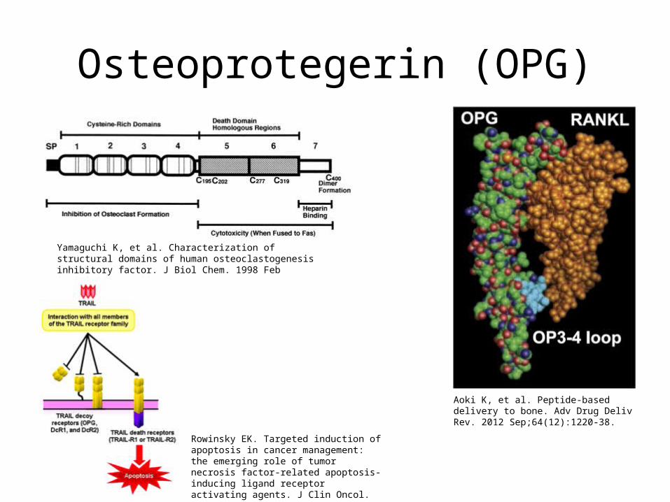
Osteoprotegerin (OPG)
Aoki K, et al. Peptide-based delivery to bone. Adv Drug Deliv Rev. 2012 Sep;64(12):1220-38.
Yamaguchi K, et al. Characterization of structural domains of human osteoclastogenesis inhibitory factor. J Biol Chem. 1998 Feb 27;273(9):5117-23.
Rowinsky EK. Targeted induction of apoptosis in cancer management: the emerging role of tumor necrosis factor-related apoptosis-inducing ligand receptor activating agents. J Clin Oncol. 2005 Dec 20;23(36):9394-407.
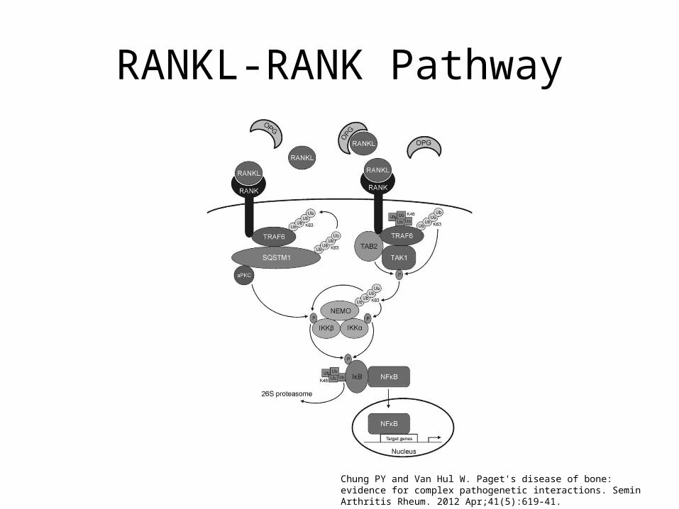
RANKL-RANK Pathway
Chung PY and Van Hul W. Paget's disease of bone: evidence for complex pathogenetic interactions. Semin Arthritis Rheum. 2012 Apr;41(5):619-41.
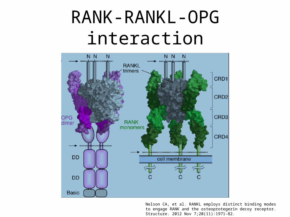
RANK-RANKL-OPG interaction
Nelson CA, et al. RANKL employs distinct binding modes to engage RANK and the osteoprotegerin decoy receptor. Structure. 2012 Nov 7;20(11):1971-82.
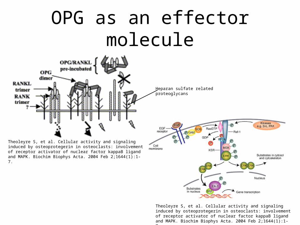
OPG as an effector molecule
Theoleyre S, et al. Cellular activity and signaling induced by osteoprotegerin in osteoclasts: involvement of receptor activator of nuclear factor kappaB ligand and MAPK. Biochim Biophys Acta. 2004 Feb 2;1644(1):1-7.
Theoleyre S, et al. Cellular activity and signaling induced by osteoprotegerin in osteoclasts: involvement of receptor activator of nuclear factor kappaB ligand and MAPK. Biochim Biophys Acta. 2004 Feb 2;1644(1):1-7.
Heparan sulfate related proteoglycans
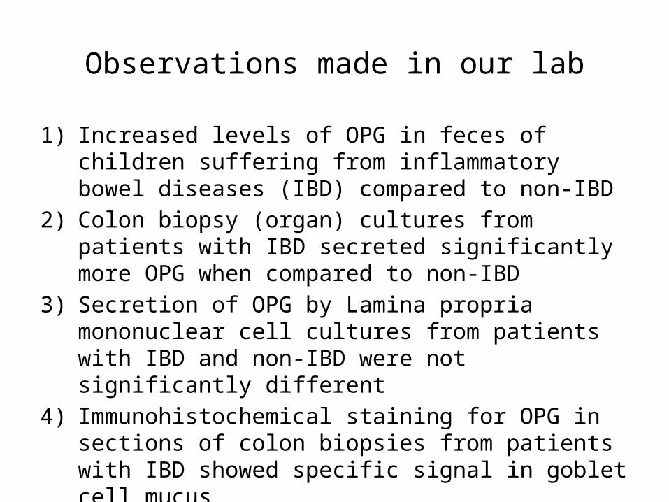
Observations made in our lab
1) Increased levels of OPG in feces of children suffering from inflammatory bowel diseases (IBD) compared to non-IBD
2) Colon biopsy (organ) cultures from patients with IBD secreted significantly more OPG when compared to non-IBD
3) Secretion of OPG by Lamina propria mononuclear cell cultures from patients with IBD and non-IBD were not significantly different
4) Immunohistochemical staining for OPG in sections of colon biopsies from patients with IBD showed specific signal in goblet cell mucus
5) Nuclear OPG signal was seen only in intestinal epithelial cells of colon biopsies from patients with out IBD
cont.
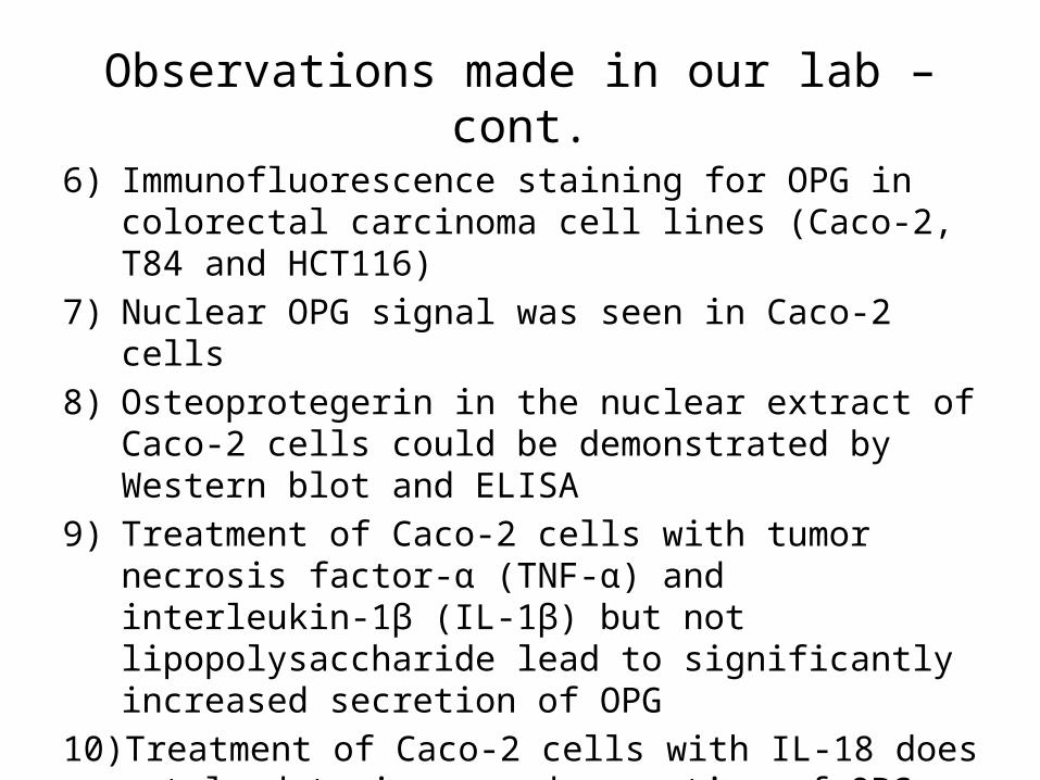
Observations made in our lab – cont.
6) Immunofluorescence staining for OPG in colorectal carcinoma cell lines (Caco-2, T84 and HCT116)
7) Nuclear OPG signal was seen in Caco-2 cells8) Osteoprotegerin in the nuclear extract of Caco-2 cells could
be demonstrated by Western blot and ELISA 9) Treatment of Caco-2 cells with tumor necrosis factor-α (TNF-
α) and interleukin-1β (IL-1β) but not lipopolysaccharide lead to significantly increased secretion of OPG
10) Treatment of Caco-2 cells with IL-18 does not lead to increased secretion of OPG
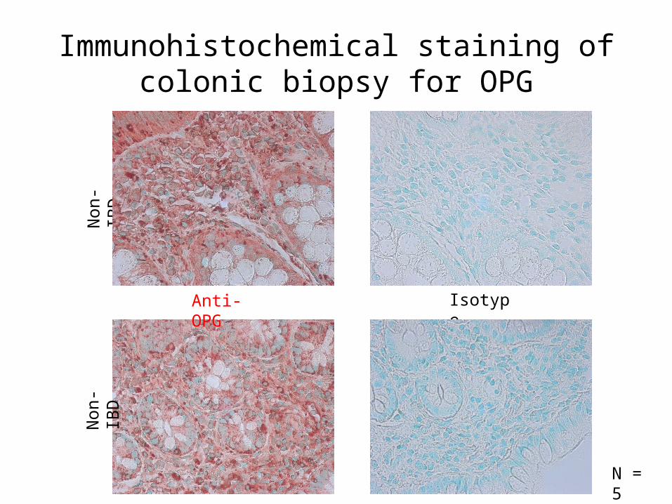
Immunohistochemical staining of colonic biopsy for OPG
Non
-IBD
Non
-IBD
Anti-OPG Isotype
N = 5
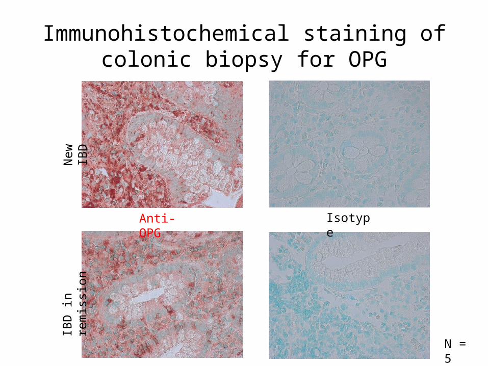
Immunohistochemical staining of colonic biopsy for OPG
Anti-OPG Isotype
New
IBD
IBD
in re
mis
sion
N = 5
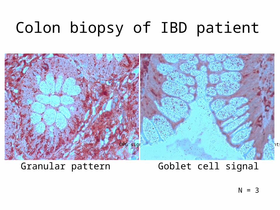
Granular pattern
OPG signal in goblet cells of colon biopsy of IBD patients
N = 3
Colon biopsy of IBD patient
Goblet cell signal
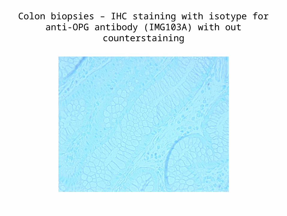
Colon biopsies – IHC staining with isotype for anti-OPG antibody (IMG103A) with out counterstaining
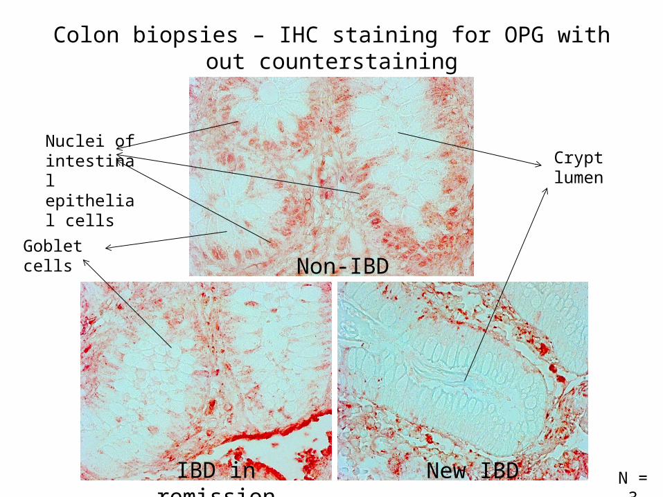
Colon biopsies – IHC staining for OPG with out counterstaining
Non-IBD
IBD in remission New IBD N = 3
Crypt lumen
Goblet cells
Nuclei of intestinal epithelial cells
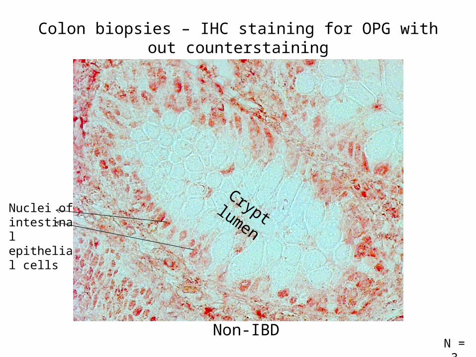
Crypt lumenNuclei of intestinal epithelial cells
Colon biopsies – IHC staining for OPG with out counterstaining
N = 3Non-IBD
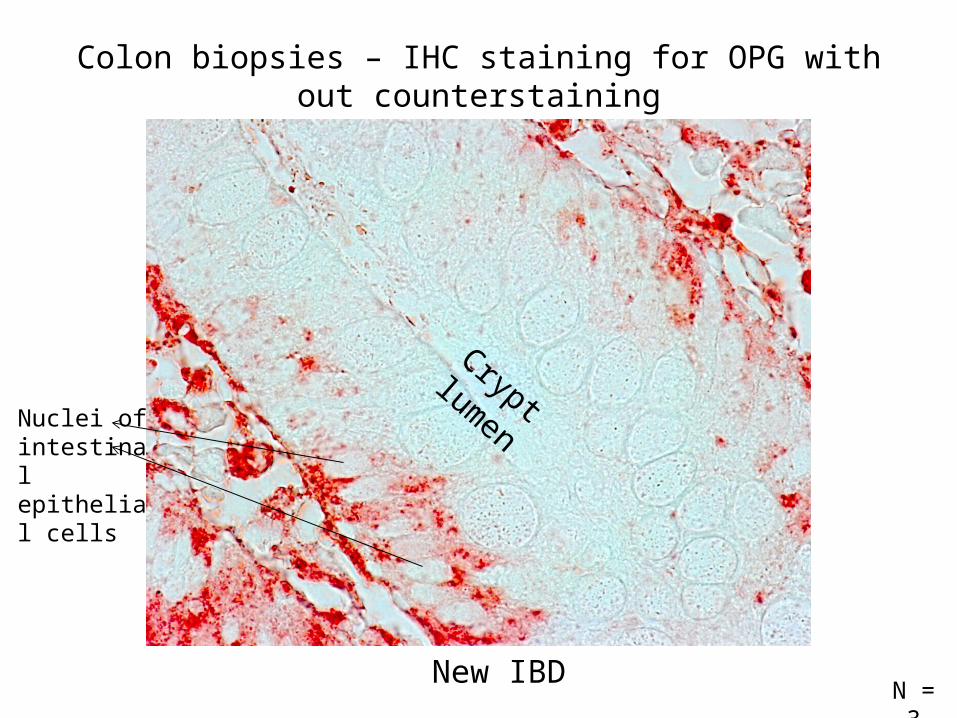
Crypt lumenNuclei of intestinal epithelial cells
New IBD
Colon biopsies – IHC staining for OPG with out counterstaining
N = 3
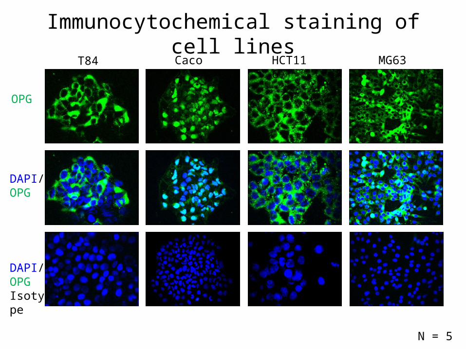
T84 Caco2 HCT116 MG63
OPG
DAPI/ OPG
DAPI/ OPGIsotype
Immunocytochemical staining of cell lines
N = 5
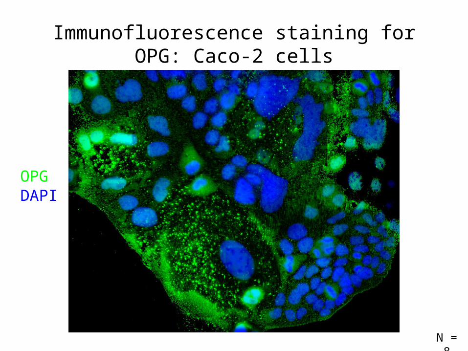
Immunofluorescence staining for OPG: Caco-2 cells
OPGDAPI
N = 8
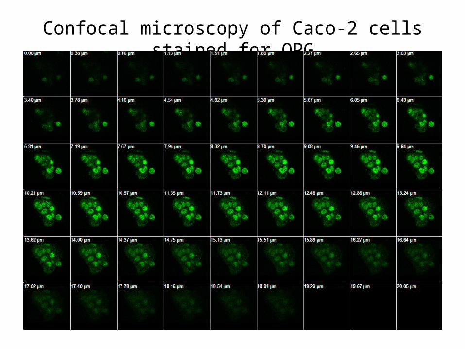
Confocal microscopy of Caco-2 cells stained for OPG
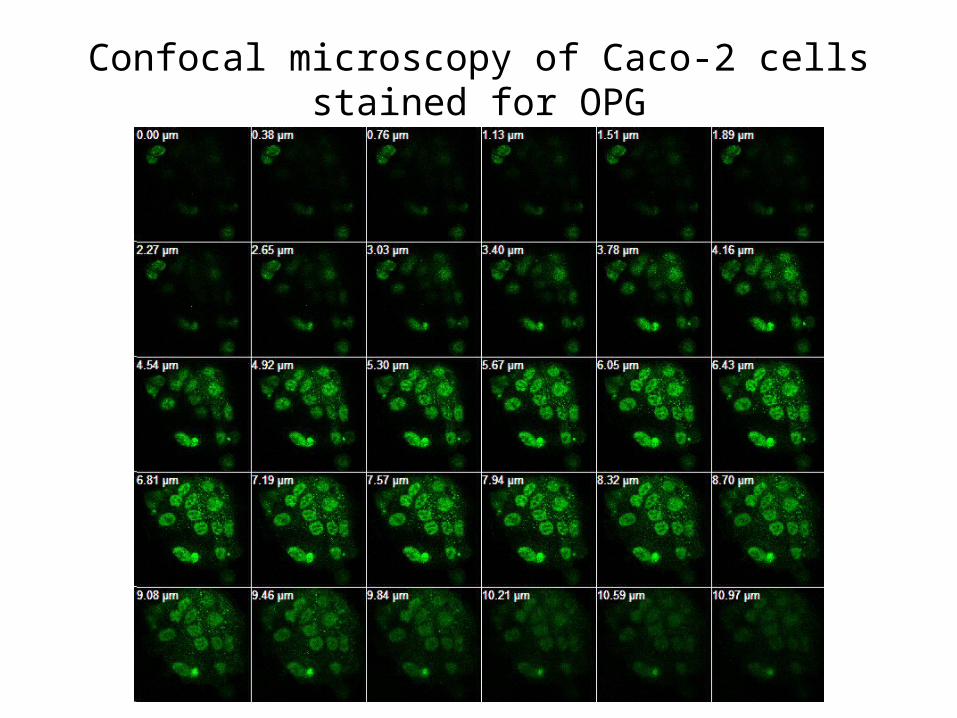
Confocal microscopy of Caco-2 cells stained for OPG
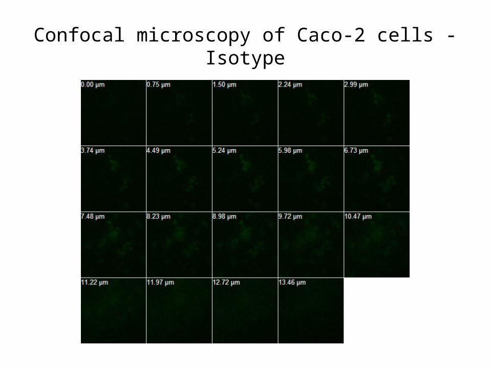
Confocal microscopy of Caco-2 cells - Isotype
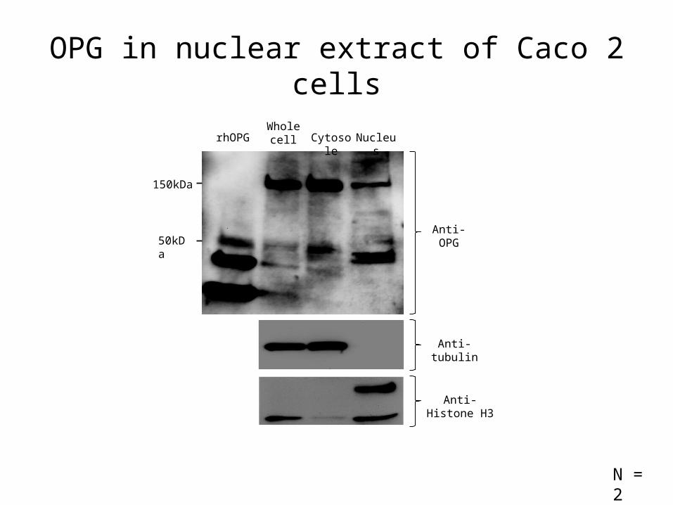
Whole cell Cytosole Nucleus
Anti-OPG50kDa
150kDa
Anti-tubulin
Anti-Histone H3
OPG in nuclear extract of Caco 2 cells
rhOPG
N = 2
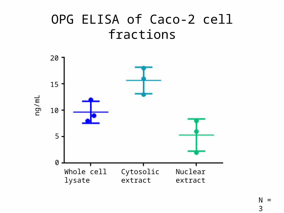
OPG ELISA of Caco-2 cell fractions
5
10
15
ng/m
L
20
0Whole cell lysate Cytosolic extract Nuclear extract
N = 3
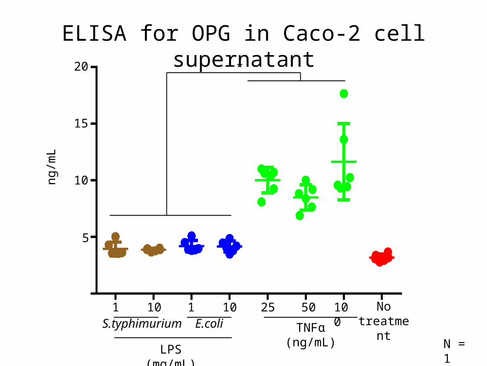
ELISA for OPG in Caco-2 cell supernatant
No treatment
5
10
15
20ng
/mL
1 10 1 10 25 50 100S.typhimurium E.coli TNFα (ng/mL)
LPS (mg/mL)
*
N = 1
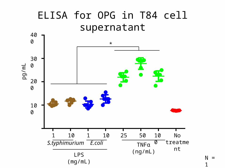
ELISA for OPG in T84 cell supernatant
100
200
300
400
pg/m
L
*
No treatment
1 10 1 10 25 50 100S.typhimurium E.coli TNFα (ng/mL)
LPS (mg/mL) N = 1
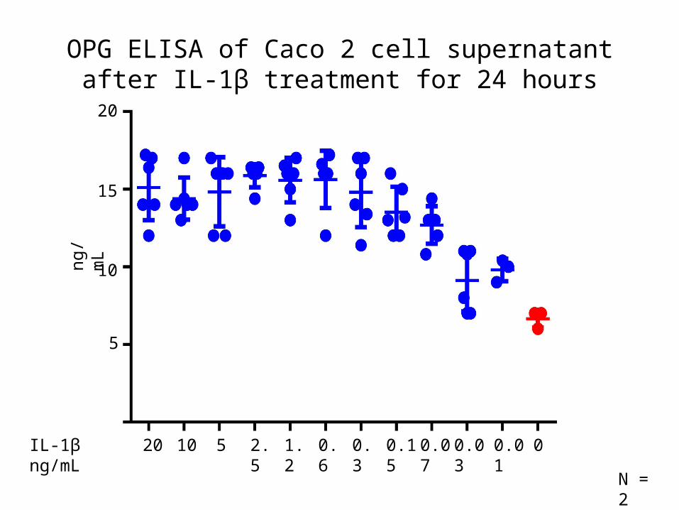
OPG ELISA of Caco 2 cell supernatant after IL-1β treatment for 24 hours
5
10
15
20
IL-1β ng/mL 20 10 5 2.5 1.2 0.6 0.3 0.15 0.07 0.03 0.01 0
ng/m
L
N = 2
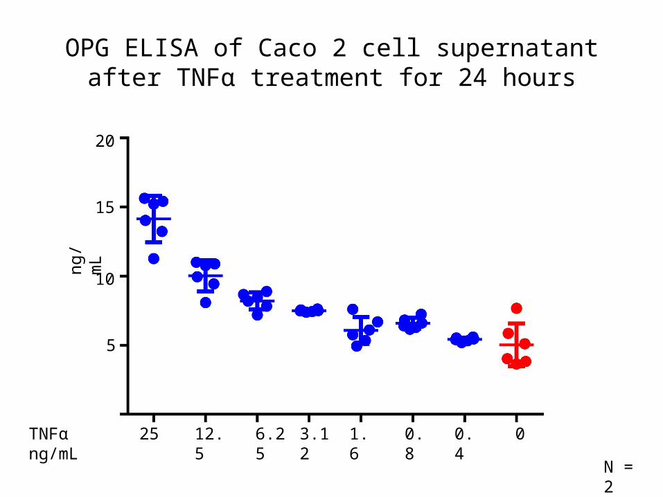
OPG ELISA of Caco 2 cell supernatant after TNFα treatment for 24 hours
ng/m
L
5
10
15
20
TNFα ng/mL 25 12.5 6.25 3.12 1.6 0.8 0.4 0
N = 2
5
10
15
20
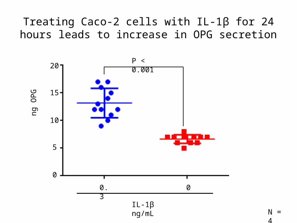
0.3
IL-1β ng/mL
0
Treating Caco-2 cells with IL-1β for 24 hours leads to increase in OPG secretion
ng O
PG
N = 4
P < 0.001
0
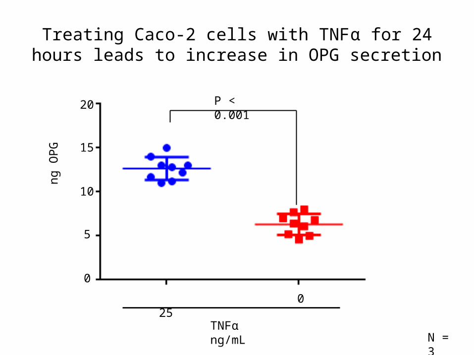
Treating Caco-2 cells with TNFα for 24 hours leads to increase in OPG secretion
5
10
15
20
ng O
PG
N = 3
25
TNFα ng/mL
0
P < 0.001
0
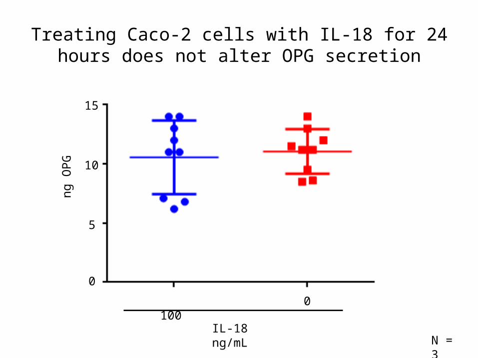
Treating Caco-2 cells with IL-18 for 24 hours does not alter OPG secretion
100
IL-18 ng/mL
0
5
10
ng O
PG
N = 3
0
15
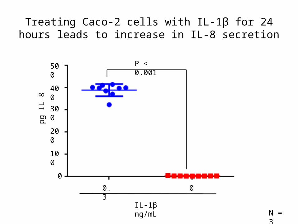
Treating Caco-2 cells with IL-1β for 24 hours leads to increase in IL-8 secretion
100
300
pg IL
-8
0
400
200
500
0.3
IL-1β ng/mL
0
N = 3
P < 0.001
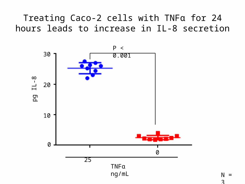
Treating Caco-2 cells with TNFα for 24 hours leads to increase in IL-8 secretion
10
20
pg IL
-8
0
30
N = 3
25
TNFα ng/mL
0
P < 0.001
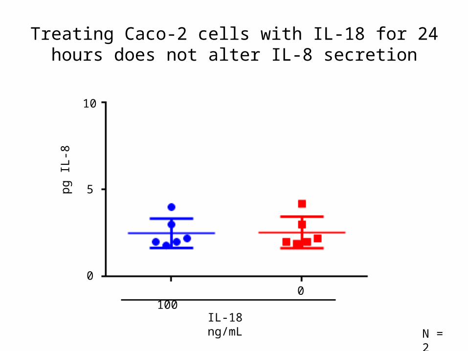
Treating Caco-2 cells with IL-18 for 24 hours does not alter IL-8 secretion
100
IL-18 ng/mL
0
N = 2
5
10
pg IL
-8
0
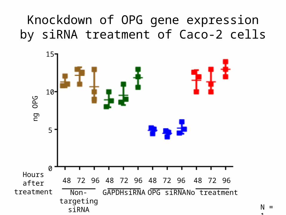
Knockdown of OPG gene expression by siRNA treatment of Caco-2 cells
48 72 96 48 72 96 48 72 96 48 72 96
5
10
15
ng O
PG
Non-targeting siRNA
GAPDHsiRNA OPG siRNA No treatment
Hours after treatment
0
N = 1
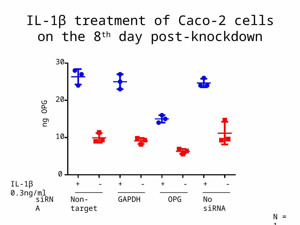
IL-1β treatment of Caco-2 cells on the 8th day post-knockdown
IL-1β 0.3ng/ml + - + - + - + -
Non-targetsiRNA GAPDH OPG No siRNA
10
20
30
ng O
PG
0
N = 1
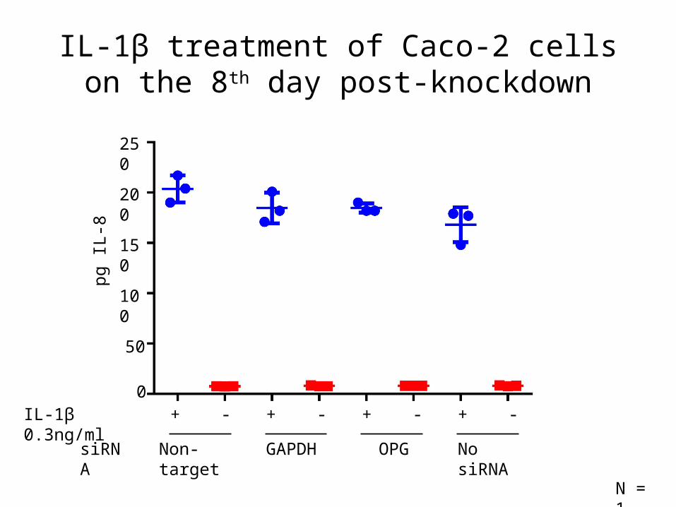
IL-1β treatment of Caco-2 cells on the 8th day post-knockdown
IL-1β 0.3ng/ml + - + - + - + -
Non-targetsiRNA GAPDH OPG No siRNA
0
N = 1
50
100
250
150
200
pg IL
-8
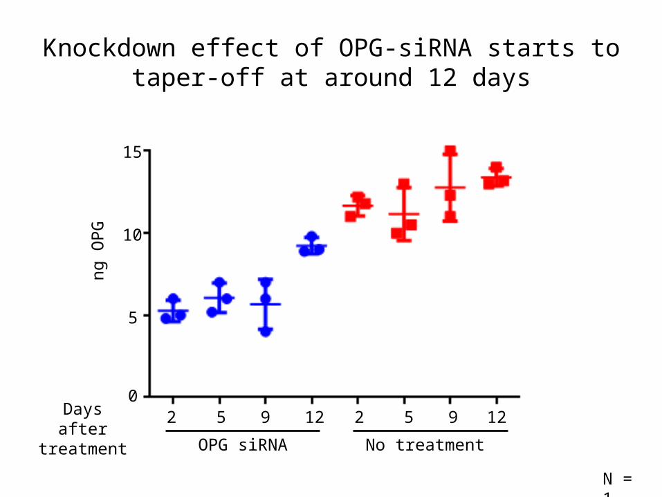
Knockdown effect of OPG-siRNA starts to taper-off at around 12 days
5
10
ng O
PG
0
15
2 5 9
No treatment
2 5 9
OPG siRNA
Days after treatment
12 12
N = 1
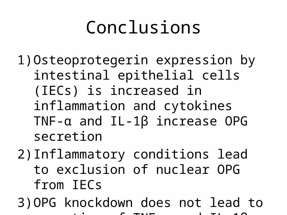
Conclusions
1) Osteoprotegerin expression by intestinal epithelial cells (IECs) is increased in inflammation and cytokines TNF-α and IL-1β increase OPG secretion
2) Inflammatory conditions lead to exclusion of nuclear OPG from IECs
3) OPG knockdown does not lead to secretion of TNF-α and IL-1β by Caco-2 cells
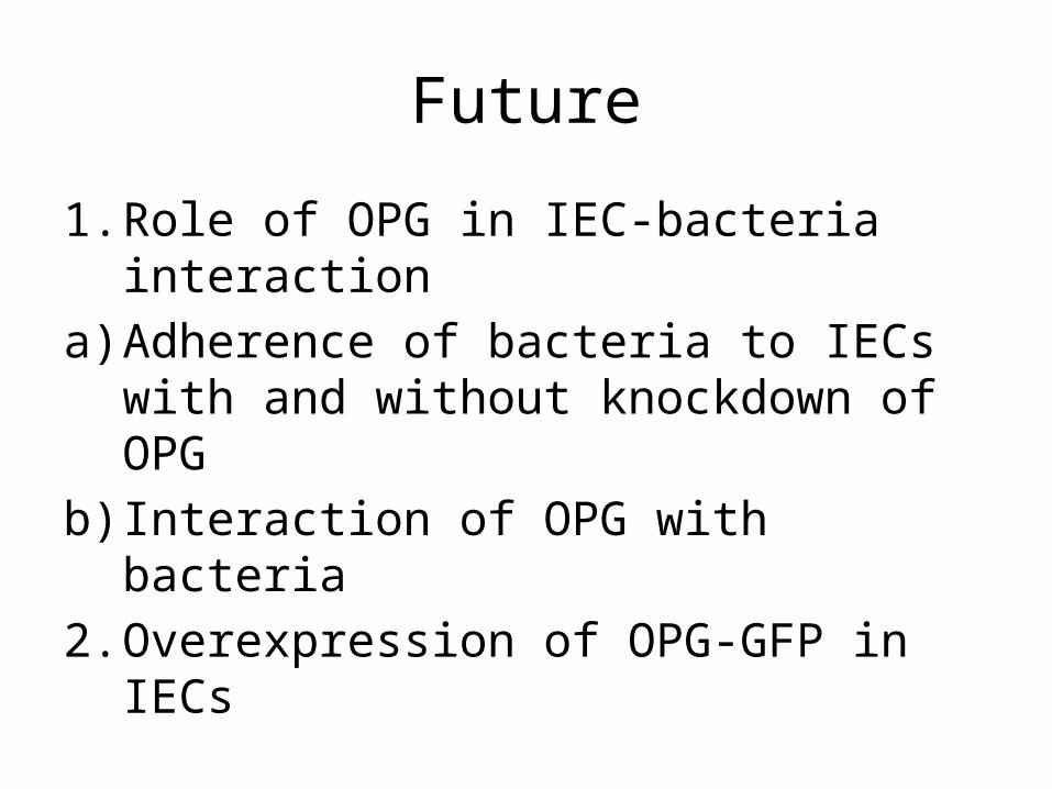
Future
1. Role of OPG in IEC-bacteria interactiona) Adherence of bacteria to IECs with and
without knockdown of OPGb) Interaction of OPG with bacteria2. Overexpression of OPG-GFP in IECs