Debridement of Bacterial Biofilms with TiO2/H2 Solutions...
9
Research Article Debridement of Bacterial Biofilms with TiO 2 /H 2 O 2 Solutions and Visible Light Irradiation Oscar Janson , 1 Maria Strømme, 2 Håkan Engqvist, 1 and Ken Welch 2 1 Division of Applied Material Science, Department of Engineering Sciences, Uppsala University, Uppsala 751 21, Sweden 2 Division of Nanotechnology and Functional Materials, Department of Engineering Sciences, Uppsala University, Uppsala 751 21, Sweden Correspondence should be addressed to Oscar Janson; [email protected] Received 10 April 2018; Accepted 6 June 2018; Published 2 July 2018 Academic Editor: Wen-Cheng Chen Copyright © 2018 Oscar Janson et al. is is an open access article distributed under the Creative Commons Attribution License, which permits unrestricted use, distribution, and reproduction in any medium, provided the original work is properly cited. Objectives. e aim of the study was to explore the debridement efficacy of different solutions of H 2 O 2 and rutile particles against Staphylococcus epidermidis and Pseudomonas aeruginosa biofilms attached to titanium surfaces when exposed to visible light. Materials and Methods. Titanium discs cultivated with biofilms of Staphylococcus epidermidis or Pseudomonas aeruginosa were subjected for 1 min to suspensions consisting of rutile particles mixed with high (950 mM) or low (2 mM) concentrations of H 2 O 2 under visible light irradiation (405 nm; 2.1 mW/cm 2 ). Discs were rinsed and the degree of debridement was determined through scanning electron microscopy and viability assessment of the remaining bacteria using luminescence measurements and/or a metabolic activity assay. Results. Cleaning mixtures containing the higher concentration of H 2 O 2 showed a significantly improved debridement compared to the negative control in all experiments. e addition of rutile particles was shown to have a statistically significant effect in one test with S. epidermidis. Limited evidence of the catalytic effect of visible light irradiation was seen, but effects were relatively small and statistically insignificant. Conclusions.H 2 O 2 at a concentration of 950 mM proved to be the strongest contribution to the debridement and bactericidal effect of the cleaning techniques tested in this study. 1. Introduction Peri-implantitis is an inflammatory disease that is defined by bleeding on probing and marginal loss of bone at the implant site [1]. e frequency of peri-implantitis has been reported to be as high as 19-22% of subjects and 9-10% of implants [2–4]. Peri-implantitis arises from bacteria attaching to the dental implants leading to the formation of bacterial biofilms, which are clusters of bacteria encased in an extracellular polymeric substance and adherent to a surface [5]. e bacteria in biofilms possess a different gene expression compared to planktonic bacteria which can make the infection resistant to antibiotics [6]. erefore nonantibiotic treatment alternatives may be required. Commonly used cleaning protocols for peri-implantitis infected implants include mechanical clean- ing with curettes or brushes, which can damage the implant surface and have difficulty reaching bacteria located in the micro roughness of the implants [7]. Chemicals like hydrogen peroxide (H 2 O 2 ), chlorhexidine gluconate, saline, and citric acid are also used for disinfection and while they all show good effect, they are not satisfactory for complete cleaning of the implants [8]. erefore there is an urgent need for more effective cleaning methods. Photocatalytic activation of titanium dioxide (TiO 2 ) has long been investigated not only as an environmental cleaning agent of waste water and air purification [9], but also for biomedical applications [10–12]. Photocatalysis occurs when an electron from the valence band in a TiO 2 crystal is excited to the conduction band by a photon of higher energy compared to the band gap between the valence and conduction bands of the crystal, thereby creating an electron-hole pair. e band gaps for the two most common crystal forms of TiO 2 , anatase and rutile, are 3.20 and 3.03 eV, respectively [13], corresponding to light wavelengths of 387 nm and 409 nm, respectively. e excited electron can react with oxygen (O 2 ) and create superoxide radicals (O 2 −∙ ) while the hole can react with water (H 2 O) and give rise to hydroxyl radicals ( ∙ OH) [14]. Reactive oxygen species Hindawi International Journal of Biomaterials Volume 2018, Article ID 5361632, 8 pages https://doi.org/10.1155/2018/5361632
Transcript of Debridement of Bacterial Biofilms with TiO2/H2 Solutions...

Research ArticleDebridement of Bacterial Biofilms with TiO
2/H
2O
2
Solutions and Visible Light Irradiation
Oscar Janson ,1 Maria Strømme,2 Håkan Engqvist,1 and KenWelch2
1Division of Applied Material Science, Department of Engineering Sciences, Uppsala University, Uppsala 751 21, Sweden2Division of Nanotechnology and Functional Materials, Department of Engineering Sciences, Uppsala University,Uppsala 751 21, Sweden
Correspondence should be addressed to Oscar Janson; [email protected]
Received 10 April 2018; Accepted 6 June 2018; Published 2 July 2018
Academic Editor: Wen-Cheng Chen
Copyright © 2018 Oscar Janson et al. This is an open access article distributed under the Creative Commons Attribution License,which permits unrestricted use, distribution, and reproduction in any medium, provided the original work is properly cited.
Objectives. The aim of the study was to explore the debridement efficacy of different solutions of H2O2and rutile particles against
Staphylococcus epidermidis and Pseudomonas aeruginosa biofilms attached to titanium surfaces when exposed to visible light.Materials and Methods. Titanium discs cultivated with biofilms of Staphylococcus epidermidis or Pseudomonas aeruginosa weresubjected for 1 min to suspensions consisting of rutile particles mixed with high (950 mM) or low (2 mM) concentrations ofH2O2under visible light irradiation (405 nm; 2.1 mW/cm2). Discs were rinsed and the degree of debridement was determined
through scanning electronmicroscopy and viability assessment of the remaining bacteria using luminescencemeasurements and/ora metabolic activity assay. Results. Cleaningmixtures containing the higher concentration of H
2O2showed a significantly improved
debridement compared to the negative control in all experiments. The addition of rutile particles was shown to have a statisticallysignificant effect in one test with S. epidermidis. Limited evidence of the catalytic effect of visible light irradiation was seen, buteffects were relatively small and statistically insignificant.Conclusions. H
2O2at a concentration of 950mMproved to be the strongest
contribution to the debridement and bactericidal effect of the cleaning techniques tested in this study.
1. Introduction
Peri-implantitis is an inflammatory disease that is defined bybleeding on probing and marginal loss of bone at the implantsite [1].The frequency of peri-implantitis has been reported tobe as high as 19-22% of subjects and 9-10% of implants [2–4].Peri-implantitis arises from bacteria attaching to the dentalimplants leading to the formation of bacterial biofilms, whichare clusters of bacteria encased in an extracellular polymericsubstance and adherent to a surface [5]. The bacteria inbiofilms possess a different gene expression compared toplanktonic bacteria which can make the infection resistant toantibiotics [6].Therefore nonantibiotic treatment alternativesmay be required. Commonly used cleaning protocols forperi-implantitis infected implants include mechanical clean-ing with curettes or brushes, which can damage the implantsurface and have difficulty reaching bacteria located in themicro roughness of the implants [7]. Chemicals like hydrogenperoxide (H
2O2), chlorhexidine gluconate, saline, and citric
acid are also used for disinfection and while they all showgood effect, they are not satisfactory for complete cleaning ofthe implants [8]. Therefore there is an urgent need for moreeffective cleaning methods.
Photocatalytic activation of titanium dioxide (TiO2) has
long been investigated not only as an environmental cleaningagent of waste water and air purification [9], but also forbiomedical applications [10–12]. Photocatalysis occurs whenan electron from the valence band in a TiO
2crystal is
excited to the conduction band by a photon of higherenergy compared to the band gap between the valenceand conduction bands of the crystal, thereby creating anelectron-hole pair. The band gaps for the two most commoncrystal forms of TiO
2, anatase and rutile, are 3.20 and 3.03
eV, respectively [13], corresponding to light wavelengths of387 nm and 409 nm, respectively. The excited electron canreact with oxygen (O
2) and create superoxide radicals (O
2
−∙)while the hole can react with water (H
2O) and give rise
to hydroxyl radicals (∙OH) [14]. Reactive oxygen species
HindawiInternational Journal of BiomaterialsVolume 2018, Article ID 5361632, 8 pageshttps://doi.org/10.1155/2018/5361632

2 International Journal of Biomaterials
Table 1: Description of debridement treatment conditions. Exposure time was 1 min for all treatment conditions.
Sampledesignation
Concentration rutile(g/l)
Concentration H2O2
(mM)Light, intensity:wavelength
(mW/cm2:nm)Ethanol (Positive control) - - Ambient lightPBS (Negative control) - - Ambient light0 Hi Amb - 950 Ambient lightR Hi Amb 0.5 950 Ambient lightR Hi Li 0.5 950 2.1:405R Low Li 0.5 2 2.1:405
(ROS) such as hydroxyl radicals, superoxide radicals, andH2O2are known to damage the cell wall of bacteria leading
to cell death [15]. Rutile has a smaller band gap but isconsidered a less active photocatalyst compared to anatase ormixed anatase/rutile phases [16].Howeverwhen adding smallamounts of H
2O2, rutile has been shown to increase hydroxyl
radical formation under light irradiation [16–18]. H2O2is in
itself an effective antiseptic and with a concentration of 3vol.% (950 mM) has been shown to be an effective treatmentagainst S. epidermidis biofilms on titanium dental implantsurfaces in vitro [19–21]. TiO
2in combination with H
2O2
has been shown to give rise to an increased synergisticbactericidal effect [19, 21]. Studies have shown that smallamounts of H
2O2together with TiO
2of the allotropic form
rutile under light exposure is the most effective combinationto degrade the organic dye Rhodamine B [22], as well asinactivate themultiresistant bacteriumKlebsiella pneumoniae[23].
We hypothesize that TiO2particles combined with H
2O2
under light irradiation can be effective in debriding peri-implantitis infected implants. Consequently, the aim of thisstudy was to explore the debridement efficacy of mixtures ofrutile TiO
2particles and H
2O2under visible light irradiation
(405 nm wavelength) on Staphylococcus epidermidis andPseudomonas aeruginosa biofilm adherent to Ti surfaces.S. epidermidis is a Gram-positive bacterium that easily cancreate biofilms [15] andP. aeruginosa is aGram-negative, rod-shaped bacterium which has a low antibiotic susceptibility,making this pair suitable for the test in the current study. Bothspecies are facultative anaerobes, which means that they cangrow both with and without oxygen.
2. Material and Methods
2.1. Titanium Discs. Commercially pure (cp), machineshaped Ti discs (diameter 6.2 mm, thickness 2 mm) weresurface-modified to mimic a rough dental implant surface.The discs were grit blasted with TiO
2particles (particle
size = 180-220 𝜇m) and further acid etched with 0.2 vol.%hydrofluoric acid. A detailed description of the modificationprocedure and a detailed characterization of the surface canbe found elsewhere [16].
2.2. Bacteria Strains and Biofilm Formation. Two bacteriastrains were selected to form biofilms in this study. First, agenetically modified Gram-positive strain of S. epidermidis
XEN 43 was used. The strain was derived from the clinicalisolate S. epidermidis 1457, where luxABCDE genes havebeen incorporated into the bacterial genome to create aconstitutive bioluminescent strain.The engineered strain hasbeen shown to be phenotypically equal to its parental strain[17] and was stored as frozen stock cultures at −20∘C in 15%glycerol (American Bioanalytic, Natick, MA, USA) to avoidphenotypic changes due to passages. Tryptic soy broth (TSB)with 10 % glucose syrup (Dan Sukker, Nordic Sugar AB,Malmo, Sweden) was used as growthmedium and incubationwas performed in an aerobic atmosphere at 37∘C. Biofilminoculum for Ti discs was prepared by adding 10 𝜇l stockto 10 ml growth medium and inoculating overnight beforeadjusting the OD at 600 nm to 0.02 (∼ 1 x 10⁷ cells ml−1). 600𝜇l of the bacterial suspension was added to Ti discs placedin a 48-well microtiter plate (Nunc, Roskilde, Denmark) andthen incubated for 24 h to allow colonization and biofilmformation on the discs. Details on determination of thecorrelation between luminescence and colony forming units(CFU), as well as OD
600and CFU, can be found elsewhere
[18].The second bacteria strain used was the Gram-negative,
rod-formed bacterial species P. aeruginosa ATCC 39324,which is amucoid clinical isolate froma cystic fibrosis patient.200 𝜇l of precultures of P. aeruginosa was added to 10 mlTryptic soy broth (TSB) with 0.2 % (v/v) of glycerol andinoculated in aerobic conditions in 37∘C overnight. Ti discswere then inoculated with 600 𝜇l of bacterial suspensiondiluted to OD
600= 0.2 and then incubated for 24 h to allow
colonization and biofilm formation on the discs.
2.3. Cleaning (Debridement) Procedure. Table 1 details thecomposition of the sample cleaning mixtures and the treat-ment conditions. TiO
2particles used were of the allotropic
form rutile. As a positive control 70% ethanol was used,prepared from mixing 96% ethanol (VWR, Fontenay-sous-Bois, France) with deionized water. Dulbecco’s phosphatebuffered saline with added Mg2+ and Ca2+ (PBS) was usedas negative control and also as solvent in cleaning mixtures.All chemicals including the rutile particles were purchasedfrom Sigma Aldrich (Sigma Aldrich, St Louis, MO, USA) ifnot stated differently.
After the biofilm formation procedure detailed inSection 2.2, discs were moved to another 48-well microtiterplate containing a solution with sterile PBS. Before debride-ment discs with biofilm were washed three times in PBS to
International Journal of Biomaterials 3
remove bacteria that were not attached to a biofilm. Thiswashing procedure was not used within a second set of testswith S. epidermidis discs as it was found that the biofilmhad too poor adherence to the surface. From there the discswere transferred to new wells and 200 𝜇l of the chemicalagents listed in Table 1 was added. Mixtures containing TiO
2
particles were vortexed immediately before use. Treatmentwas stopped after 1 min by transferring the discs to separatewells filled with 1 ml PBS. Discs were then rinsed by pipettingsterile PBS three times to remove all chemical agents beforefurther processing. All test groups consisted of 9 replicatesfrom which 1 was used for scanning electron microscopy(SEM) assessment.
2.4. Assessment of Debridement and Bacterial Viability
2.4.1. Scanning Electron Microscopy. One disc from each testgroup was removed for examination with SEM. The discswere fixated in 2.5% glutaraldehyde for 1 h, followed byimmersion in ethanol (30, 50, 70, and 96% ethanol) for10 min in each concentration. Subsequently the discs weresoaked in hexamethyldisilane (HMDS) for 15 min and thenleft to dry for a couple of hours. Afterwards the discs weresputter-coated with a thin Au/Pd layer for increased signaland reduced charging effects. The discs were then imagedusing the secondary electron detector in a Leo 1530 scanningelectronmicroscope (Zeiss, Oberkochen,Germany) operatedat 5.0 kV.
2.4.2. Metabolic Activity Assay (MAA). Thepostdebridementviability of both bacterial strains was analyzed using theMAA. 400 𝜇l of MAA solution composed of the metabolicindicator resazurin at a concentration of 12.5 𝜇g/ml in TSBwas added to eachwell containing a sample disc. Fluorescencemeasurements were made every hour for 6 hours in a TecanM200 fluorescence spectrometer, with excitation wavelengthset at 530 nm and emission wavelength at 590 nm. Betweenmeasurements the plates were placed on a shaker boardin 37∘C. Wells containing only TSB were also measuredto determine the background fluorescence, which was thensubtracted from all test groups.
2.4.3. Luminescence Measurements. Regrowth of S. epider-midis bacteria was also measured by monitoring lumines-cence after a separate debridement test. In this test, washingof the discs before the debridement procedure was not per-formed as it was observed in the previous MAA experimentthat prewashing removed too much of the biofilm. Afterthe debridement procedure, eight discs of each group weretransferred to a new 48-well plate and the wells were filledwith 600 𝜇l of TSB. Luminescence was recorded at thestart of regrowth (0 h) and once every hour from 8 h to18 h after start of regrowth in a Hidex chameleon platereader. Between measurements the plates were stored at37∘C. A well containing a sterile Ti disc was also measuredto ensure that the growth media or Ti substrate did notcontribute to the luminescent signal. For comparison ofregrowth between sample groups, the time for samples toreach 2000 luminescence units was used.
2.5. Statistical Analysis. The results are presented as themean ± standard error of the mean. Statistically significantdifferences from the mean were calculated with one-wayANOVA, followed by Dunnett and Tukey post hoc tests. TheDunnett post hoc test was used to compare each test groupagainst the PBS control while the Tukey post hoc test wasused to confirm statistically significant differences amongstthe four test groups and positive control. The Anderson-Darling test was used to verify if the data sets were normallydistributed and Levene’s test was used to assess the equality ofvariances.
3. Results
3.1. MAA: S. epidermidis. Figure 1 shows the viability of S.epidermidis after the debridement treatments as measuredwith the MAA. All five test groups exhibited statisticallysignificantly lower fluorescence intensities after 3 h regrowthcompared to the PBS treated Ti discs (Figure 1(b)). Thepositive control discs treated with ethanol showed a strongbactericidal effect in the MAA, but after three-hour reincu-bation, bacterial growth increased at a higher rate comparedto the other groups; cf. Figure 1(a). It can also be seen in theSEM micrographs in Figure 2 that the ethanol treatment didnot effectively debride the biofilm as a relatively large amountof bacteria remained on the surface compared to the discstreated with PBS. On the other hand, the R Hi Li treatmentappeared to have produced the most efficient debridementas very few bacterial cells could be observed on the discsurface, which was also supported by the lowest fluorescencevalues recorded in the MAA. However, it should be notedthat due to the relatively poor adhesion of the S. epidermidisbiofilm to the discs, a large percentage of the bacteriawas removed from all discs during the washing procedurebefore debridement, which likely contributed to the highvariability in the recorded data. Consequently, the lowerviability of R Hi Li compared to 0 Hi Amb and R Hi Ambin Figure 1(b) was not statistically significant (Tukey post hoctest, p≤ 0.05).
3.2. Luminescence Assay: S. epidermidis. Figure 3 displaysthe viability of S. epidermidis postdebridement treatment,as measured with the luminescence assay. As can be seenin panel (a), the negative control (i.e., PBS treatment) andR Low Li samples had the highest viability and both showeda peak in the regrowth curve at approximately 8 h (notethat luminescence measurements were not made betweenthe initial measurement at 0 h, just after the cleaningtreatments, and 8 h). The positive control (ethanol) had anearlier/faster regrowth than the test groups containing higherconcentrations of H
2O2, which may be expected since the
previous MAA with S. epidermidis showed that the activityor growth of bacteria on the ethanol discs increased at anaccelerated rate after three hours of reincubation. Becauseluminescence measurements were not taken between theinitial measurement at 0 h and 8 h, it is not possible to assessthe viability of the ethanol discs compared with the othersample groups in this period.

4 International Journal of Biomaterials
(a) (b)
Figure 1: Regrowth of S. epidermidis after different debridement treatments. Background fluorescence from TSB was subtracted from alltest groups. (a) Average fluorescence intensity measured during reincubation after debridement with different treatments (n=8). (b) Averagefluorescence intensity after 3 h regrowth. Groups with an asterisk indicate statistically significant difference compared to the control PBS(Dunnett post hoc test, p≤ 0.05). Groups with different letters are significantly different (five test groups compared, Tukey post hoc test, p ≤0.05).
Figure 2: SEM micrographs of S. epidermidis on the Ti surfaces after the different debridement treatments.
Similar to the previous MAA, Figure 4 shows that a rela-tively large amount of bacteria remained on the ethanol discs,suggesting that while ethanol treatment kills or inactivatesthe bacteria, it does not debride the biofilm. Note that thewashing procedure before debridement was not performed
in this test, which is why, relatively, so many more bacteriaare observed in Figure 4 compared to Figure 2.
Figure 4 also shows that the debridement treatmentwith high H
2O2concentrations resulted in efficient debride-
ment of the S. epidermidis biofilm, which is supported by

International Journal of Biomaterials 5
(a) (b)
Figure 3: Regrowth of S. epidermidis after different debridement treatments. (a) Average luminescence intensity measured duringreincubation after debridement with different treatments (n=8). (b) Average time after reincubation to reach a luminescence intensity unit of2000 for four of the test groups. Different letters associated with the bars indicate statistically significant differences (Tukey post hoc test, p ≤0.05).
Figure 4: SEM micrographs of S. epidermidis on the Ti surfaces after the different debridement treatments.
luminescence measurements. The addition of TiO2particles
to the H2O2solutions yielded a significant delay in regrowth
time; cf. Figure 3(b). However, irradiation with 405 nm lightdid not seem to have additional benefits as was suggested bythe trend in the MAA measurements.
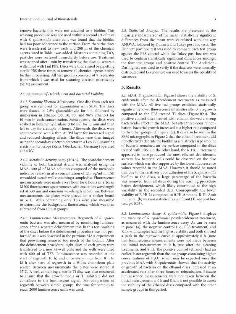
3.3. MAA: P. aeruginosa. Figure 5 displays the viability ofP. aeruginosa after the debridement treatments as measured
with the MAA. The Ti discs treated with debridement solu-tions containing the higher concentration ofH
2O2all showed
a significantly lower fluorescence signal at six hours afterdebridement compared to the Ti discs treated with onlyPBS; see Figure 5(b). An assessment time of 6 h was choseninstead of 3 h as was used with S. epidermidis due to therelatively slower growth rate of the P. aeruginosa bacteria.It is notable that the positive control ethanol did not show
6 International Journal of Biomaterials
(a) (b)
Figure 5: Regrowth ofP. aeruginosa after different debridement treatments. (a) Fluorescencemeasured during reincubation after debridementwith different treatments (n=8). (b) Average fluorescence intensity after 6 h regrowth. Groups with an asterisk indicate statistically significantdifference compared to the control PBS (Dunnett post hoc test, p≤ 0.05). Groups with different letters are significantly different (five testgroups compared, Tukey post hoc test, p ≤ 0.05).
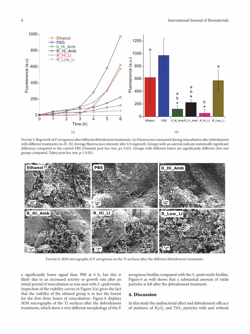
Figure 6: SEM micrographs of P. aeruginosa on the Ti surfaces after the different debridement treatments.
a significantly lower signal than PBS at 6 h, but this islikely due to an increased activity or growth rate after aninitial period of reincubation as was seen with S. epidermidis.Inspection of the viability curves in Figure 5(a) gives the factthat the viability of the ethanol group is in fact the lowestfor the first three hours of reincubation. Figure 6 displaysSEM micrographs of the Ti surfaces after the debridementtreatments, which show a very different morphology of the P.
aeruginosa biofilm compared with the S. epidermidis biofilm.Figure 6 as well shows that a substantial amount of rutileparticles is left after the debridement treatment.
4. Discussion
In this study the antibacterial effect and debridement efficacyof mixtures of H
2O2and TiO
2particles with and without

International Journal of Biomaterials 7
visible light irradiation was performed on S. epidermidis andP. aeruginosa biofilms grown on Ti surfaces. The choice ofthe Gram-positive S. epidermidis was made because of itsgood biofilm forming abilities and this special strain Xen 43provides the ability to monitor viability using luminescencemeasurements, giving a direct indication of bacterial prolif-eration [21, 24].P. aeruginosawas chosen because it is aGram-negative strain, which generally has a stronger resistanceagainst antimicrobials [25]. The higher H
2O2concentration
of 950 mM (3 vol.%) was chosen because it is used clinicallytoday to debride peri-implantitis infected implants whilethe lower concentration of 2 mM (0.006 vol.%) was chosenbecause it has been shown to be the optimal concentrationto degrade an organic substance when combined with rutileparticles under visible light irradiation [22].
It was observed that the debridement mixtures with highconcentrations of H
2O2(950mM) produced a large bubbling
effect when applied on the biofilm-covered surface. The highreduction of biofilm associated with high concentrations ofH2O2seen in the S. epidermidis test assessed with lumi-
nescence may thus be correlated to the oxygen evolutionevidenced by the bubbles that are formed [19–21]. Thesebubbles are caused by H
2O2being decomposed by catalase
produced by the bacteria. The bubbles in turn may createmechanical forces in the solution that remove the attachedbacteria. This effect was most apparent in the luminescencetest with S. epidermidis because more bacteria were presenton the surface prior to the debridement treatments comparedto the MAA test with S. epidermidis. The first debridementtest using S. epidermidis showed that the bacteria werepoorly attached to the Ti surface since a large proportionwas removed with just a gentle rinsing with PBS prior tothe debridement treatment, and thus this prewash was notperformed with the luminescence test. With the addition ofrutile particles therewas an increased bactericidal effectwhenmeasured with luminescence, possibly because the particlesadded an extramechanical force to the bubbling effect, whichis in line with several other studies [19, 21]. However, it couldalso be due to a catalytic effect of the rutile particles on H
2O2
to generate additional ROS [19, 21].The addition of light irradiation at a wavelength of 405
nm did not provide a statistically significant extra effect,although a trend for increased effect was seen in the MAAtests with both bacteria species when combined with ahigh H
2O2concentration. Theoretically this wavelength has
enough energy to induce photocatalysis in the rutile particlesused in this study, which could produce additional ROS. Thelack of a clear effect may be because hydroxyl radicals, whichare the main bactericidal ROS created in photocatalysis,are very reactive and consequently would have a reactiondistance in themagnitude ofmicrometers [10, 21].This wouldlimit the effect to the immediate surroundings of the particles,whichmay not be close enough to themajority of the bacteriabased on the particle concentrations used in this study. In theluminescence experiments, no effect of irradiation was seen,but this may partially be due to the fact that the mechanicaldebridement from the evolved oxygen bubbles removed themajority of the biofilm and with it the overlaying rutileparticles.
Ethanol had a stronger effect on S. epidermidis in theMAA experiment compared to P. aeruginosa. The smallereffect against P. aeruginosa is expected since this strain ismore resistant against ethanol compared to S. epidermidis[26]. In all tests, biofilms treated with ethanol showed anincreased activity or regrowth after a delay period duringreincubation. This highlights the advantages of the otherdebridement treatments used in this study.
In summary, H2O2at a concentration of 950 mM proved
to be the strongest contribution to the debridement and bac-tericidal effect of the cleaning techniques tested in this study.The addition of rutile particles may have additional benefitand was shown to have a statistically significant effect in onetest with S. epidermidis. Limited evidence of the catalyticeffect of visible light irradiation was seen, but effects wererelatively small and statistically insignificant. Further studieswith clinically relevant and multispecies biofilm should beperformed to confirm these results. The debridement of den-tal implants with H
2O2and rutile particles under visible light
irradiation can be used as an alternative or complementarytechnique to presently used methods such as mechanicaldebridement or chemical disinfection in the treatment ofperi-implantitis.
Data Availability
The data used to support the findings of this study areincluded within the article.
Conflicts of Interest
The authors declare that they have no conflicts of interest.
Acknowledgments
The authors acknowledge David Wiedmer (Department ofBiomaterials, Oslo University) for grit blasting and acidetching of the Ti discs, as well as providing S. epidermidisXen 43 at no cost. This research was funded by Vinnova,Sweden’s innovation agency, through the Eurostar programEureka, Grant no. [E!8320].
References
[1] N. P. Lang, T. Berglundh, and Working Group 4 of the SeventhEuropeanWorkshop on Periodontology, “Periimplant diseases:where are we now?—consensus of the Seventh EuropeanWork-shop on Periodontology,” Journal of Clinical Periodontology, vol.38, supplement 11, pp. 178–181, 2011.
[2] M. A. Atieh, N. H. M. Alsabeeha, C. M. Faggion Jr., and W. J.Duncan, “The frequency of peri-implant diseases: a systematicreview and meta-Analysis,” Journal of Periodontology, vol. 84,no. 11, pp. 1586–1598, 2013.
[3] C.-T. Lee, Y.-W.Huang, L. Zhu, andR.Weltman, “Prevalences ofperi-implantitis and peri-implant mucositis: systematic reviewand meta-analysis,” Journal of Dentistry, vol. 62, pp. 1–12, 2017.
[4] J. Derks and C. Tomasi, “Peri-implant health and disease. Asystematic review of current epidemiology,” Journal of ClinicalPeriodontology, vol. 42, supplement 16, pp. S158–S171, 2015.
8 International Journal of Biomaterials
[5] A. Han, J. K. H. Tsoi, F. P. Rodrigues, J. G. Leprince, and W.M. Palin, “Bacterial adhesion mechanisms on dental implantsurfaces and the influencing factors,” International Journal ofAdhesion and Adhesives, vol. 69, pp. 58–71, 2016.
[6] J. W. Costerton, P. S. Stewart, and E. P. Greenberg, “Bacterialbiofilms: a common cause of persistent infections,” Science, vol.284, no. 5418, pp. 1318–1322, 1999.
[7] G. R. Persson, E. Samuelsson, C. Lindahl, and S. Renvert,“Mechanical non-surgical treatment of peri-implantitis: Asingle-blinded randomized longitudinal clinical study. II.Microbiological results,” Journal of Clinical Periodontology, vol.37, no. 6, pp. 563–573, 2010.
[8] R. Burgers, C. Witecy, S. Hahnel, and M. Gosau, “The effect ofvarious topical peri-implantitis antiseptics on Staphylococcusepidermidis, Candida albicans, and Streptococcus sanguinis,”Archives of Oral Biolog, vol. 57, no. 7, pp. 940–947, 2012.
[9] M. R. Hoffmann, S. T. Martin, W. Y. Choi, and D. W.Bahnemann, “Environmental applications of semiconductorphotocatalysis,”Chemical Reviews, vol. 95, no. 1, pp. 69–96, 1995.
[10] Y. Cai, M. Strømme, A. Melhus, H. Engqvist, and K. Welch,“Photocatalytic inactivation of biofilms on bioactive dentaladhesives,” Journal of Biomedical Materials Research Part B:Applied Biomaterials, vol. 102, no. 1, pp. 62–67, 2014.
[11] E. Unosson, E. K. Tsekoura, H. Engqvist, and K. Welch, “Syner-getic inactivation of Staphylococcus epidermidis and Strepto-coccus mutansin a TiO2/H2O2/UV system,” Biomatter, vol. 3,no. 4, 2013.
[12] F. Rupp, M. Haupt, H. Klostermann et al., “Multifunctionalnature of UV-irradiated nanocrystalline anatase thin films forbiomedical applications,” Acta Biomaterialia, vol. 6, no. 12, pp.4566–4577, 2010.
[13] D. O. Scanlon, C. W. Dunnill, J. Buckeridge et al., “Band align-ment of rutile and anatase TiO
2,” Nature Materials, vol. 12, no.
9, pp. 798–801, 2013.[14] A. Fujishima, X. Zhang, and D. A. Tryk, “TiO
2photocatalysis
and related surface phenomena,” Surface Science Reports, vol.63, no. 12, pp. 515–582, 2008.
[15] P. C.Maness, S. Smolinski, D.M. Blake, Z.Huang, E. J.Wolfrum,and W. A. Jacoby, “Bactericidal activity of photocatalytic TiO2reaction: Toward an understanding of its killing mechanism,”Applied and EnvironmentalMicrobiology, vol. 6, pp. 4094–4098,1999.
[16] Y. Kakuma, A. Y. Nosaka, and Y. Nosaka, “ Difference in TiO ,”Physical Chemistry Chemical Physics, vol. 17, no. 28, pp. 18691–18698, 2015.
[17] T. Hirakawa, K. Yawata, and Y. Nosaka, “Photocatalytic reactiv-ity for O∙
2and OH∙ radical formation in anatase and rutile TiO2
suspension as the effect of H2O2addition,” Applied Catalysis A:
General, vol. 325, pp. 105–111, 2007.[18] J. Zhang and Y. Nosaka, “ Mechanism of the OH Radical Gen-
eration in Photocatalysis with TiO ,” The Journal of PhysicalChemistry C, vol. 118, no. 20, pp. 10824–10832, 2014.
[19] E. Henderson, S. Schneider, F. C. Petersen et al., “Chemicaldebridement of contaminated titanium surfaces: An in vitrostudy,”Acta Odontologica Scandinavica, vol. 71, no. 3-4, pp. 957–964, 2013.
[20] E. Gustumhaugen, J. Lonn-Stensrud, A. A. Scheie, S. P. Lyn-gstadaas, A. Ekfeldt, and S. Taxt-Lamolle, “Effect of chemicalandmechanical debridement techniques on bacterial re-growthon rough titanium surfaces: An in vitro study,” Clinical OralImplants Research, vol. 25, no. 6, pp. 707–713, 2014.
[21] D. Wiedmer, F. C. Petersen, J. Lonn-Stensrud, and H. Tiainen,“Antibacterial effect of hydrogen peroxide-titanium dioxidesuspensions in the decontamination of rough titanium sur-faces,” Biofouling, vol. 33, no. 6, pp. 451–459, 2017.
[22] O. Janson, E. Unosson,M. Strømme,H. Engqvist, andK.Welch,“Organic degradation potential of a TiO2/H2O2/UV–vis sys-tem for dental applications,” Journal of Dentistry, vol. 67, pp. 53–57, 2017.
[23] L. Korosi, M. Prato, A. Scarpellini et al., “H2O2-assisted pho-tocatalysis on flower-like rutile TiO2 nanostructures: Rapiddye degradation and inactivation of bacteria,” Applied SurfaceScience, vol. 365, pp. 171–179, 2016.
[24] C. Vuong, S. Kocianova, J. Yu, J. L. Kadurugamuwa, and M.Otto, “Development of real-time in vivo imaging of device-related Staphylococcus epidermidis infection in mice and influ-ence of animal immune status on susceptibility to infection,”TheJournal of Infectious Diseases, vol. 198, no. 2, pp. 258–261, 2008.
[25] P. S. Stewart and J. W. Costerton, “Antibiotic resistance ofbacteria in biofilms,”The Lancet, vol. 358, no. 9276, pp. 135–138,2001.
[26] P. D. Lister, D. J. Wolter, and N. D. Hanson, “Antibacterial-resistant Pseudomonas aeruginosa: clinical impact and complexregulation of chromosomally encoded resistance mechanisms,”Clinical Microbiology Reviews, vol. 22, no. 4, pp. 582–610, 2009.

CorrosionInternational Journal of
Hindawiwww.hindawi.com Volume 2018
Advances in
Materials Science and EngineeringHindawiwww.hindawi.com Volume 2018
Hindawiwww.hindawi.com Volume 2018
Journal of
Chemistry
Analytical ChemistryInternational Journal of
Hindawiwww.hindawi.com Volume 2018
Scienti�caHindawiwww.hindawi.com Volume 2018
Polymer ScienceInternational Journal of
Hindawiwww.hindawi.com Volume 2018
Hindawiwww.hindawi.com Volume 2018
Advances in Condensed Matter Physics
Hindawiwww.hindawi.com Volume 2018
International Journal of
BiomaterialsHindawiwww.hindawi.com
Journal ofEngineeringVolume 2018
Applied ChemistryJournal of
Hindawiwww.hindawi.com Volume 2018
NanotechnologyHindawiwww.hindawi.com Volume 2018
Journal of
Hindawiwww.hindawi.com Volume 2018
High Energy PhysicsAdvances in
Hindawi Publishing Corporation http://www.hindawi.com Volume 2013Hindawiwww.hindawi.com
The Scientific World Journal
Volume 2018
TribologyAdvances in
Hindawiwww.hindawi.com Volume 2018
Hindawiwww.hindawi.com Volume 2018
ChemistryAdvances in
Hindawiwww.hindawi.com Volume 2018
Advances inPhysical Chemistry
Hindawiwww.hindawi.com Volume 2018
BioMed Research InternationalMaterials
Journal of
Hindawiwww.hindawi.com Volume 2018
Na
nom
ate
ria
ls
Hindawiwww.hindawi.com Volume 2018
Journal ofNanomaterials
Submit your manuscripts atwww.hindawi.com