DDH from Infancy to Skeletal Maturity: Safe Swaddling to ... · PDF fileSafe Swaddling to...
24
DDH from Infancy to Skeletal Maturity: John R. Faust, M.D. Safe Swaddling to Periacetabular Osteotomy Disclosures John Faust, M.D., has no relationships with commercial companies to disclose (I asked Dr. Conrad for some money but he just laughed) Learning Objectives At the end of this presentation the participant will be able to: 1. Describe: • Normal development and anatomy of the hip • How instability in the hip leads to dysplasia • The clinical and radiographic findings in DDH 2. Outline the current treatment options for DDH in the: • Newborn • Young child • Skeletally mature patient Where 10 years of education and training takes you Salt Lake City, UT Washington, DC Rochester, NY San Antonio, TX Atlanta, GA Angie, Sydney (8), Holden (6), Adelaide (4), Everett (2) My daughter, Sydney
Transcript of DDH from Infancy to Skeletal Maturity: Safe Swaddling to ... · PDF fileSafe Swaddling to...

DDH from Infancy to Skeletal Maturity:
John R. Faust, M.D.
Safe Swaddling to Periacetabular Osteotomy
Disclosures John Faust, M.D., has no relationships with commercial companies to disclose (I asked Dr. Conrad for some money but he just laughed)
Learning Objectives At the end of this presentation the participant will be able to: 1. Describe:
• Normal development and anatomy of the hip • How instability in the hip leads to dysplasia • The clinical and radiographic findings in DDH
2. Outline the current treatment options for DDH in the: • Newborn • Young child • Skeletally mature patient
Where 10 years of education and training takes you
Salt Lake City, UT
Washington, DC
Rochester, NY
San Antonio, TX
Atlanta, GA
Angie, Sydney (8), Holden (6), Adelaide (4), Everett (2)
My daughter, Sydney

Trial stopped early due to bracing efficacy
Treatment success (< 50° curve at “skeletal maturity”) • Bracing: 72% • Observation: 48% Hours of brace wear associated with success rate • 0-6 hours/day: 41% success rate • Similar to observation group success rate of 48%
• 6.1-12.8 hours/day: 72% success rate • ≥ 12.9 hours/day: 90-93% success rate
The Normal Hip
• Normal acetabular version • Appropriate coverage • Not too little • Not too much
• Thin, horizontal weight-bearing zone • Round femoral head • Congruous surfaces • Symmetric, wide cartilage space
(sourcil)
The Normal Hip
• Intrinsically stable • Compare to the shoulder joint
The Normal Hip
• Intrinsically stable • Compare to the shoulder joint
• Great mobility: • Greater range of motion than needed
for normal activity
The Normal Hip
• Intrinsically stable • Compare to the shoulder joint
• Great mobility: • Greater range of motion than needed
for normal activity
• Narrow physiologic loading range • Limited peak load tolerance • Limited shear tolerance
Instability Normal Impingement
Philippe Petit Notre-Dame, 1971
FEMUR
Two main types of pathology Instability • Too little coverage • Shearing of cartilage surface • Subluxation / dislocation
Impingement • Too much coverage • Abnormal contact during physiologic motion • Can be femoral, acetabular, or both in origin
Hip Instability: biomechanics • Static overload � local stress concentration • Dynamic instability � shear forces
The Bottom Line Most osteoarthritis in the hip has a mechanical etiology • Instability and impingement are the bad actors • Symptoms may be absent before soft tissue damage occurs • Early treatment of these conditions can prolong the life of the hip
Hip Pathology
Instability • Developmental dysplasia of the
hip (DDH) • Neuromuscular hip dysplasia • Connective tissue laxity • Down syndrome
• Traumatic • Iatrogenic – osteotomy
Impingement • Femoroacetabular impingement (FAI) • Legg-Calve-Perthes disease (LCP) • Slipped capital femoral epiphysis
(SCFE) • Avascular necrosis (AVN) • ?Skeletal dysplasia • Abnormal cartilage cell function
Bone tumors oneeeeeeeeeee tumummummumumummummmmmmmmmmu o

What is Dysplasia? Definition of hip dysplasia: • Abnormal development of the femoral head and
acetabulum
dys - plasia
abnormal growth
Definition of Hip Dysplasia Abnormal development of the femoral head and acetabulum • Cartilage cell function: • Skeletal dysplasia • Abnormal muscle forces: • Cerebral palsy, spaticity • Myelomeningocele • Connective tissue disorders • Arthrogryposis • Down syndrome • External environment • DDH
DDH: definition Developmental Hip dysplasia • Femoral head and acetabulum develop abnormally due to an
abnormal relationship driven by the external environment • Starts intrauterine environment • Continues after birth
• Usually an otherwise healthy child • Includes: • Several radiographic abnormalities of inadequate formation of the
acetabulum • Partial dislocation / subluxation • Frank dislocation
• These findings may not be present at birth
DDH: spectrum of dysplasia
DDH: classification Dysplastic • Abnormal femoral head and/or acetabulum without signs of instability
Subluxatable • Rests in reduced position and can be subluxated with stress Dislocatable • Rests in reduced position and can be dislocated with stress
Reducible • Rests in a dislocated position and can be reduced
Irreducible • Rests in a dislocated position and cannot be reduced
Excludes teratologic hip dislocations • Myelodysplasia • Arthrogryposis
DDDDDDDH: classificationDysplastic• Abnormal femoral head and/or acetabulum without signs of instability
Subluxatable• Rests in reduced position and can be subluxated with stressDislocatable• Rests in reduced position and can be dislocated with stress
Reducible• Rests in a dislocated position and can be reduced
Irreducible• Rests in a dislocated position and cannot be reduced
Excludes teratologic hip dislocations• Myelodysplasia• Arthrogryposis
= Instability tests negative
Barlow +
Ortolani +
Hip Development

Hip Development The femoral head and acetabulum need each other to develop normally • Growth normalizes the hip, …in the right
environment • Our treatments are try to create that environment
Most growth occurs before 4 years old • The older the child, the harder it is to create the
right environment
Hip Development: dysplasia Normal acetabulum • Deep, wide acetabulum • Labrum extends lateral coverage • Thin capsule extends lateral • Normal pulvinar
Acetabular dysplasia • Shallow, narrow acetabulum • Thickened acetabular cartilage • Inturned hypertrophic labrum (limbus) • Thick capsule extends upward • Lateral growth plate is slanted upward • At the margin of the roof, periosteal bone growth is
retarded
Ponseti, J Bone Joint Surg Am, 1978;60:586
Hip Dysplasia: deformity Acetabulum • Oblique, shallow • Widened tear drop/medial wall • Anteversion
Femur • Anteversion • Valgus (neck-shaft angle)
Hip Dysplasia: deformity
Hip Dysplasia: deformity Hip Dysplasia: deformity
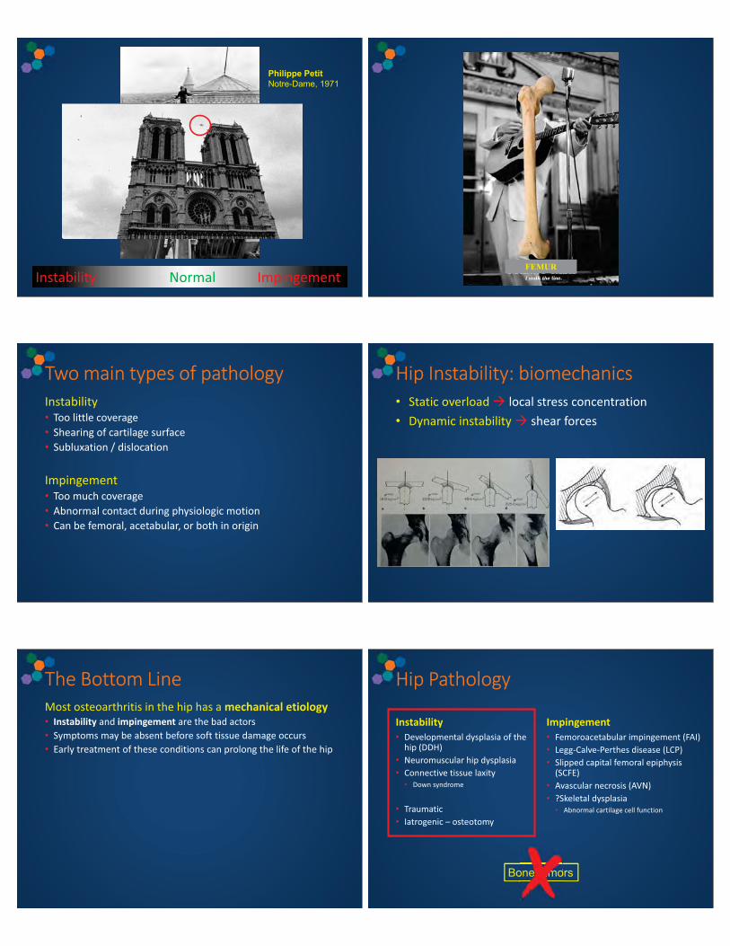
DDH: risk factors First born • More often breech, tighter uterus/abdominal contents
Female • Mother’s / endogenous estrogens
Breech • 17.3-32% of DDH children present breech
Family history
More often breech, tighter uter
Female• Mother’s / endogenous estroge
Breech• 17.3-32% of DDH children prese
Family history
DDH: risk factors Sex • Female > male • Relaxin sensitivity
Breech • 20-30% of DDH • 3% of all deliveries • Frank breech Family history
Neuromuscular abnormalities
Intra-uterine crowding • Primigravida • Left side • Oligohydramnios • Increased birth weight • Multiple births • Metatarsus adductus • Torticollis • Calcaneovalgus foot
Extra-uterine crowding • Swaddling
• Relaxin sensitivity
Breech• 20-30% of DDH• 3% of all deliveries• Frank breech
Family history
Neuromuscular abnormalities
•••••••
E•
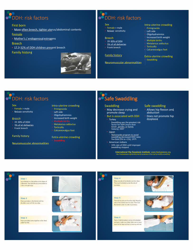
Safe Swaddling Swaddling • May decrease crying and
promote sleep • But is associated with DDH • Turkey • Swaddling was the greatest risk
factor for DDH (more than breeh, gender, or family history), 2007
• Japan • Nationwide program to avoid
swaddling decreased DDH rates five-fold (3% to <1%), 1975
• American indians • 33% rate of DDH until improper
swaddling stopped
Safe swaddling • Allows hip flexion and
abduction • Does not promote hip
dysplasia
SSSSafe SwaddlingSwaddling
d dSafe swaddling
ll h fl dMay decrease crying andpromote sleep• But is associated with DDH• Turkey• Swaddling was the greatest risk
factor for DDH (more than breeh, gender, or family history), 2007
• Japan• Nationwide program to avoid
swaddling decreased DDH rates five-fold (3% to <1%), 1975
• American indians• 33% rate of DDH until improper
swaddling stopped
Allows hip flexion and abduction
• Does not promote hip dysplasia
International Hip Dysplasia Institute: www.hipdysplasia.org http://hipdysplasia.org/developmental-dysplasia-of-the-hip/hip-healthy-swaddling

All done Physical Examination DDH is an evolving process • The physical findings on clinical examination can change
with time
Physical Examination “Orthopaedic newborn exam” • Same for all babies, for any concern • The part you are interested in most, do it last
• Overall • Head and neck • Spine • Lower extremities • Feet • Hips
Preparation and set-up: • Firm surface • Remove all clothing except diaper • Should be calm and not crying
Physical Examination Hard signs • Instability tests – “clunks” • Barlow • Ortolani • May be negative even if the hip is dysplastic • Child not calm • Soft tissue contractures • Acetabular dysplasia • Irreducible dislocation
Physical Examination Soft signs (not specific to DDH) • Limited hip abduction • Positive Galeazzi sign • Asymmetric limb lengths • Asymmetric thigh/groin creases • If walking: • Scoliosis • Asymmetric in-/out-toeing • Hyperlordosis • Waddling gait
Physical Examination General observation: • Café au lait spots • Movement in extremities • Muscle tone
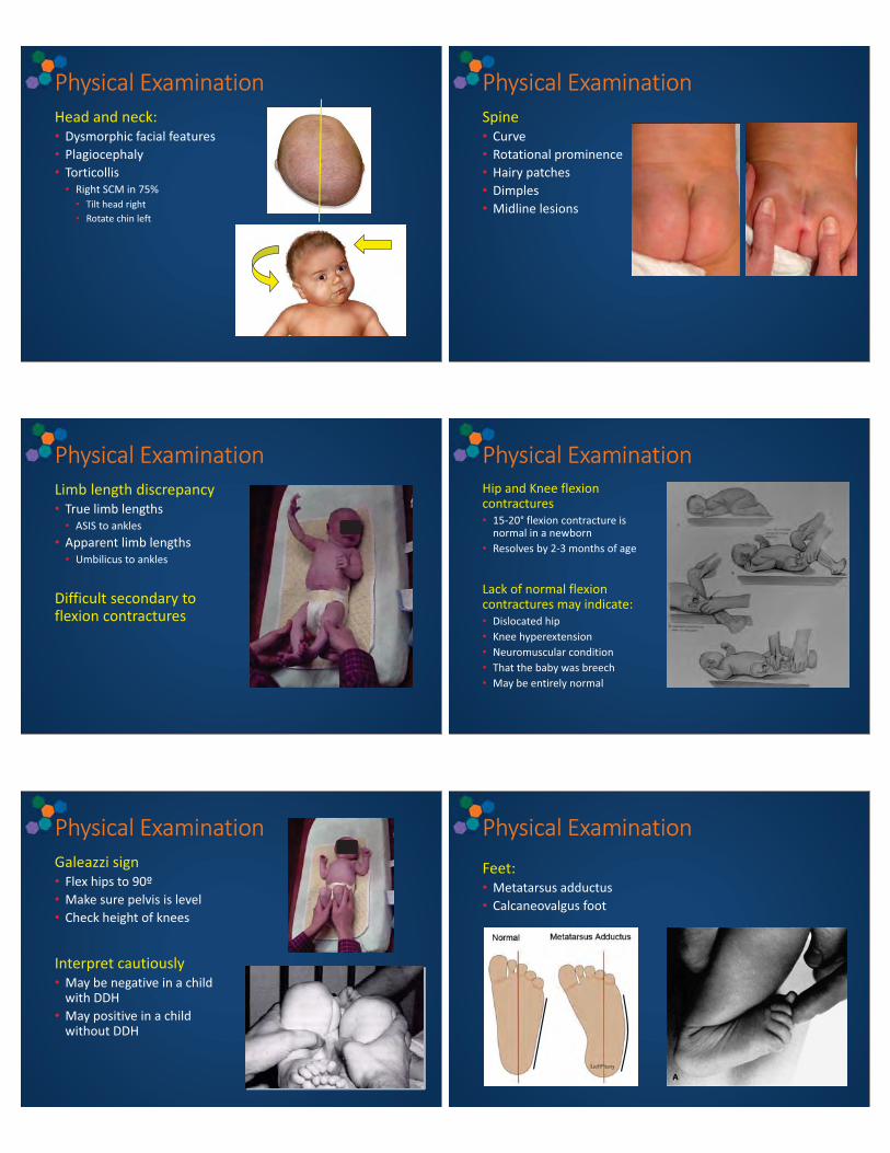
Physical Examination Head and neck: • Dysmorphic facial features • Plagiocephaly • Torticollis • Right SCM in 75% • Tilt head right • Rotate chin left
Physical Examination Spine • Curve • Rotational prominence • Hairy patches • Dimples • Midline lesions
Physical Examination Limb length discrepancy • True limb lengths • ASIS to ankles • Apparent limb lengths • Umbilicus to ankles
Difficult secondary to flexion contractures
Physical Examination Hip and Knee flexion contractures • 15-20° flexion contracture is
normal in a newborn • Resolves by 2-3 months of age
Lack of normal flexion contractures may indicate: • Dislocated hip • Knee hyperextension • Neuromuscular condition • That the baby was breech • May be entirely normal
Physical Examination Galeazzi sign • Flex hips to 90º • Make sure pelvis is level • Check height of knees
Interpret cautiously • May be negative in a child
with DDH • May positive in a child
without DDH
Feet: • Metatarsus adductus • Calcaneovalgus foot
Physical Examination
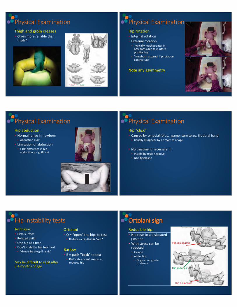
Physical Examination Thigh and groin creases • Groin more reliable than
thigh?
Physical Examination Hip rotation • Internal rotation • External rotation • Typically much greater in
newborns due to in utero positioning
• “Newborn external hip rotation contracture”
Note any asymmetry
Physical Examination Hip abduction: • Normal range in newborn • Abduction >60° • Limitation of abduction • >10° difference in hip
abduction is significant
Physical Examination Hip “click” • Caused by synovial folds, ligamentum teres, iliotibial band • Usually disappear by 12 months of age • No treatment necessary if: • Instability tests negative • Not dysplastic
Hip instability tests Technique: • Firm surface • Relaxed child • One hip at a time • Don’t grab the leg too hard • “Gentle like the girlfriends”
May be difficult to elicit after 3-4 months of age
Ortolani • O = “open” the hips to test • Reduces a hip that is “out”
Barlow • B = push “back” to test • Dislocates or subluxates a
reduced hip
Ortolani sign Reducible hip: • Hip rests in a dislocated
position • With stress can be
reduced • Flexion • Abduction • Fingers over greater
trochanter
OOOOOrtolani signReducible hip:
Hip rests in a dislocatedpositionWith stress can be reduced• Flexion• Abduction• Fingers over greater
trochanter
• HHp• W
Hip dislocated
Hip reduces
Hip dislocates

Ortolani sign Barlow sign Dislocatable or subluxatable hip • Hip reduced in the
resting position • With stress can be
dislocated/subluxated • Flexion • Adduction • Axial loading
Hip reduced
Hip dislocates
Dislocatable: “clunk”
vs. subluxatable: “glide,” “slide,” or “loose”
Barlow sign Terminology “Ortolani positive” hip Dislocated hip • Not expected to
spontaneously resolve
“Barlow positive” hip Dislocatable hip • May stabilize spontaneously
or with treatment • May convert to dislocated
without treatment
Physical Examination DDH is an evolving process • The physical findings on clinical examination can change
with time
Clinical Examination Children / adolescents – different exam than newborns • Gait • Waddling (Trendelenburg gait)
• Stance • Scoliosis: Adam’s forward bend test • Pelvic obliquity (standing limb length) • Single leg stance (Trendelenburg sign)
• Lower extremities: • Galeazzi sign • Prone galeazzi sign
• Hip: • Tenderness • Ortolani/Barlow – depends on size of the patient • ROM • Supine: flexion / abduction / internal and external rotation in flexion • Prone rotational profile • May be more than normal

Clinical Examination Gait and stance: • Hyperlordosis • Waddling gait • Trendelenburg
…think bilateral DDH
Imaging Ultrasound • What to order: bilateral hip ultrasound (static and dynamic) • When to order: <4 months of age (femoral head still
cartilaginous) • Optimal timing for screening is controversial • False positives higher when performed early • 6-8 weeks of age
• Do not need if Ortolani positive in the nursery
X-ray • What to order: AP Pelvis • When to order: >4 months of age • Femoral head ossifying and now visible
Ultrasound Diagnosis should be made by clinical examination and enhanced by US in questionable cases Ideal for: • Screening • Guiding reduction • Checking reduction on follow-up • Reducing radiation exposure
Ultrasound
Ultrasound
α
β
Ultrasound
α
α

Ultrasound: dynamic examination Ortolani and Barlow maneuvers during ultrasound • 4 to 6 mm movement is considered normal in the first few
days of life
Technique and technologist dependent
Radiographs AP pelvis • Standard
Sometimes: • Frog pelvis • Von Rosen view • 45° abduction, 25° internal rotation • Femoral shaft should point towards triradiate cartilage
Where’s the bone?
Radiographs Where’s the bone? It is still cartilage
Radiographs
Femoral head ossification Normally develops between 4 – 9 months of age
Shenton’s line Not as reliable in patients <4 years old

Useful in patients <8 yo
What should the numbers be? • Newborn < 35° • 2 yo < 20–21° • 5 yo < 17°
Very senstive to positioning, interpret with caution • Newborn: >40° significant • 2 yrs: normal <20° • Maturity: >20° is worrisome
Acetabular index (AI) Lateral Center Edge Angle of Wiberg Measure after age 5 What should the numbers be? • 5-8 yo: 19° • 9-12 yo: 25° • 13+ yo: 26-40°
Be careful interpreting the radiographs
Reynolds, J Bone Joint Surg Br, 1999;81(2) • Described two radiographic features of
acetabular retroversion • Cross-over sign • Posterior wall sign
• However, they are reproduced by simply tilting the pelvis anteriorly
Normal Retroversion?
Natural History Newborns with hip instability: • Yamamuro and Doi • 52 hips • 25% (3/12) dislocated hips at birth were normal at 5 months • 57% (24/42) subluxatable hips were normal • Barlow • 88% of Barlow (+) hips stabilized during first 2 months
Some hips correct themselves, and some do not • Subluxatable hips frequently correct themselves • 2 week observation before treatment
• Unpredictability leads to over treatment of unstable hips
Natural History Adults with DDH • False acetabulum • Well developed • Degenerative joint disease • Clinical disability
• Absent • No degenerative disease • May function well
• Low back pain • Secondary to hyperlordosis
• Scoliosis • Limb-length inequality • Ipsilateral knee deformity and pain
The right environment for growth Arthur Boorman
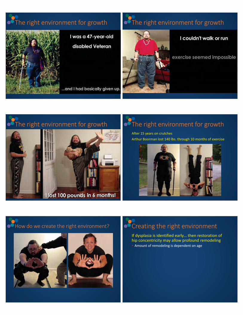
The right environment for growth The right environment for growth
The right environment for growth The right environment for growth After 15 years on crutches Arthur Boorman lost 140 lbs. through 10 months of exercise
How do we create the right environment? Creating the right environment If dysplasia is identified early… then restoration of hip concentricity may allow profound remodeling • Amount of remodeling is dependent on age

Treatment: general approach Infant: maintain reduction • Unstable hip (dislocatable/subluxatable, dislocatable but reducible) • Observation • Abduction bracing: Pavlik, Rhino
• Irreducible hip • Medial open reduction or wait
Older child: obtain reduction and maintain • Anterior open reduction (obtain) • Pelvic osteotomy (maintain) • Help acetabular remodeling
• Femoral shortening • prevent AVN
Skeltally mature: reconstruct • Ganz osteotomy • Colonna interposition arthroplasty • Pelvic support osteotomy • Total hip replacement
Treatment DDH has many aspects and remains a challenge It’s easiest to remember a few rules : • < 6mo: Pavlik • 6mo-18mo: closed Reduction and casting • If that fails, open reduction and casting • >18mo: anterior open reduction, pelvic osteotomy, femoral
shortening • Older hip dysplasia: • depends upon the patient s maturity • and the specific problem for either repositioning or salvage.
Treatment <6 months of age Observation • Some hips do stabilize without treatment • What about the ones that don’t?
Multi-diapering • Not effective except may help provide communication
between health care providers • May help some “at risk” hips
Abduction orthosis • Pavlik • Rhino cruiser brace
Pavlik harness Achieves reduction by: • preventing extension and
adduction • Allowing flexion, abduction
and motion
Success of Pavlik harness Decreased with • Diagnosis > 6 weeks • Bilateral • Acetabular index >35°
• Inappropriate indication • Muscle imbalance, joint
stiffness, ligamentous laxity • Persistence of inadequate
treatment • Pavlik harness disease
• Application related • Patient related • Proper instruction to parents • Keep it on 24 hrs/day until
stable • No swaddling, no side lying,
loose fitting clothing • Weighs about 4 ounces
Treatment
If Pavlik fails: • Abduction orthosis • Closed reduction • Open reduction

Treatment over 6 months of age Closed reduction (6-18 months of age) • Under anesthesia • Arthrogram, adductor tenotomy, spica cast • usually changed 2 more times 6 weeks apart
Open reduction (>12 months of age) • Femoral osteotomy • Pelvic osteotomy
Risks • AVN, re-dislocation, continued dysplasia • Anesthesia related risks
Older Child – Obtain and Maintain If you are going to reduce the hip, you must hit a home run. A good DDH open reduction and stabilization remains one of the more difficult orthopaedic operations. Reduction must be meticulous and gentle. Maintain the reduction with judicious osteotomies or soft tissue procedures.
The Big Unknown – What surgery in which patients improves on the natural history?
Unlike scoliosis, clubfeet, Blount s, SCFE, there are MANY DDH disasters. Only perfect results result in perfect hips. Sometimes, not even then. Once the acetabulum is fully developed, there is probably no role for reduction. Until then, the upper age limit is not well established.
8 year old girl, asymptomatic – what to do?
Possibilities Leave Alone OBTAIN • Medial open reduction • Anterior open reduction • Open reduction with shortening
MAINTAIN • Capsular plication • Femoral osteotomies • Pelvic osteotomies
What do I do?
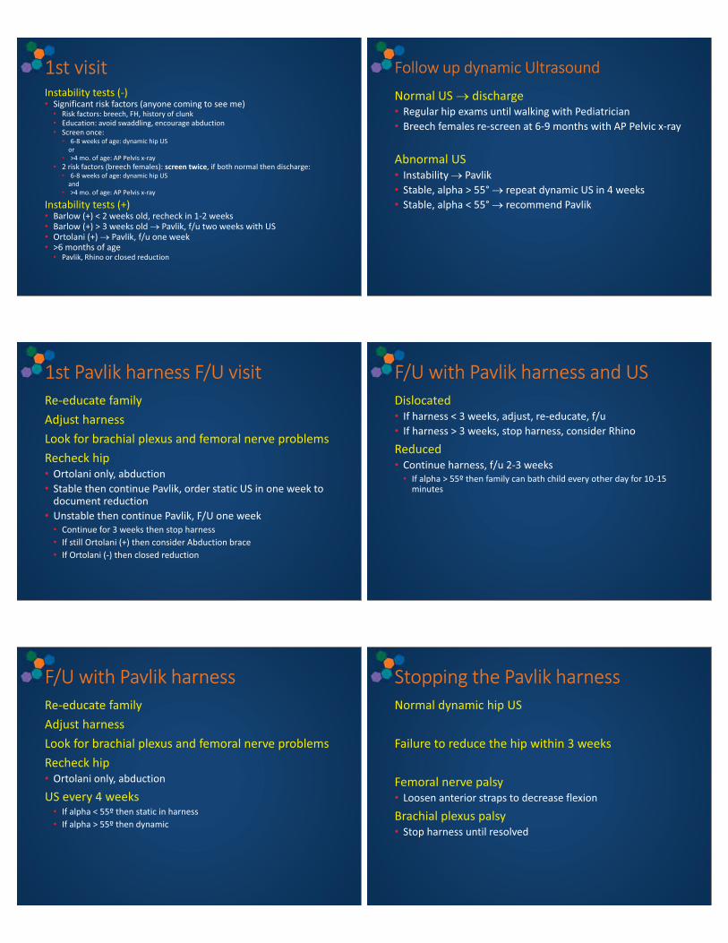
1st visit Instability tests (-) • Significant risk factors (anyone coming to see me) • Risk factors: breech, FH, history of clunk • Education: avoid swaddling, encourage abduction • Screen once: • 6-8 weeks of age: dynamic hip US
or • >4 mo. of age: AP Pelvis x-ray
• 2 risk factors (breech females): screen twice, if both normal then discharge: • 6-8 weeks of age: dynamic hip US
and • >4 mo. of age: AP Pelvis x-ray
Instability tests (+) • Barlow (+) < 2 weeks old, recheck in 1-2 weeks • Barlow (+) > 3 weeks old � Pavlik, f/u two weeks with US • Ortolani (+) � Pavlik, f/u one week • >6 months of age • Pavlik, Rhino or closed reduction
Follow up dynamic Ultrasound
Normal US � discharge • Regular hip exams until walking with Pediatrician • Breech females re-screen at 6-9 months with AP Pelvic x-ray
Abnormal US • Instability � Pavlik • Stable, alpha > 55° � repeat dynamic US in 4 weeks • Stable, alpha < 55° � recommend Pavlik
1st Pavlik harness F/U visit Re-educate family Adjust harness Look for brachial plexus and femoral nerve problems Recheck hip • Ortolani only, abduction • Stable then continue Pavlik, order static US in one week to
document reduction • Unstable then continue Pavlik, F/U one week • Continue for 3 weeks then stop harness • If still Ortolani (+) then consider Abduction brace • If Ortolani (-) then closed reduction
F/U with Pavlik harness and US Dislocated • If harness < 3 weeks, adjust, re-educate, f/u • If harness > 3 weeks, stop harness, consider Rhino
Reduced • Continue harness, f/u 2-3 weeks • If alpha > 55º then family can bath child every other day for 10-15
minutes
F/U with Pavlik harness Re-educate family Adjust harness Look for brachial plexus and femoral nerve problems Recheck hip • Ortolani only, abduction
US every 4 weeks • If alpha < 55º then static in harness • If alpha > 55º then dynamic
Stopping the Pavlik harness Normal dynamic hip US Failure to reduce the hip within 3 weeks Femoral nerve palsy • Loosen anterior straps to decrease flexion
Brachial plexus palsy • Stop harness until resolved

If the Pavlik harness fails… Take a break and re-try the harness Change to Rhino abduction brace • Have to have sufficient motion • Ortolani positive? • Only use for 2-3 weeks without documented reduction
Closed reduction
Obstacles to reduction Capsular constriction Tight transverse acetabular ligament Hypertrophic ligamentum teres Hypertrophic pulvinar Inverted labrum Iliopsoas tendon Adductor longus
Obstacles
Infolded labrum Capsular constriction
Preliminary traction Traction for ~2 weeks to stretch out tight tissues Hips flexed 45 to 90° Hips abducted 20 to 30° Complications include skin loss and limb ischemia Several studies have found no increase in AVN in closed reductions without preliminary traction Some use traction as a method of reduction by gradually increasing abduction
Arthrogram 18 or 20 gauge spinal needle Medial approach just posterior to adductor longus tendon Under fluoroscopy, needle aimed toward ipsilateral shoulder, 30-45º posteriorly Needle should enter hip joint on the medial/inferior aspect of femoral neck Water soluble contrast injected (1-2 cc)
Arthrogram Straight anterior approach Place tip of needle directly over mid femoral neck Then place needle vertical and insert onto neck
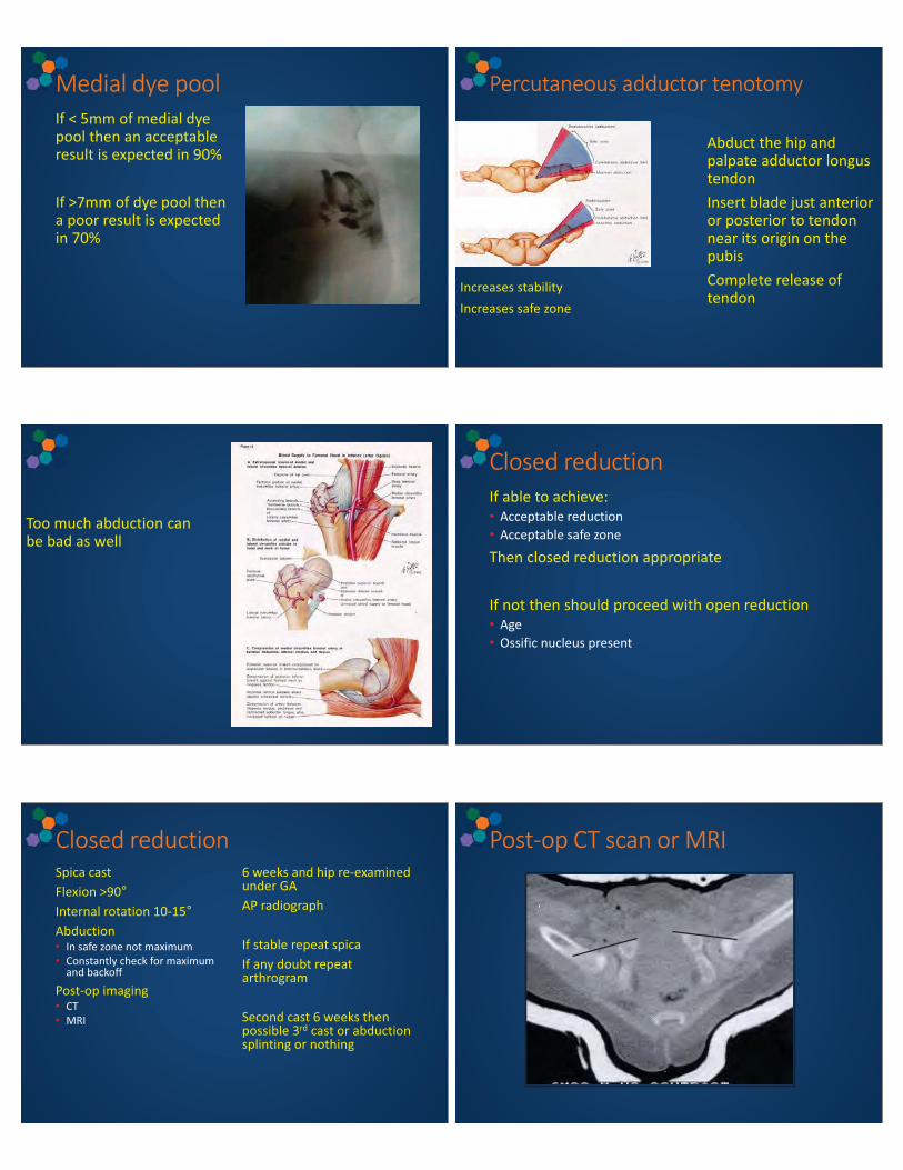
Medial dye pool If < 5mm of medial dye pool then an acceptable result is expected in 90% If >7mm of dye pool then a poor result is expected in 70%
Percutaneous adductor tenotomy
Increases stability Increases safe zone
Abduct the hip and palpate adductor longus tendon Insert blade just anterior or posterior to tendon near its origin on the pubis Complete release of tendon
Too much abduction can be bad as well
Closed reduction If able to achieve: • Acceptable reduction • Acceptable safe zone
Then closed reduction appropriate If not then should proceed with open reduction • Age • Ossific nucleus present
Closed reduction Spica cast Flexion >90° Internal rotation 10-15° Abduction • In safe zone not maximum • Constantly check for maximum
and backoff
Post-op imaging • CT • MRI
6 weeks and hip re-examined under GA AP radiograph If stable repeat spica If any doubt repeat arthrogram Second cast 6 weeks then possible 3rd cast or abduction splinting or nothing
Post-op CT scan or MRI

Open reduction Necessary when closed reduction fails to achieve a concentric reduction Must be able to deal with obstacles to reduction Must be done in a safe fashion to minimize complications
t tttttttttttttoooooooooooooo ooooooooo o oo
Open reduction Medial approach Anterolateral approach Both are able to deal with all obstacles to reduction but medial approach does not allow for capsular plication
Medial approach Useful for children under 12 months • Arthrogryposis
Not recommended in patient without contracture Medial femoral circumflex should be protected AVN around 5% and higher in older patients
Anterolateral approach More exposure Can plicate capsule Better for high dislocations Concurrent pelvic osteotomy Beware of false acetabulum
Anterolateral approach Femoral osteotomies? Used to reduce stress across hip joint • Lessen risk of AVN • Consider in any child over 2 years
old
Should be able to shuck hip 1-2 mm after open reduction, if unable forces may be too high
Incision • Separate lateral • Continue anterolateral
Shortening Derotation Varus

Pelvic Osteotomies? Added coverage and improved stability Some recommend for children over 18 months of age Hip extended, neutral rotation and abduction and if >1/3 head visible add osteotomy
Types of Osteotomies Reshaping • Congruous joint • “wandering acetabulum”
Redirectional • Congruous joint • Does not change shape
Salvage • Incongruous joint • Irreversible cartilage damage
Complications AVN Inadequate reduction Re-dislocation Residual dysplasia
Treatment of mature dysplasia Ganz osteotomy Colonna interposition arthroplasty Pelvic support osteotomy Total hip replacement
Periacetabular Osteotomy (PAO) Described by Ganz in 1988 • Bern, Switzerland
Indication: • Need to re-direct the acetabulum • Triradiate cartilage closed
Ganz, Clin Orthop Relat Res, 1988; 232:26-36 Siebenrock, J Bone Joint Surg Am, 2001;83:449-455
Periacetabular Osteotomy (PAO) Appealing features: • Single incision • Abductor-sparing approach • Less difficult than spherical
osteotomy • More blood supply to fragment • Can do capsullorhaphy and
intracapsular work • Can medialize and lengthen if
necessary • Major multidirectional corrections
possible • Allows nearly limitless correction
• Stable osteotomy leaving posterior column intact • Stable fixation • Early postop function • Better healing in skeletally mature
patients • Avoids ischial nonunion seen with Triple
osteotomy
Ganz, Clin Orthop Relat Res, 1988; 232:26-36 Siebenrock, J Bone Joint Surg Am, 2001;83:449-455

Periacetabular Osteotomy Osteotomies: • Ischium
1. Anterior ischium (infracotyloid groove)
• Pubis 2. Superior pubic ramus • Ilium
3. Ilium 4. Posterior column 5. Ischium
PAO: before and after
PAO: before and after Case example:

14 year old female complains of left hip pain
Oct 2013
Oct 2013
Intra-operative 3 weeks post-op
5 mo. Post-op 5 mo. postop
Some Hips You Can’t Help With enough growth or a young patient we can help • For everything else, there’s arthroplasty

What were the dark ages like for scoliosis treatment? Barbaric things casting and ttraction Like…
Fortunately, we now have modern treatments
Like… ccasting and ttraction Peds Ortho: Kaye Wilkins Travis Murray Sekinat McCormick John Faust • Cell: 210-245-1390 • MARC clinic daily • Appointments: 450-9300
• RBG clinic daily (except Thursday) • Appointments: 358-KIDS
• On-call at UHS a week at a time • Kid’s Bone Phone: 210-450-KIDS • Clinic hours: Allison or Mark – attending right next to them • After hours: attending on call
• Clinic: Allison (patient care coordinator): 210-376-7779 • Office: Imelda (administrative assistant): 210-567-5133
Thank you
Acknowledgements Tim Schrader

![Periacetabular Brucella Osteomyelitis - file.scirp.org · spondylitis, bursitis, tenosynovitis and osteomyelitis [3-6]. Brucella osteomyelitis may appear as a radiolucent area and](https://static.fdocuments.in/doc/165x107/5d52ce1188c993277b8b9aaa/periacetabular-brucella-osteomyelitis-filescirporg-spondylitis-bursitis.jpg)