DAVID S. LOGERSTEDT DAVID A. SCALZITTI KIM L. … pain and... · American Physical Therapy...
83
1 CLINICAL PRACTICE GUIDELINES DAVID S. LOGERSTEDT, PT, PhD • DAVID A. SCALZITTI, PT, PhD KIM L. BENNELL, PT, PhD • RANA S. HINMAN, PT, PhD HOLLY SILVERS-GRANELLI, PT, PhD • JAY EBERT, PhD KAREN HAMBLY, PT, PhD • JAMES L. CAREY, MD, MPH LYNN SNYDER-MACKLER, PT, ScD, FAPTA • MICHAEL J AXE, MD CHRISTINE M. McDONOUGH, PT, PhD Knee Pain and Mobility Impairments: Knee Meniscal/Articular Cartilage Lesions Revision 2017 Clinical Practice Guidelines Linked to the International Classification of Functioning, Disability, and Health from the Orthopaedic Section of the American Physical Therapy Association J Orthop Sports Phys Ther. 2017:47(_).A1-A_. doi:##.####/jospt.####.#### SUMMARY OF RECOMMENDATIONS……………….…….xx INTRODUCTION……………………………………….………xx METHODS…………………………………………………...….xx CLINICAL GUIDELINES: Impairment/Function-Based Diagnosis………………………….xx CLINICAL GUIDELINES: Examinations……………………………………………….…….xx CLINICAL GUIDELINES: Interventions……………………………………………………...xx AUTHOR/REVIEWER AFFILIATIONS AND CONTACTS..…xx REFERENCES……………………………………………………xx Reviewers: Roy D. Altman, MD • Paul Beattie, PT, PhD • John DeWitt, DPT • James Elliott, PT, PhD • Helene Fearon, PT • Amanda Ferland • Sandra Kaplan, PT, PhD David Killoran, PhD •Robert Marx, MD, MSc • Laura Schmitt, PT, MPT, PhD • Jonas Blach Thorlund, MSc, PhD • Leslie Torburn, DPT For author, coordinator, contributor, and reviewer affiliations see end of text. ©2017 Orthopaedic Section American Physical Therapy Association (APTA), Inc., and the Journal of
Transcript of DAVID S. LOGERSTEDT DAVID A. SCALZITTI KIM L. … pain and... · American Physical Therapy...

1
CLINICAL PRACTICE GUIDELINES
DAVID S. LOGERSTEDT, PT, PhD • DAVID A. SCALZITTI, PT, PhD
KIM L. BENNELL, PT, PhD • RANA S. HINMAN, PT, PhD
HOLLY SILVERS-GRANELLI, PT, PhD • JAY EBERT, PhD
KAREN HAMBLY, PT, PhD • JAMES L. CAREY, MD, MPH
LYNN SNYDER-MACKLER, PT, ScD, FAPTA • MICHAEL J AXE, MD
CHRISTINE M. McDONOUGH, PT, PhD
Knee Pain and Mobility Impairments:
Knee Meniscal/Articular Cartilage Lesions Revision 2017
Clinical Practice Guidelines
Linked to the International Classification
of Functioning, Disability, and Health
from the Orthopaedic Section of the
American Physical Therapy Association
J Orthop Sports Phys Ther. 2017:47(_).A1-A_. doi:##.####/jospt.####.####
SUMMARY OF RECOMMENDATIONS……………….…….xx
INTRODUCTION……………………………………….………xx
METHODS…………………………………………………...….xx
CLINICAL GUIDELINES:
Impairment/Function-Based Diagnosis………………………….xx
CLINICAL GUIDELINES:
Examinations……………………………………………….…….xx
CLINICAL GUIDELINES:
Interventions……………………………………………………...xx
AUTHOR/REVIEWER AFFILIATIONS AND CONTACTS..…xx
REFERENCES……………………………………………………xx
Reviewers: Roy D. Altman, MD • Paul Beattie, PT, PhD • John DeWitt, DPT • James Elliott,
PT, PhD • Helene Fearon, PT • Amanda Ferland • Sandra Kaplan, PT, PhD
David Killoran, PhD •Robert Marx, MD, MSc • Laura Schmitt, PT, MPT, PhD • Jonas Blach
Thorlund, MSc, PhD • Leslie Torburn, DPT
For author, coordinator, contributor, and reviewer affiliations see end of text. ©2017
Orthopaedic Section American Physical Therapy Association (APTA), Inc., and the Journal of

2
Orthopaedic & Sports Physical Therapy. The Orthopaedic Section, APTA, Inc., and the Journal
of Orthopaedic & Sports Physical Therapy consent to the reproducing and distributing this
guideline for educational purposes. Address correspondence to: Brenda Johnson, ICF-based
Clinical Practice Guidelines Coordinator, Orthopaedic Section, APTA Inc., 2920 East Avenue
South, Suite 200; La Crosse, WI 54601. Email: [email protected]

3
KNEE PAIN AND MOBILITY IMPAIRMENTS-KNEE MENISCAL/ARTICULAR
CARTILAGE LESIONS: CLINICAL PRACTICE GUIDELINES REVISION 2017
J Orthop Sports Phys Ther. 2017:47(_).A1-A_. doi:##.####/jospt.####.####
SUMMARY OF RECOMMENDATIONS*
WILL COMPLETE THIS SECTION AFTER ALL REVIEWS
*These recommendations and clinical practice guidelines are based on the scientific literature
published prior to December 2016.

4
LIST OF ACRONYMS
ACI: autologous chondrocyte implantation
ACL: anterior cruciate ligament
AE: athlete exposure
AGREE: Appraisal of Guidelines Research & Evaluation
AMIC: autologous matrix-induced chondrogenesis
APM: arthroscopic partial meniscectomy
APTA: American Physical Therapy Association
CI: confidence interval
CPG: clinical practice guideline
EQ-5: EuroQol-5
HCQ: Hughston clinic questionnaire
ICC: intraclass correlation coefficient
ICD: International Classification of Diseases
ICF: International Classification of Functioning, Disability and Health
ICRS: International Cartilage Repair Society
IKDC2000: International Knee Documentation Committee 2000 subjective knee form
JOSPT: Journal of Orthopaedic & Sports Physical Therapy
KOOS: Knee Injury and Osteoarthritis Outcome Score
KQoL-26: Knee Quality of Life 26-item questionnaire
MACI: matrix-supported autologous chondrocyte implantation
MCID: minimal clinically important difference
MRI: magnetic resonance imaging
OATS: osteochondral transplantation
OR: odds ratio
RCT: randomized controlled trial
SF-36: Medical Outcomes Study Short Form-36
SF-6D: Medical Outcomes Study Short Form-6 Dimensions
VAS: visual analogue scale
WOMAC: Western Ontario and McMaster Universities Arthritis Index
WOMET: Western Ontario Meniscal Evaluation Tool

5
INTRODUCTION
AIM OF THE GUIDELINES
The Orthopaedic Section of the American Physical Therapy Association (APTA) has an ongoing
effort to create evidence-based clinical practice guidelines (CPGs) for orthopaedic physical
therapy management of patients with musculoskeletal impairments described in the World
Health Organization’s International Classification of Functioning, Disability, and Health (ICF).93
The purposes of these clinical guidelines are to:
Describe evidence-based physical therapy practice including diagnosis, prognosis,
intervention, and assessment of outcome for musculoskeletal disorders commonly
managed by orthopaedic physical therapists
Classify and define common musculoskeletal conditions using the World Health
Organization’s terminology related to impairments of body function and body structure,
activity limitations, and participation restrictions
Identify interventions supported by current best evidence to address impairments of body
function and structure, activity limitations, and participation restrictions associated with
common musculoskeletal conditions
Identify appropriate outcome measures to assess changes resulting from physical therapy
interventions in body function and structure as well as in activity and participation of the
individual
Provide a description to policy makers, using internationally accepted terminology, of the
practice of orthopaedic physical therapists
Provide information for payers and claims reviewers regarding the practice of
orthopaedic physical therapy for common musculoskeletal conditions
Create a reference publication for orthopaedic physical therapy clinicians, academic
instructors, clinical instructors, students, interns, residents, and fellows regarding the best
current practice of orthopaedic physical therapy
STATEMENT OF INTENT
These guidelines are not intended to be construed or to serve as a standard of medical care.
Standards of care are determined on the basis of all clinical data available for an individual
patient and are subject to change as scientific knowledge and technology advance and patterns of
care evolve. These parameters of practice should be considered guidelines only. Adherence to
them will not ensure a successful outcome in every patient, nor should they be construed as
including all proper methods of care or excluding other acceptable methods of care aimed at the
same results. The ultimate judgment regarding a particular clinical procedure or treatment plan
must be made based on clinician experience and expertise in light of the clinical presentation of
the patient, the available evidence, available diagnostic and treatment options, and the patient’s
values, expectations, and preferences. However, we suggest that significant departures from
accepted guidelines should be documented in the patient’s medical records at the time the
relevant clinical decision is made.
6
Methods
Content experts were appointed by the Orthopaedic Section, APTA to conduct a review of the
literature and to develop an updated Knee Pain and Mobility Impairments-Knee
Meniscal/Articular Cartilage Lesions CPG as indicated by the current state of the evidence in the
field. The aims of the revision were to provide a concise summary of the evidence since
publication of the original guideline and to develop new recommendations or revise previously
published recommendations to support evidence-based practice. Four authors of this guideline
revision completed the Appraisal of Guidelines Research & Evaluation (AGREE) II tool to
assess the quality and reporting of the current CPGs. The authors of this guideline revision
worked with research librarians with expertise in systematic review to perform a systematic
search for concepts associated with meniscus and articular cartilage injuries of the knee in
articles published from 2008 related to classification, examination, and intervention strategies
consistent with previous guideline development methods related to ICF classification.79 Briefly,
the following databases were searched from 2008 to Dec 31, 2016 MEDLINE (PubMed; 2008 to
date); Scopus (Elsevier B.V; 2008 to date); CINAHL (EBSCO; 2008 to date); SportDiscus
(EBSCO; 2008 to date); Cochrane Library (Wiley; 2008 to date); [See APPENDIX A for full
search strategies and APPENDIX B for search dates and results, available at www.orthopt.org.]
The authors declared relationships and developed a conflict management plan which included
submitting a Conflict of Interest form to the Orthopaedic Section, APTA, Inc. Articles that were
authored by a reviewer were assigned to an alternate reviewer. Funding was provided to the
CPG development team for travel and expenses for CPG development training. The CPG
development team maintained editorial independence.
Articles contributing to recommendations were reviewed based on specified inclusion and
exclusion criteria with the goal of identifying evidence relevant to physical therapist clinical
decision-making for adult persons with knee pain and mobility impairments/knee
meniscal/articular cartilage lesions. The title and abstract of each article were reviewed
independently by 2 members of the CPG development team for inclusion. [See APPENDIX C
for Inclusion and Exclusion criteria, available at www.orthopt.org]. Full text review was then
similarly conducted to obtain the final set of articles for contribution to recommendations. The
team leader (DSL) provided the final decision for discrepancies that were not resolved by the
review team. [See APPENDIX D for flow chart of articles and APPENDIX E for articles
included in recommendations by topic, available at www.orthopt.org]. For selected relevant
topics that were not appropriate for the development of recommendations, such as incidence and
imaging, articles were not subject to systematic review process and were not included in the flow
chart. Evidence tables for this CPG are available on the Clinical Practice Guidelines page of the
Orthopaedic Section of the APTA website: www.orthopt.org.
This guideline was issued in 2017 based on the published literature up to December 2016. This
guideline will be considered for review in 2021, or sooner if new evidence becomes available
that may change the recommendations. Any updates to the guideline in the interim period will be
noted on the Orthopaedic Section of the APTA website: www.orthopt.org

7
LEVELS OF EVIDENCE
Individual clinical research articles were graded according to criteria adapted from the Centre for
Evidence-Based Medicine, Oxford, United Kingdom for diagnostic, prospective, and therapeutic
studies.101 In 3 teams of 2, each reviewer independently assigned a level of evidence and
evaluated the quality of each article using a critical appraisal tool. [See APPENDIX F and G for
Levels of Evidence table and details on procedures used for assigning levels of evidence,
available at www.orthopt.org]. The evidence update was organized from highest level of
evidence to lowest level. An abbreviated version of the grading system is provided below.
I Evidence obtained from high quality diagnostic studies, systematic reviews,
prospective studies, or randomized controlled trials
II Evidence obtained from lesser-quality diagnostic studies, systematic reviews,
prospective studies, or, randomized controlled trials (eg, weaker diagnostic criteria and
reference standards, improper randomization, no blinding, less than 80% follow-up)
III Case controlled studies or retrospective studies
IV Case series
V Expert opinion
GRADES OF EVIDENCE The strength of the evidence supporting the recommendations was graded according to the
previously established methods for the original guideline and those provided below. Each team
developed recommendations based on the strength of evidence, including how directly the
studies addressed the question on knee pain and mobility impairments/meniscus and articular
cartilage lesion population. In developing their recommendations, the authors considered the
strengths and limitations of the body of evidence and the health benefits, side effects, and risks of
tests and interventions.
GRADES OF
RECOMMENDATION
STRENGTH OF EVIDENCE
A Strong evidence
A preponderance of level I and/or level II studies
support the recommendation. This must include
at least 1 level I study
B Moderate evidence
A single high-quality randomized controlled trial
or a preponderance of level II studies support the
recommendation
C Weak evidence
A single level II study or a preponderance of
level III and IV studies, including statements of
consensus by content experts, support the
recommendation
D Conflicting evidence
Higher-quality studies conducted on this topic
disagree with respect to their conclusions. The
recommendation is based on these conflicting
studies
E Theoretical/
foundational evidence
A preponderance of evidence from animal or
cadaver studies, from conceptual

8
models/principles, or from basic sciences/bench
research support this conclusion
F Expert opinion Best practice based on the clinical experience of
the guidelines development team
DESCRIPTION OF GUIDELINE VALIDATION
Identified reviewers who are experts in knee meniscus and articular cartilage injury management
and rehabilitation reviewed this CPG content and methods for integrity, accuracy, and that it
fully represents the condition. Any comments, suggestions, or feedback from the expert
reviewers was delivered to the author and editors to consider and make appropriate revisions.
These guidelines were also posted for public comment and review on the orthopt.org web site
and a notification of this posting was sent to the members of the Orthopaedic Section, APTA,
Inc. Any comments, suggestions, and feedback gathered from public commentary was sent to the
authors and editors to consider and make appropriate revisions in the guideline. In addition, a
panel of consumer/patient representatives and external stakeholders, such as claims reviewers,
medical coding experts, academic educators, clinical educators, physician specialists, and
researchers also reviewed the guideline and provided feedback and recommendations that were
given to the authors and editors for further consideration and revisions. Lastly, a panel of
consumer/patient representatives and external stakeholders and a panel of experts in physical
therapy practice guideline methodology annually review the Orthopaedic Section, APTA’s ICF-
based Clinical Practice Guideline Policies and provide feedback and comments to the Clinical
Practice Guideline Coordinator and Editors to improve the Association’s guideline development
and implementation processes.
DISSEMINATION AND IMPLEMENTATION TOOLS
In addition to publishing these guidelines in the Journal of Orthopaedic & Sports Physical
Therapy (JOSPT), these guidelines will be posted on CPG areas of both the JOSPT and the
Orthopaedic Section, APTA websites, which are free access website areas, and submitted to be
available free access on the Agency for Healthcare Quality and Research’s website
(www.guideline.gov). The implementation tools planned to be available for patients, clinicians,
educators, payors, policy makers, and researchers, and the associated implementation strategies
are:
Tool Strategy
“Perspectives for Patients” and/or
“Perspectives for Practice”
Patient-oriented guideline summary available on
jospt.org and orthopt.org
Mobile app of guideline based exercises for
patient/clients and healthcare practitioners
Marketing and distribution of app using
www.orthopt.org
Clinician’s Quick-Reference Guide Summary or guideline recommendations available
on www.orthopt.org
Read-for-credit continuing education units
Continuing Education Units available for physical
therapists and athletic trainers

9
Webinars educational offering for healthcare
practitioners
Guideline-based instruction available for
practitioners on www.orthopt.org
Mobile and web-based app of guideline for
training of healthcare practitioners
Marketing and distribution of app using
www.orthopt.org
Physical Therapy National Outcomes Data
Registry
Support the ongoing usage of data registry for
common musculoskeletal conditions of the
knee
Logical Observation Identifiers Names and
Codes mapping
Publication of minimal data sets and their
corresponding Logical Observation Identifiers
Names and Codes for the knee region on
www.orthopt.org
Non-English versions of the guidelines and
guideline implementation tools
Development and distribution of translated
guidelines and tools to JOSPT’s international
partners and global audience
CLASSIFICATION
The International Classification of Disease-10 (ICD-10) codes and conditions associated with knee
pain and mobility disorders are S83.2 Tear of meniscus, current, M23.2 Derangement of meniscus
due to old tear or injury, S83.3 Tear of articular cartilage of knee, current, and M93.2
Osteochondritis dissecans.
The corresponding ICD-9 Clinical Modification (CM) codes and conditions, which are used in the
USA, associated with knee pain and mobility disorders are 836.0 Tear of medial cartilage or
meniscus of knee, current, 836.1 Tear of lateral cartilage or meniscus of knee, current, 717.0 Old
bucket handle tear of medial meniscus, 717.1 Derangement of anterior horn of medial meniscus,
717.2 Derangement of posterior horn of medial meniscus, 717.3 Other and unspecified
derangement of medial meniscus, 717.40 Derangement of lateral meniscus unspecified, 717.41
Bucket handle tear of lateral meniscus, 717.42 Derangement of anterior horn of lateral meniscus,
717.43 Derangement of posterior horn of lateral meniscus, 717.49 Other derangement of lateral
meniscus, 717.89 Other internal derangement of knee, and 732.7 Osteochondritis dissecans.
The primary ICF body functions codes associated with the above noted ICD-10 conditions are
b28016 Pain in joints, b7100 Mobility of a single joint, and b770 Gait pattern functions.
The primary ICF body structures codes associated with knee pain and mobility disorders are
s75000 Bones of thigh, s75010 Bones of lower leg, s75011 Knee joint, and s75018 Structure of
lower leg, specified as fibrocartilage or hyaline cartilage of the knee. The primary ICF activities and participation codes associated with knee pain and mobility
disorders are d2302 Completing the daily routine and d4558 Moving around, specified as
quick direction changes while walking or running.
A comprehensive list of codes was published in the previous guideline.79
ORGANIZATION OF THE GUIDELINE
For each topic, the summary recommendation and grade of evidence from the 2010 guideline are
presented followed by a synthesis of the recent literature with the corresponding evidence levels.
Each topic concludes with the 2017 summary recommendation and its updated grade of
evidence.

10

11
CLINICAL PRACTICE GUIDELINES
Impairment/Function-Based Diagnosis
INCIDENCE
2010 Summary
Meniscus. Injuries to the menisci are the second most common injury to the knee with a
prevalence of 12 to 14% and an incidence of 61 cases per 100 000 persons.84, 115 A high
incidence of meniscal tears occurs with injury to the anterior cruciate ligament (ACL), ranging
from 22% to 86%.91 In the United States, 10% to 20% of all orthopaedic surgeries consist of
surgery to the meniscus on an estimated 850 000 patients each year.104
Articular Cartilage. Based on studies of knee arthroscopies, the prevalence of articular
cartilage pathologies is reported between 60% and 70%.8, 58 The incidence of isolated articular
cartilage lesions (30%) is lower than that of nonisolated cartilage lesions.126 Thirty-two to 58%
of all articular cartilage lesions are the result of a traumatic, non-contact mechanism of injury.62,
126 Sixty-four percent of all chondral lesions were less than 1 square cm.126 Thirty-three to sixty
percent of articular cartilage lesions are greater than grade 3 lesions on the International
Cartilage Repair Society (ICRS) grading system.32, 118 The ICRS cartilage injury classification
consists of 5 grading levels, from grade 0 (normal cartilage without notable defects) to grade 4
(severely abnormal, full-thickness osteochondral injury).20 The most frequent localization of
cartilage lesions were to the medial femoral condyle and the patella articular surface.126 Medial
meniscal tears (37%) and ACL ruptures (36%) were the most common injuries concomitant with
articular cartilage injuries.
Evidence Update
Meniscus
II
In active-duty United States military service personnel, Jones et al63 reported an unadjusted
incidence rate of 8.27 per 1000 person-years (95% CI: 8.22, 8.32) for acute meniscal injury. For
men, the adjusted rate per 1000 person-years was 7.08 and for women was 6.02. Oldest service
personnel (older than 40 years of age) had more than 4 times (4.25) the adjusted rate of meniscus
tears compared to youngest (less than 20 years of age) service personnel.
III
Yeh et al132 identified 129 isolated meniscus tears over a 21-season span in 1797 professional
basketball players. One-hundred eleven injuries (86.7%) were from a single incident. Lateral
meniscus tears were involved in 59.2% and medial meniscus tears were involved in 40.8% of
cases. Isolated tears accounted for 87.8% of cases, whereas 12.2% of cases were concomitant
with a ligamentous injury. They reported an overall clinical incidence of 8.2 meniscus tears per
100 athletes. Lateral meniscus tears were more likely to occur in younger athletes (less than or
equal to 30 years of age) whereas medial meniscus tears were more prevalent in older athletes
(older than 30 years of age).

12
III
In a case series of primary and subsequent revision ACL reconstruction, meniscus tear
prevalence decreased from 54.8% at primary ACL reconstruction to 43.7% at revision ACL
reconstruction, lateral meniscus tear prevalence decreased from 37.2% at primary ACL
reconstruction to 18.4% at revision ACL reconstruction, and medial meniscus tear prevalence
was unchanged (32.6% at primary ACL reconstruction to 32.6% at revision ACL
reconstruction).130
IV
In a retrospective review, Ralles et al102 reported a delay in ACL reconstruction (greater than 12
months from the index injury) was associated with increased incidence of medial meniscus
lesions and cartilage lesions. Additionally, less active patients (based on Marx Activity Scale less
than 7) were more likely to have cartilage lesions and medial meniscus tears compared to those
who were more active.
IV
In an injury surveillance study of high school athletes, the meniscus was involved in 23.0% of all
knee injuries in all reported sports, corresponding to 0.51 injuries per 10,000 athlete exposures
(AEs).117 In sex-comparable sports, boys had 0.22 injuries per 10 000 AEs and girls had 0.42
injuries per 10 000 AEs, resulting in girls having a higher rate of meniscus injuries compared to
boys (rate ratio: 1.88 (95% confidence interval (CI): 1.48, 2.40).
IV
In a claims analysis study, Abrams et al1 reported that from 2005 to 2011, 387 833
meniscectomies and 23 640 meniscus repairs were performed in the United States. They reported
only a small increase in the number of yearly meniscectomies from 2005 to 2011 (4.7%) but
there was a larger increase (11.4%) in the number of yearly meniscus repairs. The overall
incidence of meniscectomies went from 0.21% per year to 0.24% per year whereas the incidence
of meniscal repairs went from 0.01% per year to 0.02% per year.
IV
Similarly, in Denmark from 2000-2011, the number of yearly meniscus procedures doubled from
8750 to 17 368.122 The largest increases in incidence rate in the same time period were seen in
older patients (3-fold increase) and in middle-aged patients (2-fold increase).
Articular Cartilage
II
A systematic review of 11 studies (931 participants) of the prevalence of chondral lesions in
athletes’ knees identified by arthroscopy or magnetic resonance imaging (MRI) found the overall
prevalence of full-thickness focal chondral lesions was 36% (range: 2.4% to 75%).45 Thirty-five
percent of lesions were located in the femoral condyles, 37% in the patella and trochlea, and 25%
in the tibial plateaus. The prevalence of full-thickness focal chondral lesions in asymptomatic
individuals was 14% but was substantially higher in basketball players and endurance runners
(59% (range: 18% to 63%)).

13
II
Brophy et al21 examined 725 participants with revision ACL reconstructions to determine the
presence of chondral lesions and their relationship with prior meniscus surgery. After adjusting
for patient age, knees with prior partial meniscectomy were more likely to have cartilage
deterioration -than knees with prior meniscus repair or no previous history of meniscus surgery.
IV
Nepple et al90 identified 432 articular cartilage abnormalities in 704 knee MRI scans from 594
participants from the National Football League Scouting Combine. Full-thickness lesions were
present in 17% of knees with the lateral compartment being the most common site. Previous
surgery to the knee was significantly associated with full-thickness articular cartilage lesions.
Meniscus and Articular Cartilage
III
Wyatt et al130 investigated the prevalence of meniscus and cartilage lesions in a sample of 261
patients who had primary and subsequent revision ACL reconstruction. The prevalence of
cartilage injuries was twice more common among those undergoing revision ACL reconstruction
(31.8%) compared to those undergoing primary ACL reconstruction (14.9%). There was a higher
prevalence of meniscus tears at primary ACL reconstruction (54.8%) compared to revision ACL
reconstruction (43.7%). There was a higher prevalence of lateral meniscus tears at primary ACL
reconstruction (37.2%) compared to revision ACL reconstruction (18.4%) but no difference in
prevalence of medial meniscus tears between primary (32.6%) and revision reconstruction
(32.6%).
IV
Kuikka et al74 reported on population-based incidence in young military men. They reported an
incidence of 3.1 per 1000 person-years (95% CI: 2.7, 3.4) for old meniscus tears, 2.2 per 1000
person-years (95% CI: 1.9, 2.5) for new meniscus tears, and 0.2 per 1000 person-years (95% CI:
0.1, 0.3) for fresh chondral lesions. Twenty-seven percent of individuals were hospitalized for
old meniscus tear, 19.9% for new meniscus tears, and 1.7% for chondral lesions. They reported
that one-third of service class changes were the result of meniscal tears and new chondral
lesions.
2017 Summary
Meniscus lesions account for almost one-quarter of all knee injuries. Women may have higher
incidence of meniscus tears than men. Older individuals have a higher rate of meniscus tears
compared to younger individuals. Lateral meniscus tears are more likely to occur in younger
athletes and medial meniscus tears are more likely to occur in older participants. The incidence
rate of meniscus procedures (partial meniscectomies and meniscus repairs) has substantially
increased over the past decade.
The prevalence of articular cartilage lesions in athletes’ knees ranges from 17% to 59%, some of
those athletes being asymptomatic. The incidence rate of articular cartilage lesions is high after
partial meniscectomy or second ACL injury.

14
PATHOANATOMICAL FEATURES
2010 Summary
Meniscus
The medial and lateral menisci cover the superior aspect of the tibia.19 Each meniscus is
comprised of fibrocartilage and is wedge-shaped. The lateral meniscus is more circular,
whereas the medial meniscus is more crescent-shaped. The lateral meniscus is more
mobile than the medial meniscus. The menisci function to distribute stress across the
knee during weight bearing, provide shock absorption, serve as secondary joint
stabilizers, provide articular cartilage nutrition and lubrication, facilitate joint gliding,
prevent hyperextension, and protect the joint margins.19 Individuals who sustain a
meniscal tear report a similar history as an individual with an ACL tear, such as feeling
a “pop” while suddenly changing direction with or without contact.19 The rate of medial
meniscal tears increases over time, whereas lateral meniscal tears do not.64, 91, 118
Prolonged delays in ACL reconstruction is related to increased occurrence of meniscus
injuries.91
Articular Cartilage
The articular cartilage that covers the gliding surfaces of the knee joint is hyaline in nature.15, 75
Hyaline cartilage decreases the friction between gliding surfaces, withstands compression by
acting as a shock absorber, and resists wear during normal situations.15, 23 Injuries to the articular
cartilage can be the result of acute trauma or repetitive minor trauma.15, 62, 126 Some individuals
who sustain articular surface injury do not seek treatment. Many lesions are nonprogressive and
remain asymptomatic, while some experts believe that even small asymptomatic lesions may
increase in size and eventually become painful if left untreated.48 Four methods of operative care
that are most widely used are arthroscopic lavage and debridement, microfracture, autologous
chondrocyte implantation (ACI), and osteochondral transplantation (OATS).75
No evidence update
2017 Summary
Partial meniscectomies and meniscal repairs are the primary surgical procedures used to treat
meniscus tears.. Microfracture procedures for articular cartilage lesions are largely used for
young patients with good outcomes and has been combined with an extrinsic matrix known as
autologous matrix-induced chrondrogenesis (AMIC). ACI procedures continue to progress with
matrix-associated ACI and other cartilage engineering techniques.
CLINICAL COURSE
2010 Recommendation
C
Knee pain and mobility impairments associated with meniscal and articular cartilage tears can be
the result of a contact or noncontact incident, which can result in damage to one or more

15
structures. Clinicians should assess for impairments in range of motion, motor control, strength,
and endurance of the limb associated with the identified meniscal or articular cartilage pathology
or following meniscal or chondral surgery.
Evidence Update
Meniscus
I
Katz et al66 randomized 351 patients with a meniscus tear and mild to moderate knee
osteoarthritis into either arthroscopic partial meniscectomy (APM) and rehabilitation, or
rehabilitation only. Patients were followed up at 6 and 12 months and results were similar for
the 2 groups. In the intention-to-analysis (adjusted for study site), at 6 months, the mean
Western Ontario and McMaster Universities Arthritis Index (WOMAC) physical function score
improved by 20.9 points for the surgical group and 18.5 points for the rehabilitation group. At
12 months, the mean scores improved by 23.5 and 22.8 points for the surgical and rehabilitation
groups respectively. Similar improvements in both groups were reported in Knee Injury and
Osteoarthritis Outcome Score (KOOS) pain subscale scores at both time points. At 6 months,
30% of the patients assigned to the rehabilitation group crossed over to the surgery group
whereas 5% assigned to the surgery group chose not to undergo surgery.
II
A systematic review of 4 studies (prospective and cross-sectional) assessing quadriceps strength
after APM reported large quadriceps strength deficits less than 1 month after surgery (Cohen’s d
= -1.01 to -1.62), small to large deficits 1 to 3 months after surgery (d = -0.40 to -8.04), small to
large deficits 3 to 6 months after surgery (d = -0.40 to -5.11), and small deficits (d = -0.30 to -
0.37) greater than 6 months after surgery.85
II
A systematic review of 367 participants from 7 studies (included randomized control trials
(RCT) and retrospective observational trials) evaluated outcomes comparing meniscal repair to
meniscectomy.131 Patients post meniscus repair reported similar long-term International Knee
Documentation Committee 2000 knee examination form clinical scores, higher Lysholm scores
(mean difference 5.24), and less change in Tegner scores (median difference -0.81) as compared
to patients post meniscectomy. Patients post meniscus repair had better self-reported knee
function and less activity loss compared to those post meniscectomy.
II
Al-Dadah et al3 investigated proprioception and self-reported knee function pre-operatively
(baseline) and 3 months later (follow-up) in patients undergoing knee arthroscopy. At baseline,
the group scheduled for APM (n=50) had impaired proprioception compared to healthy controls
and the contralateral uninjured knee. At follow-up, despite improvements in perceived knee
function according to Lysholm, Cincinnati, and IKDC2000 scores compared to pre-operative
scores, the APM leg continued to demonstrate impaired proprioception compared to their normal
contralateral knee and to healthy controls.

16
II
Busija et al24 assessed the change in Medical Outcomes Study Short Form-36 (SF-36) in patients
undergoing 4 types of orthopedic surgeries. In 63 patients (85%) who underwent APM and
completed 3-month follow-up assessment, a large effect size (1.0) was observed for
improvement in body pain and a moderate effect size (0.70) for the Physical Component
Summary of the SF-36.
II
Fabricant et al42 studied factors related to patient recovery 12 months following APM. There was
141 patients included at baseline (tested 2-6 weeks prior to surgery) and 126 (89%) completed
the study. Pain and knee function were rated by the surgeon. Variables assessed to predict
recovery rate included: osteoarthritis severity (Modified Outerbridge Score), meniscal excision
depth, involvement of both menisci, extent of tear, sex, age, body mass index, and time
(preoperative, and 1, 3, 8, 16, 24, and 48 weeks post surgery). Of the variables assessed, female
sex and greater osteoarthritis severity were associated with slower rate of short- to intermediate-
term recovery.
II
In patients with degenerative meniscus lesions, Osteras and colleagues95 randomized 17 patients
to either specialized exercise therapy or APM. The exercise therapy group had similar to better
adjusted differences in change from baseline to 3 months follow-up compared to the APM group
for visual analog scale (VAS) pain scores (exercise therapy: -1.1; APM: -1.1), total KOOS scores
(exercise therapy: -10.7; APM: -8.9), Hospital Anxiety and Depression Scale scores (exercise
therapy: -1.7; APM: -0.7), and quadriceps muscle strength with maximal external load using 5
repetitions (exercise therapy: 10.5; APM: 4.1).
II
In this 10-year study, Zaffagnini and colleagues133 compared clinical and structural outcomes in
patients receiving a medial collagen meniscus implant (MCMI) compared to patients undergoing
APM. Thirty-three of the 36 patients returned for re-assessment (92%) and results showed that
on average patients receiving MCMI (n=17) compared to the APM group (n=16) had similar
pain (VAS: 1.2 versus 1.8), higher physical activity levels (Tegner activity scale: 7.5 versus
5.0), and less joint space narrowing (radiographs: 0.48 mm versus 2.13 mm).
II
Kijowski et al69 evaluated whether preoperative MRI features were associated with clinical
outcomes 1 year later. In 100 patients undergoing APM, clinical outcomes were assessed using
the IKDC2000 and structural integrity using the Boston Leads Osteoarthritis Knee scoring
system. Poorer clinical outcome after surgery was associated with greater severity of cartilage
loss and bone edema, specific to the compartment of the meniscal tear. Meniscal root tears were
associated with an increased risk for limited improvement in middle-aged and older patients
following APM.
II

17
Thorlund et al120 assessed knee muscle strength included maximal isometric knee extension and
flexion, one leg-hop for distance, and maximum number of one leg hops in 30 seconds and found
no difference in change in knee muscle strength from 2 year post APM to 4 years post APM in
patients who had undergone APM compared to healthy controls. KOOS-quality of life subscale
was lower in patients 4 years after APM compared to healthy controls with no differences in the
other 4 KOOS subscale scores between patients and controls.
II
A series of publications from a 2-year longitudinal cohort study assessed 82 patients 3 months
post APM of the medial meniscus (baseline) with 66 (80%) who returned 2 years later for re-
assessment (follow-up).52, 54, 121 Thirty-eight healthy controls were assessed at baseline and 23
(61%) returned for re-assessment 2 years later. At baseline, the operated leg had a lower
maximum loading rate during early stance phase of walking compared to healthy controls. The
peak vertical force during stance increased (relative to baseline) in the operated leg compared to
healthy controls over time.53 Knee muscle weakness in the operated leg compared to controls
that had been observed in patients 3 months following surgery had recovered 2 years later, such
that no differences were observed at follow-up between groups.54 Higher peak knee adduction
moment and knee adduction moment impulse (indicators of knee joint loading) during walking
were found in patients 3 months following surgery compared to healthy controls. Knee muscle
weakness 3 months following APM was not associated with change in the knee adduction
moment over the subsequent 2 years.52 At baseline, in a sub-group of these patients (n=66),
greater varus, valgus, and total knee joint angular laxity was found compared to healthy controls.
No differences were observed in change in stiffness over the 2-year period between the operated
legs and controls.121
III
Stein et al113 investigated clinical and radiographic outcomes in patients with an isolated
traumatic medial meniscal tear, who had undergone a meniscal repair (n=42) or partial
meniscectomy (n=39). At long-term follow-up (5-8 years after surgery), 56% of the cohort
(meniscal repair: 62%; partial meniscectomy: 51%) returned for follow-up and osteoarthritis
progression was greater in the meniscectomy group (40%) compared to the meniscal repair
group (20%). There was no difference between groups in knee function using the Lysholm score
(meniscal repair: 91.5; partial meniscectomy: 88.4). Pre-injury activity levels according to the
Tegner activity scale were reported in over 95% of the repair group compared to 50% of
meniscectomy group.
III
Scanzello et al109 investigated whether synovitis in patients undergoing APM (n=33) predicted
post-operative symptoms. Synovitis and hyperplasia were assessed via surgical biopsies. In
patients with inflammation, Lysholm scores and the physical component summary of the
Medical Outcomes Study Short Form-12 were worse pre-operatively. However, there was no
association between synovial inflammation and self-reported symptoms at 16 weeks, 1 year, and
2 year post-operatively.
III

18
Sung-Gon et al116 evaluated return-to-sport after surgery in 56 athletes undergoing APM.
Athletes younger than 30 years returned to sport on average 54 days following surgery while
those older than 30 years returned to sports later, on average 89 days following surgery. Patients
with medial meniscus tears had a longer return to sport time (79 days) than those with lateral
meniscus tears (61 days). Elite and competitive athletes had shorter return to sport time (53-54
days), than recreational athletes (88 days). Therefore age, level of physical activity, and which
meniscus is torn may influence time to return-to-sport.
Articular Cartilage
II
In a systematic review, Filardo et al44 reported on failure rates after ACI surgeries (5-12 years
post-surgery) in 193 patients. They reported that failure rates varied based on the definition
criteria: (1) surgical: the percentage of patients needing revision surgery (10.4% failure rate), (2)
clinical improvement based on minimally clinically important difference (MCID) on IKDC2000
(21.2% failure rate), (3) absolute IKDC2000 scores less than 60 (24.4% failure rate), or (4)
IKDC clinical knee scores that were “severely abnormal” (3.6% failure rate). When all criteria
were combined, the failure rate was 33.7% at a mean follow of 8.5 years.
2017 Summary The clinical course for most patients after meniscus injury managed with or without surgery is
satisfactory. Patients who have non-operative management for meniscus repair have similar to
better outcomes in terms of strength and perceived knee function in the short-term and
intermediate term compared to those who had APM.
Impairments in proprioception and muscle strength, and poor patient-reported outcomes (PROs)
are present early after meniscal injury and in the short-term time period (less than 6 months) after
APM. Most of these impairments and PROs resolve within 2 years after APM, however,
perceived quality of life is lower than healthy controls 4 years after APM. Demographics,
meniscus tear location, physical impairments, and functional levels as determined by
performance-based tests and PROs can influence return to sport rates after APM.
Patients who have meniscus repair have similar to better perceived knee function, less activity
loss and higher rates of return to activity compared to those who have APM. Elite and competitive
athletes or Athletes younger than 30 years or are likely to return to sport less than 2 months after APM
and athletes older than 30 are likely to return by 3 months after APM.
Failure rates for ACI are higher with over 1/3 failing by 8.5 years after surgery.
RISK FACTORS
2010 Recommendation
C
Clinicians should consider age and greater time from injury as predisposing factors for having a
meniscal injury. Patients who participated in high-level sports or had increased knee laxity after
an ACL injury are more likely to have late meniscal surgery. (Recommendation based on weak
evidence.)

19
Clinicians should consider the patients’ age and presence of a meniscal tear for the odds of
having a chondral lesion subsequent to having an ACL injury. The greater a patient’s age and
longer time from initial ACL injury are predictive factors of the severity of chondral lesions, and
time from initial ACL injury is significantly associated with the number of chondral lesions.
(Recommendation based on weak evidence.)
Evidence Update
Meniscus
II
A systematic review of 11 studies of risk factors for meniscus tears found strong evidence that
older age (greater than 60 years) (odds ratio (OR) = 2.32), male sex (OR = 2.98), work-related
kneeling and squatting (OR = 2.69), and climbing more than 30 flights of stairs per day (OR =
2.28) were associated with the occurrence of degenerative meniscus tears.112 Playing soccer (OR
= 3.58) and rugby (OR = 2.84) were strong risk factors for acute meniscus tears. Additionally,
delayed ACL reconstruction (OR = 3.50) was a strong risk factor for future medial meniscus
tears.
II
Papalia et al97 performed a systematic review of 32 studies to identify risk factors of knee
osteoarthritis after meniscectomy. The overall mean prevalence of knee osteoarthritis was 53.5%
(range: 16% to 92.9%). They found strong evidence that medial and lateral meniscectomy, and
duration of pre-operative symptoms were associated with knee osteoarthritis. Consistent
evidence was found that extent of meniscectomy was associated with knee osteoarthritis. Age at
surgery, sex, duration of follow-up, cartilage status, body mass index, functional results, and
objective symptoms were inconsistent in their association with knee osteoarthritis.
II
A systematic review of 5 studies on factors associated with knee osteoarthritis after partial
meniscectomy found normal or nearly normal clinical results in 80% to 100% of patients.100
Radiographic evidence of joint degeneration after partial meniscectomy was present in up to
60% of patients.
II
Rosenberger et al105 found that women had poorer knee function on the Lysholm Scale than men
until 48 weeks post-surgery. Among women, previous knee injury or impairment and lower
preoperative fitness level were risk factors for slower postoperative recovery after partial
meniscectomy.
II
In a study of all meniscal repairs and any concomitant procedures from a New York statewide
database, risk factors for meniscectomy after meniscal repairs were identified.82 Older age
(hazard ratio = 0.53), lateral meniscus injury (hazard ratio = 0.71), and surgeon characteristics

20
(high annual volume of meniscus repairs) (hazard ratio = 0.37) were associated with lower risk
of subsequent meniscectomy after an initial isolated meniscus repair.
III
Brambilla et al18 retrospectively examined the prevalence of associated meniscus and cartilage
lesions in ACL reconstruction. They reported an increase of an average of 0.6% of associated
lesion for each month of delay of ACL reconstruction. A delay in 12 months of ACL
reconstruction increased the odds of developing a medial meniscus tear (OR, 1.81 (95% CI: 1.32,
2.48)) and developing a cartilage lesion on the medial femoral condyle (OR, 2.35 (95% CI: 1.50,
3.68)) and on the medial tibial plateau (OR, 5.57, (95% CI: 1.91, 16.26)). Male sex increased the
odds for developing lateral meniscal tears (OR, 2.29, (95% CI: 1.60, 3.28)) and medial meniscal
tears (OR, 1.75, (95% CI: 1.28, 2.40)).
III
In a retrospective analysis, Hwang et al59 investigated the risk factors associated with medial
meniscus posterior root tears. Patients with medial meniscus posterior root tears were older,
more likely to be female, and had a higher body mass index (greater than 30 kg/m2), greater
varus mechanical axis angle, lower sports activity level, and higher Kellgren-Lawrence grade
than patients with other types of meniscus tears.
III
In a case-control study, Englund et al41 reported that a meniscus tear, independent of
meniscectomy and adjusted for patient demographics, physical activity, and mechanical
alignment, is highly predictive (OR = 5.7) of the development of radiographic tibiofemoral
osteoarthritis.
III
In a retrospective analysis of 1252 patients on the Kaiser Permanente Anterior Cruciate
Ligament Reconstruction Registry, time from injury to ACL reconstruction of greater than 12
months increased the risk of medial meniscus injury at the time of ACL reconstruction. At the
time of ACL reconstruction, women had a lower risk of lateral meniscus injury as compared to
men.27 Increasing age and greater delay in time to ACL reconstruction increased the risk for
cartilage injury at the time of ACL reconstruction. A decrease in the rate of medial meniscus
repairs relative to medial meniscus injury were associated with delayed time to ACL
reconstruction and increasing age.
III
In a cross-sectional analysis of 2131 knees from the MultiCenter Osteoarthritis Study,31
meniscus tears in the medial and lateral compartments had OR of 6.3 in the medial compartment
and OR of 10.3 in the lateral compartment for meniscus extrusion (meniscal margin extending
beyond the tibial margin). Varus and valgus malalignment, and cartilage damage in the medial
and lateral compartments, respectively were also associated with meniscus extrusion.
IV
In a retrospective analysis of 309 consecutive patients by Wu et al,129 the prevalence of radial
tears in the posterior horn of the medial meniscus was 25.3% and of horizontal tears in the

21
posterior horn was 26.3%. Higher static valgus angle of the knee (OR, 12.58 (95% CI: 2.83,
55.90)), older age (OR, 0.88, (95% CI: 0.78, 0.94)), and higher Outbridge grade were risk factors
for radial tears in the posterior horn of the medial meniscus.
IV
In a retrospective analysis of 129 patients with ACL reconstruction, delay in ACL reconstruction
of greater than 24 weeks was identified as a risk factor of medial, lateral, or both meniscus tears
at time of surgery.60
Articular Cartilage
I
Pestka et al99 evaluated clinical outcomes after matrix-supported autologous chondrocyte
implantation (MACI) using IKDC2000 questionnaire. They reported that patients with good to
excellent IKDC2000 scores 6 and 12 months after surgery were more likely to have good to
excellent scores at 36 months, whereas patients with poor IKDC2000 scores 12 and 24 months
after surgery were more likely to not show improvement by 36 months.
I
In a retrospective analysis of 454 patients, Salzmann and colleagues108 found that absence of
previous knee trauma, longer symptom duration, female sex, and previous surgery to the index
knee predicted lower IKDC2000 scores in all patients undergoing microfracture surgery. In
patients who failed microfracture surgery, absence of previous knee trauma, longer symptom
duration, lower preoperative pain and function, smoking, and follow-up time were predictive of
lower IKDC2000 scores. Lower preoperative pain and function, smoking, and patellofemoral
lesions were related to higher probability of reoperation.
I
Jungmann65 in a study of 88 patients reported that women (OR = 1.7) and having previous
multiple knee surgeries (OR = 4.0), previous bone marrow stimulation procedures (OR = 1.9),
and periosteum patch-covered ACI (OR = 2.0 to 2.4) were associated with significantly higher
risk of surgical revision of the index knee.
II
Ebert et al37 reported that higher pre-operative SF-36 mental and physical component summary
scores, and shorter duration of symptoms were associated with higher KOOS Sports/Recreation
scores 5 years after MACI. Younger age, higher SF-36 mental component scores, shorter
duration of symptoms, fewer previous knee procedures, and smaller graft size predicted better 5-
year MRI scores. Earlier return to full weight-bearing were associated with higher 5-year patient
satisfaction scores.
Meniscus and Articular Cartilage
I
22
In a prospective, longitudinal observational study of 152 women older than 40 years of age,
Crema et al30 reported that cartilage loss in the medial tibia was associated with complex medial
meniscus tears or medial meniscus maceration. However, cartilage loss in the medial femoral
condyle was not associated with single medial meniscus tears.
III
Kluczynski et al,71 in a prospective case-control study of 541 patients, reported that male sex was
associated with overall lateral meniscus tears in patients undergoing ACL reconstruction, male
sex and delayed surgery up to 6 weeks were associated with lateral meniscus tear surgical
management. Male sex, obesity, sports injuries, and a greater number of instability episodes were
identified as risk factors for medial meniscus tears in patients undergoing ACL reconstruction
and medial meniscus tear surgical management. Older age, obesity, and delayed surgery up to
12 weeks were associated with chondral lesions in patients undergoing ACL reconstruction.
III
In a case-control study of 122 patients, people with a higher body mass index prior to ACI
procedure were more likely to have poorer knee function as reported by the modified Cincinnati
scores 24 months after surgery, independent of other demographic and lesion characteristics.61
IV
Among 103 patients (range: 14-85 years of age) prospectively followed, individuals with isolated
root and radial/flap meniscus tears had greater articular cartilage degeneration on the medial
femoral condyle.57 Those with isolated root and complex meniscus tears had more articular
cartilage degeneration on the medial tibial plateau, whereas those with isolated radial/flap
meniscus tears had more articular cartilage degeneration on the lateral tibial plateau. An increase
in age and body mass index decreased the Noyes lateral-compartment score, and an increase in
age decreased the Noyes medial-compartment score.
IV
In a case series of 97 patients, symptoms lasting more than 6 months after initial injury and a
wedge shaped (asymmetrical) discoid lateral meniscus was associated with an increased
incidence of articular cartilage damage as observed on arthroscopy.35
2017 Summary Cutting and pivoting sports are risk factors for acute meniscus tears. Increased age and delayed
ACL reconstruction are risk factors for future meniscus tears. Female sex, older age, higher
body mass index, lower physical activity, and delayed ACL reconstruction are risk factors for
medial meniscus tears. Female sex, older age, higher body mass index, longer symptom
duration, previous procedures and surgeries, and lower knee function are associated with higher
failures with articular cartilage repair surgical procedures.
DIAGNOSIS/CLASSIFICATION
2010 Summary

23
The ICD diagnosis of a meniscal tear and the associated ICF diagnosis of joint pain and mobility
impairments are made with a fair level of certainty when the patient presents with the following
clinical findings:9, 13, 20, 56, 81, 86, 106
Twisting injury
Tearing sensation at time of injury
Delayed effusion (6-24 hours postinjury)
History of “catching” or “locking”
Pain with forced hyperextension
Pain with maximum passive knee flexion
Pain or audible click with McMurray’s maneuver
o Sensitivity: 55% (95% CI: 50%, 80%)
Medial meniscus: 50% (95% CI: 38%, 62%)
Lateral meniscus: 21% (95% CI: 9%, 43%)
o Specificity: 77% (95% CI: 62%, 87%)
Medial meniscus: 77% (95% CI: 57%, 90%)
Lateral meniscus: 94% (95% CI: 85%, 98%)
Joint line tenderness
o Sensitivity: 76% (95% CI: 73%, 80%)
Medial meniscus: 83% (95% CI: 71%, 90%)
Lateral meniscus: 68% (95% CI: 46%, 85%)
o Specificity: 77% (95% CI: 64%, 87%)
Medial meniscus: 76% (95% CI: 55%, 89%)
Lateral meniscus: 97% (95% CI: 89%, 99%)
Discomfort or a sense of locking or catching in the knee over either the medial or lateral joint
line during the Thessaly Test when performed at 20° of knee flexion
o Sensitivity:
Medial meniscus: 59%-89%
Lateral meniscus: 67%-92%
o Specificity:
Medial meniscus: 83-97%
Lateral meniscus: 95%-96%
Meniscal Pathology Composite Score: The combination of history of “catching” or
“locking”, pain with forced hyperextension, pain with maximum passive knee flexion, and
pain or audible click with McMurray’s maneuver
o Greater than 5 positive findings
Sensitivity: 11.2%
Specificity: 99.0%
o Greater than 3 positive findings
Sensitivity: 30.8%
Specificity: 90.2%
o Greater than 1 positive findings
Sensitivity: 76.6%
Specificity: 43.1%
o Zero (0) positive findings
Sensitivity: 23.4%
Specificity: 56.9%

24
The ICD diagnosis of an articular cartilage defect and the associated ICF diagnosis of joint pain
and mobility impairments is made with a low level of certainty when the patient presents with the
following clinical findings:22
Acute trauma with hemarthrosis (0-2 hours) (associated with osteochondral fracture)
Insidious onset aggravated by repetitive impact
Intermittent pain and swelling
History of “catching” or “locking”
Joint line tenderness
No Evidence Update
2017 Recommendation
C
Clinicians should use the clinical finding of knee pain, history of twisting knee mechanism
injury, history of “catching” or “locking, effusion, and a composite score of Meniscal Pathology
Composite Score greater than 3 positive findings for classifying a patient with knee pain and
mobility disorders into the following International Statistical Classification of Diseases and
Related Health Problems (ICD) categories: tear of the meniscus and the associated International
Classification of Functioning, Disability, and Health (ICF) impairment-based category knee pain
(b28016 Pain in joint) and mobility impairments (b7100 Mobility of a single joint).
D
Clinicians may use the clinical finding of intermittent knee pain, history of acute trauma to the
knee, history of “catching” or “locking, effusion, and joint line tenderness for classifying a
patient with knee pain and mobility disorders into the following International Statistical
Classification of Diseases and Related Health Problems (ICD) categories: tear of the articular
cartilage and the associated International Classification of Functioning, Disability, and Health
(ICF) impairment-based category knee pain (b28016 Pain in joint) and mobility impairments
(b7100 Mobility of a single joint).
A pathoanatomical/medical diagnosis of meniscus/articular cartilage lesions can provide
valuable information in describing tissue pathology and may assist in preoperative planning and
predicting prognosis. The proposed model for examination, diagnosis, and treatment planning
for patients with Knee Pain and Mobility Impairments associated with knee meniscus/articular
cartilage lesions uses the following components: (1) medical screening; (2) classify condition
through evaluation of clinical findings suggestive of musculoskeletal impairments of body
functioning (ICF) and associated tissue pathology/disease (ICD); (3) determination of irritability
stage; (4) determination of evaluative outcome measure instruments; (5) intervention strategies
for patients with meniscus/articular cartilage lesions. This model is depicted in the FIGURE.
Figure. Model of Diagnosis, Examination, and Treatment
Component 1: medical screening

25
Diagnostic Classification Criteria
Appropriate for
physical therapy
evaluation and
intervention
Appropriate for physical therapy
evaluation and intervention along
with consultation with another
healthcare provider
Not appropriate
for physical
therapy evaluation
and intervention
versus versus
Consultation with
appropriate
healthcare
provider
Component 2: classify condition through differential evaluation of clinical findings suggestive of
musculoskeletal impairments of body functioning (ICF) and the associated tissue pathology/disease (ICD)
Articular Cartilage
Clinical Findings Acute trauma with hemarthrosis (0-2 hours)
(associated with osteochondral fracture)
Insidious onset aggravated by repetitive
impact
Intermittent pain and swelling
History of “catching” or “locking”
Joint line tenderness
Meniscus
Clinical Findings Twisting injury
Tearing sensation at time of injury
Delayed effusion (6-24 hours postinjury)
History of “catching” or “locking”
Pain with forced hyperextension
Pain with maximum passive knee flexion
Pain or audible click with McMurray’s
maneuver
Joint line tenderness
Discomfort or a sense of locking or catching
in the knee over either the medial or lateral
joint line during the Thessaly Test when
performed at 20° of knee flexion
Meniscal Pathology Composite Score: The
combination of history of “catching” or
“locking”, pain with forced hyperextension,
pain with maximum passive knee flexion,
and pain or audible click with McMurray’s
maneuver
Component 3: Determination of irritability stage
Diagnosis of tissue irritability is important for guiding the clinical decisions regarding treatment frequency, intensity,
duration, and type, with the goal of matching the optimal dosage of treatment to the status of the tissue being treated. There
are cases where the alignment of irritability and the duration of symptoms does not match, requiring clinicians to make
judgments when applying time-based research results on a patient-by-patient basis.
26
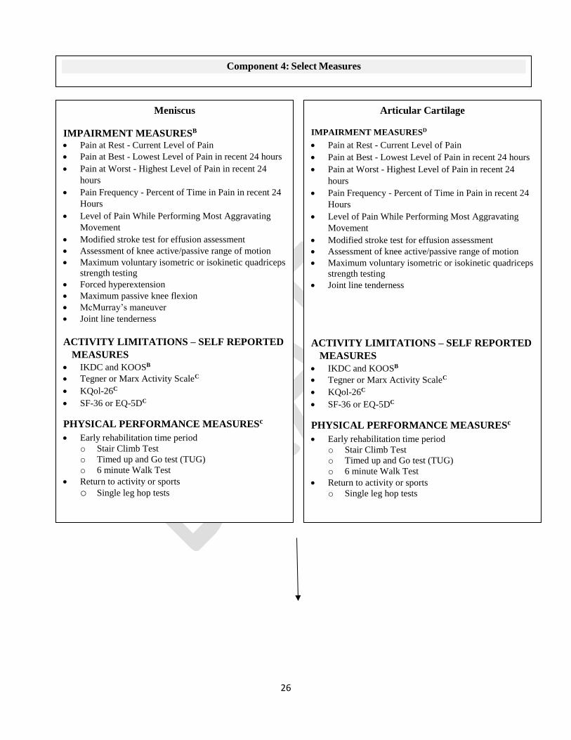
Component 4: Select Measures
Meniscus
IMPAIRMENT MEASURESB
Pain at Rest - Current Level of Pain
Pain at Best - Lowest Level of Pain in recent 24 hours
Pain at Worst - Highest Level of Pain in recent 24
hours
Pain Frequency - Percent of Time in Pain in recent 24
Hours
Level of Pain While Performing Most Aggravating
Movement
Modified stroke test for effusion assessment
Assessment of knee active/passive range of motion
Maximum voluntary isometric or isokinetic quadriceps
strength testing
Forced hyperextension
Maximum passive knee flexion
McMurray’s maneuver
Joint line tenderness
ACTIVITY LIMITATIONS – SELF REPORTED
MEASURES
IKDC and KOOSB
Tegner or Marx Activity ScaleC
KQol-26C
SF-36 or EQ-5DC
PHYSICAL PERFORMANCE MEASURESc
Early rehabilitation time period
o Stair Climb Test
o Timed up and Go test (TUG)
o 6 minute Walk Test
Return to activity or sports
o Single leg hop tests
Articular Cartilage
IMPAIRMENT MEASURESD
Pain at Rest - Current Level of Pain
Pain at Best - Lowest Level of Pain in recent 24 hours
Pain at Worst - Highest Level of Pain in recent 24
hours
Pain Frequency - Percent of Time in Pain in recent 24
Hours
Level of Pain While Performing Most Aggravating
Movement
Modified stroke test for effusion assessment
Assessment of knee active/passive range of motion
Maximum voluntary isometric or isokinetic quadriceps
strength testing
Joint line tenderness
ACTIVITY LIMITATIONS – SELF REPORTED
MEASURES
IKDC and KOOSB
Tegner or Marx Activity ScaleC
KQol-26C
SF-36 or EQ-5DC
PHYSICAL PERFORMANCE MEASURESc
Early rehabilitation time period
o Stair Climb Test
o Timed up and Go test (TUG)
o 6 minute Walk Test
Return to activity or sports
o Single leg hop tests

27
Component 5: Select Intervention strategies
Meniscus
Early Rehabilitation Strategies
PROGRESSIVE MOTION
progressive active and passive knee motion
following knee meniscal surgeryC
Early to Late Rehabilitation Strategies
PROGRESSIVE WEIGHT BEARINGD
PROGRESSIVE RETURN TO ACTIVITYC
SUPERVISED REHABILITATIONB
THERAPEUTIC EXERCISESB
supervised, progressive range of motion exercises,
progressive strength training of the knee and hip
muscles, and neuromuscular training
NEUROMUSCULAR ELECTRICAL
STIMULATIONC
provide neuromuscular stimulation/reeducation to
increase quadriceps strength, functional
performance, and knee function
Articular Cartilage
Early Rehabilitation Strategies
PROGRESSIVE MOTION progressive active and passive knee motion
following knee meniscal and articular cartilage
surgeryC
Early to Late Rehabilitation Strategies
PROGRESSIVE WEIGHT BEARINGB
reach full weight-bearing by 6-8 weeks after
matrix-supported autologous chondrocyte
implantation (MACI)
PROGRESSIVE RETURN TO ACTIVITYE
Dependent upon type of surgery
THERAPEUTIC EXERCISESB
supervised, progressive range of motion exercises,
progressive strength training of the knee and hip
muscles, and neuromuscular training

28
Guidelines based on strong evidence
A- Guidelines based on moderate evidence
B- Guidelines based on weak evidence
C- Conflicting evidence
D- Guidelines based upon theoretical/foundational evidence
E- Guidelines based on expert opinion
RE-EVALUATE
Patient Goals Met
Discharge to Self-Management
Successful Recovery varies depending
on the type of surgery and extent of
impairments Physical Impairment resolved
High self-reported knee function
Normal limb-to-limb symmetry or meets
age- and sex-matched population norms
Patient Goals Not Met
Continue with treatment
interventions or Modify as
needed

29
Component 1
Medical screening incorporates the findings of the history and physical examination to determine
whether the patient’s symptoms originate from a condition that requires referral to another health
care provider. The Ottawa Knee Rules, discussed later, is an example of tools that may be
helpful in this decision-making process. In addition to these conditions, clinicians should screen
for the presence of psychosocial issues that may affect prognostication and treatment decision
making for rehabilitation. Psychological stress negatively influences recovery. Fear of reinjury is
a frequently cited reason that athletes do not return to sport or reduce their level of physical
activity.5, 6 Low internal health locus of control (the belief in one’s ability to control one’s life),
lower self-efficacy, and depressive symptoms prior to surgery results in worse outcomes after
ACL reconstruction.46, 119 Athletes who did not return to sport after ACL reconstruction had
significantly lower preoperative motivation and more negative psychological response than those
who did return.7 Accordingly, identifying cognitive behavioral tendencies during the patient’s
evaluation can direct the therapist to employ specific patient education strategies to optimize
patient outcomes to physical therapy interventions and potentially provide indications for
referring the patient for consultation with another medical or mental health practitioner.14
Component 2
Differential evaluation of musculoskeletal clinical findings is used to determine the most relevant
physical impairments associated with the patient’s reported activity limitations and medical
diagnosis.67 Clusters of these clinical findings are described as impairment patterns in the
physical therapy literature and are labeled according to the key impairment(s) of body function
associated with that cluster. The ICD-10 and primary and secondary ICF codes associated with
meniscus/articular cartilage lesions are provided in the 2010 ICF-based meniscus/articular
cartilage lesions CPG.78 These impairment patterns are useful in driving the interventions, which
focus on normalizing the key impairments of body function, which in turn improves the
movement and function of the patient and lessens or alleviates the activity limitations commonly
reported by the patients who meet the diagnostic criteria of that specific pattern. Key clinical
findings to rule in and rule out the common impairment patterns, and their associated medical
conditions, are shown in the FIGURE. Impairment-based classification is critical for matching
the intervention strategy that is most likely to provide the optimal outcome for a patient’s clinical
findings.67 However, it is important for clinicians to understand that the impairment pattern and
that the most relevant impairments of body function and the associated intervention strategies
often change during the patient’s episode of care. Thus, continual re-evaluation of the patient’s
response to treatment and the patient’s emerging clinical findings is important for providing the
optimal interventions throughout the patient’s episode of care.16
Component 3
Irritability is a term used by rehabilitation practitioners to reflect the tissue’s ability to handle
physical stress,88 and is presumably related to physical status and the extent of inflammatory
activity that is present. There are cases where the alignment of irritability and the duration of
symptoms does not match, requiring clinicians to make judgments when applying time-based
research results on a patient-by-patient basis.16 Diagnosis of tissue irritability is important for
guiding the clinical decisions regarding treatment frequency, intensity, duration, and type, with
the goal of matching the optimal dosage of treatment to the status of the tissue being treated.16, 67
There are other biopsychosocial elements that may relate to staging of the condition, including,
30
but not limited to, the level of disability reported by the patient and activity avoidance.28
Component 4
Outcome measure instrument are standardized instruments for measuring a specific end point,
whether it is a body structure or function, activity limitation, or participation restriction. They
are Important in direct management of individual patient care and for the opportunity they can
collectively compare care and determine effectiveness through the repeated application of
standardized measurement. Outcomes in clinical practice provide the mechanism by which the
health care provider, the patient, the public, and the payer are able to assess the end results of
care and its effect upon the health of the patient and society. Outcome measurement can identify
baseline pain, function and disability, assess global knee function, determine readiness to return
to activities, and monitor changes in status throughout treatment. Outcome measure instruments
can be classified as patient-reported outcome measures (PROMs), physical performance
measures, and physical impairment measures.
Component 5
Interventions are listed by phase of rehabilitation (early, early to late phase). Because irritability
level often reflects the tissue’s ability to accept physical stress, clinicians should match the most
appropriate intervention strategies to the irritability level of the patient’s condition.16, 67
Additionally, clinicians should attend to influences from psychosocial factors5-7 in patients with
conditions in all stages of recovery.
DIFFERENTIAL DIAGNOSIS
2017 Summary (unchanged from 2010)
C
Clinicians should consider diagnostic classifications associated with serious pathological condi-
tions or psychosocial factors when the patient’s reported activity limitations or impairments of
body function and structure are inconsistent with those presented in the diagnosis/classification
section of this guideline, or, when the patient’s symptoms are not resolving with appropriate
interventions.
IMAGING STUDIES
2017 Summary (unchanged from 2010)
When a patient reports a history of knee trauma, the therapist needs to be alert for the presence
of knee fracture. The Ottawa Knee rule has been developed and validated to assist clinicians in
determining when to order radiographs in individuals with acute knee injury.11, 114 Clinical
examination by well-trained clinicians appears to be as accurate as magnetic resonance
imaging (MRI) in regards to the diagnosis of cruciate or meniscal lesions.72, 83 A lower
threshold of suspicion of a meniscal tear is warranted in middle aged and elderly patients.50, 83
MRI may be reserved for more complicated or confusing cases.72 MRI may assist an
orthopaedic surgeon in pre-operative planning and prognosis.72, 83

31
CLINICAL GUIDELINES
Examination
OUTCOME MEASURES-
ACTIVITY LIMITATIONS-SELF-REPORTED MEASURES
2010 Recommendation
B
Clinicians should use a validated patient-reported outcome measure, a general health
questionnaire, and a validated activity scale for patients with knee pain and mobility impairments.
These tools are useful for identifying a patient’s baseline status relative to pain, function, and
disability and for monitoring changes in the patient’s status throughout the course of treatment.
Evidence Update
II
The KOOS has been evaluated for its reliability and validity in people with articular cartilage
lesions.40 Using qualitative methodology, content validity of the KOOS was demonstrated in
people who had undergone, or were candidates for, articular cartilage repair. In the quantitative
analysis, KOOS subscales showed excellent test-retest reliability (all ICC>0.70) and construct
validity was demonstrated against the SF-36, although correlation between the KOOS quality of
life subscale and SF-36 General Health was non-significant. The KOOS showed excellent
sensitivity to change from baseline to 12 months after baseline, with standardized response means
from 0.8 to 1.2 and minimal detectable change estimates ranging between 7.4 and 12.1.
II
The psychometric properties (internal consistency, convergent validity, sensitivity to change and
floor and ceiling effects) of the generic Euroqol-5 (EQ-5D) and Medical Outcomes Study Short
Form-6 Dimensions (SF-6D) were compared using the knee-specific Hughston Clinic
Questionnaire (HCQ) in 84 patients on average 5 days, 6 weeks, and 6 months following APM.49
The results showed that the EQ-5D was more consistently responsive to change over time, was
better at distinguishing differences between groups, and better reflected the results of the joint–
specific HCQ than the SF-6D. Thus in this patient population, the EQ-5D is preferable to the SF-
6D when used alongside a knee-specific instrument such as the HCQ.
II
This study reported the development and validation of the Knee Quality of Life 26-item
questionnaire (KQoL-26) for patients with a suspected ligamentous or meniscal injury.47 The
questionnaire contains 26 items with 3 subscales of knee-related quality of life: physical
functioning, activity limitations, and emotional functioning. The KQoL-26 was found to have
good evidence for internal reliability (Cronbach’s alpha 0.91-0.94), test re-test reliability
(estimates of 0.80-0.93), construct validity (correlations with other knee scales including
Lysholm Knee Scale, (r=0.58 to 0.76 with the 3 KQoL-26 subscales), EQ-5D questionnaire
(r=0.21 to 0.54 with the 3 KQoL-26 subscales), SF-36 (r=0.39 to 0.64 with the 3 KQoL-26
subscales), and knee symptom questions), and responsiveness (higher effect sizes and
responsiveness statistics than the EQ-5D and SF-36).

32
III
The KOOS has been cross-culturally adapted for use in both the Persian and Arabic languages. In
Iranian patients with ACL, meniscus, and combined meniscus and ACL injuries, the Persian
version showed acceptable test-retest reliability on all subscales (ICC>0.70) except the KOOS
Sports/Recreation subscale (ICC=0.61), and adequate construct validity against the SF-36.107 The
Arabic version showed acceptable test-retest reliability (ICC>0.7) for all subscales, as well as good
construct validity against subscales of the RAND-36 (Arabic version of SF-36) (r=0.61 to 0.78)
scores of pain in Egyptian people with ACL, meniscus, and combined knee injuries.4
III
A study compared the measurement properties of the Dutch-language versions of the IKDC2000,
Knee Form, KOOS, and WOMAC in patients with meniscal tears.123 Findings showed good to
excellent measurement properties for the IKDC2000. The KOOS and WOMAC dimensions
performed suboptimally with respect to internal consistency, measurement error, ability to measure
true change, and content validity.
III
In a study of 53 individuals obtained from a sports injury database and electronic medical
records system, Balain et al12 investigated responses to 4 self-report measures (patient
satisfaction, Lysholm scale, VAS for worst pain, and the modified IKDC2000 scale). On all 4
measures, when asked to indicate their current status prior to microfracture surgery for cartilage
defects, patients rated themselves better at least 6 months later than they did prior to surgery.
III
A Rasch model was used to assess the internal construct validity of the Lysholm knee scale in
157 patients with chondral pathology.110 Fit to the Rasch model with 7 remaining items was
achieved after removal of the swelling item. There was a high degree of agreement between the
patient and health professional scoring (ICC 0.90). By removing the swelling item and using
unweighted scores, a modified version of the Lysholm knee scale can be used as an outcome
measure for knee chondral damage.
III
A study translated and culturally adapted the Western Ontario Meniscal Evaluation Tool
(WOMET) into Turkish and evaluated the reliability and validity of the translated tool in 96
patients with meniscal pathology.26 Validity of the tool was compared against the Lysholm knee
scale and the SF-36. Test-retest reliability of the Turkish-version of the WOMET was good to
excellent, and it demonstrated good correlation with the Lysholm knee scale. Weaker
correlations were observed with several SF-36 domains, predominantly mental component and
emotional role scores.
III
After cross-cultural adaptation of the KOOS into Spanish, the Spanish version was evaluated in
20 patients who underwent arthroscopic surgery for knee cartilage defects with a microfracture
technique.124 Validity was assessed against the SF-36. The Spanish KOOS demonstrated
adequate test-retest reliability with ICCs exceeding 0.8 for all domains. Significant agreement

33
between the Spanish-version KOOS and the SF-36 domains of physical function and pain was
observed.
2017 Recommendation
B
For knee-specific outcomes, clinicians should use the IKDC2000 or KOOS (or a culturally-
appropriate version for patients whose primary language is not English) and may use the
Lysholm scale (with removal of swelling item and using unweighted scores).
C
Clinicians may use the Tegner Scale or Marx Activity Level Scale to assess activity level, before
and after interventions intended to alleviate the physical impairments, activity limitations, and
participation restrictions associated with meniscus or articular cartilage lesions, however, these
have less robust evidence about measurement properties. The SF-36 or the EQ-5D are
appropriate general health measures in this population. The KQol-26 may be used to assess knee-
related quality of life.
PHYSICAL PERFORMANCE MEASURES
Refer to the 2010 Knee Pain and Mobility Impairment CPG for a list of activity limitation
measures and their measurement properties.79
2010 Recommendation
C
Clinicians should utilize easily reproducible physical performance measures, such as single limb
hop tests, 6-minute walk test, or timed up-and-go test to assess activity limitations and
participation restrictions associated with their patient’s knee pain or mobility impairment and to
assess the changes in the patient’s level of function over the episode of care.
No Evidence Update
2017 Recommendation
C
Clinicians may administer appropriate clinical or field tests, such as single-legged hop tests, (eg,
single hop for distance, cross-over hop for distance, triple hop for distance, and 6-meter timed
hop), that can identify a patient’s baseline status relative to pain, function, and disability; detect
side-to-side asymmetries; assess global knee function; determine a patient's readiness to return to
activities; and monitor changes in the patient’s status throughout the course of treatment.
PHYSICAL IMPAIRMENT MEASURES
Refer to the 2010 Knee Pain and Mobility Impairment CPG for a list of physical impairment
measures and their measurement properties.80
Evidence Update

34
II
A systematic review of 4 articles examined the validity and reliability of tests to assess meniscus
tears.33 They reported that the Thessaly test had fair reliability (kappa=0.54) based on 1 study of
moderate quality. The McMurray and joint line tenderness tests had poor reliability (kappa <
0.38) based on 3 studies of low to moderate quality.
II
In a large diagnostic study of 292 patients with knee pathology and 75 healthy controls, Blyth et
al17 examined the diagnostic accuracy of several meniscal tear clinical tests compared to MRI in
primary care clinicians. McMurray’s test had fair diagnostic accuracy, while all other tests had
poor diagnostic accuracy. The McMurray’s test had sensitivity: 0.58 (95% CI: 0.49, 0.67);
specificity: 0.56 (95% CI: 0.45, 0.66); OR: 1.79 (95% CI: 1.04, 3.09) compared to MRI. The
Thessaly test had sensitivity: 0.66 (95% CI: 0.57, 0.74); specificity: 0.39 (95% CI: 0.29, 0.50);
OR: 1.24 (95% CI: 0.71, 2.18) compared to MRI. The Apley’s test had sensitivity: 0.53 (95% CI:
0.44, 0.62); specificity: 0.53 (95% CI: 0.42, 0.63); OR: 1.24 (95% CI: 0.73, 2.12) compared to
MRI. The joint line tenderness test had sensitivity: 0.77 (95% CI: 0.68, 0.84); specificity: 0.26
(95% CI: 0.18, 0.36); OR: 1.16 (95% CI: 0.63, 2.13) compared to MRI.
III
Haviv et al55 investigated the accuracy of joint line tenderness of meniscus tears in 134 men and
61 women. Medial and lateral meniscus tears in men had sensitivity of 0.50-0.58, specificity of
0.74-1.00, and diagnostic accuracy of 0.63-0.86. Medial and lateral meniscus tears in women had
sensitivity of 0.40-0.49, specificity of 0.71-0.98, and diagnostic accuracy of 0.57-0.93.
III
Snoeker et al111 investigated the reliability and diagnostic accuracy of deep squat, Thessaly test,
and the joint line tenderness test. The Thessaly test had a kappa of 0.54, sensitivity of 0.52-0.67,
specificity of 0.38-0.44, positive likelihood ratio of 0.91-1.07, and negative likelihood ratio of
0.88-1.12. The deep squat test had a kappa of 0.46, sensitivity of 0.75-0.77, specificity of 0.36-
0.42, positive likelihood ratio of 1.20-1.29, and negative likelihood ratio of 0.60-0.64. The joint
line tenderness test had a kappa of 0.17.
IV
Campbell and colleagues25 examined the association between patients’ pain symptom location
with arthroscopy findings in patients with meniscus tear. They reported that pain symptom
location was not correlated with the location of the meniscus tear (P=.98).
2017 Recommendation
B
Clinicians should administer appropriate physical impairment assessments of body structure and
function, at least at baseline and at 1 follow-up point, for all patients with meniscus tears to
support standardization for quality improvement in clinical care and research, including:
Modified stroke test for effusion assessment
Assessment of knee active range of motion
Maximum voluntary isometric or isokinetic quadriceps strength testing

35
Forced hyperextension
Maximum passive knee flexion
McMurray’s maneuver
Joint line tenderness
D
Clinicians may administer the appropriate physical impairment assessments of body structure
and function, at least at baseline and at 1 follow-up point, for all patients with articular cartilage
lesions to support standardization for quality improvement in clinical care and research,
including:
Modified stroke test for effusion assessment
Assessment of knee active range of motion
Maximum voluntary isometric or isokinetic quadriceps strength testing
Joint line tenderness
BEST-PRACTICE POINT
Essential Data Elements
Clinicians should use the following measures, at least at baseline and at 1 follow-up point, for all
patients with meniscus tears to support standardization for quality improvement in clinical care
and research:
Activity Limitation – Self Report Measures
IKDC and KOOS
Activity Limitation – Physical Performance measures
Early rehabilitation time period
o 30 second Chair Stand Test
o Stair Climb Test
o Timed up and Go test (TUG)
o 6 minute Walk Test
Return to activity or sports
o Single leg hop tests
Physical Impairment Measures
Modified stroke test for effusion assessment
Assessment of knee active range of motion
Maximum voluntary isometric or isokinetic quadriceps strength testing
Forced hyperextension
Maximum passive knee flexion
McMurray’s maneuver
Joint line tenderness

36
Clinicians should use the following measures, at least at baseline and at 1 follow-up point, for all
patients with articular cartilage lesions to support standardization for quality improvement in
clinical care and research:
Activity Limitation – Self Report Measures
IKDC and KOOS
Activity Limitation – Physical Performance measures
Early rehabilitation time period
o 30 second Chair Stand Test
o Stair Climb Test
o Timed up and Go test (TUG)
o 6 minute Walk Test
Return to activity or sports
o Single leg hop tests
Physical Impairment Measures
Modified stroke test for effusion assessment
Assessment of knee active range of motion
Maximum voluntary isometric or isokinetic quadriceps strength testing
Joint line tenderness

37
CLINICAL GUIDELINES
Interventions
PROGRESSIVE KNEE MOTION
2010 Recommendation
C
Clinicians may utilize early progressive knee motion following knee meniscal and articular
cartilage surgery.
Evidence update
II
A systematic review of 4 Level III studies on clinical effectiveness of continuous passive motion
after articular lesion surgery did not find improved histological outcomes on second-look
arthroscopic biopsies or improved radiographic findings greater than 1 year after surgery.43
Mixed results in clinical outcomes were reported between the continuous passive motion groups
and the active range of motion groups, although, the study results were heterogeneous.
II
In a randomized controlled trial, patients randomized to the supervised active range of motion
group (n=14) using an adjustable pedal arm stationary cycle ergometer had significantly better
gait measures (presence or absence of antalgic gait and limp during gait) early after partial
meniscectomy compared to the control group (n=14) who did not have supervised therapy.68 No
differences were reported between the groups over time in range of motion, effusion, or
IKDC2000 scores.
2017 Recommendation
C
Clinicians may use early progressive active and passive knee motion following knee meniscal
and articular cartilage surgery.
PROGRESSIVE WEIGHT BEARING
2010 Recommendation
D
There are conflicting opinions regarding the best use of progressive weight bearing in patients
with meniscal repairs or chondral lesions.
Evidence update
I
Ebert and colleagues36 randomized 62 patients after MACI to an accelerated weight-bearing
group (stepwise progression in weight-bearing with full weight-bearing by 8 weeks) or to a
standard of care weight-bearing group (5 weeks of 20% partial weight-bearing followed by
stepwise progression in weight-bearing with full weight-bearing by week 11). Three months after

38
MACI, patients in the accelerated group had better KOOS scores compared to those in the
standard of care group (range KOOS subscales: 11.84 to 83.32 versus 6.82 to 78.55). Both
groups demonstrated progressive graft tissue healing over time with no difference between
groups at any time period (no complete graft de-lamination).
I
Twenty-eight consecutive patients after MACI were randomized to an accelerated weight-
bearing group (stepwise progression in weight-bearing with full weight-bearing by 6 weeks)
(n=14) or to a standard of care weight-bearing group (stepwise progression in weight-bearing
with full weight-bearing by 8 weeks) (n=14).38 Six and 12 months after MACI, patients in the
accelerated group had better KOOS quality of life scores compared to those in the standard of
care group (6 months: 62 versus 50; 12 months: 77 versus 58). Both groups demonstrated
progressive graft tissue healing over time with no difference between groups at any time period.
I
Thirty-one patients after ACI were randomized to an accelerated weight-bearing group (stepwise
progression in weight-bearing with full weight-bearing after 6 weeks) or to a standard of care
weight-bearing group (stepwise progression in weight-bearing with full weight-bearing after 8
weeks).128 Both groups showed improvement in clinical scores (IKDC2000 and Tegner scale)
and MRI scores over 2 years but no significant difference between groups were noted at 1 year
and 2 years after ACI.
IV
A retrospective analysis of 34 patients with degenerative medial meniscus tear and knee
osteoarthritis using a foot-worn biomechanical device during activities of daily living were
assessed before and 3 months and 12 months of wearing the device.39 Using a gait mat, patients
had significant improvement in gait velocity, step length, and single-limb support of the involved
knee and improved limb symmetry 3 months after device use. These results were maintained 12
months after device use.
2017 Recommendation
B
Clinicians should use a stepwise progression of weight bearing to reach full bearing by 6-8
weeks after matrix-supported autologous chondrocyte implantation (MACI) for articular
cartilage lesions.
2017 Recommendation (unchanged from 2010)
D
Conflicting evidence exists for the best use of progressive weight bearing in patients with
meniscal repairs.
PROGRESSIVE RETURN TO ACTIVITY
2017 Recommendation (unchanged from 2010)
C

39
Clinicians may utilize early progressive return to activity following knee meniscal repair surgery.
E (unchanged from 2010) Clinicians may need to delay return to activity depending on the type of articular cartilage
surgery.
SUPERVISED REHABILITATION
2010 Recommendation
D
There are conflicting opinions regarding the best use of clinic-based programs for patients
following meniscectomy to increase quadriceps strength and functional performance.
Evidence update
II
A systematic review of 18 randomized controlled trials (RCTs) and meta-analysis of 6 RCTs
conducted by Dias et al34 supports the utilization of outpatient physical therapy with a home
exercise program compared to a home exercise program alone to improve knee range of motion
and self-reported knee function, and reduce knee joint effusion in patients after PAM. However,
the studies were of moderate to high risk of bias.
II
In a systematic review of 12 articles conducted by Reid and colleagues,103 supervised clinic-
based rehabilitation or well-structured home exercise program demonstrated improvements in
knee muscle performance and knee function early after partial meniscectomy. However, the
evidence is limited on the use of exercise to prevent the development of osteoarthritis or total
knee joint arthroplasty.
II
In a systematic review by Coppola et al,29 5 RCTs were identified comparing outcomes of home-
based versus supervised outpatient rehabilitation after meniscectomy. In early and intermediate
follow-ups, there was no difference between groups in patient-reported outcomes at 3 weeks and
1 year after meniscectomy. However, the mean scores for these groups were lower than
population norm, which may suggest that patients in both groups were not fully rehabilitated.
Two studies87, 125 reported on higher vertical jump height and single hop distances in the
supervised rehabilitation group (vertical jump: 22.5 cm; single hop: 113.8 cm) compared to the
home-based group (vertical jump: 20.1 cm; single hop distance: 94.7 cm), though, both studies
had short follow-ups (less than 4 weeks).
II
Papalia and colleagues,98 in a systematic review, evaluated the same 5 RCTs as Coppola et al29
comparing outcomes between home-based versus supervised outpatient rehabilitation after
meniscectomy. They reached similar conclusions that differences were demonstrated in
performance-based outcomes (vertical jump height, single hop distance, and knee extensor
strength) but not in patient-reported outcomes (Lysholm, Tegner, Hughston questionnaire).

40
2017 Recommendation
B
Clinicians should use exercises as part of the in-clinic supervised rehabilitation program after
arthroscopic meniscectomy and should provide and supervise the progression of a home-based exercise
program, providing education to ensure independent performance.
THERAPEUTIC EXERCISES
2010 Recommendation
B
Clinicians should consider strength training and functional exercise to increase quadriceps and
hamstrings strength, quadriceps endurance, and functional performance following meniscectomy
Evidence update
I
Osteras et al94 randomized 42 participants after degenerative meniscectomy to receive either 12
weeks of specialized exercise therapy (n=22) or no exercise therapy (n=20). Four participants (2
in each group) were lost to follow-up. Improvements in pain (VAS: 1.9), muscle strength
(quadriceps peak torque: 38.1 Nm), and KOOS scores (18.0 points) were significantly higher in
the specialized exercise therapy group compared to the no exercise therapy group (VAS: 0.6;
quadriceps peak torque: 10.4 Nm; KOOS: 6.5) after the intervention period and 12 months later.
I
In a similar study, Osteras et al96 randomized 75 participants with degenerative meniscus tear to
receive either 12 weeks of specialized exercise therapy (n=38) or no physical therapy (n=37).
Eleven participants (5 in the exercise group, 6 in the no-therapy group) were lost to follow-up.
Improvements in pain, muscle strength, and patient-reported measures were significantly higher
in the exercise therapy group compared to the no therapy group after the intervention period and
12 months later.
I
Assche and colleagues10 implemented the same standardized rehabilitation protocol to patients
who were initially randomized into an ACI surgery group (n=57) or a microfracture surgery
group (n=61). Both groups received the same rehabilitation program consisting of progressive,
stepwise weight bearing, joint mobilization exercises, progressive strength training to the knee
muscles, neuromuscular training, and return to sports integration. The authors reported no
differences in recovery between the 2 groups at 2-year follow-up. When assessing patient
recovery, activities that were repetitive movements of active modalities in low-load conditions
(range of motion, non-weight bearing strengthening exercises, proprioceptive exercises) were
considered low-load modalities. Patients that had low levels of activity (<12 minutes per day of
activity) in these low-load modalities had poorer outcomes in quadriceps strength and single-
legged hop performance than patients that had high levels of activity (>12 minutes per day of
activity) in low-load modalities.

41
I
Hall et al51 performed a RCT to investigate the effects of a neuromuscular training program on
knee kinetics, cartilage quality, and physical function during walking and single-legged sit to
stand after APM. Groups were randomly assigned to the neuromuscular training group or a
control group. The authors reported no differences in peak knee adduction moment, cartilage
quality, and physical function. The neuromuscular group was more likely to demonstrate
improvements in physical function and overall improvement compared to the control group.
I
Kise and colleagues70 randomized 140 participants into 2 treatment groups: exercise therapy
(n=70) or APM. Thirteen (19%) of 70 participants crossed over to the APM group and were
analyzed in the as treated group. The authors reported no clinically relevant differences in KOOS
change scores from baseline to 2-year follow-up between groups (0.9 points, 95% CI: -4.3, 6.1).
Both groups demonstrated similar improvements from baseline to 2-year follow-up (exercise
group: 25.3 points (95% CI: 21.6, 29.0): APM group: 24.4 point (95% CI: 20.7, 28.0). The
exercise group had greater improvement in muscle strength at 3 and 12 months (P<.03)
II
Koutras and colleagues73 randomized 20 male patients after APM to either receive standard
rehabilitation augmented with progressive isokinetic muscle strength training or progressive
isotonic muscle strength training. Both groups demonstrated a significant improvement in knee
extensor and flexor isokinetic strength and single-legged hop limb-to-limb symmetry (knee
extensor at 60o/sec: 17% improvement; knee flexor at 60o/sec: 12% improvement; single hop:
14% improvement; triple hop: 17% improvement; vertical hop: 18% improvement) and in
Lysholm scores (17% improvement) over time but no significant differences were noted between
groups.
II
Lind et al77 randomized 60 patients after isolated meniscal repair to receive either free
rehabilitation (restricted range of motion and toe-touch weight-bearing and no brace for 2 weeks
with unrestricted activity and free range of motion afterwards) or restricted rehabilitation (braced
toe-touch weight bearing and progressive restricted range of motion for 6 weeks). Patients were
followed at 3 months, and 1 and 2 years on KOOS and Tegner measures. Patients who
underwent repeat arthroscopy demonstrated little to partial healing in approximately one-third of
patients in each group (n=19). KOOS and Tegner scores was similar in both groups at 1 and 2
years.
IV
In a retrospective study, 30 patients with non-traumatic posterior root tear of the medial
meniscus had supervised physical therapy, focusing on knee range of motion and knee muscle
strength for at least 8 weeks and were prescribed non-steroidal anti-inflammatory drugs for 8 to
12 weeks.76 Patients demonstrated significant and clinically meaningful improvements in pain
levels (4 point improvement on VAS) and self-reported knee function (13 points improvement in
Lysholm scores).
IV

42
Neogi et al89 reported benefit in symptoms and function with 12-week rehabilitation and
analgesics (up to 6 weeks) in 37 patients with degenerative meniscus. Patients demonstrated
improvements in Lysholm scores from pre-treatment to final follow-up (56 to 79), Tegner scores
(2 to 4), VAS of pain at rest (2- to 0). Despite the improvement, the number of participants with
radiographic osteoarthritis had increased by the final follow-up from 24 knees with Kellgren
Lawrence classifications at grades 0 and 1 and 9 at stage 2 or greater at pre-treatment to 12 knees
with grade 0 and 1 and 21 at stage 2 or greater at final follow-up.
IV
Forty-eight patients with full-thickness articular cartilage lesions with poor knee function
participated in a 3-month rehabilitation program consisting of cardiovascular training,
progressive strength training of the knee and hip muscles, and neuromuscular training.127
Primary outcome measures were KOOS and IKDC2000 scores, and isokinetic muscle strength
and hop test scores. The authors reported an 83% adherence rate to the rehabilitation program
and 4 patients showed increases in pain and effusion. They reported clinically significant
increases in KOOS-sports/recreation and KOOS-quality of life subscales. Patients also had large
effects in standardized response means for muscle strength (0.99 to 1.22) and hop performance
(0.53 to 0.75).
2017 Recommendation
B
Clinicians should provide supervised, progressive range of motion exercises, progressive
strength training of the knee and hip muscles, and neuromuscular training in patients with knee
meniscus tears and articular cartilage lesions and after meniscus or articular cartilage surgery.
NEUROMUSCULAR ELECTRICAL STIMULATION
2010 Recommendation
B
Neuromuscular electrical stimulation can be used with patients following meniscal or chondral
injuries to increase quadriceps muscle strength.
Evidence update
II
Akkaya et al2 conducted a 3-arm RCT in 45 patients comparing a home exercise program
(without any biofeedback or electrical stimulation) to electromyographic biofeedback to the
quadriceps in addition to home exercise program and to electrical stimulation to the quadriceps
in addition to home exercise program after partial meniscectomy. All 3 groups similar gait
measures muscle performance values 2 and 6 weeks after surgery. All groups had significant
improvement in pain during walking and Lysholm scores early after partial meniscectomy.
II
In a RCT, 64 participants were randomized to receive either electromyographic biofeedback
(n=33) or usual care (n=31) early after meniscal repair.92 Electromyographic values and KOOS
Sports/Recreation scores were significantly better in the biofeedback group (Electromyographic:

43
16 to 25% higher; KOOS Sports/Recreation: 6% higher) compared to the usual care group 8
weeks after meniscal repair. However, these differences may not be clinically meaningful.
2017 Recommendation
C Clinicians may provide neuromuscular stimulation/reeducation in patients following meniscus
procedures to increase quadriceps strength, functional performance, and knee function.

44
AFFILIATIONS & CONTACTS
Authors Reviewers
David Logerstedt, PT, PhD
Assistant Professor
Department of Physical Therapy
University of the Sciences
Philadelphia, PA
David Scalzitti, PT, PhD
Assistant Professor
Department of Physical Therapy
George Washington University
Washington, DC
Kim L. Bennell, PT, PhD
Professor
Department of Physiotherapy
University of Melbourne
Parkville, Victoria, Australia
Rana S. Hinman, PT, PhD
PT, PhD
Associate Professor
Department of Physiotherapy
University of Melbourne
Parkville, Victoria, Australia
Holly Silvers-Granelli, PT, PhD
Physical Therapist
Velocity Physical Therapy
Santa Monica, CA
Jay Ebert, PhD
Lecturer
School of Sport Science, Exercise and Health
The University of Western Australia
Crawely, West Australia, Australia
Karen Hambly, PT, PhD
Senior Lecturer
Medway Building
University of Kent
Roy D. Altman, MD
Professor of Medicine
Division of Rheumatology and Immunology
David Geffen School of Medicine at UCLA
Los Angeles, CA
Paul Beattie, PT, PhD
Clinical Professor
Division of Rehabilitative Sciences
University of South Carolina
Columbia, SC
John DeWitt, DPT
Director of Physical Therapy Sports & Orthopaedic
Residencies
The Ohio State University
Columbus, OH
James M. Elliott, PT, PhD
Assistant Professor
NUPTHMS, Feinberg School of Medicine
Northwestern University
Chicago, IL
Helene Fearon, PT
Arizona Physical Therapy Specialists
Phoenix, AZ
Amanda Ferland, DPT
Clinical Faculty
Intech Rehabilitation Group / USC Division of
Biokinesiology and Physical Therapy
Orthopaedic Physical Therapy Residency and
Spine Rehabilitation Fellowship
Beijing, China
Jennifer S. Howard, ATC, PhD
Assistant Professor, Athletic Training
Department of Health and Exercise Science

45
Chatham Maritime, Kent, United Kingdom
James Carey, PT, PhD
Professor and
Co-Director, Brain Plasticity Laboratory
Division of Rehabilitation Medicine
University of Minnesota
Lynn Snyder-Mackler, PT, ScD
Alumni Distinguished Professor
Department of Physical Therapy
Faculty Athletics Representative
University of Delaware
Newark, DE
Michael Axe, MD
First State Orthopaedics
Newark, DE
Christine M. McDonough, PT, PhD
Research Assistant Professor
Health & Disability Research Institute
Boston University School of Public Health
Boston, MA
and
Adjunct Clinical Assistant Professor
Department of Orthopaedic Surgery
Geisel School of Medicine at Dartmouth
Dartmouth-Hitchcock Medical Center
Lebanon, NH
and
ICF-based Clinical Practice Guidelines Editor
Orthopaedic Section, APTA Inc.
La Crosse, WI
Beaver College of Health Sciences
Appalachian State University
Sandra Kaplan, PT, PhD
Clinical Practice Guidelines Coordinator
Academy of Pediatric Physical Therapy, APTA Inc.
and
Professor, Doctoral Programs in Physical Therapy
Rutgers University
Newark, NJ
David Killoran, PhD
Patient/Consumer Representative for the
ICF-based Clinical Practice Guidelines
Orthopaedic Section, APTA Inc.
La Crosse, WI
and
Professor Emeritus
Loyola Marymount University
Los Angeles, CA
Robert Marx, MD, MSc
Department of Orthopaedics
Hospital for Special Surgery
New York, NY
Laura Schmitt, PT, MPT, PhD
Assistant Professor
School of Health and Rehabilitation Sciences
The Ohio State University
Columbus, OH
Jonas Bloch Thorlund, PT, PhD
Associate Professor
University of Southern Denmark
Department of Sports Science and Clinical
Biomechanics
Research Unit for Musculoskeletal Function and
Physiotherapy
Odense M, Denmark
Leslie Torburn, DPT
Principal and Consultant
Silhouette Consulting, Inc.
Sacramento, CA

46
ICF-based Clinical Practice Guidelines Editor
Christine M. McDonough, PT, PhD
Research Assistant Professor
Health & Disability Research Institute
Boston University School of Public Health
Boston, MA
and
Adjunct Clinical Assistant Professor
Department of Orthopaedic Surgery
Geisel School of Medicine at Dartmouth
Dartmouth-Hitchcock Medical Center
Lebanon, NH
and
ICF-based Clinical Practice Guidelines Editor
Orthopaedic Section, APTA Inc.
La Crosse, WI
Guy G. Simoneau, PT, PhD
ICF-based Clinical Practice Guidelines Editor
Orthopaedic Section, APTA Inc.
La Crosse, WI
and
Professor
Physical Therapy Department
Marquette University
Milwaukee, WI
Joseph J. Godges, DPT, MA
Adjunct Associate Professor of Clinical Physical
Therapy
Division of Biokinesiology and Physical Therapy
University of Southern California
Los Angeles, CA
and
ICF-based Clinical Practice Guidelines Editor
Orthopaedic Section, APTA Inc.
La Crosse, WI
References

47
1. Abrams GD, Frank RM, Gupta AK, Harris JD, McCormick FM, Cole BJ. Trends in
meniscus repair and meniscectomy in the United States, 2005-2011. Am J Sports Med.
2013;41:2333-2339.
2. Akkaya N, Ardic F, Ozgen M, Akkaya S, Sahin F, Kilic A. Efficacy of
electromyographic biofeedback and electrical stimulation following arthroscopic partial
meniscectomy: a randomized controlled trial. Clinical rehabilitation. 2012;26:224-236.
3. Al-Dadah O, Shepstone L, Donell ST. Proprioception following partial meniscectomy in
stable knees. Knee Surgery, Sports Traumatology, Arthroscopy. 2011;19:207-213.
4. Almangoush A, Herrington L, Attia I, et al. Cross-cultural adaptation, reliability, internal
consistency and validation of the Arabic version of the Knee injury and Osteoarthritis
Outcome Score (KOOS) for Egyptian people with knee injuries. Osteoarthritis &
Cartilage. 2013;21:1855-1864.
5. Ardern CL, Osterberg A, Tagesson S, Gauffin H, Webster KE, Kvist J. The impact of
psychological readiness to return to sport and recreational activities after anterior cruciate
ligament reconstruction. British journal of sports medicine. 2014;48:1613-1619.
6. Ardern CL, Taylor NF, Feller JA, Webster KE. Fear of re-injury in people who have
returned to sport following anterior cruciate ligament reconstruction surgery. Journal of
science and medicine in sport / Sports Medicine Australia. 2012;15:488-495.
7. Ardern CL, Taylor NF, Feller JA, Whitehead TS, Webster KE. Psychological responses
matter in returning to preinjury level of sport after anterior cruciate ligament
reconstruction surgery. The American journal of sports medicine. 2013;41:1549-1558.
8. Aroen A, Loken S, Heir S, et al. Articular cartilage lesions in 993 consecutive knee
arthroscopies. Am J Sports Med. 2004;32:211-215.
9. Arroll B, Robb G, Sutich E, et al. The Diagnosis and Management of Soft Tissue Knee
Injuries: Internal Derangements. Wellington, NZ: New Zealand Guidelines Group; 2003.
10. Assche DV, Caspel DV, Staes F, et al. Implementing one standardized rehabilitation
protocol following autologous chondrocyte implantation or microfracture in the knee
results in comparable physical therapy management. Physiotherapy Theory & Practice.
2011;27:125-136.
11. Bachmann LM, Haberzeth S, Steurer J, ter Riet G. The accuracy of the Ottawa knee rule
to rule out knee fractures: a systematic review. Annals of internal medicine.
2004;140:121-124.
12. Balain B, Ennis O, Kanes G, et al. Response shift in self-reported functional scores after
knee microfracture for full thickness cartilage lesions. Osteoarthritis and cartilage /
OARS, Osteoarthritis Research Society. 2009;17:1009-1013.
13. Bansal P, Deehan DJ, Gregory RJ. Diagnosing the acutely locked knee. Injury.
2002;33:495-498.
14. Bergbom S, Boersma K, Overmeer T, Linton SJ. Relationship among pain
catastrophizing, depressed mood, and outcomes across physical therapy treatments.
Physical therapy. 2011;91:754-764.
15. Bhosale AM, Richardson JB. Articular cartilage: structure, injuries and review of
management. Br Med Bull. 2008;87:77-95.
16. Blanpied PR, Gross AR, Elliott JM, et al. Neck Pain: Revision 2017. The Journal of
orthopaedic and sports physical therapy. 2017;47:A1-A83.
17. Blyth M, Anthony I, Francq B, et al. Diagnostic accuracy of the Thessaly test,
standardised clinical history and other clinical examination tests (Apley's, McMurray's

48
and joint line tenderness) for meniscal tears in comparison with magnetic resonance
imaging diagnosis. Health technology assessment (Winchester, England). 2015;19:1-62.
18. Brambilla L, Pulici L, Carimati G, et al. Prevalence of Associated Lesions in Anterior
Cruciate Ligament Reconstruction: Correlation With Surgical Timing and With Patient
Age, Sex, and Body Mass Index. Am J Sports Med. 2015;43:2966-2973.
19. Brindle T, Nyland J, Johnson DL. The Meniscus: Review of Basic Principles With
Application to Surgery and Rehabilitation. J Athl Train. 2001;36:160-169.
20. Brittberg M, Winalski CS. Evaluation of cartilage injuries and repair. J Bone Joint Surg
Am. 2003;85-A 58-69.
21. Brophy RH, Wright RW, David TS, et al. Association between previous meniscal surgery
and the incidence of chondral lesions at revision anterior cruciate ligament
reconstruction. The American Journal of Sports Medicine. 2012;40:808-814.
22. Brukner P, Kahn K. Clinical Sports Medicine. Sydney, Australia: McGraw-Hill; 2006.
23. Buckwalter JA, Mankin HJ. Articular cartilage: tissue design and chondrocyte-matrix
interactions. Instructional course lectures. 1998;47:477-486.
24. Busija L, Osborne RH, Nilsdotter A, Buchbinder R, Roos EM. Magnitude and
meaningfulness of change in SF-36 scores in four types of orthopedic surgery. Health &
Quality of Life Outcomes. 2008;6:55-55.
25. Campbell J, Harte A, Kerr DP, Murray P. The location of knee pain and pathology in
patients with a presumed meniscus tear: preoperative symptoms compared to arthroscopic
findings. Ir J Med Sci. 2014;183:23-31.
26. Celik D, Demirel M, Kus G, Erdil M, Ozdincler AR. Translation, cross-cultural
adaptation, reliability and validity of the Turkish version of the Western Ontario
Meniscal Evaluation Tool (WOMET). Knee Surg Sports Traumatol Arthrosc.
2015;23:816-825.
27. Chhadia AM, Inacio MC, Maletis GB, Csintalan RP, Davis BR, Funahashi TT. Are
meniscus and cartilage injuries related to time to anterior cruciate ligament
reconstruction? The American Journal of Sports Medicine. 2011;39:1894-1899.
28. Cook C. Orthopedic manual therapy : an evidence based approach. 2nd. Upper Saddle
River, NJ: Pearson Education; 2012.
29. Coppola SM, Collins SM. Is physical therapy more beneficial than unsupervised home
exercise in treatment of post surgical knee disorders? A systematic review. Knee.
2009;16:171-175.
30. Crema MD, Guermazi A, Li L, et al. The association of prevalent medial meniscal
pathology with cartilage loss in the medial tibiofemoral compartment over a 2-year
period. Osteoarthritis and cartilage / OARS, Osteoarthritis Research Society.
2010;18:336-343.
31. Crema MD, Roemer FW, Felson DT, et al. Factors associated with meniscal extrusion in
knees with or at risk for osteoarthritis: the Multicenter Osteoarthritis study. Radiology.
2012;264:494-503.
32. Curl WW, Krome J, Gordon ES, Rushing J, Smith BP, Poehling GG. Cartilage injuries: a
review of 31,516 knee arthroscopies. Arthroscopy. 1997;13:456-460.
33. Décary S, Ouellet P, Vendittoli PA, Desmeules F. Reliability of physical examination
tests for the diagnosis of knee disorders: Evidence from a systematic review. Manual
Therapy. 2016;26:172-182.

49
34. Dias JM, Mazuquin BF, Mostagi FQRC, et al. The effectiveness of postoperative
physical therapy treatment in patients who have undergone arthroscopic partial
meniscectomy: Systematic review with meta-analysis. Journal of Orthopaedic and Sports
Physical Therapy. 2013;43:560-576.
35. Ding J, Zhao J, He Y, Huangfu X, Zeng B. Risk factors for articular cartilage lesions in
symptomatic discoid lateral meniscus. Arthroscopy : The Journal of Arthroscopic &
Related Surgery : Official Publication of the Arthroscopy Association of North America
and the International Arthroscopy Association. 2009;25:1423-1426.
36. Ebert JR, Robertson WB, Lloyd DG, Zheng MH, Wood DJ, Ackland T. Traditional vs
accelerated approaches to post-operative rehabilitation following matrix-induced
autologous chondrocyte implantation (MACI): comparison of clinical, biomechanical and
radiographic outcomes. Osteoarthritis and cartilage / OARS, Osteoarthritis Research
Society. 2008;16:1131-1140.
37. Ebert JR, Smith A, Edwards PK, Hambly K, Wood DJ, Ackland TR. Factors predictive
of outcome 5 years after matrix-induced autologous chondrocyte implantation in the
tibiofemoral joint. The American Journal of Sports Medicine. 2013;41:1245-1254.
38. Edwards PK, Ackland TR, Ebert JR. Accelerated weightbearing rehabilitation after
matrix-induced autologous chondrocyte implantation in the tibiofemoral joint: early
clinical and radiological outcomes. The American Journal of Sports Medicine.
2013;41:2314-2324.
39. Elbaz A, Beer Y, Rath E, et al. A unique foot-worn device for patients with degenerative
meniscal tear. Knee surgery, sports traumatology, arthroscopy : official journal of the
ESSKA. 2013;21:380-387.
40. Engelhart L, Nelson L, Lewis S, et al. Validation of the knee injury and osteoarthritis
outcome score subscales for patients with articular cartilage lesions of the knee.
American Journal of Sports Medicine. 2012;40:2264-2272.
41. Englund M, Guermazi A, Roemer FW, et al. Meniscal tear in knees without surgery and
the development of radiographic osteoarthritis among middle-aged and elderly persons:
The Multicenter Osteoarthritis Study. Arthritis and Rheumatism. 2009;60:831-839.
42. Fabricant PD, Rosenberger PH, Jokl P, Ickovics JR. Predictors of short-term recovery
differ from those of long-term outcome after arthroscopic partial meniscectomy.
Arthroscopy: The Journal of Arthroscopy & Related Surgery. 2008;24:769-778.
43. Fazalare JA, Griesser MJ, Siston RA, Flanigan DC. The use of continuous passive
motion following knee cartilage defect surgery: a systematic review. Orthopedics.
2010;33:878-878.
44. Filardo G, Andriolo L, Balboni F, Marcacci M, Kon E. Cartilage failures. Systematic
literature review, critical survey analysis, and definition. Knee Surgery, Sports
Traumatology, Arthroscopy. 2015;23:3660-3669.
45. Flanigan DC, Harris JD, Trinh TQ, Siston RA, Brophy RH. Prevalence of chondral
defects in Athletes' Knees: A systematic review. Medicine and Science in Sports and
Exercise. 2010;42:1795-1801.
46. Garcia GH, Wu HH, Park MJ, et al. Depression Symptomatology and Anterior Cruciate
Ligament Injury: Incidence and Effect on Functional Outcome--A Prospective Cohort
Study. The American journal of sports medicine. 2016;44:572-579.

50
47. Garratt AM, Brealey S, Robling M, et al. Development of the Knee Quality of Life
(KQoL-26) 26-item questionnaire: Data quality, reliability, validity and responsiveness.
Health and Quality of Life Outcomes. 2008;6:
48. Gobbi A, Nunag P, Malinowski K. Treatment of full thickness chondral lesions of the
knee with microfracture in a group of athletes. Knee Surg Sports Traumatol Arthrosc.
2005;13:213-221.
49. Goodwin PC, Ratcliffe J, Morris J, Morrissey MC. Using the knee-specific Hughston
Clinic Questionnaire, EQ-5D and SF-6D following arthroscopic partial meniscectomy
surgery: a comparison of psychometric properties. Quality of Life Research.
2011;20:1437-1446.
50. Habata T, Uematsu K, Hattori K, Takakura Y, Fujisawa Y. Clinical features of the
posterior horn tear in the medial meniscus. Arch Orthop Trauma Surg. 2004;124:642-
645.
51. Hall M, Hinman RS, Wrigley TV, et al. Neuromuscular Exercise post Partial Medial
Meniscectomy: Randomized Controlled Trial. Med Sci Sports Exerc. 2015;47:1557-1566.
52. Hall M, Wrigley TV, Metcalf BR, et al. Knee muscle strength after recent partial
meniscectomy does not relate to 2-year change in knee adduction moment. Clinical
orthopaedics and related research. 2014;472:3114-3120.
53. Hall M, Wrigley TV, Metcalf BR, et al. A longitudinal study of impact and early stance
loads during gait following arthroscopic partial meniscectomy. Journal of Biomechanics.
2014;47:2852-2857.
54. Hall M, Wrigley TV, Metcalf BR, et al. A Longitudinal Study of Strength and Gait after
Arthroscopic Partial Meniscectomy. Medicine & Science in Sports & Exercise.
2013;45:2036-2043.
55. Haviv B, Bronak S, Kosashvili Y, Thein R. Gender differences in the accuracy of joint
line tenderness for arthroscopically confirmed meniscal tears. Archives of orthopaedic
and trauma surgery. 2015;135:1567-1570.
56. Hegedus EJ, Cook C, Hasselblad V, Goode A, McCrory DC. Physical examination tests
for assessing a torn meniscus in the knee: a systematic review with meta-analysis. J
Orthop Sports Phys Ther. 2007;37:541-550.
57. Henry S, Mascarenhas R, Kowalchuk D, Forsythe B, Irrgang JJ, Harner CD. Medial
meniscus tear morphology and chondral degeneration of the knee: is there a relationship?
Arthroscopy : The Journal of Arthroscopic & Related Surgery : Official Publication of
the Arthroscopy Association of North America and the International Arthroscopy
Association. 2012;28:1124-1134.e1122.
58. Hjelle K, Solheim E, Strand T, Muri R, Brittberg M. Articular cartilage defects in 1,000
knee arthroscopies. Arthroscopy. 2002;18:730-734.
59. Hwang BY, Kim SJ, Lee SW, et al. Risk factors for medial meniscus posterior root tear.
The American Journal of Sports Medicine. 2012;40:1606-1610.
60. Jacob KM, Oommen AT. A retrospective analysis of risk factors for meniscal co-
morbidities in anterior cruciate ligament injuries. Indian Journal of Orthopaedics.
2012;46:566-569.
61. Jaiswal PK, Bentley G, Carrington RW, Skinner JA, Briggs TW. The adverse effect of
elevated body mass index on outcome after autologous chondrocyte implantation. The
Journal of bone and joint surgery.British volume. 2012;94:1377-1381.

51
62. Johnson-Nurse C, Dandy DJ. Fracture-separation of articular cartilage in the adult knee.
The Journal of bone and joint surgery. 1985;67:42-43.
63. Jones JC, Burks R, Owens BD, Sturdivant RX, Svoboda SJ, Cameron KL. Incidence and
risk factors associated with meniscal injuries among active-duty US military service
members. Journal of athletic training. 2012;47:67-73.
64. Joseph C, Pathak SS, Aravinda M, Rajan D. Is ACL reconstruction only for athletes? A
study of the incidence of meniscal and cartilage injuries in an ACL-deficient athlete and
non-athlete population: an Indian experience. Int Orthop. 2008;32:57-61.
65. Jungmann PM, Salzmann GM, Schmal H, Pestka JM, Südkamp NP, Niemeyer P.
Autologous Chondrocyte Implantation for Treatment of Cartilage Defects of the Knee:
What Predicts the Need for Reintervention? American Journal of Sports Medicine.
2012;40:58-67.
66. Katz JN, Brophy RH, Chaisson CE, et al. Surgery versus physical therapy for a meniscal
tear and osteoarthritis. The New England journal of medicine. 2013;368:1675-1684.
67. Kelley MJ, Shaffer MA, Kuhn JE, et al. Shoulder pain and mobility deficits: adhesive
capsulitis. The Journal of orthopaedic and sports physical therapy. 2013;43:A1-31.
68. Kelln BM, Ingersoll CD, Saliba S, Miller MD, Hertel J. Effect of early active range of
motion rehabilitation on outcome measures after partial meniscectomy. Knee surgery,
sports traumatology, arthroscopy : official journal of the ESSKA. 2009;17:607-616.
69. Kijowski R, Woods MA, McGuine TA, Wilson JJ, Graf BK, De Smet AA. Arthroscopic
partial meniscectomy: MR imaging for prediction of outcome in middle-aged and elderly
patients. Radiology. 2011;259:203-212.
70. Kise NJ, Risberg MA, Stensrud S, Ranstam J, Engebretsen L, Roos EM. Exercise therapy
versus arthroscopic partial meniscectomy for degenerative meniscal tear in middle aged
patients: randomised controlled trial with two year follow-up. BMJ. 2016;354:i3740.
71. Kluczynski MA, Marzo JM, Bisson LJ. Factors associated with meniscal tears and
chondral lesions in patients undergoing anterior cruciate ligament reconstruction: a
prospective study. The American Journal of Sports Medicine. 2013;41:2759-2765.
72. Kocabey Y, Tetik O, Isbell WM, Atay OA, Johnson DL. The value of clinical
examination versus magnetic resonance imaging in the diagnosis of meniscal tears and
anterior cruciate ligament rupture. Arthroscopy. 2004;20:696-700.
73. Koutras G, Letsi M, Papadopoulos P, Gigis I, Pappas E. A Randomized Trial of
Isokinetic Versus Isotonic Rehabilitation Program After Arthroscopic Meniscectomy.
International Journal of Sports Physical Therapy. 2012;7:31-38.
74. Kuikka PI, Pihlajamaki HK, Mattila VM. Knee injuries related to sports in young adult
males during military service - incidence and risk factors. Scandinavian Journal of
Medicine & Science in Sports. 2013;23:281-287.
75. Lewis PB, McCarty LP, 3rd, Kang RW, Cole BJ. Basic science and treatment options for
articular cartilage injuries. J Orthop Sports Phys Ther. 2006;36:717-727.
76. Lim HC, Bae JH, Wang JH, Seok CW, Kim MK. Non-operative treatment of
degenerative posterior root tear of the medial meniscus. Knee surgery, sports
traumatology, arthroscopy : official journal of the ESSKA. 2010;18:535-539.
77. Lind M, Nielsen T, Fauno P, Lund B, Christiansen SE. Free rehabilitation is safe after
isolated meniscus repair: a prospective randomized trial comparing free with restricted
rehabilitation regimens. The American Journal of Sports Medicine. 2013;41:2753-2758.

52
78. Logerstedt DS, Snyder-Mackler L, Ritter RC, Axe MJ. Knee pain and mobility
impairments: meniscal and articular cartilage lesions. The Journal of orthopaedic and
sports physical therapy. 2010;40:A1-A35.
79. Logerstedt DS, Snyder-Mackler L, Ritter RC, Axe MJ, Godges JJ. Knee stability and
movement coordination impairments: knee ligament sprain. J Orthop Sports Phys Ther.
2010;40:A1-A37.
80. Logerstedt DS, Snyder-Mackler L, Ritter RC, Axe MJ, Orthopedic Section of the
American Physical Therapy A. Knee pain and mobility impairments: meniscal and
articular cartilage lesions. J Orthop Sports Phys Ther. 2010;40:A1-A35.
81. Lowery DJ, Farley TD, Wing DW, Sterett WI, Steadman JR. A clinical composite score
accurately detects meniscal pathology. Arthroscopy. 2006;22:1174-1179.
82. Lyman S, Hidaka C, Valdez AS, et al. Risk factors for meniscectomy after meniscal
repair. The American Journal of Sports Medicine. 2013;41:2772-2778.
83. Madhusudhan T, Kumar T, Bastawrous S, Sinha A. Clinical examination, MRI and
arthroscopy in meniscal and ligamentous knee Injuries - a prospective study. J Orthop
Surg. 2008;3:19.
84. Majewski M, Susanne H, Klaus S. Epidemiology of athletic knee injuries: A 10-year
study. Knee. 2006;13:184-188.
85. McLeod MM, Gribble P, Pfile KR, Pietrosimone BG. Effects of Arthroscopic Partial
Meniscectomy on Quadriceps Strength: A Systematic Review. Journal of Sport
Rehabilitation. 2012;21:285-295.
86. Meserve BB, Cleland JA, Boucher TR. A meta-analysis examining clinical test utilities
for assessing meniscal injury. Clin Rehabil. 2008;22:143-161.
87. Moffet H, Richards CL, Malouin F, Bravo G, Paradis G. Early and intensive
physiotherapy accelerates recovery postarthroscopic meniscectomy: results of a
randomized controlled study. Arch Phys Med Rehabil. 1994;75:415-426.
88. Mueller MJ, Maluf KS. Tissue adaptation to physical stress: a proposed "Physical Stress
Theory" to guide physical therapist practice, education, and research. Physical therapy.
2002;82:383-403.
89. Neogi DS, Kumar A, Rijal L, Yadav CS, Jaiman A, Nag HL. Role of nonoperative
treatment in managing degenerative tears of the medial meniscus posterior root. Journal
of orthopaedics and traumatology : official journal of the Italian Society of Orthopaedics
and Traumatology. 2013;14:193-199.
90. Nepple JJ, Wright RW, Matava MJ, Brophy RH. Full-thickness knee articular cartilage
defects in national football league combine athletes undergoing magnetic resonance
imaging: prevalence, location, and association with previous surgery. Arthroscopy: The
Journal of Arthroscopy & Related Surgery. 2012;28:798-806.
91. O'Connor DP, Laughlin MS, Woods GW. Factors related to additional knee injuries after
anterior cruciate ligament injury. Arthroscopy. 2005;21:431-438.
92. Oravitan M, Avram C. The Effectiveness of Electromyographic Biofeedback as Part of a
Meniscal Repair Rehabilitation Programme. Journal of Sports Science & Medicine.
2013;12:526-532.
93. Organization WH. International Classification of Functioning, Disability, and Health.
Geneva, Switzerland; 2010.
94. Osteras H. A 12-week medical exercise therapy program leads to significant
improvement inknee function after degenerative meniscectomy: A randomized controlled

53
trial with one year follow-up. Journal of Bodywork and Movement Therapies.
2014;18:374-382.
95. Osteras H, Osteras B, Torstensen TA. Medical exercise therapy, and not arthroscopic
surgery, resulted in decreased depression and anxiety in patients with degenerative
meniscus injury. Journal of Bodywork and Movement Therapies. 2012;16:456-463.
96. Østerås H, Østerås B, Torstensen TA. Is postoperative exercise therapy necessary in
patients with degenerative meniscus? A randomized controlled trial with one year follow-
up. Knee Surgery, Sports Traumatology, Arthroscopy. 2014;22:200-206.
97. Papalia R, Del Buono A, Osti L, Denaro V, Maffulli N. Meniscectomy as a risk factor for
knee osteoarthritis: A systematic review. British Medical Bulletin. 2011;99:89-106.
98. Papalia R, Vasta S, Tecame A, D'Adamio S, Maffulli N, Denaro V. Home-based vs
supervised rehabilitation programs following knee surgery: A systematic review. British
Medical Bulletin. 2013;108:55-72.
99. Pestka JM, Bode G, Salzmann G, et al. Clinical outcomes after cell-seeded autologous
chondrocyte implantation of the knee: When can success or failure be predicted?
American Journal of Sports Medicine. 2014;42:208-215.
100. Petty CA, Lubowitz JH. Does arthroscopic partial meniscectomy result in knee
osteoarthritis? A systematic review with a minimum of 8 years' follow-up. Arthroscopy -
Journal of Arthroscopic and Related Surgery. 2011;27:419-424+e459.
101. Phillips B, Ball C, Sackett D, et al. Levels of Evidence. Available at:
http://www.cebm.net/index.aspx?o=4590. Accessed August 4, 2009.
102. Ralles S, Agel J, Obermeier M, Tompkins M. Incidence of Secondary Intra-articular
Injuries With Time to Anterior Cruciate Ligament Reconstruction. The American Journal
of Sports Medicine. 2015;43:1373-1379.
103. Reid D, Rydwanski J, Hing W, White S. The effectiveness of post-operative
rehabilitation following partial meniscectomy of the knee. Physical Therapy Reviews.
2012;17:45-54.
104. Renstrom P, Johnson RJ. Anatomy and biomechanics of the menisci. Clin Sports Med.
1990;9:523-538.
105. Rosenberger PH, Dhabhar FS, Epel E, Jokl P, Ickovics JR. Sex differences in factors
influencing recovery from arthroscopic knee surgery. Clinical orthopaedics and related
research. 2010;468:3399-3405.
106. Ryzewicz M, Peterson B, Siparsky PN, Bartz RL. The diagnosis of meniscus tears: the
role of MRI and clinical examination. Clin Orthop Relat Res. 2007;455:123-133.
107. Salavati M, Mazaheri M, Negahban H, et al. Validation of a Persian-version of Knee
injury and Osteoarthritis Outcome Score (KOOS) in Iranians with knee injuries.
Osteoarthritis and Cartilage. 2008;16:1178-1182.
108. Salzmann GM, Sah B, Sudkamp NP, Niemeyer P. Reoperative characteristics after
microfracture of knee cartilage lesions in 454 patients. Knee surgery, sports
traumatology, arthroscopy : official journal of the ESSKA. 2013;21:365-371.
109. Scanzello CR, Albert AS, DiCarlo E, et al. The influence of synovial inflammation and
hyperplasia on symptomatic outcomes up to 2 years post-operatively in patients
undergoing partial meniscectomy. Osteoarthritis and Cartilage. 2013;21:1392-1399.
110. Smith HJ, Richardson JB, Tennant A. Modification and validation of the Lysholm Knee
Scale to assess articular cartilage damage. Osteoarthritis and cartilage / OARS,
Osteoarthritis Research Society. 2009;17:53-58.

54
111. Snoeker BA, Lindeboom R, Zwinderman AH, Vincken PW, Jansen JA, Lucas C.
Detecting Meniscal Tears in Primary Care: Reproducibility and Accuracy of 2 Weight-
Bearing Tests and 1 Non-Weight-Bearing Test. The Journal of orthopaedic and sports
physical therapy. 2015;45:693-702.
112. Snoeker BAM, Bakker EWP, Kegel CAT, Lucas C. Risk factors for meniscal tears: A
systematic review including meta-analysis. Journal of Orthopaedic and Sports Physical
Therapy. 2013;43:352-367.
113. Stein T, Mehling AP, Welsch F, von Eisenhart-Rothe R, Jäger A. Long-term outcome
after arthroscopic meniscal repair versus arthroscopic partial meniscectomy for traumatic
meniscal tears. American Journal of Sports Medicine. 2010;38:1542-1548.
114. Stiell IG, Greenberg GH, Wells GA, et al. Derivation of a decision rule for the use of
radiography in acute knee injuries. Annals of emergency medicine. 1995;26:405-413.
115. Stocker BD, Nyland JA, Caborn DN, Sternes R, Ray JM. Results of the Kentucky high
school football knee injury survey. J Ky Med Assoc. 1997;95:458-464.
116. Sung-Gon K, Nagao M, Kamata K, Maeda K, Nozawa M. Return to sport after
arthroscopic meniscectomy on stable knees. BMC Sports Science, Medicine &
Rehabilitation. 2013;5:1-8.
117. Swenson DM, Collins CL, Best TM, Flanigan DC, Fields SK, Comstock RD.
Epidemiology of knee injuries among U.S. high school athletes, 2005/2006-2010/2011.
Medicine and Science in Sports and Exercise. 2013;45:462-469.
118. Tandogan RN, Taser O, Kayaalp A, et al. Analysis of meniscal and chondral lesions
accompanying anterior cruciate ligament tears: relationship with age, time from injury,
and level of sport. Knee Surg Sports Traumatol Arthrosc. 2004;12:262-270.
119. te Wierike SC, van der Sluis A, van den Akker-Scheek I, Elferink-Gemser MT, Visscher
C. Psychosocial factors influencing the recovery of athletes with anterior cruciate
ligament injury: a systematic review. Scandinavian journal of medicine & science in
sports. 2013;23:527-540.
120. Thorlund JB, Aagaard P, Roos EM. Muscle strength and functional performance in
patients at high risk of knee osteoarthritis: a follow-up study. Knee Surg Sports
Traumatol Arthrosc. 2012;20:1110-1117.
121. Thorlund JB, Creaby MW, Wrigley TV, Metcalf BR, Bennell KL. Knee joint laxity and
passive stiffness in meniscectomized patients compared with healthy controls. Knee.
2014;21:886-890.
122. Thorlund JB, Hare KB, Lohmander LS. Large increase in arthroscopic meniscus surgery
in the middle-aged and older population in Denmark from 2000 to 2011. Acta
Orthopaedica. 2014;85:287-292.
123. van de Graaf VA, Wolterbeek N, Scholtes VA, Mutsaerts EL, Poolman RW. Reliability
and Validity of the IKDC, KOOS, and WOMAC for Patients With Meniscal Injuries. Am
J Sports Med. 2014;42:1408-1416.
124. Vaquero J, Longo UG, Forriol F, Martinelli N, Vethencourt R, Denaro V. Reliability,
validity and responsiveness of the Spanish version of the Knee Injury and Osteoarthritis
Outcome Score (KOOS) in patients with chondral lesion of the knee. Knee Surg Sports
Traumatol Arthrosc. 2014;22:104-108.
125. Vervest AM, Maurer CA, Schambergen TG, de Bie RA, Bulstra SK. Effectiveness of
physiotherapy after meniscectomy. Knee Surg Sports Traumatol Arthrosc. 1999;7:360-
364.

55
126. Widuchowski W, Widuchowski J, Trzaska T. Articular cartilage defects: study of 25,124
knee arthroscopies. Knee. 2007;14:177-182.
127. Wondrasch B, Aroen A, Rotterud JH, Hoysveen T, Bolstad K, Risberg MA. The
feasibility of a 3-month active rehabilitation program for patients with knee full-thickness
articular cartilage lesions: the Oslo Cartilage Active Rehabilitation and Education Study.
The Journal of orthopaedic and sports physical therapy. 2013;43:310-324.
128. Wondrasch B, Zak L, Welsch GH, Marlovits S. Effect of accelerated weightbearing after
matrix-associated autologous chondrocyte implantation on the femoral condyle on
radiographic and clinical outcome after 2 years: a prospective, randomized controlled
pilot study. The American Journal of Sports Medicine. 2009;37 Suppl 1:88S-96S.
129. Wu J, Huang JM, Zhao B, Cao JG, Chen X. Risk Factors Comparison for Radial and
Horizontal Tears. J Knee Surg. 2016;29:679-683.
130. Wyatt RWB, Inacio MCS, Liddle KD, Maletis GB. Prevalence and incidence of cartilage
injuries and meniscus tears in patients who underwent both primary and revision anterior
cruciate ligament reconstructions. American Journal of Sports Medicine. 2014;42:1841-
1846.
131. Xu C, Zhao J. A meta-analysis comparing meniscal repair with meniscectomy in the
treatment of meniscal tears: the more meniscus, the better outcome? Knee surgery, sports
traumatology, arthroscopy : official journal of the ESSKA. 2015;23:164-170.
132. Yeh PC, Starkey C, Lombardo S, Vitti G, Kharrazi FD. Epidemiology of isolated
meniscal injury and its effect on performance in athletes from the National Basketball
Association. American Journal of Sports Medicine. 2012;40:589-594.
133. Zaffagnini S, Marcheggiani Muccioli GM, Lopomo N, et al. Prospective long-term
outcomes of the medial collagen meniscus implant versus partial medial meniscectomy: a
minimum 10-year follow-up study. American journal of sports medicine. 2011;39:977-
985.

56
APPENDIX A: SEARCH STRATEGIES FOR ALL DATABASES SEARCHED
MEDLINE
((“Menisci, Tibial” [MH]) OR (knee joint [MH] AND (menisc* [TW] OR “articular cartilage”
[TW] OR chondral [TW]))) AND (classif* [TW])
(("Menisci, Tibial"[MH]) OR (knee joint[MH] AND (menisc*[TW] OR "articular
cartilage"[TW] OR chondral[TW]))) AND (sensitiv*[Title/Abstract] OR sensitivity and
specificity[MeSH Terms] OR diagnos*[Title/Abstract] OR diagnosis[MeSH:noexp] OR
diagnostic[MeSH:noexp] OR diagnosis, differential[MeSH:noexp] OR
diagnosis[Subheading:noexp] OR questionnaires[Mesh] OR "disability evaluation"[mesh:noexp]
OR questionnaire[tiab] OR questionnaires[tiab] OR instrument[tiab] OR instruments[tiab] OR
scale[tiab] OR scales[tiab] OR measurement[tiab] OR measurements[tiab] OR index[tiab] OR
indices[tiab] OR score[tiab] OR scores[tiab])
((“Menisci, Tibial” [MH]) OR (knee joint [MH] AND (menisc* [TW] OR “articular cartilage”
[TW] OR chondral [TW]))) AND (physical therapy modalities [MH] OR recovery of function
[MH] OR rehabilitation [MH] OR therapeutics [MH] OR “physical therapy” [TW] OR
physiother* [TW] OR recovery [TW] OR restoration [TW] OR re-education [TW] OR early
ambulation [MH] OR strengthening [TW] OR resistance training [MH] OR “resistance methods”
[TW] OR exercise therapy [MH] OR biofeedback, psychology [MH] OR “neuromuscular
electrical stimulation” [TW] OR pain management [MH] OR pain measurement [MH] OR
mobilization* [TW] OR “continuous passive motion” [TW] OR manipulation, spinal [MH] OR
ultrasonography [TW] OR ultrasound [TW] OR acupuncture [TW] OR laser* [TW] OR patient
education as topic [MH] OR electrical stimulation [MH] OR electrical stimulation therapy [MH]
OR Transcutaneous electric nerve stimulation [MH] OR taping [TW] OR bracing [TW] OR
orthotic* [TW] OR weight-bearing [MH] OR Range of motion [MH] OR Treatment Outcome
[MH] OR Exercise [MH] OR “physical therapy treatments” [TW] OR “training program” [TW])
((“Menisci, Tibial” [MH]) OR (knee joint [MH] AND (menisc* [TW] OR “articular cartilage”
[TW] OR chondral [TW]))) AND (prognos* [tw] OR return to work [tw] OR return to work
[MH] OR return to sport [tw])
((“Menisci, Tibial” [MH]) OR (knee joint [MH] AND (menisc* [TW] OR “articular cartilage”
[TW] OR chondral [TW]))) AND (preval* [tw] OR incidenc* [tw] OR epidem* [tw])
((“Menisci, Tibial” [MH]) OR (knee joint [MH] AND (menisc* [TW] OR “articular cartilage”
[TW] OR chondral [TW]))) AND (associat* [tw] OR risk* [tw] OR probabil* [tw] OR odds*
[tw] OR relat* [tw] OR prevalen* [tw] OR predict* [tw] OR caus* [tw] OR etiol* [tw] OR
interact* [tw])
SCOPUS
((TITLE-ABS-KEY (“menisc*”) AND (TITLE-ABS-KEY (tibial) OR TITLE-ABS-KEY
(medial) OR TITLE-ABS-KEY (lateral))) OR (TITLE-ABS-KEY (semilunar) AND TITLE-
ABS-KEY (cartilage*)) OR (TITLE-ABS-KEY (“knee joint”) AND (TITLE-ABS-KEY

57
(menisc*) OR TITLE-ABS-KEY (“articular cartilage”) OR TITLE-ABS-KEY (chondral))))
AND (TITLE-ABS-KEY (classif*))
((TITLE-ABS-KEY (“menisc*”) AND (TITLE-ABS-KEY (tibial) OR TITLE-ABS-KEY
(medial) OR TITLE-ABS-KEY (lateral))) OR (TITLE-ABS-KEY (semilunar) AND TITLE-
ABS-KEY (cartilage*)) OR (TITLE-ABS-KEY (“knee joint”) AND (TITLE-ABS-KEY
(menisc*) OR TITLE-ABS-KEY (“articular cartilage”) OR TITLE-ABS-KEY (chondral))))
AND (TITLE-ABS-KEY (sensitiv*) OR TITLE-ABS-KEY (sensitivity and specificity) OR
TITLE-ABS-KEY (diagnos*) OR TITLE-ABS-KEY (questionnaires) OR TITLE-ABS-KEY
("disability evaluation") OR TITLE-ABS-KEY (questionnaire) OR TITLE-ABS-KEY
(questionnaires) OR TITLE-ABS-KEY (instrument) OR TITLE-ABS-KEY (instruments) OR
TITLE-ABS-KEY (scale) OR TITLE-ABS-KEY (scales) OR TITLE-ABS-KEY (measurement)
OR TITLE-ABS-KEY (measurements) OR TITLE-ABS-KEY (index) OR TITLE-ABS-KEY
(indices) OR TITLE-ABS-KEY (score) OR TITLE-ABS-KEY (scores))
((TITLE-ABS-KEY (“menisc*”) AND (TITLE-ABS-KEY (tibial) OR TITLE-ABS-KEY
(medial) OR TITLE-ABS-KEY (lateral))) OR (TITLE-ABS-KEY (semilunar) AND TITLE-
ABS-KEY (cartilage*)) OR (TITLE-ABS-KEY (“knee joint”) AND (TITLE-ABS-KEY
(menisc*) OR TITLE-ABS-KEY (“articular cartilage”) OR TITLE-ABS-KEY (chondral))))
AND (TITLE-ABS-KEY (“physical therapy modalities”) OR TITLE-ABS-KEY (“recovery of
function”) OR TITLE-ABS-KEY (rehabilitation) OR TITLE-ABS-KEY (therapeutics) OR
TITLE-ABS-KEY (“physical therapy”) OR TITLE-ABS-KEY (physiother*) OR TITLE-ABS-
KEY (recovery) OR TITLE-ABS-KEY (restoration) OR TITLE-ABS-KEY (re-education) OR
TITLE-ABS-KEY (“early ambulation”) OR TITLE-ABS-KEY (strengthening) OR TITLE-ABS-
KEY (“resistance training”) OR TITLE-ABS-KEY (“resistance methods”) OR TITLE-ABS-
KEY (“exercise therapy”) OR TITLE-ABS-KEY (biofeedback) OR TITLE-ABS-KEY
(“neuromuscular electrical stimulation”) OR TITLE-ABS-KEY (“pain management”) OR
TITLE-ABS-KEY (“pain measurement”) OR TITLE-ABS-KEY (mobilization*) OR TITLE-
ABS-KEY (“continuous passive motion”) OR TITLE-ABS-KEY (“spinal manipulation”) OR
TITLE-ABS-KEY (ultrasonography) OR TITLE-ABS-KEY (ultrasound) OR TITLE-ABS-KEY
(acupuncture) OR TITLE-ABS-KEY (laser*) OR TITLE-ABS-KEY (“patient education”) OR
TITLE-ABS-KEY (“electrical stimulation”) OR TITLE-ABS-KEY (“electrical stimulation
therapy”) OR TITLE-ABS-KEY (“Transcutaneous electric nerve stimulation”) OR TITLE-ABS-
KEY (taping) OR TITLE-ABS-KEY (bracing) OR TITLE-ABS-KEY (orthotic*) OR TITLE-
ABS-KEY (weight-bearing) OR TITLE-ABS-KEY (“Range of motion”) OR TITLE-ABS-KEY
(“Treatment Outcome”) OR TITLE-ABS-KEY (Exercise) OR TITLE-ABS-KEY (“physical
therapy treatments”) OR TITLE-ABS-KEY (“training program”))
((TITLE-ABS-KEY (“menisc*”) AND (TITLE-ABS-KEY (tibial) OR TITLE-ABS-KEY
(medial) OR TITLE-ABS-KEY (lateral))) OR (TITLE-ABS-KEY (semilunar) AND TITLE-
ABS-KEY (cartilage*)) OR (TITLE-ABS-KEY (“knee joint”) AND (TITLE-ABS-KEY
(menisc*) OR TITLE-ABS-KEY (“articular cartilage”) OR TITLE-ABS-KEY (chondral))))
AND (TITLE-ABS-KEY (prognos*) OR TITLE-ABS-KEY (return to work) OR TITLE-ABS-
KEY (return to sport))

58
((TITLE-ABS-KEY (“menisc*”) AND (TITLE-ABS-KEY (tibial) OR TITLE-ABS-KEY
(medial) OR TITLE-ABS-KEY (lateral))) OR (TITLE-ABS-KEY (semilunar) AND TITLE-
ABS-KEY (cartilage*)) OR (TITLE-ABS-KEY (“knee joint”) AND (TITLE-ABS-KEY
(menisc*) OR TITLE-ABS-KEY (“articular cartilage”) OR TITLE-ABS-KEY (chondral))))
AND ((TITLE (prevalence) OR KEY (prevalence)) OR (TITLE (incidence) OR KEY
(incidence)) OR (TITLE (epidemiology) OR KEY (epidemiology)))
((TITLE-ABS-KEY (“menisc*”) AND (TITLE-ABS-KEY (tibial) OR TITLE-ABS-KEY
(medial) OR TITLE-ABS-KEY (lateral))) OR (TITLE-ABS-KEY (semilunar) AND TITLE-
ABS-KEY (cartilage*)) OR (TITLE-ABS-KEY (“knee joint”) AND (TITLE-ABS-KEY
(menisc*) OR TITLE-ABS-KEY (“articular cartilage”) OR TITLE-ABS-KEY (chondral))))
AND (TITLE-ABS-KEY (associat*) OR TITLE-ABS-KEY (risk*) OR TITLE-ABS-KEY
(probabil*) OR TITLE-ABS-KEY (odds*) OR TITLE-ABS-KEY (relat*) OR TITLE-ABS-KEY
(prevalen*) OR TITLE-ABS-KEY (predict*) OR TITLE-ABS-KEY (caus*) OR TITLE-ABS-
KEY (etiol*) OR TITLE-ABS-KEY (interact*))
CINAHL
((TX (“menisc*”) AND (TX (tibial) OR TX (medial) OR TX (lateral))) OR (TX (semilunar)
AND TX (cartilage*)) OR (TX (“knee joint”) AND (TX (menisc*) OR TX (“articular cartilage”)
OR TX (chondral)))) AND (TX (classif*))
((TX (“menisc*”) AND (TX (tibial) OR TX (medial) OR TX (lateral))) OR (TX (semilunar)
AND TX (cartilage*)) OR (TX (“knee joint”) AND (TX (menisc*) OR TX (“articular cartilage”)
OR TX (chondral)))) AND (TX (sensitiv*) OR TX (sensitivity and specificity) OR TX
(diagnos*) OR TX (questionnaires) OR TX ("disability evaluation") OR TX (questionnaire) OR
TX (questionnaires) OR TX (instrument) OR TX (instruments) OR TX (scale) OR TX (scales)
OR TX (measurement) OR TX (measurements) OR TX (index) OR TX (indices) OR TX (score)
OR TX (scores))
((TX (“menisc*”) AND (TX (tibial) OR TX (medial) OR TX (lateral))) OR (TX (semilunar)
AND TX (cartilage*)) OR (TX (“knee joint”) AND (TX (menisc*) OR TX (“articular cartilage”)
OR TX (chondral)))) AND (TX (“physical therapy modalities”) OR TX (“recovery of function”)
OR TX (rehabilitation) OR TX (therapeutics) OR TX (“physical therapy”) OR TX (physiother*)
OR TX (recovery) OR TX (restoration) OR TX (re-education) OR TX (“early ambulation”) OR
TX (strengthening) OR TX (“resistance training”) OR TX (“resistance methods”) OR TX
(“exercise therapy”) OR TX (biofeedback) OR TX (“neuromuscular electrical stimulation”) OR
TX (“pain management”) OR TX (“pain measurement”) OR TX (mobilization*) OR TX
(“continuous passive motion”) OR TX (“spinal manipulation”) OR TX (ultrasonography) OR
TX (ultrasound) OR TX (acupuncture) OR TX (laser*) OR TX (“patient education”) OR TX
(“electrical stimulation”) OR TX (“electrical stimulation therapy”) OR TX (“Transcutaneous
electric nerve stimulation”) OR TX (taping) OR TX (bracing) OR TX (orthotic*) OR TX
(weight-bearing) OR TX (“Range of motion”) OR TX (“Treatment Outcome”) OR TX
(Exercise) OR TX (“physical therapy treatments”) OR TX (“training program”))

59
((TX (“menisc*”) AND (TX (tibial) OR TX (medial) OR TX (lateral))) OR (TX (semilunar)
AND TX (cartilage*)) OR (TX (“knee joint”) AND (TX (menisc*) OR TX (“articular cartilage”)
OR TX (chondral)))) AND (TX (prognos*) OR TX (return to work) OR TX (return to sport))
((TX (“menisc*”) AND (TX (tibial) OR TX (medial) OR TX (lateral))) OR (TX (semilunar)
AND TX (cartilage*)) OR (TX (“knee joint”) AND (TX (menisc*) OR TX (“articular cartilage”)
OR TX (chondral)))) AND ((TI (prevalence) OR SU (prevalence)) OR (TI (incidence) OR SU
(incidence)) OR (TI (epidemiology) OR SU (epidemiology)))
((TX (“menisc*”) AND (TX (tibial) OR TX (medial) OR TX (lateral))) OR (TX (semilunar)
AND TX (cartilage*)) OR (TX (“knee joint”) AND (TX (menisc*) OR TX (“articular cartilage”)
OR TX (chondral)))) AND (TX (associat*) OR TX (risk*) OR TX (probabil*) OR TX (odds*)
OR TX (relat*) OR TX (prevalen*) OR TX (predict*) OR TX (caus*) OR TX (etiol* ) OR
TX (interact*))
SportDiscus
((TX (“menisc*”) AND (TX (tibial) OR TX (medial) OR TX (lateral))) OR (TX (semilunar)
AND TX (cartilage*)) OR (TX (“knee joint”) AND (TX (menisc*) OR TX (“articular cartilage”)
OR TX (chondral)))) AND (TX (classif*))
((TX (“menisc*”) AND (TX (tibial) OR TX (medial) OR TX (lateral))) OR (TX (semilunar)
AND TX (cartilage*)) OR (TX (“knee joint”) AND (TX (menisc*) OR TX (“articular cartilage”)
OR TX (chondral)))) AND (TX (sensitiv*) OR TX (sensitivity and specificity) OR TX
(diagnos*) OR TX (questionnaires) OR TX ("disability evaluation") OR TX (questionnaire) OR
TX (questionnaires) OR TX (instrument) OR TX (instruments) OR TX (scale) OR TX (scales)
OR TX (measurement) OR TX (measurements) OR TX (index) OR TX (indices) OR TX (score)
OR TX (scores))
((TX (“menisc*”) AND (TX (tibial) OR TX (medial) OR TX (lateral))) OR (TX (semilunar)
AND TX (cartilage*)) OR (TX (“knee joint”) AND (TX (menisc*) OR TX (“articular cartilage”)
OR TX (chondral)))) AND (TX (“physical therapy modalities”) OR TX (“recovery of function”)
OR TX (rehabilitation) OR TX (therapeutics) OR TX (“physical therapy”) OR TX (physiother*)
OR TX (recovery) OR TX (restoration) OR TX (re-education) OR TX (“early ambulation”) OR
TX (strengthening) OR TX (“resistance training”) OR TX (“resistance methods”) OR TX
(“exercise therapy”) OR TX (biofeedback) OR TX (“neuromuscular electrical stimulation”) OR
TX (“pain management”) OR TX (“pain measurement”) OR TX (mobilization*) OR TX
(“continuous passive motion”) OR TX (“spinal manipulation”) OR TX (ultrasonography) OR
TX (ultrasound) OR TX (acupuncture) OR TX (laser*) OR TX (“patient education”) OR TX
(“electrical stimulation”) OR TX (“electrical stimulation therapy”) OR TX (“Transcutaneous
electric nerve stimulation”) OR TX (taping) OR TX (bracing) OR TX (orthotic*) OR TX
(weight-bearing) OR TX (“Range of motion”) OR TX (“Treatment Outcome”) OR TX
(Exercise) OR TX (“physical therapy treatments”) OR TX (“training program”))

60
((TX (“menisc*”) AND (TX (tibial) OR TX (medial) OR TX (lateral))) OR (TX (semilunar)
AND TX (cartilage*)) OR (TX (“knee joint”) AND (TX (menisc*) OR TX (“articular cartilage”)
OR TX (chondral)))) AND (TX (prognos*) OR TX (return to work) OR TX (return to sport))
((TX (“menisc*”) AND (TX (tibial) OR TX (medial) OR TX (lateral))) OR (TX (semilunar)
AND TX (cartilage*)) OR (TX (“knee joint”) AND (TX (menisc*) OR TX (“articular cartilage”)
OR TX (chondral)))) AND ((TI (prevalence) OR SU (prevalence)) OR (TI (incidence) OR SU
(incidence)) OR (TI (epidemiology) OR SU (epidemiology)))
((TX (“menisc*”) AND (TX (tibial) OR TX (medial) OR TX (lateral))) OR (TX (semilunar)
AND TX (cartilage*)) OR (TX (“knee joint”) AND (TX (menisc*) OR TX (“articular cartilage”)
OR TX (chondral)))) AND (TX (associat*) OR TX (risk*) OR TX (probabil*) OR TX (odds*)
OR TX (relat*) OR TX (prevalen*) OR TX (predict*) OR TX (caus*) OR TX (etiol* ) OR
TX (interact*))
Cochrane Library
(((“menisc*”) AND ((tibial) OR (medial) OR (lateral))) OR ((semilunar) AND (cartilage*)) OR
((“knee joint”) AND ((menisc*) OR (“articular cartilage”) OR (chondral)))) AND (classif*)
(((“menisc*”) AND ((tibial) OR (medial) OR (lateral))) OR ((semilunar) AND (cartilage*)) OR
((“knee joint”) AND ((menisc*) OR (“articular cartilage”) OR (chondral)))) AND ((sensitiv*)
OR (sensitivity and specificity) OR (diagnos*) OR (questionnaires) OR ("disability evaluation")
OR (questionnaire) OR (questionnaires) OR (instrument) OR (instruments) OR (scale) OR
(scales) OR (measurement) OR (measurements) OR (index) OR (indices) OR (score) OR
(scores))
(((“menisc*”) AND ((tibial) OR (medial) OR (lateral))) OR ((semilunar) AND (cartilage*)) OR
((“knee joint”) AND ((menisc*) OR (“articular cartilage”) OR (chondral)))) AND ((“physical
therapy modalities”) OR (“recovery of function”) OR (rehabilitation) OR (therapeutics) OR
(“physical therapy”) OR (physiother*) OR (recovery) OR (restoration) OR (re-education) OR
(“early ambulation”) OR (strengthening) OR (“resistance training”) OR (“resistance methods”)
OR (“exercise therapy”) OR (biofeedback) OR (“neuromuscular electrical stimulation”) OR
(“pain management”) OR (“pain measurement”) OR (mobilization*) OR (“continuous passive
motion”) OR (“spinal manipulation”) OR (ultrasonography) OR (ultrasound) OR (acupuncture)
OR (laser*) OR (“patient education”) OR (“electrical stimulation”) OR (“electrical stimulation
therapy”) OR (“Transcutaneous electric nerve stimulation”) OR (taping) OR (bracing) OR
(orthotic*) OR (weight-bearing) OR (“Range of motion”) OR (“Treatment Outcome”) OR
(Exercise) OR (“physical therapy treatments”) OR (“training program”))
(((“menisc*”) AND ((tibial) OR (medial) OR (lateral))) OR ((semilunar) AND (cartilage*)) OR
((“knee joint”) AND ((menisc*) OR (“articular cartilage”) OR (chondral)))) AAND ((prognos*)
OR (return to work) OR (return to sport))

61
(((“menisc*”) AND ((tibial) OR (medial) OR (lateral))) OR ((semilunar) AND (cartilage*)) OR
((“knee joint”) AND ((menisc*) OR (“articular cartilage”) OR (chondral)))) AND ((prevalence)
OR (incidence) OR (epidemiology))
(((“menisc*”) AND ((tibial) OR (medial) OR (lateral))) OR ((semilunar) AND (cartilage*)) OR
((“knee joint”) AND ((menisc*) OR (“articular cartilage”) OR (chondral)))) AND ((associat*)
OR (risk*) OR (probabil*) OR (odds*) OR (relat*) OR (prevalen*) OR (predict*) OR (caus*)
OR (etiol* ) OR (interact*))
SEARCH RESULTS
Database Date
Conducted Results, n
Date
Conducted Results, n
Total
MEDLINE November 2014 3773 December 2016 1900 5673
SCOPUS November 2014 6692 December 2016 3879 10571
CINAHL November 2014 2207 December 2016 672 2879
SportDiscus November 2014 5573 December 2016 3044 8617
Cochrane Library November 2014 244 218 462
Cochrane reviews
Other reviews
Trials
Technology assessments
Economic evaluations
6
15
221
1
1
3
3
204
7
1
9
18
425
8
2
Total 18489 9713 28202
Total with duplicates removed 4990 2690 7680

62
APPENDIX C. CRITERIA FOR INCLUSION AND EXCLUSION OF STUDIES FOR
REVIEW
Articles published in peer-reviewed journals that include studies of the following types:
systematic reviews, meta-analyses, experimental and quasi-experimental, cohort, case series, and
cross-sectional studies will be included.
Exclusions: meeting abstracts, press releases, theses, non-systematic review articles, case reports,
and articles that cannot be retrieved in English
Inclusion criteria We will include articles reporting on isolated and combined injuries for meniscus and articular
cartilage injuries:
the functional anatomy of the menisci and articular cartilage of the tibiofemoral joint
OR
tests and measures for diagnosis and/or differential diagnosis of meniscal and chondral
lesions within the scope of physical therapist practice, including but not limited to
“specific tests and measures”.
OR
measurement properties of instruments and tests specific to measuring meniscal and
chondral lesions-related outcomes (including but not limited to symptoms, functions,
activity and participation)
OR
measurement properties of instruments that are not specific to meniscal and chondral
lesions BUT are specific to lower extremity outcomes.
OR
measurement properties of instruments using data from a sample of patients with
meniscal and chondral lesions
OR
primarily adolescents and adults (>=12 years old)
o studies reporting on persons <12 years old IF the proportion in the sample is
small (<5%) OR that separate data are available for adults
AND
meniscal and chondral lesions, including the following topics:
risk of meniscal and chondral lesions, including but not limited to.
diagnostic characteristics of meniscal and chondral lesions, including but not limited to
location, duration, and quality, and related impairments and functional limitations
interventions within the scope of practice of physical therapists for meniscal and
chondral lesions, to include:

63
We will include all outcomes.
Exclusion criteria
We will exclude articles reporting on:
degenerative/OCD lesions
primarily infants and children (<12 years old)
ligament-related injuries of the tibiofemoral joint
patellofemoral pain, patellar tendinopathy/tendon pain, or iliotibial band
Non-musculoskeletal tibiofemoral pain:
o diabetes
o ulcers
o primary peripheral nerve entrapment
topics outside the scope of physical therapist practice
o decisions to order radiologic tests (MRIs etc.)
o pharmacological interventions

64
APPENDIX D: Flow Chart of Articles
Records identified through
database search
(n =28202)
Scre
enin
g In
clu
ded
El
igib
ility
Id
enti
fica
tio
n
Records screened (title and abstract)
(n =7680)
Full-text articles assessed
for eligibility
(n =617)
Records excluded
(n =7063)
Relevant articles
appraised
(n =106)
Full-text articles excluded,
(n =511)
Methodology (n =415)
Outside scope (n =70)
Redundant (n = 26)
Studies included in
recommendations
(n = 87)
Duplicates removed
(n =20522)
Appraised articles
excluded, (n =19)

65
+
APPENDIX E. ARTICLES INCLUDED IN RECOMMENDATIONS BY TOPIC
Impairment/Function-Based Diagnosis
Incidence
Jones JC, Burks R, Owens BD, Sturdivant RX, Svoboda SJ, Cameron KL. Incidence and risk
factors associated with meniscal injuries among active-duty US military service members.
Journal of athletic training. 2012;47:67-73.
Yeh PC, Starkey C, Lombardo S, Vitti G, Kharrazi FD. Epidemiology of isolated meniscal injury
and its effect on performance in athletes from the National Basketball Association. American
Journal of Sports Medicine. 2012;40:589-594.
Ralles, S., J. Agel, M. Obermeier and M. Tompkins (2015). "Incidence of Secondary Intra-
articular Injuries With Time to Anterior Cruciate Ligament Reconstruction." The American
Journal of Sports Medicine 43(6): 1373-1379.
Swenson DM, Collins CL, Best TM, Flanigan DC, Fields SK, Comstock RD. Epidemiology of
knee injuries among U.S. high school athletes, 2005/2006-2010/2011. Medicine and Science
in Sports and Exercise. 2013;45:462-469.
Abrams GD, Frank RM, Gupta AK, Harris JD, McCormick FM, Cole BJ. Trends in meniscus
repair and meniscectomy in the United States, 2005-2011. The American Journal of Sports
Medicine. 2013;41:2333-2339.
Thorlund JB, Hare KB, Lohmander LS. Large increase in arthroscopic meniscus surgery in the
middle-aged and older population in Denmark from 2000 to 2011. Acta Orthopaedica.
2014;85:287-292.
Flanigan DC, Harris JD, Trinh TQ, Siston RA, Brophy RH. Prevalence of chondral defects in
Athletes' Knees: A systematic review. Medicine and Science in Sports and Exercise.
2010;42:1795-1801.
Brophy RH, Wright RW, David TS, et al. Association between previous meniscal surgery and
the incidence of chondral lesions at revision anterior cruciate ligament reconstruction. The
American Journal of Sports Medicine. 2012;40:808-814.
Nepple JJ, Wright RW, Matava MJ, Brophy RH. Full-thickness knee articular cartilage defects in
national football league combine athletes undergoing magnetic resonance imaging:
prevalence, location, and association with previous surgery. Arthroscopy: The Journal of
Arthroscopy & Related Surgery. 2012;28:798-806.
Wyatt RWB, Inacio MCS, Liddle KD, Maletis GB. Prevalence and incidence of cartilage injuries
and meniscus tears in patients who underwent both primary and revision anterior cruciate
ligament reconstructions. American Journal of Sports Medicine. 2014;42:1841-1846.
Kuikka PI, Pihlajamaki HK, Mattila VM. Knee injuries related to sports in young adult males
during military service - incidence and risk factors. Scandinavian Journal of Medicine &
Science in Sports. 2013;23:281-287.
Clinical Course
Katz JN, Brophy RH, Chaisson CE, et al. Surgery versus physical therapy for a meniscal tear and
osteoarthritis. The New England journal of medicine. 2013;368:1675-1684.
66
McLeod MM, Gribble P, Pfile KR, Pietrosimone BG. Effects of Arthroscopic Partial
Meniscectomy on Quadriceps Strength: A Systematic Review. Journal of Sport
Rehabilitation. 2012;21:285-295.
Osteras H, Osteras B, Torstensen TA. Medical exercise therapy, and not arthroscopic surgery,
resulted in decreased depression and anxiety in patients with degenerative meniscus injury.
Journal of Bodywork and Movement Therapies. 2012;16:456-463.
Al-Dadah O, Shepstone L, Donell ST. Proprioception following partial meniscectomy in stable
knees. Knee Surgery, Sports Traumatology, Arthroscopy. 2011;19:207-213.
Busija L, Osborne RH, Nilsdotter A, Buchbinder R, Roos EM. Magnitude and meaningfulness of
change in SF-36 scores in four types of orthopedic surgery. Health & Quality of Life
Outcomes. 2008;6:55-55.
Fabricant PD, Rosenberger PH, Jokl P, Ickovics JR. Predictors of short-term recovery differ
from those of long-term outcome after arthroscopic partial meniscectomy. Arthroscopy: The
Journal of Arthroscopy & Related Surgery. 2008;24:769-778.
Zaffagnini S, Marcheggiani Muccioli GM, Lopomo N, et al. Prospective long-term outcomes of
the medial collagen meniscus implant versus partial medial meniscectomy: a minimum 10-
year follow-up study. American journal of sports medicine. 2011;39:977-985.
Kijowski R, Woods MA, McGuine TA, Wilson JJ, Graf BK, De Smet AA. Arthroscopic partial
meniscectomy: MR imaging for prediction of outcome in middle-aged and elderly patients.
Radiology. 2011;259:203-212.
Hall, M., T. V. Wrigley, B. R. Metcalf, R. S. Hinman, A. R. Dempsey, P. M. Mills, F. M.
Cicuttini, D. G. Lloyd and K. L. Bennell (2014). "Knee muscle strength after recent partial
meniscectomy does not relate to 2-year change in knee adduction moment." Clinical
orthopaedics and related research 472(10): 3114-3120.
Hall, M., T. V. Wrigley, B. R. Metcalf, R. S. Hinman, A. R. Dempsey, P. M. Mills, F. M.
Cicuttini, D. G. Lloyd and K. L. Bennell (2014). "A longitudinal study of impact and early
stance loads during gait following arthroscopic partial meniscectomy." Journal of
Biomechanics 47(12): 2852-2857.
Hall M, Wrigley TV, Metcalf BR, et al. A Longitudinal Study of Strength and Gait after
Arthroscopic Partial Meniscectomy. Medicine & Science in Sports & Exercise.
2013;45:2036-2043.
Thorlund JB, Creaby MW, Wrigley TV, Metcalf BR, Bennell KL. Knee joint laxity and passive
stiffness in meniscectomized patients compared with healthy controls. Knee. 2014;21:886-
890.
Thorlund JB, Aagaard P, Roos EM. Muscle strength and functional performance in patients at
high risk of knee osteoarthritis: a follow-up study. Knee surgery, sports traumatology,
arthroscopy : official journal of the ESSKA. 2012;20:1110-1117.
Stein T, Mehling AP, Welsch F, von Eisenhart-Rothe R, Jäger A. Long-term outcome after
arthroscopic meniscal repair versus arthroscopic partial meniscectomy for traumatic meniscal
tears. American Journal of Sports Medicine. 2010;38:1542-1548.
Scanzello CR, Albert AS, DiCarlo E, et al. The influence of synovial inflammation and
hyperplasia on symptomatic outcomes up to 2 years post-operatively in patients undergoing
partial meniscectomy. Osteoarthritis and Cartilage. 2013;21:1392-1399.
Sung-Gon K, Nagao M, Kamata K, Maeda K, Nozawa M. Return to sport after arthroscopic
meniscectomy on stable knees. BMC Sports Science, Medicine & Rehabilitation. 2013;5:1-8.

67
Filardo, G., L. Andriolo, F. Balboni, M. Marcacci and E. Kon (2015). "Cartilage failures.
Systematic literature review, critical survey analysis, and definition." Knee Surgery, Sports
Traumatology, Arthroscopy 23(12): 3660-3669.
Xu, C. and J. Zhao (2015). "A meta-analysis comparing meniscal repair with meniscectomy in
the treatment of meniscal tears: the more meniscus, the better outcome?" Knee surgery,
sports traumatology, arthroscopy : official journal of the ESSKA 23(1): 164-170.
Risk Factors
Snoeker BAM, Bakker EWP, Kegel CAT, Lucas C. Risk factors for meniscal tears: A systematic
review including meta-analysis. Journal of Orthopaedic and Sports Physical Therapy.
2013;43:352-367.
Papalia R, Del Buono A, Osti L, Denaro V, Maffulli N. Meniscectomy as a risk factor for knee
osteoarthritis: A systematic review. British Medical Bulletin. 2011;99:89-106.
Petty CA, Lubowitz JH. Does arthroscopic partial meniscectomy result in knee osteoarthritis? A
systematic review with a minimum of 8 years' follow-up. Arthroscopy - Journal of
Arthroscopic and Related Surgery. 2011;27:419-424+e459.
Rosenberger PH, Dhabhar FS, Epel E, Jokl P, Ickovics JR. Sex differences in factors influencing
recovery from arthroscopic knee surgery. Clinical orthopaedics and related research.
2010;468:3399-3405.
Lyman S, Hidaka C, Valdez AS, et al. Risk factors for meniscectomy after meniscal repair. The
American Journal of Sports Medicine. 2013;41:2772-2778.
Brambilla, L., L. Pulici, G. Carimati, A. Quaglia, E. Prospero, C. Bait, E. Morenghi, N.
Portinaro, M. Denti and P. Volpi (2015). "Prevalence of Associated Lesions in Anterior
Cruciate Ligament Reconstruction: Correlation With Surgical Timing and With Patient Age,
Sex, and Body Mass Index." Am J Sports Med 43(12): 2966-2973.
Hwang BY, Kim SJ, Lee SW, et al. Risk factors for medial meniscus posterior root tear. The
American Journal of Sports Medicine. 2012;40:1606-1610.
Englund M, Guermazi A, Roemer FW, et al. Meniscal tear in knees without surgery and the
development of radiographic osteoarthritis among middle-aged and elderly persons: The
Multicenter Osteoarthritis Study. Arthritis and Rheumatism. 2009;60:831-839.
Chhadia AM, Inacio MC, Maletis GB, Csintalan RP, Davis BR, Funahashi TT. Are meniscus
and cartilage injuries related to time to anterior cruciate ligament reconstruction? The
American Journal of Sports Medicine. 2011;39:1894-1899.
Crema MD, Roemer FW, Felson DT, et al. Factors associated with meniscal extrusion in knees
with or at risk for osteoarthritis: the Multicenter Osteoarthritis study. Radiology.
2012;264:494-503.
Wu, J., J. M. Huang, B. Zhao, J. G. Cao and X. Chen (2016). "Risk Factors Comparison for
Radial and Horizontal Tears." J Knee Surg 29(8): 679-683.
Jacob KM, Oommen AT. A retrospective analysis of risk factors for meniscal co-morbidities in
anterior cruciate ligament injuries. Indian Journal of Orthopaedics. 2012;46:566-569.
Pestka JM, Bode G, Salzmann G, et al. Clinical outcomes after cell-seeded autologous
chondrocyte implantation of the knee: When can success or failure be predicted? American
Journal of Sports Medicine. 2014;42:208-215.
Salzmann GM, Sah B, Sudkamp NP, Niemeyer P. Reoperative characteristics after microfracture
of knee cartilage lesions in 454 patients. Knee surgery, sports traumatology, arthroscopy :
official journal of the ESSKA. 2013;21:365-371.

68
Ebert JR, Smith A, Edwards PK, Hambly K, Wood DJ, Ackland TR. Factors predictive of
outcome 5 years after matrix-induced autologous chondrocyte implantation in the
tibiofemoral joint. The American Journal of Sports Medicine. 2013;41:1245-1254.
Jungmann PM, Salzmann GM, Schmal H, Pestka JM, Südkamp NP, Niemeyer P. Autologous
Chondrocyte Implantation for Treatment of Cartilage Defects of the Knee: What Predicts the
Need for Reintervention? American Journal of Sports Medicine. 2012;40:58-67.
Crema MD, Guermazi A, Li L, et al. The association of prevalent medial meniscal pathology
with cartilage loss in the medial tibiofemoral compartment over a 2-year period.
Osteoarthritis and cartilage / OARS, Osteoarthritis Research Society. 2010;18:336-343.
Kluczynski MA, Marzo JM, Bisson LJ. Factors associated with meniscal tears and chondral
lesions in patients undergoing anterior cruciate ligament reconstruction: a prospective study.
The American Journal of Sports Medicine. 2013;41:2759-2765.
Jaiswal PK, Bentley G, Carrington RW, Skinner JA, Briggs TW. The adverse effect of elevated
body mass index on outcome after autologous chondrocyte implantation. The Journal of bone
and joint surgery.British volume. 2012;94:1377-1381.
Henry S, Mascarenhas R, Kowalchuk D, Forsythe B, Irrgang JJ, Harner CD. Medial meniscus
tear morphology and chondral degeneration of the knee: is there a relationship? Arthroscopy
: The Journal of Arthroscopic & Related Surgery : Official Publication of the Arthroscopy
Association of North America and the International Arthroscopy Association. 2012;28:1124-
1134.e1122.
Ding J, Zhao J, He Y, Huangfu X, Zeng B. Risk factors for articular cartilage lesions in
symptomatic discoid lateral meniscus. Arthroscopy : The Journal of Arthroscopic & Related
Surgery : Official Publication of the Arthroscopy Association of North America and the
International Arthroscopy Association. 2009;25:1423-1426.
Diagnosis/Classification
Differential Diagnosis/
Examination
Outcome Measures
Engelhart L, Nelson L, Lewis S, et al. Validation of the knee injury and osteoarthritis outcome
score subscales for patients with articular cartilage lesions of the knee. American Journal of
Sports Medicine. 2012;40:2264-2272.
Goodwin PC, Ratcliffe J, Morris J, Morrissey MC. Using the knee-specific Hughston Clinic
Questionnaire, EQ-5D and SF-6D following arthroscopic partial meniscectomy surgery: a
comparison of psychometric properties. Quality of Life Research. 2011;20:1437-1446.
Garratt AM, Brealey S, Robling M, et al. Development of the Knee Quality of Life (KQoL-26)
26-item questionnaire: Data quality, reliability, validity and responsiveness. Health and
Quality of Life Outcomes. 2008;6:
Salavati M, Mazaheri M, Negahban H, et al. Validation of a Persian-version of Knee injury and
Osteoarthritis Outcome Score (KOOS) in Iranians with knee injuries. Osteoarthritis and
Cartilage. 2008;16:1178-1182.

69
van de Graaf, V. A., N. Wolterbeek, V. A. Scholtes, E. L. Mutsaerts and R. W. Poolman (2014).
"Reliability and Validity of the IKDC, KOOS, and WOMAC for Patients With Meniscal
Injuries." Am J Sports Med 42(6): 1408-1416.
Almangoush A, Herrington L, Attia I, et al. Cross-cultural adaptation, reliability, internal
consistency and validation of the Arabic version of the Knee injury and Osteoarthritis
Outcome Score (KOOS) for Egyptian people with knee injuries. Osteoarthritis & Cartilage.
2013;21:1855-1864.
Balain B, Ennis O, Kanes G, et al. Response shift in self-reported functional scores after knee
microfracture for full thickness cartilage lesions. Osteoarthritis and cartilage / OARS,
Osteoarthritis Research Society. 2009;17:1009-1013.
Smith HJ, Richardson JB, Tennant A. Modification and validation of the Lysholm Knee Scale to
assess articular cartilage damage. Osteoarthritis and cartilage / OARS, Osteoarthritis
Research Society. 2009;17:53-58.
Celik, D., M. Demirel, G. Kus, M. Erdil and A. R. Ozdincler (2015). "Translation, cross-cultural
adaptation, reliability and validity of the Turkish version of the Western Ontario Meniscal
Evaluation Tool (WOMET)." Knee Surg Sports Traumatol Arthrosc 23(3): 816-825.
Vaquero, J., U. G. Longo, F. Forriol, N. Martinelli, R. Vethencourt and V. Denaro (2014).
"Reliability, validity and responsiveness of the Spanish version of the Knee Injury and
Osteoarthritis Outcome Score (KOOS) in patients with chondral lesion of the knee." Knee
Surg Sports Traumatol Arthrosc 22(1): 104-108.
Physical Impairment Measures
Décary, S., P. Ouellet, P. A. Vendittoli and F. Desmeules (2016). "Reliability of physical
examination tests for the diagnosis of knee disorders: Evidence from a systematic review."
Manual Therapy 26: 172-182.
Blyth, M., I. Anthony, B. Francq, K. Brooksbank, P. Downie, A. Powell, B. Jones, A. MacLean,
A. McConnachie and J. Norrie (2015). "Diagnostic accuracy of the Thessaly test,
standardised clinical history and other clinical examination tests (Apley's, McMurray's and
joint line tenderness) for meniscal tears in comparison with magnetic resonance imaging
diagnosis." Health technology assessment (Winchester, England) 19(62): 1-62.
Haviv, B., S. Bronak, Y. Kosashvili and R. Thein (2015). "Gender differences in the accuracy of
joint line tenderness for arthroscopically confirmed meniscal tears." Archives of orthopaedic
and trauma surgery 135(11): 1567-1570.
Snoeker, B. A., R. Lindeboom, A. H. Zwinderman, P. W. Vincken, J. A. Jansen and C. Lucas
(2015). "Detecting Meniscal Tears in Primary Care: Reproducibility and Accuracy of 2
Weight-Bearing Tests and 1 Non-Weight-Bearing Test." The Journal of orthopaedic and
sports physical therapy 45(9): 693-702.
Campbell, J., A. Harte, D. P. Kerr and P. Murray (2014). "The location of knee pain and
pathology in patients with a presumed meniscus tear: preoperative symptoms compared to
arthroscopic findings." Ir J Med Sci 183(1): 23-31.
Intervention
Progressive Knee Motion
Fazalare JA, Griesser MJ, Siston RA, Flanigan DC. The use of continuous passive motion
following knee cartilage defect surgery: a systematic review. Orthopedics. 2010;33:878-878.

70
Kelln BM, Ingersoll CD, Saliba S, Miller MD, Hertel J. Effect of early active range of motion
rehabilitation on outcome measures after partial meniscectomy. Knee surgery, sports
traumatology, arthroscopy : official journal of the ESSKA. 2009;17:607-616.
Progressive Weight Bearing
Ebert JR, Robertson WB, Lloyd DG, Zheng MH, Wood DJ, Ackland T. Traditional vs
accelerated approaches to post-operative rehabilitation following matrix-induced autologous
chondrocyte implantation (MACI): comparison of clinical, biomechanical and radiographic
outcomes. Osteoarthritis and cartilage / OARS, Osteoarthritis Research Society.
2008;16:1131-1140.
Edwards PK, Ackland TR, Ebert JR. Accelerated weightbearing rehabilitation after matrix-
induced autologous chondrocyte implantation in the tibiofemoral joint: early clinical and
radiological outcomes. The American Journal of Sports Medicine. 2013;41:2314-2324.
Wondrasch B, Zak L, Welsch GH, Marlovits S. Effect of accelerated weightbearing after matrix-
associated autologous chondrocyte implantation on the femoral condyle on radiographic and
clinical outcome after 2 years: a prospective, randomized controlled pilot study. The
American Journal of Sports Medicine. 2009;37 Suppl 1:88S-96S.
Elbaz A, Beer Y, Rath E, et al. A unique foot-worn device for patients with degenerative
meniscal tear. Knee surgery, sports traumatology, arthroscopy : official journal of the
ESSKA. 2013;21:380-387.
Progressive Return to Activity
Dias JM, Mazuquin BF, Mostagi FQRC, et al. The effectiveness of postoperative physical
therapy treatment in patients who have undergone arthroscopic partial meniscectomy:
Systematic review with meta-analysis. Journal of Orthopaedic and Sports Physical Therapy.
2013;43:560-576.
Reid D, Rydwanski J, Hing W, White S. The effectiveness of post-operative rehabilitation
following partial meniscectomy of the knee. Physical Therapy Reviews. 2012;17:45-54.
Supervised Rehabilitation
Coppola SM, Collins SM. Is physical therapy more beneficial than unsupervised home exercise
in treatment of post surgical knee disorders? A systematic review. Knee. 2009;16:171-175.
Papalia R, Vasta S, Tecame A, D'Adamio S, Maffulli N, Denaro V. Home-based vs supervised
rehabilitation programs following knee surgery: A systematic review. British Medical
Bulletin. 2013;108:55-72.
Therapeutic Exercises
Osteras H. A 12-week medical exercise therapy program leads to significant improvement
inknee function after degenerative meniscectomy: A randomized controlled trial with one
year follow-up. Journal of Bodywork and Movement Therapies. 2014;18:374-382.
Østerås H, Østerås B, Torstensen TA. Is postoperative exercise therapy necessary in patients
with degenerative meniscus? A randomized controlled trial with one year follow-up. Knee
Surgery, Sports Traumatology, Arthroscopy. 2014;22:200-206.
Assche DV, Caspel DV, Staes F, et al. Implementing one standardized rehabilitation protocol
following autologous chondrocyte implantation or microfracture in the knee results in

71
comparable physical therapy management. Physiotherapy Theory & Practice. 2011;27:125-
136.
Hall, M., R. S. Hinman, T. V. Wrigley, E. M. Roos, P. W. Hodges, M. P. Staples and K. L.
Bennell (2015). "Neuromuscular Exercise post Partial Medial Meniscectomy: Randomized
Controlled Trial." Med Sci Sports Exerc 47(8): 1557-1566.
Kise, N. J., M. A. Risberg, S. Stensrud, J. Ranstam, L. Engebretsen and E. M. Roos (2016).
"Exercise therapy versus arthroscopic partial meniscectomy for degenerative meniscal tear in
middle aged patients: randomised controlled trial with two year follow-up." BMJ 354: i3740.
Koutras G, Letsi M, Papadopoulos P, Gigis I, Pappas E. A Randomized Trial of Isokinetic
Versus Isotonic Rehabilitation Program After Arthroscopic Meniscectomy. International
Journal of Sports Physical Therapy. 2012;7:31-38.
Lind M, Nielsen T, Fauno P, Lund B, Christiansen SE. Free rehabilitation is safe after isolated
meniscus repair: a prospective randomized trial comparing free with restricted rehabilitation
regimens. The American Journal of Sports Medicine. 2013;41:2753-2758.
Lim HC, Bae JH, Wang JH, Seok CW, Kim MK. Non-operative treatment of degenerative
posterior root tear of the medial meniscus. Knee surgery, sports traumatology, arthroscopy :
official journal of the ESSKA. 2010;18:535-539.
Neogi DS, Kumar A, Rijal L, Yadav CS, Jaiman A, Nag HL. Role of nonoperative treatment in
managing degenerative tears of the medial meniscus posterior root. Journal of orthopaedics
and traumatology : official journal of the Italian Society of Orthopaedics and Traumatology.
2013;14:193-199.
Wondrasch B, Aroen A, Rotterud JH, Hoysveen T, Bolstad K, Risberg MA. The feasibility of a
3-month active rehabilitation program for patients with knee full-thickness articular cartilage
lesions: the Oslo Cartilage Active Rehabilitation and Education Study. The Journal of
orthopaedic and sports physical therapy. 2013;43:310-324.
Neuromuscular Electrical Stimulation
Akkaya N, Ardic F, Ozgen M, Akkaya S, Sahin F, Kilic A. Efficacy of electromyographic
biofeedback and electrical stimulation following arthroscopic partial meniscectomy: a
randomized controlled trial. Clinical rehabilitation. 2012;26:224-236.
Oravitan M, Avram C. The Effectiveness of Electromyographic Biofeedback as Part of a
Meniscal Repair Rehabilitation Programme. Journal of Sports Science & Medicine.
2013;12:526-532.
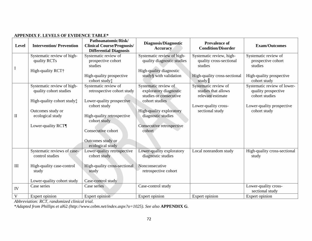
72
APPENDIX F. LEVELS OF EVIDENCE TABLE*
Level Intervention/ Prevention
Pathoanatomic/Risk/
Clinical Course/Prognosis/
Differential Diagnosis
Diagnosis/Diagnostic
Accuracy
Prevalence of
Condition/Disorder Exam/Outcomes
I
Systematic review of high-
quality RCTs
High-quality RCT†
Systematic review of
prospective cohort
studies
High-quality prospective
cohort study‡
Systematic review of high-
quality diagnostic studies
High-quality diagnostic
study§ with validation
Systematic review, high-
quality cross-sectional
studies
High-quality cross-sectional
study║
Systematic review of
prospective cohort
studies
High-quality prospective
cohort study
II
Systematic review of high-
quality cohort studies
High-quality cohort study‡
Outcomes study or
ecological study
Lower-quality RCT¶
Systematic review of
retrospective cohort study
Lower-quality prospective
cohort study
High-quality retrospective
cohort study
Consecutive cohort
Outcomes study or
ecological study
Systematic review of
exploratory diagnostic
studies or consecutive
cohort studies
High-quality exploratory
diagnostic studies
Consecutive retrospective
cohort
Systematic review of
studies that allows
relevant estimate
Lower-quality cross-
sectional study
Systematic review of lower-
quality prospective
cohort studies
Lower-quality prospective
cohort study
III
Systematic reviews of case-
control studies
High-quality case-control
study
Lower-quality cohort study
Lower-quality retrospective
cohort study
High-quality cross-sectional
study
Case-control study
Lower-quality exploratory
diagnostic studies
Nonconsecutive
retrospective cohort
Local nonrandom study High-quality cross-sectional
study
IV Case series Case series Case-control study Lower-quality cross-
sectional study
V Expert opinion Expert opinion Expert opinion Expert opinion Expert opinion
Abbreviation: RCT, randomized clinical trial.
*Adapted from Phillips et al62 (http://www.cebm.net/index.aspx?o=1025). See also APPENDIX G.

73
†High quality includes RCTs with greater than 80% follow-up, blinding, and appropriate randomization procedures.
‡High-quality cohort study includes greater than 80% follow-up.
§High-quality diagnostic study includes consistently applied reference standard and blinding.
║High-quality prevalence study is a cross-sectional study that uses a local and current random sample or censuses.
¶Weaker diagnostic criteria and reference standards, improper randomization, no blinding, and less than 80% follow-up may add bias and threats to
validity.

74
APPENDIX G. PROCEDURES FOR ASSIGNING LEVELS OF EVIDENCE
• Level of evidence is assigned based on the study design using the Levels of Evidence table
(APPENDIX F), assuming high quality (eg, for intervention, randomized clinical trial starts at
level I)
• Study quality is assessed using the critical appraisal tool, and the study is assigned 1 of 4 overall
quality ratings based on the critical appraisal results
• Level of evidence assignment is adjusted based on the overall quality rating:
– High quality (high confidence in the estimate/results): study remains at assigned level of
evidence (eg, if the randomized clinical trial is rated high quality, its final assignment is
level I). High quality should include:
Randomized clinical trial with greater than 80% follow-up, blinding, and
appropriate randomization procedures
Cohort study includes greater than 80% follow-up
Diagnostic study includes consistently applied reference standard and blinding
Prevalence study is a cross-sectional study that uses a local and current random
sample or censuses
– Acceptable quality (the study does not meet requirements for high quality and
weaknesses limit the confidence in the accuracy of the estimate): downgrade 1 level
Based on critical appraisal results
– Low quality: the study has significant limitations that substantially limit confidence in the
estimate: downgrade 2 levels
Based on critical appraisal results
– Unacceptable quality: serious limitations - exclude from consideration in the guideline
Based on critical appraisal results

75
APPENDIX H. Ligament sprain CPG revision – Critical Appraisal Scores
Clinical Course-Levels of Evidence adapted from Phillips
Systematic review of
prospective cohort
studies
High-quality prospective
cohort study
Systematic review of
retrospective cohort
studies
Lower-quality
prospective cohort study
High-quality
retrospective cohort
study
Consecutive cohort
Outcomes study or
ecological study
Lower quality
retrospective cohort
study
High-quality cross-
sectional study
Case-control study Case series Expert opinion
Katz 2013 X
McLeod 2012 X
Osteras 2012 X
Al-Dadah 2011 X
Busija 2008 X
Fabricant 2008 X
Zaffagnini 2011 X
Kijowski 2011 X
Hall 2014 X
Hall M 2014 X
Hall 2013 X
Thorlund 2014 X
Thorlund 2012 X
Stein 2010 X
Scanzello 2013 X
Sung-Gon 2013 X
Filardo 2015 X
Xu 2015 X

76
Risk factors – AMSTAR systematic reviews
Was an 'a
priori'
design
provided?
Was there
duplicate
study
selection
and data
extraction?
Was a
comprehen
sive
literature
search
performed?
Was the
status of
publication
(i.e. grey
literature)
used as an
inclusion
criterion?
Was a list
of studies
(included
and
excluded)
provided?
Were the
characterist
ics of the
included
studies
provided?
Was the
scientific
quality of
the
included
studies
assessed
and
documente
d?
Was the
scientific
quality of
the
included
studies
used
appropriate
ly in
formulating
conclusions
?
Were the
methods
used to
combine
the findings
of studies
appropriate
?
Was the
likelihood
of
publication
bias
assessed?
Was the
conflict of
interest
included?
What is
your
overall
assessment
of the
methodolog
ical quality
of this
review?
(High,
Acceptable,
Low,
Unacceptab
le)
Snoeker
2013 Y Y Y N Y Y Y CA Y CA Y HQ
Papalia
2011 Y Y Y N N N Y Y Y CA N AQ
Petty 2011 Y N N Y N N N N Y N N LQ
Risk factors – SIGN Cross-sectional
The
study
addresses
an
appropria
te and
clearly
focused
question
The two
groups
being
studied
are
selected
from
source
populatio
ns that
are
compara
ble in all
respects
other
than the
factor
The
study
indicates
how
many of
the
people
asked to
take part
did so, in
each of
the
groups
being
studied
The
likelihoo
d that
some
eligible
subjects
might
have the
outcome
at the
time of
enrolmen
t is
assessed
and
taken
into
What
percenta
ge of
individua
ls or
clusters
recruited
into each
arm of
the study
dropped
out
before
the study
was
complete
d
Compari
son is
made
between
full
participa
nts and
those lost
to follow
up, by
exposure
status
The
outcomes
are
clearly
defined
The
assessme
nt of
outcome
is made
blind to
exposure
status. If
the study
is
retrospec
tive this
may not
be
applicabl
e
Where
blinding
was not
possible,
there is
some
recogniti
on that
knowled
ge of
exposure
status
could
have
influence
d the
assessme
The
method
of
assessme
nt of
exposure
is
reliable
Evidence
from
other
sources
is used to
demonstr
ate that
the
method
of
outcome
assessme
nt is
valid and
reliable
Exposure
level or
prognosti
c factor
is
assessed
more
than
once
The main
potential
confound
ers are
identified
and
taken
into
account
in the
design
and
analysis
Have
confiden
ce
intervals
been
provided
?
How
well was
the study
done to
minimise
the risk
of bias or
confound
ing?
(High,
Acceptab
le, Low,
Unaccept
able)

77
under
investiga
tion
account
in the
analysis
nt of
outcome
Chhadia
2011 Y Y DNA DNA DNA Y CS CS Y CS DNA Y Y AQ
Risk factors – SIGN Cohort
The
study
addresses
an
appropria
te and
clearly
focused
question
The two
groups
being
studied
are
selected
from
source
populatio
ns that
are
compara
ble in all
respects
other
than the
factor
under
investiga
tion
The
study
indicates
how
many of
the
people
asked to
take part
did so, in
each of
the
groups
being
studied
The
likelihoo
d that
some
eligible
subjects
might
have the
outcome
at the
time of
enrolmen
t is
assessed
and
taken
into
account
in the
analysis
What
percenta
ge of
individua
ls or
clusters
recruited
into each
arm of
the study
dropped
out
before
the study
was
complete
d
Compari
son is
made
between
full
participa
nts and
those lost
to follow
up, by
exposure
status
The
outcomes
are
clearly
defined
The
assessme
nt of
outcome
is made
blind to
exposure
status. If
the study
is
retrospec
tive this
may not
be
applicabl
e
Where
blinding
was not
possible,
there is
some
recogniti
on that
knowled
ge of
exposure
status
could
have
influence
d the
assessme
nt of
outcome
The
method
of
assessme
nt of
exposure
is
reliable
Evidence
from
other
sources
is used to
demonstr
ate that
the
method
of
outcome
assessme
nt is
valid and
reliable
Exposure
level or
prognosti
c factor
is
assessed
more
than
once
The main
potential
confound
ers are
identified
and
taken
into
account
in the
design
and
analysis
Have
confiden
ce
intervals
been
provided
?
How
well was
the study
done to
minimise
the risk
of bias or
confound
ing?
(High,
Acceptab
le, Low,
Unaccept
able)
Pestka
2014 Y Y N N N Y N Y Y Y Y N N AQ
Salzman
n 2013 Y Y Y N N Y DNA N Y Y N N N AQ
Ebert
2013 Y Y N Y N N DNA CS Y Y N N Y AQ
Jungman
n 2012 Y Y N N N Y Y Y Y Y N N N AQ

78
Hwang
2012 Y Y DNA DNA N Y DNA CS Y Y N N Y AQ
Lyman
2013 Y Y N DNA DNA Y DNA N Y Y N Y Y AQ
Jaiswal
2012 Y Y N DNA N Y N N Y Y Y N Y AQ
Rosenber
ger 2010 Y Y N N N Y Y Y Y Y Y N Y AQ
Wu 2016 Y CS Y Y CS Y NA N Y Y N N N AQ
Risk factors – SIGN Case Control
The study
addresses
an
appropriate
and clearly
focused
question
The cases
and
controls are
taken from
comparable
populations
The same
exclusion
criteria are
used for
both cases
and
controls
What
percentage
of each
group
(cases and
controls)
participated
in the
study?
Compariso
n is made
between
participants
and non-
participants
to establish
their
similarities
or
differences
Cases are
clearly
defined and
differentiat
ed from
controls
It is clearly
established
that
controls are
non-cases
Measures
will have
been taken
to prevent
knowledge
of primary
exposure
influencing
case
ascertainme
nt
Exposure
status is
measured
in a
standard,
valid and
reliable
way
The main
potential
confounder
s are
identified
and taken
into
account in
the design
and
analysis
Confidence
intervals
are
provided
How well
was the
study done
to minimise
the risk of
bias or
confoundin
g?
Englund
2009 Y Y Y Y Y Y Y Y Y N HQ
Kluczynski
2013 Y Y Y N Y Y CS Y N Y AQ
Risk factors – Modified Case Series
Did the
study
address a
clearly
focused
question /
issue?
Is the
research
method
(study
design)
appropriate
for
answering
the
research
question?
Are both the
setting and the
subjects
representative
with
regard to the
population to
which the
findings will be
referred?
Is the
researcher’s
perspective
clearly
described
and taken
into
account?
Are the
methods for
collecting
data clearly
described?
Are the
methods for
analyzing the
data likely to
be valid and
reliable? Are
quality
control
measures
used?
Was the
analysis
repeated
by more
than one
researcher
to
ensure
reliability?
Are the
results
credible, and
if so, are they
relevant for
practice?
Are the
conclusions
drawn
justified by
the results?
Are the
findings of
the study
transferabl
e to other
settings?
How well
was the
study done
to minimise
the risk of
bias or
confounding
?

79
Henry 2012 Y Y Y CS Y Y CS Y Y Y HQ
Crema 2012 Y Y Y CS Y Y Y Y Y Y HQ
Crema 2010 Y Y Y CS Y Y CS Y Y Y HQ
Ding 2009 N Y Y CS N Y CS Y Y Y AQ
Guenther
2014 Y Y Y CS Y Y CS Y Y Y HQ
Jacob 2012 N Y Y CS N Y CS Y Y CS AQ
Examination – Outcome measures- Levels of Evidence adapted from Phillips
Systematic review of
prospective cohort
studies
High-quality prospective
cohort study
Systematic review of
lower-quality
prospective cohort
studies
Lower-quality
prospective cohort study
High-quality cross-
sectional study
Lower-quality cross-
sectional study Expert opinion
What is your overall
assessment of the
methodological quality
of this review? (High,
Acceptable, Low,
Unacceptable)
Engelhart 2012 X AQ
Goodwin 2011 X AQ
Garratt 2008 X AQ
Salavati 2008 X AQ
Van de Graaf 2014 X AQ
Almangoush 2013 X AQ
Balain 2009 X AQ
Smith 2009 X AQ
Celik 2015 X AQ
Vaquero 2014 X AQ
Examination – Physical Impairment Measures- Levels of Evidence adapted from Phillips

80
Systematic review of
prospective cohort
studies
High-quality prospective
cohort study
Systematic review of
lower-quality
prospective cohort
studies
Lower-quality
prospective cohort study
High-quality cross-
sectional study
Lower-quality cross-
sectional study Expert opinion
What is your overall
assessment of the
methodological quality
of this review? (High,
Acceptable, Low,
Unacceptable)
Décary 2016 X AQ
Blyth 2015 X AQ
Haviv 2015 X AQ
Snoeker 2015 X AQ
Campbell 2014 X LQ
Interventions- AMSTAR systematic review
The study
addresses a clearly
defined
research question
At least two
people should
select
studies and extract data
A
comprehensive
literature
search is carried out
The authors clearly state
if or how
they limited their review
by
publication type
The
included and
excluded
studies are listed
The
characteristics of the
included
studies are provided
The scientific
quality of
the included studies is
assessed
and documented
The scientific
quality of
the included studies was
assessed
appropriate-ly
Appropriate
methods are
used to combine the
individual
study findings
The
likelihood of
publication
bias is assessed
Conflicts of
interest are declared
What is your
overall
assessment of
the
methodologic
al quality of
this review?
(High
Quality ≥
8;Acceptable
≥5; Low ≤4
Fazalare 2010 CA N Y N N Y Y Y Y N N AQ
Wasielewski 2011 CA
Y Y N N Y Y Y Y N N AQ
Imoto 2011 CA
Y Y Y Y Y Y N Y N N AQ
Papalia 2013 CA
Y Y N N Y Y Y CA N N AQ
Dias 2013 CA
Y Y N Y Y Y Y Y N N AQ
Harston 2012 CA
CA Y N N Y Y Y NA N N LQ
Coppola 2009 CA
Y Y N N Y Y Y CA N N AQ
McLeod 2012 CA
CA Y N N Y Y Y Y Y N AQ
Reid 2012 CA
Y Y N N Y Y Y Y N N AQ

81
Interventions- PEDro
The study
addresses a
clearly defined
research question
At least two
people
should select
studies and extract data
A
comprehens
ive literature
search is carried out
The authors
clearly state if or how
they limited
their review by
publication type
The
included
and excluded
studies are listed
The
characteristi
cs of the included
studies are provided
The
scientific quality of
the included
studies is assessed
and documented
The
scientific quality of
the included
studies was assessed
appropriate-ly
Appropriate methods are
used to
combine the individual
study findings
The
likelihood
of publication
bias is assessed
Conflicts of
interest are declared
What is your
overall
assessment of
the
methodologic
al quality of
this review?
(High
Quality ≥
8;Acceptable
≥5; Low ≤4
Kelln 2009 Y Y Y N N N N Y Y Y Y AQ
Edwards 2013 Y Y Y Y N N Y Y Y Y Y HQ
Wondrasch 2009
AJSM Y Y Y CA N N Y Y Y Y Y HQ
Akkaya 2012 N Y Y Y N N Y Y Y Y Y HQ
Lind 2013 Y Y Y CA N N N N N Y Y AQ
Katz 2013
Y Y N Y N N N Y Y Y Y AQ
Osteras Bodywork
2014 Y Y Y CA N Y N Y Y Y Y HQ
Østerås KSSTA
2014 Y Y N CA N N N Y N Y Y AQ
Østerås Bodywork
2012 Y Y Y Y N Y N Y Y Y Y HQ
Ebert 2008 Y Y N Y N N Y Y Y Y Y HQ
Oravitan 2013 Y Y N Y N N N Y Y Y Y AQ
Koutras 2012 Y Y Y Y Y N N Y Y Y Y HQ
Kise 2016 Y Y Y CA N N Y Y Y Y Y HQ
Hall 2015 Y Y Y Y N N Y Y Y Y Y HQ

82
Interventions- Modified Case Series
Did the
study
address a
clearly
focused
question /
issue?
Is the
research
method
(study
design)
appropriat
e for
answering
the
research
question?
Are both
the setting
and the
subjects
representa
tive with
regard to
the
population
to which
the
findings
will be
referred?
Is the
researcher
’s
perspective
clearly
described
and taken
into
account?
Are the
methods
for
collecting
data
clearly
described
?
Are the
methods
for
analyzing
the data
likely to be
valid and
reliable?
Are
quality
control
measures
used?
Was the
analysis
repeated
by more
than one
researcher
to
ensure
reliability
?
Are the
results
credible,
and if so,
are they
relevant
for
practice?
Are the
conclusion
s drawn
justified
by the
results?
Are the
findings of
the study
transferab
le to other
settings?
How well
was the
study done
to
minimise
the risk of
bias or
confoundi
ng?
Wondrasch
2013 Y Y Y CA Y Y CA Y Y Y HQ
Assche 2011 Y Y Y CA Y Y CA Y Y Y HQ
Neogi 2013 Y Y Y CA
Y Y CA
Y Y Y HQ
Lim 2010 Y Y CA CA
N CA CA
Y Y Y AQ
Elbaz 2013 N Y Y Y Y CA CA
Y Y Y AQ

83
Acknowledgments: The authors would like to acknowledge the contributions of George Washington University Himmelfarb Health
Science librarian, Tom Harrod, for his guidance and assistance in the design and implementation of the literature search. The authors
would also like to acknowledge the assistance in screening articles provided by Nicholas Ienni, Doctor of Physical Therapy student.