
David Pendergrast Auckland Eye - gpcme.co.nz North/0832 room 7 Thurs... · –No loss of red reflex...
48
Eye Emergencies David Pendergrast Auckland Eye
Transcript of David Pendergrast Auckland Eye - gpcme.co.nz North/0832 room 7 Thurs... · –No loss of red reflex...

Eye Emergencies
David Pendergrast
Auckland Eye
Ophthalmic Emergencies
• Patients will often present to their GP with ophthalmic symptoms and signs and these may simply indicate normal visual experience or some possible pathology from minor to major.
• A small number of patients will present to you with conditions where your immediate management will influence the final outcome.
• Your recognition of these and appropriate referral may save vision or even life.

Outline
• Eye trauma
– PEI, Blunt Injury, Chemical injury
• Acute loss of vision
– Keratitis, Angle closure glaucoma, Retinal detachment, Vascular occlusion, Endophthalmitis

Trauma: Penetrating Eye Injury
• Compromised integrity of cornea or sclera• Not always obvious so always consider it if
history suggestive• Delay in treatment can lead to worse outcome:
– Loss of or damage to intra-ocular contents– Severe infection: Endophthalmitis
• ALSO
– Sympathetic ophthalmia if treatment delayed– Intra-ocular toxicity if IOFB not recognised

Intra ocular foreign body??
• High velocity: hammer on steel, nail gun
• High level of suspicion
• Look carefully for entry wound: may be on lids
• X-ray orbits: upgaze, downgaze, lateral

PEI Slitlamp Signs• Shallow anterior chamber (cf uninjured eye)
• Irregular or displaced pupil
• Prolapsed uveal tissue

PEI: Seidel Test• Undiluted fluorescein
• When diluted by aqueous fluoresces brightly
• Stream of aqueous revealed
• Don’t press on eye
• Don’t bother doing if PEI obvious

Suspected PEI Management:• Eye shield
• Tonometry contraindicated
• Leave embedded foreign object in place
• Analgesia and antiemetics: prevent vomiting / squeezing
• Update tetanus immunization
• Immediate referral to an ophthalmologist
• NBM usually as surgery likely

Any questions?
Trauma: Blunt Injury
• Assault, sporting, airbags, bungees
• Consider other injury may also be present :
– globe rupture
– blow out fracture
• Refer for S/L exam urgently
• Even without PEI the consequences for vision can be severe

Trauma: Blunt Injury• Hyphaema:
– Bleeding from torn iris or angle vessels
– Secondary bleed often more severe
– Bed rest essential
– IOP control

Trauma: Blunt Injury• Hyphaema:
– Corneal bloodstaining
– Risk if high pressure and repeat bleeding
– May take months to clear
Pupil margin Bloodstaining

Trauma: Blunt Injury• Iris injury:
– Compressive force can tear fragile iris tissue
– Most common : sphincter splits
– More severe: iridodialysis

Trauma: Blunt Injury• Angle recession:
– Root of iris and trabecular meshwork torn off sclera
– Gonioscopy on all hyphaemas when settled
– Risk of late glaucoma if recession present as trabecular meshwork damaged
– All significant blunt injury needs referral

Trauma: Blunt Injury• Lens dislocation and cataract
– Often marked change in vision
– Sometimes not obvious
– Subtle A/C deepening
– Iridodonesis
– Important to identify history of trauma in any patient with cataract as zonular damage likely

Trauma: Blunt Injury
• Commotio retinae:
• Posteriorly transmitted force– Haemorrhage
– Oedema
– Traumatic macular hole
– Long term vision reduction

Trauma: Chemical Injury
• True ocular emergency: don’t check VA• Time to irrigation and duration influences final
outcome• Topical anaesthetic• Saline or Ringer’s• IV tube or Morgan lens • At least 1-2 litres• At least 30 minutes
Trauma: Chemical Injury
• Don’t attempt to neutralise alkali with acid.
• Transfer urgently to ophthalmology dept.
• If dry / particulate chemical and delay in transfer, may sweep superior fornix with cotton bud to remove solid debris.
• Patients should be instructed to bring the container of the chemical that caused their eye injury.

Trauma: Chemical injury• Prognosis depends on degree of limbal damage
• Alkali usually worse than acid as it penetrates more
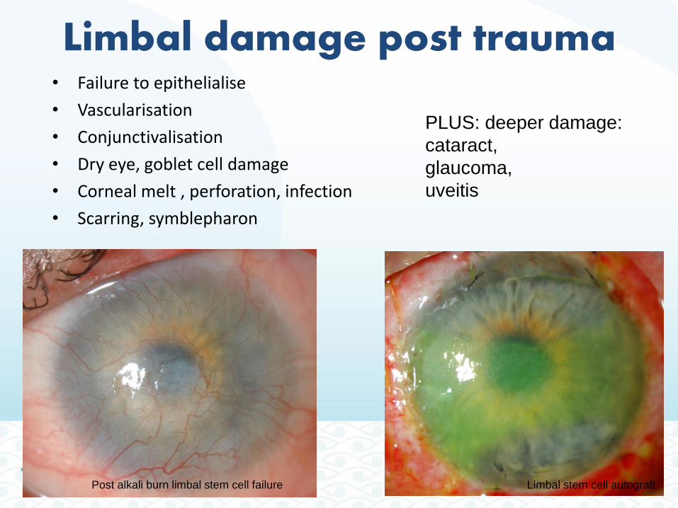
Limbal damage post trauma• Failure to epithelialise
• Vascularisation
• Conjunctivalisation
• Dry eye, goblet cell damage
• Corneal melt , perforation, infection
• Scarring, symblepharon
Post alkali burn limbal stem cell failure Limbal stem cell autograft
PLUS: deeper damage:
cataract,
glaucoma,
uveitis

Any questions ?

Sudden Visual Loss:– Urgent appropriate management may be vision saving.
• Cornea
• Glaucoma
• Retina
• Vascular
• Endophthalmitis
– PLUS: recent awareness of chronic loss:
• Refractive error and amblyopia
• Unilateral cataract
• End stage open angle glaucoma

Microbial keratitis• Symptoms:
– Lacrimation
– Photophobia
– Irritation or pain
– Reduced vision
• Signs:
– Intense injection (circumcorneal)
– Corneal infiltrate
– Epithelial defect (fl. +ve)
– Hypopyon

Microbial Keratitis
• Viral• Bacterial• Fungal• Amoebic• Features seldom
pathognomonic• Most need urgent
admission, corneal scrape for culture
• Intensive Rx, adjusted once results available

Risk factors for microbial keratitis
• Does not “just happen” (except HSV)
• C/L wear especially any overnight wear
• Trauma including surgery.
• Reduced ocular surface defences: lid abnormalities, corneal anesthesia, dry eye, poor blink.
• Reduced systemic defences: diabetes, immunocompromise, malnutrition, old age.

Hydrops in Keratoconus
• Another corneal cause of sudden vision loss
• Keratoconus may be undiagnosed
• Split in DM allows aqueous into stroma
• Gradual resolution over a few months

Acute Angle Closure Glaucoma
• If acute angle-closure glaucoma is not treated immediately, damage to the optic nerve and significant and permanent vision loss can occur within hours.
• Patients often present with
– blurred vision
– severe eye pain / frontal headache
– colored halos around lights
– nausea and vomiting
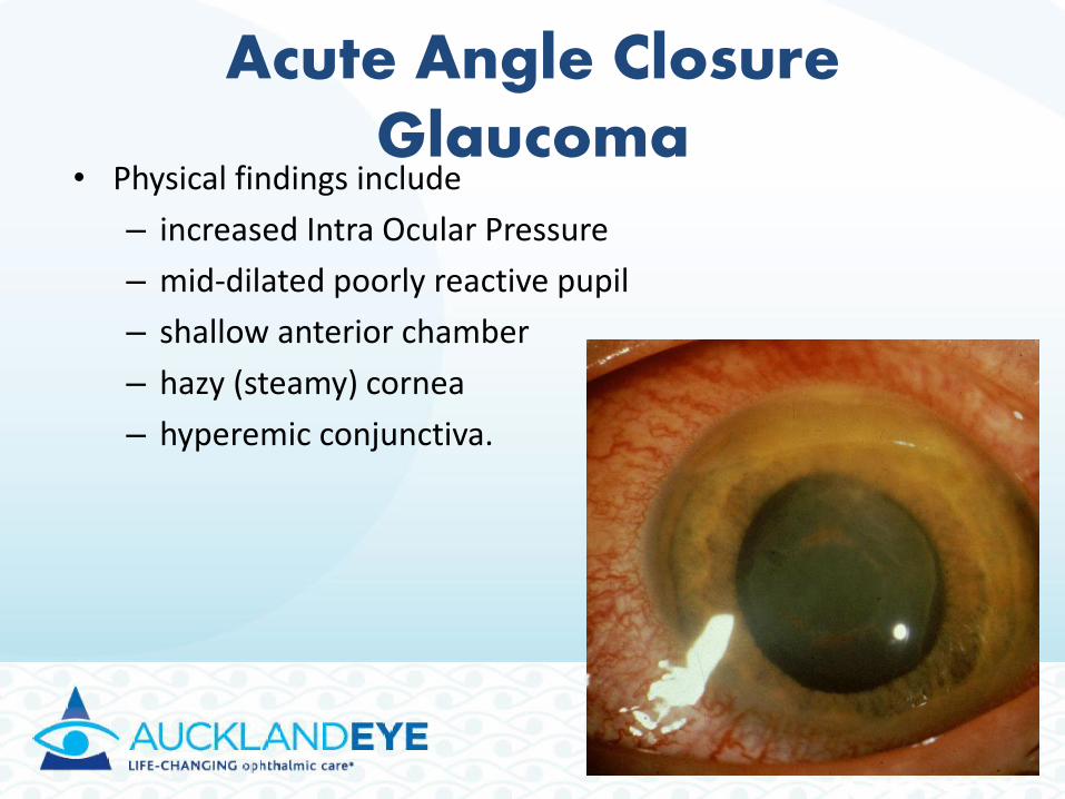
Acute Angle Closure Glaucoma
• Physical findings include
– increased Intra Ocular Pressure
– mid-dilated poorly reactive pupil
– shallow anterior chamber
– hazy (steamy) cornea
– hyperemic conjunctiva.

Acute Angle Closure Glaucoma
• Risk factors include:
– Enlargement or anterior placement of the lens
– Hypermetropia
– Narrow angle, and shallow anterior chamber.

Acute Angle Closure Glaucoma• Therapy is initiated to lower the
intraocular pressure, reduce pain, and clear corneal oedema in preparation for iridotomy.
• Topical pressure lowering agents:
– 0.5% timolol maleate (Timoptol)
– 1% apraclonidine (Iopidine)
– 2% pilocarpine (Isopto Carpine)
• Oral Acetazolamide
• Definitive treatment is laser iridotomy.
• Surgical iridectomy if laser iridotomy not successful.
Sudden Visual Loss: Retinal
• Retinal detachment
• Retinal artery occlusion
• Retinal vein occlusion
• Vitreous haemorrhage
• Wet macular degeneration

Retinal Detachment:• Separation of neural retina from the RPE
• Separates photoreceptors from their blood supply
• Early diagnosis and treatment essential
• Treated within days often full return of vision
• Delayed treatment may lead to permanent vision loss even NPL in the eye

Retinal Detachment:
• 1 in 10,000 per year• 1 to 5 per week at Greenlane Clinical Centre• Risk factors:
– Myopia : 55% of non traumatic RRD– Cataract surgery (esp. complicated ) – Diabetic retinopathy (tractional)– Family history of retinal detachment– Older age ie degenerative retinal holes– Trauma

Retinal Detachment:• Flashing lights (vitreous traction on peripheral retina)
• Floaters ( vitreous condensation or haemorrhage)
• Curtain like progressive field defect
• Reduced central vision once macula detached
• VA may be 6/6 to PL
• Not usually loss of RR or APD

Retinal Detachment:
• Difficult to identify with direct ophthalmoscope• Referral is mandatory for symptoms• The more recent the onset the more urgent• Surgery usually involves;
– Vitrectomy– Drainage of sub retinal fluid– Intra-ocular gas or oil to flatten the retina– Laser or cryotherapy to seal off the hole– External scleral indents less often used now

Retinal artery occlusion• CRAO or BRAO
• Embolic: carotid or cardiac: will need ECHO and carotid imaging
• Difficult to see with direct ophthalmoscope
• GCA if >60 years old
• All need ESR, CRP urgently
• All need referral
• Bilateral, transient consider:– Migraine
– Vertebrobasilar

Central Retinal Artery Occlusion
• Painless and sudden loss of vision in one eye.
• Amaurosis fugax (transient) may precede loss.
• Signs:
– Reduced VA
– APD usually
– No loss of red reflex
– Cherry red spot

Risk factors• Older patients:
– atherosclerosis, diabetes, hypercholesterolaemia, hypertension, hypercoagulable state, cardiac arrhythmias.
• Giant cell arteritis in 5 to 10 percent of cases
• Younger patients:
– collagen vascular diseases, hypercoagulopathies, cardiac valvular disease, syphilis, sickle cell disease.
• Glaucoma, eye surgery: post elevated IOP.

Retinal vein occlusion
• Central or branch vein occlusion
• Often a delay to present
• Blood and thunder fundus
• Measure BP, FBC, glucose, lipids
• Refer for FFA and OCT
• Late neovascularisation:– Retina or iris
– 90 day glaucoma
• May need PRP or anti-VEGF
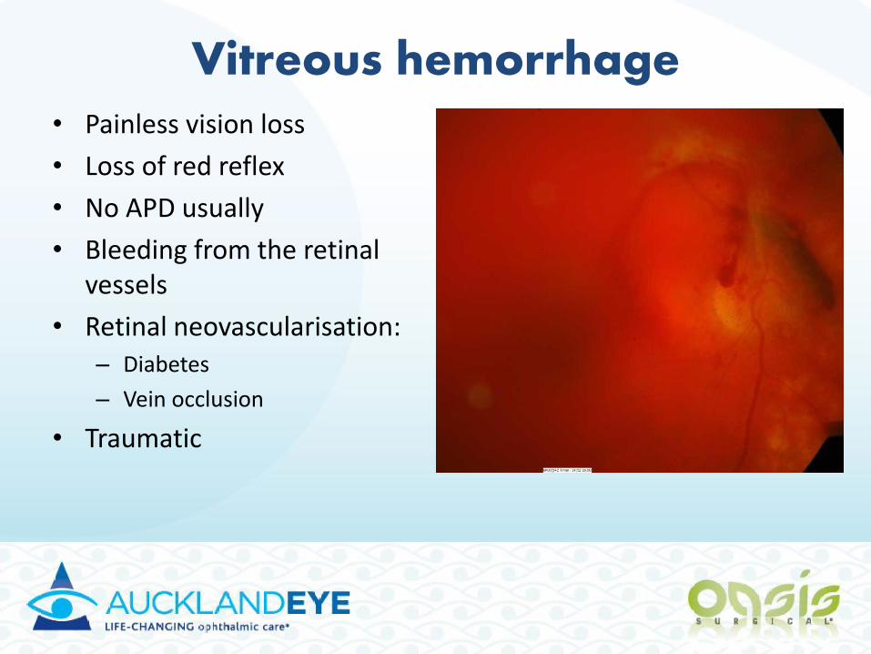
Vitreous hemorrhage• Painless vision loss
• Loss of red reflex
• No APD usually
• Bleeding from the retinal vessels
• Retinal neovascularisation:– Diabetes
– Vein occlusion
• Traumatic

Macular Degeneration
• Central loss, peripheral vision maintained
• No APD
• Good red reflex
• Painless
• Preceded by metamorphopsia

Macular Degeneration:
• Gradual loss: not urgent but needs referral
• Sudden loss or metamorphopsia: urgent referral
• Previously little therapy to reverse vision loss
• Now : intravitreal VEGF inhibitors:– Halt deterioration
– Improve vision in many cases
• But: temporary benefit and need repeat usually weeks to months later

Sudden visual loss: Endophthalmitis
• Severe pain
• Reduced vision
• Recent eye surgery
• Hypopyon
• Intense injection
• Poor red reflex

Sudden Visual Loss: Endophthalmitis
• Post cataract: 1 in 1000 incidence
• Post intravitreal avastin injection: 1 in 200
• Emergency referral
• Risk of severe vision loss high
• Reduced by early intra-vitreal antibiotics asap. (hours count)
• Then will require vitrectomy
Any questions?

Summary 1• Prompt recognition and appropriate
treatment essential
• The outcome may depend on timely management
• Refer ocular emergencies immediately to the emergency department or ophthalmologist
• Most frequent conditions:
– PEI, Retinal detachment, CRAO, Acute angle-closure glaucoma, and Chemical injury

Summary 2• Eye injury from high-velocity trauma should be
immediately evaluated by ophthalmologist.
• Suspected globe rupture should be immediately referred to an ophthalmologist.
• An eye exposed to chemicals should be irrigated with at least 1 to 2 liters of normal saline or other solution suitable for eye irrigation.
• Lowering intraocular pressure in acute angle-closure glaucoma may save vision; laser iridotomy is the definitive treatment for acute angle-closure glaucoma.
Thank you!