
Hiding Data in Images Using Cryptography and Deep Neural ...
Data Hiding in Medical ImagesArindam Dasa, Saiful Islama, Sandesh Guptab, Phalguni Guptaa
a Department of Computer Science and Engineering,Indian Institute of Technology Kanpur, Kanpur, India 208016
Email: {dasa, sislam, pg}@cse.iitk.ac.inb Department of Computer Science and Engineering,
U.I.E.T., C.S.J.M University Kanpur, Kanpur, India 208016Email: [email protected]
Abstract— This paper presents an efficient technique to hidetext information in 3D medical images such as MRI andPET. It embeds text information only in the non-anatomicalpixels. It ensures that no anatomical part of the 3D medicalimage gets contaminated. It makes the retrieval of 100%data. The technique has been tested on several MRI andPET images.
Index Terms— MRI, PET, Segmentation, Region Growing,Data Hiding
I. INTRODUCTION
Image is one of the most preferred multimedia whichcan be used to carry overt information between communi-cating entities. There exist considerably good number oftechniques to hide data within an image. They have beendeveloped based on the specific needs over time. Aim ofhiding techniques is to provide the least change in thecover image with the best possible quality of the hiddendata. For example, in medical domain, there is often arequirement by physicians to discuss among themselvesabout the status of the patient’s health. Consider thefollowing situation where a doctor who has a brain MRIimage of a patient wants to send it to the patient to see hisMRI scan and also to communicate his observations to anexpert for consultation. There is a necessity of secretlyembedding text data in a medical image which can beaccessed only by the intended recipient.
This paper presents a new technique of informationhiding in the field of medical imaging. The proposed tech-nique is different from watermarking and steganographybecause in this present work data can not be embeddedby distorting region of interest of cover work. In medicaldomain, distortion of medical images is not permissibledue to legal and technical reasons. Hence, embedding inmedical images should not distort region of interest in anyway. The paper is organized as follows: Section II brieflyreviews some well known segmentation algorithms. InSection III, a new information hiding algorithm has been
Manuscript received January 2, 2013; revised September 12, 2013.Corresponding author: Saiful IslamAuthors like to acknowledge the support provided by the Department
of Information Technology, Government of India to carry out thisresearch work and All India Institute of Medical Sciences(AIIMS), NewDelhi, India for providing MRI and PET images.
presented. The simulation results are analyzed in the nextsection. Conclusions are given in Section V.
II. RELATED WORKS
In the context of medical images, segmentation isrequired for distinguishing organs by different textures inmedical images. There exist several segmentation tech-niques for medical images which can be classified intofour categories. First one is based on Region Growing [1].It is an effective approach for segmenting a diverse classof images. A region growing algorithm generally startsfrom a seed region (which is typically one or more pixels)which is considered to be within the region of the objectto be segmented in the image. Pixels of the neighboringregion are tested to decide whether they should also beconsidered as a part of the region or not. If so, the pixelsare added to the region and the process is continueduntil no more new pixels can be included in the region.Techniques of this kind mainly differ depending on thefactors used to decide the possibility to include a pixel ina region, the connectivity used to determine neighbors, orthe method of visiting the neighboring pixels.
The second category of segmentation techniques isbased on Watersheds [1] which classify pixels into regionsby virtue of gradient descent on image features andanalysis of weak points along region boundaries. It isbased on the principle of water raining onto a landscapeand flowing due to gravity towards lower basins. Size ofthose basins grows with increasing amount of water untilwater gets spilled into one another resulting in smallerbasins merging into bigger ones. Regions are formedusing local geometric shapes to relate pixels in imagedomain with local extrema in some feature measurement.This technique is less affected by user-defined thresh-old unlike any region-growing method. The watershedtechniques are also flexible in terms of not producing asingle segmentation but a hierarchy of segmentation asdescribed in [2], [3]. The drawback of this approach isthat it produces too many regions by over segmenting theimage with redundancy in regions.
Level Set Segmentation [1] is another category whichdetects evolution of contours and surfaces on the image.The main advantage of using level sets in segmentation
JOURNAL OF COMPUTERS, VOL. 9, NO. 3, MARCH 2014 513
© 2014 ACADEMY PUBLISHERdoi:10.4304/jcp.9.3.513-518

is that arbitrarily complex shapes can be modeled andtopological changes can be handled implicitly. Thesetechniques can be used for segmentation by using imagebased features such as mean intensity, gradient and edgesin the governing differential equation by which the levelset function is evolved. In this technique, a contour isinitialized by the user to evolve until it finds a perfect fitof an anatomical structure. An overview of this categoryof segmentation is given in [4]. Techniques under thiscategory are found to be computationally expensive.
Hybrid Segmentation [5], [6] is another category thatintegrates boundary-based and region-based segmentationmethods. This kind of techniques is used when thecategory of problem is too exceptional to achieve propersegmentation by using any of the above techniques. Thesetechniques are appropriate only when a highly customizedsolution is required for the segmentation of images.
III. THE PROPOSED SCHEME
Figure 1. Data Flow diagram for embedding text data into 3D medicalbrain image (a) Source Image (b) Segmentation unit (c) Mask todistinguish brain area (d) Embedding unit (e) Text data to be embedded(f) Text embedded brain image.
Figure 2. Data Flow diagram for retrieving text data from 3D medicalbrain image (a) Image with embedded text (b) Segmentation unit (c)Mask to distinguish brain area (d) De-embedding unit (e) Retrieved textdata.
This section presents an efficient technique to hide textinformation inside 3D medical images. It consists of twomodules; segmentation of non-anatomical part from brainimage followed by embedding secret information. Figure
1 depicts the flow diagram of the technique. The 3Dimage is segmented to crop out the anatomical regionand to form a mask which marks the volumetric pixels(voxels) of the anatomical region so that anatomical andnon-anatomical voxels can be distinguished. The textinformation is embedded in the non-anatomical part ofthe image with help of the mask. Similarly, at the retrievalstage, the text embedded medical image is segmentedinto the anatomical and non-anatomical parts and the textinformation is retrieved from the non-anatomical part.Figure 2 shows the retrieving mechanism.
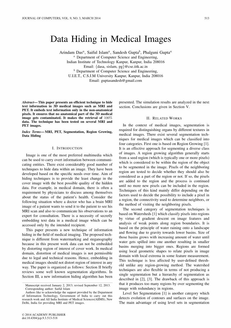
Medical images are 3D and of gray-scale type withvalues ranging from 0 to infinite; however, typically thehighest gray-scale value of any voxel in an image is foundto be 2000. Unlike the 2D images (carrying no sensitivedata), the 3D medical images generally carry sensitivepatient information and under no circumstance the voxelintensity (in the anatomical regions) can be changed.Hence, the anatomical region should be kept isolated fromthe embedding process. One might come up with thetrivial idea of simply distinguishing the voxel with value0 or lesser than some threshold as the non-anatomicalregion of the image; but it can be noted that certain imagesmay contain noise and even non-anatomical region can beconsidered as anatomical region with this approach. As aresult, the embedding capacity becomes considerably low.Figure 3 shows an example of such an image.
Figure 3. Brain image of PET modality having a lot of noise surroundingthe brain.
The anatomical region cannot be found by ruling outthe voxels below some threshold since a noisy voxeloutside the anatomical region can always have value equalto the value of a voxel in the anatomical region. Thetechnique should be able to exploit the non-anatomicalregion of the image optimally to achieve the maximumembedding capacity. More the embedding capacity, moretext information can be stored.
514 JOURNAL OF COMPUTERS, VOL. 9, NO. 3, MARCH 2014
© 2014 ACADEMY PUBLISHER

The proposed segmentation technique is shown in Al-gorithm 1. It belongs to a region-growing category. Itstarts from a voxel (which has to be one of the voxelsof the anatomical region) and it goes on checking forits neighbors that whether they fall between a lower andupper threshold so that one can include them in the region.In a 3-dimensional space, every voxel has 28 neighborsand hence for each voxel, all its neighbors are checkedfor candidacy. If it is found to satisfy the criteria, theprocess is recursively called for each such neighbor. Thealgorithm traverses the connected neighbors starting froma seed coordinate to detect the anatomical region. Thetime complexity (worst case) of the technique is equalto the total number of voxels in the image. The initialparameters play an important role in the functioning ofthe technique. The seed coordinate, upper threshold (β)and lower threshold (α) values are provided to segmentout the desired region.
Algorithm 1 : SegmentationRequire: A 3D matrix M representing the voxel values
of the medical image of size (m× n× p)Seed coordinate S = (x, y, z) denoting the startingpoint for the algorithmLower Threshold α, a real value denoting theminimum allowable value for anatomical voxels.Upper Threshold β, a real value denoting themaximum allowable value for anatomical voxel
Ensure: A 3D matrix M ′ with values 0 or 1 representingthe mask image for anatomical region of M
1: V isited(S)← True2: if Intensity(S) ≥ α ∧ Intensity(S) ≤ β then3: M ′
x,y,z ← 14: for all neighbor N of S do5: if V isited(N) = False then6: Segmentation(N,α, β)7: end if8: end for9: else
10: M ′x,y,z ← 0
11: end if12: return M ′
1) Seed Coordinate: It cannot be trivially desig-nated by choosing the mid-voxel viz. the voxel(m/2, n/2, p/2) of an image of dimension (m ×n × p). This is because such a voxel may not bein an anatomical region but a in hollow part of theanatomical region which might turn out to be ofgray-scale value of zero. On the other hand, onemight have the intuition to select the voxel withthe highest gray-scale value as the seed coordinate.But that may not necessarily succeed since therecan be cases where the image might have noisesurrounding the anatomical region and coinciden-tally one of the noisy voxels (totally disconnected
from the anatomical region) may have the highestgray-scale value which may be mistakenly taken asthe seed-coordinate. In the medical images of anymodality, it is seen that the anatomical componentcertainly occupies the central 20% of the image andbecause of this, the seed-coordinate can be foundin the central 20% of the image. Hence, exploitingthis property, the voxel with the highest gray-scalevalue is found in the central 20% of the image andis used as the seed coordinate for the segmentationtechnique.
2) Upper/Lower Threshold: Ranges of intensity val-ues of different modalities are generally found tobe different. Also, two images of the same modalitymay have different gray-scale ranges. Hence, a con-stant lower/upper threshold may not provide goodresult. Threshold values (specific to the input image)are to be selected dynamically from the images. Theupper threshold can be determined from the highestgray-scale value of the image. But the selectionof the lower threshold value is not trivial. Certainobservations can be made from Figure 4. The lowerthreshold value is very much image specific andthus to be computed dynamically. Also it can beobserved that lesser the lower threshold value, morethe extraneous voxels selected as the anatomicalregion. Hence, it is more conservative in terms offlagging a voxel as a non-anatomical voxel. Morethe conservative segmentation, lesser the embeddingcapacity and lesser the conservative segmentations,more the chances of contaminating the anatomicalregion in the image. Through experiments on aconsiderable number of medical images of differentmodalities, it is inferred that the lower thresholdis nearly equal to 1
18
th of the value of the upperthreshold. By such upper and lower threshold val-ues, it is found to segment the anatomical regioncompletely without inclusion of extraneous voxels.
A. Data Embedding
The embedding process computes mask using segmen-tation and embeds text in the least significant bits of non-anatomical voxels. Seed coordinate, upper (β) and lower(α) thresholds are computed to segment input medicalimage M . Segmentation of M is done through the Al-gorithm 1. This segmented image S is used to computemask of the image to differentiate between anatomicaland non-anatomical part of any medical image. With themask S for the medical image, embedding of data canbe done in the non-anatomical region. Size of secretmessage is computed and is appended at the beginningof the secret message to form the augmented message asshown in Figure 5. Initial twenty bits of the augmentedmessage are reserved for storing message size. After this,the mask S and medical image M are randomly permutedthrough key p to get S′ and M ′. The key k is used todistribute message randomly over the image so that onlythe intended recipient can extract the secret text. Each of
JOURNAL OF COMPUTERS, VOL. 9, NO. 3, MARCH 2014 515
© 2014 ACADEMY PUBLISHER

(a) MRI Image (b) Lower Threshold = 30 (c) Lower Threshold = 142.5
(d) PET Image (e) Lower Threshold = 30 (f) Lower Threshold = 69.11
Figure 4. Brain image Segmentation with various Lower Threshold values.
8 bits of the text character is stored in least significantbit (LSB) of eight contiguous permuted non-anatomicalvoxels in the M ′. Finally, the permuted image M ′ isrearranged to get the resultant image M ′′. Algorithm 2delineates the technique.
Figure 5. Secret message format.
B. Data ExtractingData extraction is a process to retrieve the embedded
message from a image. Steps of extraction are similarto embedding process. Seed coordinate, upper (β) andand lower (α) thresholds are computed to segment theinput image M . Segmented image S is used as mask todistinguish the non-anatomical voxels. Image and maskare permutated using same key p to get M ′ and S′.Twenty bits of message length are extracted to get the sizeof message (msg size). Depending upon the message sizeall remaining bits of the augmented message are retrievedfrom the LSB of every non-anatomical voxels to form theoutput text (T ). The technique is given in Algorithm 3.
IV. PERFORMANCE EVALUATION
The proposed technique to embed text information in3D brain images has been tested on 3D images of brain
Algorithm 2 : Embedding3DRequire: A 3D matrix M representing the voxel values
of the medical image of size (m× n× p)Text to be embedded Tkey p
Ensure: M with text T and key p embedded in non-anatomical region
1: Let Seed coordinate (x, y, z) be the voxel with thehighest gray-scale value in central 20% of inputimage M
2: Let β (Upper Threshold) be the maximum voxel valuein M
3: Let α (Lower Threshold) be β18 ;
4: Let Embedding Mask S be the result of Segmenta-tion( M , (x, y, z) , α , β)
5: Let msg size be the size of Text to be embedded;6: Let T ′ be the augmented text (size and text )to be
embedded;7: Let M ′ ← permute(M,p); {Permutated Image}8: Let S′ ← permute(S,p); {Permutated Mask}9: for all character c in T ′ do
10: for i = 1 to 8 do11: (x, y, z) ← next voxel coordinate in S′ with
value 0; {Embedding Text}12: M ′
(x,y,z) ← ci; {ci is ith bit of character c}13: end for14: end for15: M ′′ ← permute(M ′, p); {Re-arranging Image}16: return M ′′
516 JOURNAL OF COMPUTERS, VOL. 9, NO. 3, MARCH 2014
© 2014 ACADEMY PUBLISHER

Algorithm 3 : DataExtracting3DRequire: A 3D matrix M representing the voxel values
of the medical image of size (m× n× p)key p
Ensure: Text T retrieved from non-anatomical region inM
1: Let Seed coordinate (x, y, z) be the voxel with thehighest gray-scale value in central 20% of inputimage M
2: Let β (Upper Threshold) be the maximum voxel valuein M
3: Let α (Lower Threshold) be β18 ;
4: Let Embedding Mask S be the result of Segmenta-tion( M , (x, y, z) , α , β)
5: Let M ′ ← permute(M,p); {Permutated Image}6: Let S′ ← permute(S,p); {Permutated Mask}7: let msg size be the message size retrieved from
consecutive voxels in S′ with value 08: for j = 21 to msg size+ 20 do9: Messagej ← LSB of M ′
Xj{Retrieving Message}
10: end for11: T ← Message;12: return T
of 44 subjects. Out of these images, there are 14 MRIimages and remaining are PET images. Each MRI imageis of size 192*256*176 while the size of PET images is128*128*47. A slice of MRI image and a PET image ofa brain are shown in Figure 6 and Figure 7. Segmentationand embedding text in the segmented image are done insuch a way that there is no contamination or loss of infor-mation in the brain image. Thus, the embedding capacityis much lesser than the actual capacity. Maximum size ofmessage that can be embedded in a image is known asembedding capacity. If an image of dimension (m×n×p)has x non-anatomical voxels, then its embedding capacity(in terms of number of bits) is be given by
EmbeddingCapacity =x
m× n× p(1)
where x8 − 2 is the maximum number of characters that
can be embedded.The capacity, however, is considerablylarge to fit a large amount of text data into 3D images.Each image is segmented to detect the brain region and theembedding capacity is calculated to be 18% or 1,038,666characters. The mask for the brain region is shown inFigure 8 and corresponding MRI image with embeddedtext is shown in Figure 9. The proposed technique suc-cessfully embedded text information for all the test casesin non-anatomical voxels of a medical image.
V. CONCLUSIONS
This paper has proposed an efficient technique to hidedata in 3D medical brain image. It is novel due to thefact that there has been no prior attempt to exploit the
Figure 6. MRI Image with he Text to be Embedded.
Figure 7. PET Image of Dimension (128 × 128 × 47) and size 1.47MB.
Figure 8. MRI Image Mask for Brain Region.
non-anatomical voxels of a medical image to embeddata for esoteric communication. The conventional wayof storing information in image header is accessible toanyone and hence, any data hiding technique of this typecertainly helps to maintain secrecy of information withinusers viz. doctors and patients. On the other hand, theproposed segmentation technique proposed can be furtherextended to segment different organs of the brain viz.cerebrum, medulla, etc. Further, it can be used as a tool
JOURNAL OF COMPUTERS, VOL. 9, NO. 3, MARCH 2014 517
© 2014 ACADEMY PUBLISHER

Figure 9. MRI Image Embedded with Text Data.
to remove noise from a medical images. It can be donesimply by embedding continuous sequence of characterswith ASCII 0 i.e. all pixels which do not belong tothe anatomical region can be set to a value zero. Thistechnique can also be used to embed information in otheranatomical images like lungs, limbs, etc. In that case, thesegmentation algorithm may be customized and properlytuned for desirable results.
ACKNOWLEDGMENT
The authors are grateful to the anonymous referees fortheir valuable comments and suggestions to improve thepresentation of this paper.
REFERENCES
[1] L. Ibanez, W. Schroeder, L. Ng, J. Cates, and T. I. S.Consortium, The Insight Tool Kit Software Guide, 2nd ed.Insight Tool Kit, 2005.
[2] T. Yoo, U. Neumann, H. Fuchs, S. Pizer, T. Cullip,J. Rhoades, and R. Whitaker, “Direct visualization ofvolume data,” IEEE Journal on Computer Graphics andApplications, vol. 12, no. 4, pp. 63 –71, 1992.
[3] T. S. Yoo, U. Neumann, H. Fuchs, S. M. Pizer,T. Cullip, J. Rhoades, and R. Whitaker, “Achievingdirect volume visualization with interactive semantic regionselection,” in Proceedings of the 2nd conference onVisualization(VIS ’91). Los Alamitos, CA, USA: IEEEComputer Society Press, 1991, pp. 58–65. [Online].Available: http://dl.acm.org/citation.cfm?id=949607.949617
[4] A. M. Andrew, “Level set methods and fast marchingmethods: Evolving interfaces in computational geometry,fluid mechanics, computer vision, and materials science,”Robotica, vol. 18, no. 1, pp. 89–92, 2000. [Online].Available: http://dl.acm.org/citation.cfm?id=980371.980382
[5] C. Imielinska, P. D, D. Metaxas, P. D, J. Udupa, P. D, Y. Jin,and T. Cheng, “Hybrid segmentation of the visible humandata,” in Proceedings of the Third Visible Human ProjectConference, Bethesda, MD, 2000.
[6] C. Imielinska, D. N. Metaxas, J. K. Udupa, Y. Jin,and T. Chen, “Hybrid segmentation of anatomical data,”in Proceedings of the 4th International Conferenceon Medical Image Computing and Computer-AssistedIntervention(MICCAI ’01). London, UK: Springer-Verlag,2001, pp. 1048–1057. [Online]. Available: http://dl.acm.org/citation.cfm?id=646924.709334
Arindam Das has graduated from the Department of ComputerScience and Engineering, Indian Institute of Technology Kanpur,India in 2012. He is working in a software firm located inBangaluru India. His area of research includes Medical Imageregistration and Data Hiding.
Saiful Islam is a research scholar in the Department of Com-puter Science and Engineering, Indian Institute of TechnologyKanpur, India. He has completed his graduation from AligarhMuslim University, Aligarh, India. He has worked as SeniorLecturer in the department of computer engineering, AligarhMuslim University. His area of research includes InformationHiding and Data Security.
Sandesh Gupta received his Bachelors in Technology from theUniversity Institute of Engineering and Technology, C.S.J.MUniversity Kanpur in 2001. Currently he is an Assistant Pro-fessor in the Department of Computer Science and Engineeringin the same institution. His areas of interest are Medical ImageProcessing, Computer Graphics and Biometric Security. He isoperation in-charge of ACM International Collegiate Program-ming Contest Asia Region, Kanpur Site from 2001. He hasseveral publications and one International patent. Recently hehas submitted his PhD thesis to the Uttar Pradesh TechnicalUniversity, Lucknow.
Phalguni Gupta is associated with Department of ComputerScience and Engineering, Indian Institute of Technology Kanpur,India, where currently he is a Professor. He received his PhDdegree in computer science and engineering from Indian Insti-tute of Technology Kharagpur, India, in 1986. Prior to joiningIIT Kanpur, he was with the Image Processing and Data ProductGroup of the Space Applications Centre (ISRO), Ahmedabad,India (19831987) and was responsible for correcting image datareceived from Indian Remote Sensing Satellite. His research in-terest includes sequential algorithms, parallel algorithms, onlinealgorithms, image processing, biometrics, identity and infras-tructure management. Currently, he is responsible for severalresearch projects in the area of image processing, biometrics andparallel processing. He has published over 150 peer-reviewedjournals and conference papers. He is also the co-author of sixbooks. He is a member of the Association Computing Machinery(ACM) and recipient of 2007 IBM Faculty Award.
518 JOURNAL OF COMPUTERS, VOL. 9, NO. 3, MARCH 2014
© 2014 ACADEMY PUBLISHER


![Parametric reversible data hiding in encrypted images ... · parametric reversible data hiding method in encrypted images (PRDHEI). When parameter ε = 1 (ε∈[1, 255]), PRDHEI is](https://static.fdocuments.in/doc/165x107/5f38f6212c686b4a6a792e01/parametric-reversible-data-hiding-in-encrypted-images-parametric-reversible.jpg)