Damagedligaments at craniocervical junction …jnnp.bmj.com/content/jnnp/54/9/817.full.pdf · In...
5
Journal of Neurology, Neurosurgery, and Psychiatry 1991;54:817-821 Damaged ligaments at the craniocervical junction presenting as an extradural tumour: a differential diagnosis in the elderly H A Crockard, P Sett, J F Geddes, J M Stevens, B E Kendall, J A S Pringle Abstract An extradural mass at the craniocer- vical junction causing progressive neurological disability in five elderly patients is described. The lesion, which might be confused with a meningioma or other tumour, is composed of amor- phous degenerate fibrocartilaginous material and could be due to degenera- tion of the ligaments responsible for atlanto-axial stability. Recognition of the condition early is important as the patient's clinical condition will deteriorate without decompression. Anterior transoral removal is relatively simple, unlike surgery for tumours in the area, and will not destabilise the craniovertebral junction. It is likely that a proportion of these lesions are undetected, misdiagnosed or untreated to the detriment of the patient. The National Hospital for Neurology and Neurosurgery, Maida Vale, London, UK Department of Surgical Neurology H A Crockard P Sett Department of Neuropathology J F Geddes Department of Neuroradiology J M Stevens B E Kendall Department of Morbid Anatomy, The Institute of Orthopaedics, Brockley Hill, Stanmore, Middlesex, UK J A S Pringle Correspondence to: Alan Crockard, Department of Surgical Neurology, The National Hospital for Neurology and Neurosurgery, Maida Vale, London W9 1TL, UK. Received 11 January 1991. Accepted 19 February 1991 Tumours at the craniocervical junction are uncommon' and difficult to diagnose clini- cally.' Symptoms and signs often result in a presumptive diagnosis of multiple sclerosis in the young and brain stem vascular insuf- ficiency in the elderly.'` The tumours most often encountered are usually neoplasms; meningiomas when bone erosion is absent, and neurofibromas, chordomas and meastases when bone erosion or destruction is present.'5 Many non-neoplastic mass lesions also have been reported. Recently, relatively invasive tests such as myelography and computed myelography were necessary for diagnosis, but since most of these lesions occur more commonly in the elderly such tests often were avoided or at least delayed. MRI has the advantage of being non-invasive, and its increasing availability is leading to more patients being investigated when symptoms are mild or equivocal. One outcome has been the realisation that non-tumoural conditions are more common than was formerly recog- nised,57'10 and at least one new pathological entity has been described recently.8 We de- scribe five elderly patients, each with a non- inflammatory, acellular mass, histologically resembling material from a degenerate intervertebral disc, posterior to the odontoid which was severely compressing the upper spinal cord. Material and methods All the patients had had plain radiographs of the craniocervical junction. Three had MRI studies of the area and two had CT myelo- graphy. In four the radiological diagnosis was meningioma, and in one the possibility of ossifying posterior longitudinal ligament was raised. In four patients a transoral approach was used to remove the retrodental mass with minimal bone resection so as not to com- promise atlanto-axial stability. Post operatively all cases were reassessed for common clinical and radiological features (table), and all pathological material was examined with particular attention to bone and connective tissue stains. Case histories Case 1 A 67 year old woman presented initially with pain in her left hand and a "bursting feeling" which was diagnosed as median nerve com- pression and which did not respond to a carpal tunnel decompression. The condition progres- sed to numbness and weakness in both upper limbs and a fluctuating level of spastic quadriparesis two months before her final presentation. Several weeks before her referral she had transient episodes of respiratory dif- ficulty, diminished bladder sensation and a weak voice. On examination there was a depressed gag reflex, fluctuating weakness in upper and lower limbs, wasting of the small muscles of both hands, hyperreflexia in the arms and legs, bilateral extensor plantars. There was a loss of pinprick sensation up to the second cervical vertebra but joint position sensation was intact. Respiratory function and sleep studies demon- strated an FEV, of 1-04. FEVI/FVC of 1 excluded an obstructive airways disease. The tests pointed to a neurological cause for her respiratory depression. Plain radiographs showed some osteoarth- rosis in the atlanto-axial joints but no erosions or subluxation. The MRI revealed a smooth extradural mass behind the odontoid and an unusually posterior location of the neural axis which otherwise did not appear compressed. Axial images from a subsequent computed myelogram (fig la) showed that it consisted of two lateral lobula- tions which were indenting the antero-lateral surfaces of the spinal cord, and causing con- siderable compressive deformity. She deteriorated rapidly with a bulbar palsy and an aspiration pneumonitis and an urgent transoral procedure was carried out. The arch of the atlas and the odontoid peg were normal. 817 on 26 August 2018 by guest. Protected by copyright. http://jnnp.bmj.com/ J Neurol Neurosurg Psychiatry: first published as 10.1136/jnnp.54.9.817 on 1 September 1991. Downloaded from
Transcript of Damagedligaments at craniocervical junction …jnnp.bmj.com/content/jnnp/54/9/817.full.pdf · In...

Journal ofNeurology, Neurosurgery, and Psychiatry 1991;54:817-821
Damaged ligaments at the craniocervical junctionpresenting as an extradural tumour: a differentialdiagnosis in the elderly
H A Crockard, P Sett, J F Geddes, J M Stevens, B E Kendall, J A S Pringle
AbstractAn extradural mass at the craniocer-vical junction causing progressiveneurological disability in five elderlypatients is described. The lesion, whichmight be confused with a meningioma orother tumour, is composed of amor-phous degenerate fibrocartilaginousmaterial and could be due to degenera-tion of the ligaments responsible foratlanto-axial stability. Recognition of thecondition early is important as thepatient's clinical condition willdeteriorate without decompression.Anterior transoral removal is relativelysimple, unlike surgery for tumours inthe area, and will not destabilise thecraniovertebral junction. It is likely thata proportion of these lesions areundetected, misdiagnosed or untreatedto the detriment of the patient.
The National Hospitalfor Neurology andNeurosurgery, MaidaVale, London, UKDepartment ofSurgical NeurologyH A CrockardP SettDepartment ofNeuropathologyJ F GeddesDepartment ofNeuroradiologyJ M StevensB E Kendall
Department ofMorbid Anatomy, TheInstitute ofOrthopaedics,Brockley Hill,Stanmore, Middlesex,UKJ A S PringleCorrespondence to:Alan Crockard, Departmentof Surgical Neurology,The National Hospitalfor Neurology andNeurosurgery, Maida Vale,London W9 1TL, UK.Received 11 January 1991.Accepted 19 February 1991
Tumours at the craniocervical junction are
uncommon' and difficult to diagnose clini-cally.' Symptoms and signs often result in a
presumptive diagnosis of multiple sclerosis inthe young and brain stem vascular insuf-ficiency in the elderly.'` The tumours mostoften encountered are usually neoplasms;meningiomas when bone erosion is absent,and neurofibromas, chordomas and meastaseswhen bone erosion or destruction is present.'5Many non-neoplastic mass lesions also havebeen reported. Recently, relatively invasivetests such as myelography and computedmyelography were necessary for diagnosis,but since most of these lesions occur more
commonly in the elderly such tests often were
avoided or at least delayed. MRI has theadvantage of being non-invasive, and itsincreasing availability is leading to more
patients being investigated when symptomsare mild or equivocal. One outcome has beenthe realisation that non-tumoural conditionsare more common than was formerly recog-nised,57'10 and at least one new pathologicalentity has been described recently.8 We de-scribe five elderly patients, each with a non-
inflammatory, acellular mass, histologicallyresembling material from a degenerateintervertebral disc, posterior to the odontoidwhich was severely compressing the upperspinal cord.
Material and methodsAll the patients had had plain radiographs of
the craniocervical junction. Three had MRIstudies of the area and two had CT myelo-graphy. In four the radiological diagnosis wasmeningioma, and in one the possibility ofossifying posterior longitudinal ligament wasraised. In four patients a transoral approachwas used to remove the retrodental mass withminimal bone resection so as not to com-promise atlanto-axial stability.
Post operatively all cases were reassessed forcommon clinical and radiological features(table), and all pathological material wasexamined with particular attention to bone andconnective tissue stains.
Case historiesCase 1A 67 year old woman presented initially withpain in her left hand and a "bursting feeling"which was diagnosed as median nerve com-pression and which did not respond to a carpaltunnel decompression. The condition progres-sed to numbness and weakness in both upperlimbs and a fluctuating level of spasticquadriparesis two months before her finalpresentation. Several weeks before her referralshe had transient episodes of respiratory dif-ficulty, diminished bladder sensation and aweak voice.On examination there was a depressed gag
reflex, fluctuating weakness in upper and lowerlimbs, wasting of the small muscles of bothhands, hyperreflexia in the arms and legs,bilateral extensor plantars. There was a loss ofpinprick sensation up to the second cervicalvertebra but joint position sensation was intact.Respiratory function and sleep studies demon-strated an FEV, of 1-04. FEVI/FVC of 1excluded an obstructive airways disease. Thetests pointed to a neurological cause for herrespiratory depression.
Plain radiographs showed some osteoarth-rosis in the atlanto-axial joints but no erosionsor subluxation.The MRI revealed a smooth extradural mass
behind the odontoid and an unusually posteriorlocation of the neural axis which otherwise didnot appear compressed. Axial images from asubsequent computed myelogram (fig la)showed that it consisted of two lateral lobula-tions which were indenting the antero-lateralsurfaces of the spinal cord, and causing con-siderable compressive deformity.
She deteriorated rapidly with a bulbar palsyand an aspiration pneumonitis and an urgenttransoral procedure was carried out. The archof the atlas and the odontoid peg were normal.
817
on 26 August 2018 by guest. P
rotected by copyright.http://jnnp.bm
j.com/
J Neurol N
eurosurg Psychiatry: first published as 10.1136/jnnp.54.9.817 on 1 S
eptember 1991. D
ownloaded from

Crockard, Sett, Geddes, Stevens, Kendall, Pringle
Figure la) Sagittal MRI of case 1, Ti weighted spin echo image (TR 500, TE 26 at 0-26 Tesla). In the mid sagittal plane the retrodental masspresents a smoothly convex posterior contour, whose compressive effect on the spinal cord is manifest only by the abnormally posterior location of theneural axis as it traverses the craniocervicaljunction. b) Axial image of the computed myelogram on case 1, through Cl. The lateral lobulations of theretrodental mass are shown, and the anterolateral concavities in the spinal cordfrom the compression they are causing.
The "tumour" was firm, yellowish, amorphousmaterial, entirely extradural without capsulebut very adherent to the cruciate and posteriorlongitudinal ligaments. A complete excisionwas possible and a bone graft inserted.
Despite the decompression, the broncho-pneumonia progressed and caused her death.A necropsy confirmed the severe broncho-pneumonia with a right lung abscess.
Case 2This extremely fit 83 year old man complainedof a seven month history of paraesthesia in theleft hand which progressed to numbness in theleft upper and lower limb. He had difficulty indoing buttons and holding objects in the affec-ted hand and there was a progressive left sidedweakness. He had some neck pain and par-ticularly pain in the C2 distribution for aboutthe same time but denied any trauma.
Figure 2 Sagittal MRIof case 2, Tl weighted spinecho image (TR 500, TE30 at 0O5 Tesla). Thelarge posteriorly convexretrodental mass is showncausing marked mid-sagittal compression of thespinal cord. (The natureand significance of the darkband across the superiorpart of the odontoid isuncertain; it was notvisible on plainradiographs or computedmyelography). Tissue ofapparently similar textureand signal is visible withinand below the atlanto-dental interval.
Plain radiographs of the cervical spinerevealed marked degenerative changes withanterior and posterior osteophytes in the midand lower cervical region. There was noatlanto-axial subluxation or erosion of the firsttwo vertebrae, but osteoarthritic changes werepresent at C1/C2. MRI showed moderate midcervical spondylotic cord compression, and anextensive mass at the atlanto-axial levelanterior to the cord (fig 2).A presumptive diagnosis ofmeningioma was
made and he was observed for a period of sixmonths. The clinical condition deteriorated tothe point where he was no longer able to walkaround freely by himself without falling and arepeat MRI scan revealed a slight increase insize of the anterior extradural mass, and sug-gested, in addition, focal oedema in the cord atthe site of maximum compression. In view ofthe deterioration, a transoral approach wascarried out to the mass, with excision of somenormal bone from the odontoid, which was notfractured. Yellowish amorphous materialextruded from between the odontoid and thearch of Cl when the anterior capsule had beenincised, and similar tissue was removed frombehind the odontoid by suction and rongeurs,and a good decompression of the dura wasobtained with minimal bone removal.
Postoperatively the patient recovered fromthe procedure and is now fully ambulant with-out a hemiparesis.
Case 3A 79 year old man presented with a historyof a progressive spastic quadriparesis of threemonths' duration. Over the two weeks beforeadmission, he had developed intense extensorspasms in the lower limbs. More recently, hehad had some difficulty with swallowing andhad experienced disturbing nightmares.
818
on 26 August 2018 by guest. P
rotected by copyright.http://jnnp.bm
j.com/
J Neurol N
eurosurg Psychiatry: first published as 10.1136/jnnp.54.9.817 on 1 S
eptember 1991. D
ownloaded from
Damaged ligaments at the craniocervicaljunction presenting as an extradural tumour: a differential diagnosis in the elderly
Table Radiologicalfeatures of the "extradural tumours"
The massType of Atlanto-axial Subaxial Cord
Patient study subluxation Erosions changes compression Site (max) Extent Calafication Lobulation NMR signal
1 (F) CT MRI Mild anterior Nil Mild spondylosis + + Atlas ring Basion to base ofC2 Nil Yes Similar to brain2(M) MRI Nil Nil Nil + + + Atlas ring Basionto base ofC2 Nil Nil Similar to brain3(M) CTM Nil Yes Severe spondylosis + + Atlas ring Basion to base ofC2 Yes Yes4 (M) CTM Nil Nil Mild spondylosis + + Atlas ring Basion to base of C2 Yes Nil5(F) CT MRI Nil Yes Severe spondylosis + + + Atlas ring Basion to base ofC2 Yes Yes Low
The features in common:1) Site ofmaximal mass (C1)2) Extent (basion to base of C2)3) Some evidence of spondylosis in cervical spine (severe in two; virtually absent in one)4) Absence ofatlanto-axial instability (only one had mild widening ofatlanto-dental interval)5) Moderate or severe cord compression
Variable features:1) Corticated erosions at ligament attachment-transverse ligament and atlas-two
-posterior surfiae ofdens-two (both types in D.B.)2) Lobubr mDs (not smoothly convex) 2/53) Calfii2/54) NMR signa on TI and T2 weighted imuges (three cases) low (1); brain-hke (2).
Two years before, he had presented with aspastic quadriparesis and, at that stage, wasfound to have significat ervical spondylosiswith cord compresson at C3 a-nd C4. For thishe had an anterir microsurgical spinal decom-pression with bone grafting which had resultedin an mpwvnent of his condition. He hadremained wel until his recut deterioration.Hisgeral physicalcwdiition was extremely
poor. He was in left ventricular failure, hyper-tensive with cardiac arrhythmias.
Plain radiographs excluded atlanto-axialsubluxation. There were marked spondyloticchanges and evidence of osteoarthrosis at Cl/C2. The site of the previous Clowardprocedure revealed a firm union.A CT myelogram confirmed that there was
no further compression at the previous level.What was considered to be early ossifyingposterior longitudinal ligament noted on themyelogram two years before was now largerand was compressing the cord at the cranio-cervical junction.A transoral procedure was carried out and
this revealed a normal bone ofthe odontoid pegand arch of Cl. Degenerative amorphousmaterial with occasional calcified areas wasremoved and a good decompression effected inthis way.
Postoperatively, after temporary cardiovas-cular problems, he made a good recovery withimprovement in his spastic quadriparesis.
Case 4A 78 year old man presented with a two yearhistory of progressive paraesthesia, weaknessand clumsiness of both hands which caused hiswriting to deteriorate. Three months beforeadmission he had noticed a deterioration in hisgait. More recently he complained of shootingpains down the left side.On examination he was confined to a wheel-
chair with spastic quadriparesis with obviousincrease in tone and reflexes and bilateral exten-sor plantars. He was unable to support himself.Joint position was absent in all four limbs. Hehad no sphincter problems.Computed myelotomography showed a
smooth extradural mass behind the odontoidwhich was not calcified and lacked the lobula-
tion shown in case 1. There was no evidence ofatlanto-axial instability.A transoral removal of the exuradund mass
was carried out. Most of the fibwus avascuLarlesion was removed. HistoiogicaUy, the lesionconsisted ofamorphous,acdei ma al-taining fibrous tissue andc
Plain radiographs of the craniocervicalregion showed no instability postoperativelyand initially he progressed very well followingthe operation. One weck from the day ofoperation his condition deteriorated. His con-dition was too poor for further surgical man-agement to be considered and he finally died ofbronchopneumonia.
Case SAn 82 year old woman presented with a longhistory of neck pain and evidence of cervicalspondylosis, and more recently a ten monthhistory ofprogressive weakness ofthe arms andlegs. Her hand function was particularly affec-ted and there was obvious wasting of the smallmuscles of the hand. She had becomeincreasingly disabled and unable to look afterherself. Surgery has been deferred in view ofher age.
Plain radiographs of the craniocervical junc-tion excluded any instability. MRI showed alarge irregular retrodental extradural intra-spinal mass with marked spinal cord compres-sion. Plain CT showed that the mass had areasof calcium (fig 3a, b and c).
DiscussionAll the patients reported in this series wereelderly, with a relatively short history ofneurological disability. In none was there anyhistory of relevant antecedent trauma. Four ofthe five patients had difficulty using their hands,and spastic tetraparesis was present at the timeof their referral for neurosurgical management.Two had signs of bulbar palsy, includingrespiratory insufficiency in one. Clinicalfeatures were progressive, and it was likely thatwithout operative intervention the patientswould have died. Since the cause of the com-pression was not neoplastic, and amenable tosurgical excision, the recognition of this new
819
on 26 August 2018 by guest. P
rotected by copyright.http://jnnp.bm
j.com/
J Neurol N
eurosurg Psychiatry: first published as 10.1136/jnnp.54.9.817 on 1 S
eptember 1991. D
ownloaded from

Crockard, Sett, Geddes, Stevens, Kendall, Pringle
Figure 3a) SagittalMRI of the cervical spinein case 5, TI weighted spinecho image (TR 500, TE26, 0 26 Tesla). A largeirregular low signal mass isshown behind the odontoidcompressing the spinalcord. It appears to besurrounded by epiduralfat. The cervicalintervetebral discs showmarked resorptive changes.b) Sagittal MRI in case 5,T2 weighted spin echoimage (TR 1800, TE 120,0 26 Tesla). Theretrodental mass is shownto consist of materials ofdifferent signal intensity.The dark areaspresumably representacellularfibrous tissue andcalcification, but the natureof brighter areas is lesscertain. Histologicalconfirmation was notobtained in this case. c)Axial CT image throughthe atlas, mainlyhyperdense mass is shownbehind the odontoid whichcontains calcified or ossifieddebris. At the sites ofattachment of thetransverse ligament thereare deep, but wellcorticated erosions of theatlas, and there is minimalerosion of the posteriorsurface of the odontoid.
pathological entity is of importance.Many types of non-neoplastic mass have
been described at or near the craniocervicaljunction. These have included rheumatoidarthritis,24 hypertrophy of the ligamentumflavum,18 synovial cyst,"9 ossifying posteriorlongitudinal ligament,7 elastofibroma,20tophaceous gout,2 calcium pyrophosphatedihydrate deposition disease,23 hypertrophicnon-union of odontoid fracture,25 and post-traumatic peri-odontal cicatrix.26 Also Sze etal8 recently described several elderly patientswith retro-odontoid "pseudotumours",osteoarthritic changes in the adjacent joints,and chronic atlanto-axial subluxation. His-tological features differed a little from the casesreported here in that inflammatory cells werepresent and the process was attributed to areactive soft tissue response to the chronicsubluxation.
The peri-odontoid "pseudotumours" in ourcases consisted of amorphous yellowishmaterial, sometimes friable, sometimes grittyin texture. The histological appearance in eachwas identical, and consisted of degenerateligament, fibrocartilage, much of which wasacellular and necrotic, and fibrin. No inflam-matory cells were present. In some areasfibrovascular ingrowths were seen. Fragmentsofligamentous insertions were also included (fig4), and the ligaments appeared fibrillated anddisintegrating although the underlying bonewas normal. There was also evidence of earlyjoint damage, with amorphous calcified debrisand small pieces of bone incorporated in thesynovial fragments. However, this was not asynovial disease like pigmented villonodularsynovitis and synovial chondromatosis2' and nocalcium pyrophosphate dihydrate or uric acidcrystals were seen. In fact, the material closely
820
on 26 August 2018 by guest. P
rotected by copyright.http://jnnp.bm
j.com/
J Neurol N
eurosurg Psychiatry: first published as 10.1136/jnnp.54.9.817 on 1 S
eptember 1991. D
ownloaded from
Damaged ligaments at the craniocervicaljunction presenting as an extradural tumour: a differential diagnosis in the elderly
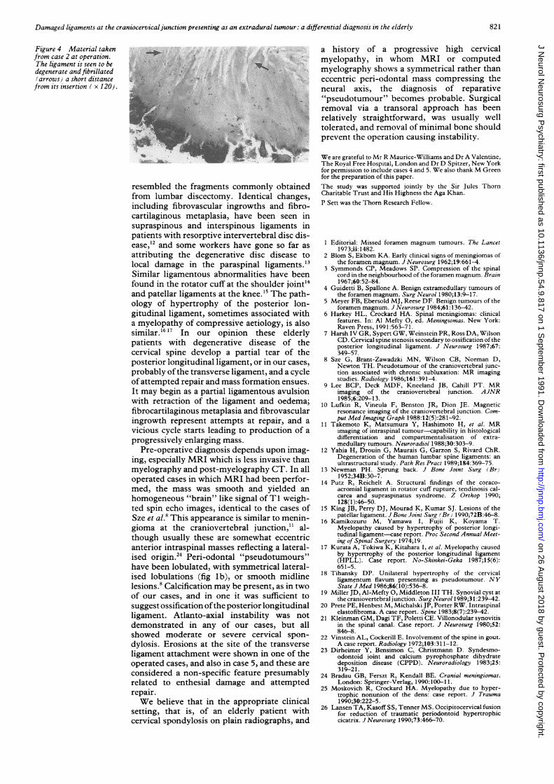
Figure 4 Material takenfrom case 2 at operation.The ligament is seen to bedegenerate and fibrillated(arrows) a short distancefrom its insertion ( x 120).
v ..r --i,.1 .....0 VI QOA
-.n!op
resembled the fragments commonly obtainedfrom lumbar discectomy. Identical changes,including fibrovascular ingrowths and fibro-cartilaginous metaplasia, have been seen insupraspinous and interspinous ligaments inpatients with resorptive intervertebral disc dis-ease,12 and some workers have gone so far as
attributing the degenerative disc disease to
local damage in the paraspinal ligaments. "3Similar ligamentous abnormalities have beenfound in the rotator cuff at the shoulder joint"4and patellar ligaments at the knee.15 The path-ology of hypertrophy of the posterior lon-gitudinal ligament, sometimes associated witha myelopathy of compressive aetiology, is alsosimilar.16 17 In our opinion these elderlypatients with degenerative disease of thecervical spine develop a partial tear of theposterior longitudinal ligament, or in our cases,
probably ofthe transverse ligament, and a cycleof attempted repair and mass formation ensues.
It may begin as a partial ligamentous avulsionwith retraction of the ligament and oedema;fibrocartilaginous metaplasia and fibrovascularingrowth represent attempts at repair, and a
vicious cycle starts leading to production of a
progressively enlarging mass.
Pre-operative diagnosis depends upon imag-ing, especially MRI which is less invasive thanmyelography and post-myelography CT. In alloperated cases in which MRI had been perfor-med, the mass was smooth and yielded an
homogeneous "brain" like signal of TI weigh-ted spin echo images, identical to the cases ofSze et al.8 This appearance is similar to menin-gioma at the craniovertebral junction,11 al-though usually these are somewhat eccentricanterior intraspinal masses reflecting a lateral-ised origin.24 Peri-odontal "pseudotumours"have been lobulated, with symmetrical lateral-ised lobulations (fig lb), or smooth midlinelesions.8 Calcification may be present, as in twoof our cases, and in one it was sufficient tosuggest ossification ofthe posterior longitudinalligament. Atlanto-axial instability was notdemonstrated in any of our cases, but allshowed moderate or severe cervical spon-
dylosis. Erosions at the site of the transverseligament attachment were shown in one of theoperated cases, and also in case 5, and these are
considered a non-specific feature presumablyrelated to enthesial damage and attemptedrepair.We believe that in the appropriate clinical
setting, that is, of an elderly patient withcervical spondylosis on plain radiographs, and
-. d a history of a progressive high cervicalmyelopathy, in whom MRI or computedmyelography shows a symmetrical rather thaneccentric peri-odontal mass compressing theneural axis, the diagnosis of reparative"pseudotumour" becomes probable. Surgicalremoval via a transoral approach has beenrelatively straightforward, was usually welltolerated, and removal of minimal bone shouldprevent the operation causing instability.
We are grateful to Mr R Maurice-Williams and Dr A Valentine,The Royal Free Hospital, London and Dr D Spitzer, New Yorkfor permission to include cases 4 and 5. We also thank M Greenfor the preparation of this paper.The study was supported jointly by the Sir Jules ThornCharitable Trust and His Highness the Aga Khan.P Sett was the Thorn Research Fellow.
1 Editorial: Missed foramen magnum tumours. The Lancet1973;ii: 1482.
2 Blom S, Ekbom KA. Early clinical signs of meningiomas ofthe foramen magnum. J Neurosurg 1962;19:661-4.
3 Symmonds CP, Meadows SP. Compression of the spinalcord in the neighbourhood of the foramen magnum. Brain1967;60:52-84.
4 Guidetti B, Spallone A. Benign extramedullary tumours ofthe foramen magnum. Surg Neurol 1980;13:9-17.
5 Meyer FB, Ebersold MJ, Reese DF. Benign tumours of theforamen magnum. J Neurosurg 1984;61:136-42.
6 Harkev HL, Crockard HA. Spinal meningiomas: clinicalfeatures. In: Al Mefty 0, ed. Meningiomas. New York:Raven Press, 1991:563-71.
7 Harsh IV GR, Sypert GW, Weinstein PR, Ross DA, WilsonCD. Cervical spine stenosis secondary to ossification of theposterior longitudinal ligament. J Neurosurg 1987;67:349-57.
8 Sze G, Brant-Zawadzki MN, Wilson CB, Norman D,Newton TH. Pseudotumour of the craniovertebral junc-tion associated with chronic subluxation: MR imagingstudies. Radiology 1986;161:391-4.
9 Lee BCP, Deck MDF, Kneeland JB, Cahill PT. MRimaging of the craniovertebral junction. AJNR1985;6:209-13.
10 Lufkin R, Vineula F, Benston JR, Dion JE. Magneticresonance imaging of the craniovertebral junction. Com-put Med Imaging Graph 1988:12(5):281-92.
11 Takemoto K, Matsumura Y, Hashimoto H, et al. MRimaging of intraspinal tumour-capability in histologicaldifferentiation and compartmentalisation of extra-medullary tumours. Neuroradiol 1988;30:303-9.
12 Yahia H, Drouin G, Maurais G, Garzon S, Rivard ChR.Degeneration of the human lumbar spine ligaments: anultrastructural study. Path Res Pract 1989;184:369-75.
13 Newman PH. Sprung back. J Bone Joint Surg (Br)1 952;34B:30-7.
14 Putz R, Reichelt A. Structural findings of the coraco-acromial ligament in rotator cuff rupture, tendinosis cal-carea and supraspinatus syndrome. Z Orthop 1990;128(1):46-50.
15 King JB, Perry DJ, Mourad K, Kumar SJ. Lesions of thepatellar ligament. J Bone Joint Surg (Br 1990;72B:46-8.
16 Kamikozuru M, Yamawa I, Fujii K, Koyama T.Myelopathy caused by hypertrophy of posterior longi-tudinal ligament-case report. Proc Second Annual Meet-ing of Spinal Surgery 1974;19.
17 Kurata A, Tokiwa K, Kitahara 1, et al. Myelopathy causedby hypertrophy of the posterior longitudinal ligament(HPLL). Case report. No-Shinkei-Geka 1987;15(6):651-5.
18 Tihansky DP. Unilateral hypertrophy of the cervicalligamentum flavum presenting as pseudotumour. NYState JMed 1986;86(10):536-8.
19 Miller JD, Al-Meftv 0, Middleton III TH. Synovial cyst atthe craniovertebral junction. Surg Neurol 1989;31:239-42.
20 Prete PE, Henbest M, Michalski JP, Porter RW. Intraspinalelastofibroma. A case report. Spine 1983;8(7):239-42.
21 Kleinman GM, Dagi TF, Poletti CE. Villonodular synovitisin the spinal canal. Case report. J Neurosurg 1980;52:846-8.
22 Vinstein AL, Cockerill E. Involvement of the spine in gout.A case report. Radiology 1972;103:31 1-12.
23 Dirheimer Y, Bensimon C, Christmann D. Syndesmo-odontoid joint and calcium pyrophosphate dihydratedeposition disease (CPPD). Neuroradiology 1983;25:319-21.
24 Bradau GB, Ferszt R, Kendall BE. Cranial meningiomas.London: Springer-Verlag, 1990:100-11.
25 Moskovich R, Crockard HA. Myelopathy due to hyper-trophic nonunion of the dens: case report. J Trauma1990;30:222-5.
26 Lansen TA, Kasoff SS, Tenner MS. Occipitocervical fusionfor reduction of traumatic periodontoid hypertrophiccicatrix. J Neurosurg 1990;73:466-70.
821
IM.
...... ...
.K
-e...
k.-L A
on 26 August 2018 by guest. P
rotected by copyright.http://jnnp.bm
j.com/
J Neurol N
eurosurg Psychiatry: first published as 10.1136/jnnp.54.9.817 on 1 S
eptember 1991. D
ownloaded from

![Ultrasound of the Neonatal Craniocervical Junction · 2014-03-28 · Ultrasound of the Neonatal Craniocervical Junction ... and more recently by magnetic resonance [4]. Direct ultrasound](https://static.fdocuments.in/doc/165x107/5f03fc4a7e708231d40bbfcf/ultrasound-of-the-neonatal-craniocervical-2014-03-28-ultrasound-of-the-neonatal.jpg)