Daima
261
Towards fine-tuning the surface corona of inorganic and organic nanomaterials to control their properties at nano-bio interface A thesis submitted in fulfilment of the requirements for the degree of Doctor of Philosophy By Hemant Kumar Daima B.Sc. (Life Sciences), M.Sc. (Biotechnology) School of Applied Sciences College of Science, Engineering and Health RMIT University March 2013
-
Upload
jayabrata-das -
Category
Documents
-
view
27 -
download
0
Transcript of Daima

Towards fine-tuning the surface corona of inorganic and organic nanomaterials to control
their properties at nano-bio interface
A thesis submitted in fulfilment of the requirements for the degree of Doctor of Philosophy
By
Hemant Kumar Daima B.Sc. (Life Sciences), M.Sc. (Biotechnology)
School of Applied Sciences
College of Science, Engineering and Health RMIT University
March 2013

Towards fine-tuning the surface corona of inorganic and organic nanomaterials to control their properties
at nano-bio interface
A thesis submitted in fulfilment of the requirements for the degree of Doctor of Philosophy (Applied Biology and Biotechnology)
By
Hemant Kumar Daima B.Sc. (Life Sciences), M.Sc. (Biotechnology), GATE (Life Sciences)
(National Overseas Scholar, Government of India)
School of Applied Sciences College of Science, Engineering and Health RMIT University, Melbourne (Australia)
March 2013

PhD Thesis Hemant Kumar Daima RMIT University, Australia
Page i
Declaration
I, Hemant Kumar Daima, declare that except where due acknowledgements
have been made, this work is that of myself alone; this work has not been
submitted previously, in whole or in part, to qualify for any other academic
award; and the content of the thesis is the result of work that has been
carried out since the official commencement date of the approved research
programme; and, any editorial work, paid or unpaid, carried out by a third
party is acknowledged.
Hemant Kumar Daima
Date 14.06.2013

PhD Thesis Hemant Kumar Daima RMIT University, Australia
Page ii
Acknowledgements
Working toward Ph.D. has been a wonderful and overwhelming experience and I
am indebted to many people, who supported me during this marathon event. I would like
to thank all of them for their generous support and help throughout this experience.
First of all, I sincerely express my gratitude to my research supervisors, Prof. S. K.
Bhargava and Associate Prof. Vipul Bansal, for sharing their knowledge, and constantly
motivating me to strive for greater heights in life. I feel humbled to have worked under
your guidance and both of you have been great mentors. Along with my research guides,
I must thank Dr. PR. Selvakannan for introducing me to the fascinating area of
Nanobiotechnology and helping me throughout this research marathon. Today, I feel
lucky to have you all in my life because whenever, I was in dark, I found that you were
standing with me to support and encourage. Once I read that “you are not alone during
your PhD, your supervisor are with you and your success is their success.” You made
these words true to me. I am thankful to you for making the time working on my PhD an
unforgeable experience.
Dr. Shiv, Dr. Anthony, Dr. Ravi and Ahmad, I am also thankful to you for helping
and assisting me with some experiments and data analysis presented in this thesis. Dr.
Andrew and Dr. Selvakannan thank you very much for your considerable time to proof-
read this thesis. Also, I must thank Prof. Andrew Smith (Dean, School of Applied
Sciences) for his priceless suggestions during thesis writing. Special words of
indebtedness are due to technical staff Zahra, Paul, Phil, Frank, Peter, Diana, and to all
the duty microscopists, who were available whenever I needed them. Thank you all for
your technical expertise and support throughout the years. I also express my gratitude to
administrative and laboratory staff Karl, Karina, Dr. Lisa, Alexendra, Ruth, Nadia,
Howard and Samira for all the help provided over the years.
During my stay in Melbourne, I have met some fantastic individuals, I thank Dr.
James, Dr. Mamad, Dr. Sarvesh, Dr. Sampa, Dr. Lily, Dr. Helen, Dr. Devendra, Dr. Frew,
Dr. Samuel, Dr. Steven, Dr. Kshudiram, Dr. Neda, Dr. Gopa, Dr. Ylias, Dr. Mukund, Dr.

PhD Thesis Hemant Kumar Daima RMIT University, Australia
Page iii
Andrew, Dr. Rajesh, Dr. Julie, Dr. Sheshnath, Dr. Jammi, Dr. Colin, Dr. Deepa, Dr.
Travor, Dr. Greg, Dr. Srinivash, Dr. Jyoti, Dr. Jarrod, Hailey, Jackie, Jos, Emma, Andrew,
Andreas, Vivian, Blake, Rahul, Katie, Scott, Fiona, Vishal, Kavitha, Nicola, Ilija, Manika,
Akshi, Mahasha, Nafisha, Nura, Addi, Vinni, Sami, Chewe, Blagoj, Kae, Grace, ST, Kim,
Linda, Rose, Ben, Den, Meth, Melissa and Adam. You all have unique personality and I
will always treasure our friendship and it was a pleasant time for me. You all have been
very kind, welcoming and accommodating and it was truly fantastic working and staying
with you. Also, I must thank my house owner Chalie and current and previous house
mates (Kasif, Mohammad, Tsion, Parul, Ali, Polin, Thakashi, Sonam, Hadi and Yumi) for
their cooperation and support at home. Thank you all for always being just a phone call
away.
I extend my genuine gratitude to MSJE, Government of India, New Delhi (India) for
National Overseas Scholarship, which enabled me to pursue doctoral studies at RMIT
University, Melbourne (Australia). Also, I would like to convey my special gratitude to all
the staff members of the Indian Consulate at Melbourne, for their constant support and
encouragement throughout my stay in Australia. I am also thankful to the DPVC travel
grants for funding my two international trips to USA and India for giving me opportunity
to present my work at international conferences and RACI graduate travel award.
Finally, I would like to acknowledge the people, who mean the world to me, my
parents Shri R. C. Daima and Smt. S. Devi for everything. Many thanks to my naughty
brother Lokesh and lovely sister in-law Aarti, for their infinite love. At the end, special
words are due to Kalpana for her love, encouragement, patience & above all her support.
Your support and inspiration made me to believe in myself & helped me to achieve my
dream. I cannot pay back the sacrifices done by you all. Thank you for being in my life.
Last but not least, I thank all those who have helped me directly & indirectly for
the successful completion of my research work. Finally, I thank the Almighty for giving
me the strength & patience to work through all these years so that today I can stand
proud. Hemant Kumar Daima
PhD Thesis Hemant Kumar Daima RMIT University, Australia
Page iv
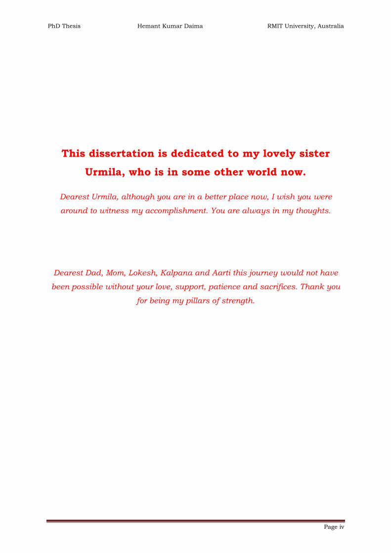
This dissertation is dedicated to my lovely sister
Urmila, who is in some other world now.
Dearest Urmila, although you are in a better place now, I wish you were
around to witness my accomplishment. You are always in my thoughts.
Dearest Dad, Mom, Lokesh, Kalpana and Aarti this journey would not have
been possible without your love, support, patience and sacrifices. Thank you
for being my pillars of strength.

PhD Thesis Hemant Kumar Daima RMIT University, Australia
Page v
Table of Contents
List of figures ………………..…………………………………………..……………… xi
List of tables ………………..…………………………………..………..…………… xvii
List of abbreviation and acronyms ………………..…………..………………… xviii
Abstract ………………..………………………………………………………………… xx
Chapter I. Introduction ………………………………………………………… 1-51
1.1. Nanosciences to nanobiotechnology ………….………………………….... 2
1.2. What is ‘NANO’ and why it is different? …………….…………………….. 4
1.3. Nano-bio interface, toxicity and fabrication of nanomaterials ……….. 5
1.4. Interactions between nanoparticles and bacterial cells ……………….. 9
1.4.1. Structure of Gram positive and negative bacterial cell wall … 10
1.4.2. Antibiotics, antimicrobial peptides and nanomaterial as
antibacterial agents …………………………………………………………… 12
1.5. Other applications of nanobiotechnology ……………………………...… 15
1.6. Production of nanoparticles by top-down or bottom-up approach … 17
1.7. Metal nanoparticles formation and their stability ………………..……. 18
1.8. Surface plasmon of metallic Au and Ag nanoparticles ………..…...... 20
1.9. Objective of the thesis ……………………………………………………….. 22
1.10. Outline of thesis ………………………………………………………………. 24
1.11. References …………………………………………………………………….… 26
Chapter II. Characterization techniques ………………………………… 52-71
2.1. Introduction ……………………………………………………………..…….. 53
2.2. Microscopy techniques ………………………………………………………. 54
2.2.1. Transmission electron microscopy (TEM) ……..……………….. 54

PhD Thesis Hemant Kumar Daima RMIT University, Australia
Page vi
2.2.2. Scanning electron microscopy (SEM) ………………..………….. 56
2.3. Spectroscopy techniques ……………………………………………………. 59
2.3.1. Ultraviolet-visible (UV-vis) spectroscopy ….…………….….…… 59
2.3.2. Fourier transform infrared (FTIR) spectroscopy ………..…..…. 60
2.3.3. Other spectroscopic techniques ………….………………..……… 61
2.3.3.1. Atomic absorption spectroscope (AAS) …………….….. 61
2.3.3.2. Inductively coupled plasma mass spectroscopy …..… 62
2.4. X-ray based characterization techniques ……….……………………….. 63
2.4.1. X-Ray photoemission spectroscopy (XPS) ….……………..……. 63
2.4.2. Energy dispersive X-ray analysis (EDX) …………..……….……. 65
2.5. Particles size analyser ……………………………………………………….. 65
2.5.1. Dynamic light scattering (DLS) ………….………………...……… 66
2.6. Zeta potential measurement (particles charge analyser) …………….. 67
2.7. Electrochemical studies using cyclic voltammetry (CV) ………..……. 68
2.8. References ………………………………………………………………………. 69
Chapter III. Influence of composition and surface functionalization of
nanomaterials towards their biological activities …………………… 72-141
3.1. Introduction ……………………………………………………………………. 73
3.2. Scheme of the work …………………………………………………………… 79
3.3. Experimental section …………………………………………………………. 81
3.3.1. Synthesis of Au, Ag and Au-Ag alloy nanoparticles ………..… 81
3.3.2. Processing of nanoparticles by concentration and dialysis .… 82
3.3.3. Peroxidase-like activity of Au, Ag and Au-Ag nanoparticles … 82
3.3.4. Antibacterial assays of Au, Ag and Au-Ag alloy nanoparticles
…………………………………………………………………………………….. 83

PhD Thesis Hemant Kumar Daima RMIT University, Australia
Page vii
3.4. Results and discussion ……………………………………….……………… 84
3.4.1. UV-visible spectral studies of Au, Ag and Au-Ag alloy
nanoparticles …………………………………………………………….…….. 85
3.4.2. TEM studies of amino acid reduced Au, Ag and Au-Ag alloy
nanoparticles ..…………………………………………………………………. 88
3.4.3. Estimation of the metal content present in dialysed
nanoparticle solutions …..…………………………………………..………. 91
3.4.4. Zeta potential measurements of Au, Ag and Au-Ag alloy
nanoparticles ……………………………………………………….………….. 92
3.4.5. X-ray photoemission spectroscopy analysis of Au, Ag and
Au-Ag alloy nanoparticles ………………………………………..…………. 93
3.4.6. FTIR analysis for the confirmation of amino acid shell ………. 98
3.4.7. Electrochemical behaviour of amino acid capped Au, Ag and
their alloy nanoparticles ………………………………….…….…………. 100
3.5. Rational behind employing these nanomaterials for biological
applications …………………………..………………..………………………. 104
3.5.1. In vitro peroxidase-like behaviour of amino acids capped Au, Ag
and Au-Ag alloy nanoparticles …………………….……………………… 104
3.5.2. Antibacterial assays of amino acid capped nanoparticles …. 112
3.5.2.1. Antibacterial effects of nanoparticles and influence of
surface functionalization …………………………………….….…. 113
3.5.2.2. Effect of metal composition on antibacterial activity
employing Au-Ag alloy nanoparticles …………………………… 116
3.5.2.3. Morphological studies of Au and Ag nanoparticles
treated E. coli and S. albus bacterial cells ……………………… 119
PhD Thesis Hemant Kumar Daima RMIT University, Australia
Page viii
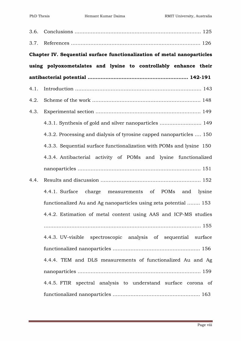
3.6. Conclusions …………………………………………………………………… 125
3.7. References ………………………………………………..…………………… 126
Chapter IV. Sequential surface functionalization of metal nanoparticles
using polyoxometalates and lysine to controllably enhance their
antibacterial potential …………………………………….………….…… 142-191
4.1. Introduction …………………………………………………………………… 143
4.2. Scheme of the work ……………………………………………………….… 148
4.3. Experimental section ……………………………..………………………… 149
4.3.1. Synthesis of gold and silver nanoparticles ……….……..…….. 149
4.3.2. Processing and dialysis of tyrosine capped nanoparticles .… 150
4.3.3. Sequential surface functionalization with POMs and lysine 150
4.3.4. Antibacterial activity of POMs and lysine functionalized
nanoparticles ……………………………………………………….………… 151
4.4. Results and discussion …………………………………………………..… 152
4.4.1. Surface charge measurements of POMs and lysine
functionalized Au and Ag nanoparticles using zeta potential …….. 153
4.4.2. Estimation of metal content using AAS and ICP-MS studies
……………………………………………………………………………………. 155
4.4.3. UV-visible spectroscopic analysis of sequential surface
functionalized nanoparticles ……………………………………………… 156
4.4.4. TEM and DLS measurements of functionalized Au and Ag
nanoparticles …………………………………………………………………. 159
4.4.5. FTIR spectral analysis to understand surface corona of
functionalized nanoparticles …………………………………….……….. 163

PhD Thesis Hemant Kumar Daima RMIT University, Australia
Page ix
4.4.6. XPS analysis of POMs and lysine functionalized nanoparticles
…………………………..………………………………………………….……. 167
4.5. Antibacterial applications of functionalized nanoparticles ………… 170
4.5.1. Assessment of antibacterial activities of gold, silver and their
POMs and lysine functionalized nanoparticles …………………..…… 170
4.5.2. Morphological changes in bacterial cell after their treatments
with gold, silver and functionalized nanoparticles …………………… 176
4.6. Conclusions …………………………………………………………………… 180
4.7. References ……………………………………………………………..……… 181
Chapter V. Self-assembled soft nanostructures of plasmid DNA and tri-
block copolymer as non-viral DNA delivery vehicle …….…….…. 192-221
5.1. Introduction ………………………………………………………………...… 193
5.2. Scheme of the work ……………………………………………………….… 197
5.3. Experimental section ……………………………………………………….. 198
5.3.1. Isolation of pDNA and purification ………………….……..…… 198
5.3.2. Preparation of pDNA and PEO-PPO-PEO complex …..…..…. 199
5.3.3. Preparation of competent cells ………..…………………….…… 200
5.3.4. Transformation and gene expression ………………….……..… 201
5.4. Results and discussion ………………………………………….…………. 201
5.4.1. TEM studies of self-assembled structures of pDNA and PEO-
PPO-PEO with varying weight ratios ………………………………….… 202
5.4.2. FTIR spectral analysis to recognize chemical interaction of self-
assembled pDNA and PEO-PPO-PEO complex ……………………..… 204
5.4.3. XPS spectral analysis of self-assembled structures of pDNA and
PEO-PPO-PEO ………………………………………………………………... 206

PhD Thesis Hemant Kumar Daima RMIT University, Australia
Page x
5.5. PEO20-PPO69-PEO20 mediated transformation and expression of GFP in
transformed colonies ……………………………………………………………….. 209
5.6. Conclusions …………………………………………………………………… 213
5.7. References ………………………………………………………………..…… 214
Chapter VI. Conclusions & scope for the future research ………. 222-228
6.1. Summary and conclusions ……………………………………….…… 223-226
6.2. Scope for future work …………………………………………………… 226-228
Appendices ……………………………..…………………………………… 229-236

PhD Thesis Hemant Kumar Daima RMIT University, Australia
Page xi
List of Figures
Figure.1.1. Comparative sizes of biological components at nanoscale
dimension ………………………………………………………………………………… 2
Figure.1.2. Schematic representation of various dimensions of materials at
nanoscale …………………………………………………………………………………. 5
Figure.1.3. Schematic representation of various physicochemical properties
of nanomaterials which influences nanoparticle-cell interaction ……………. 6
Figure.1.4. Schematic representation of peptidoglycan structure …………. 10
Figure.1.5. Schematic diagram of the structure of Gram positive bacterial
cell wall ………………………………………………………………………………….. 11
Figure.1.6. Schematic representation of Gram negative bacterial cell wall 11
Figure.1.7. Schematic representation of mechanism of antibacterial
activities exerted by nanoparticles ……………………………………………..…. 14
Figure.1.8. Schematic representation of the ‘bottom-up’ and ‘top-down’
synthesis processes of nanomaterials with the popular techniques …….… 17
Figure.1.9. Scheme illustrating the stabilization of metal nanoparticles by (A)
electrostatic and (B) steric interactions ……………………………….…………. 19
Figure.1.10. Schematic representation of the interaction of light with metal
nanoparticles ………………………………………………………………….……….. 21
Figure.2.1. Schematic representation of the major components of the optical
systems of a TEM (equivalent terms of light and electron microscopy are also
shown) …………………………………………………………………………………... 55
Figure.2.2. Schematic representation of electron-sample interaction in SEM
……………………………………………………………………………………………… 57
Figure.2.3. Schematic representation of scanning electron microscope …. 58

PhD Thesis Hemant Kumar Daima RMIT University, Australia
Page xii
Figure.2.4. Schematic representation of the XPS process, showing photo-
ionization of an atom by the ejection of a 1s electron ………………..………. 63
Figure.2.5. Schematic representation of the electrical double layer on the
surface of nanoparticle …………………………………….………………..………. 67
Figure.3.1. Chemical structures of tryptophan and tyrosine amino acids . 79
Figure.3.2. Schematic representation of synthesis of Au, Ag and their alloy
nanoparticles with different compositions and surface functionality …..… 80
Figure.3.3. UV-visible absorbance spectra of (A) tryptophan (Trp) and
tyrosine (Tyr), (B) tryptophan- and (C) tyrosine- reduced Au, Ag and Au-Ag
alloy nanoparticles, respectively ……………………………………………..……. 85
Figure.3.4. Digital photographs of metal nanoparticle solutions synthesised
using (A) tryptophan and (B) tyrosine ………………………………………….… 87
Figure.3.5. TEM images and particle size distribution histogram of
tryptophan reduced Au, Ag and their alloy nanoparticles. Average particle
sizes along with standard deviations are also shown …….…………………… 89
Figure.3.6. TEM images and particle size distribution histogram of tyrosine
reduced Au, Ag and Au-Ag alloy nanoparticles. Corresponding average
particle sizes along with standard deviations are also shown …..………….. 90
Figure.3.7. Core level Au4f and Ag3d spectra of tryptophan and tyrosine
synthesised Au and Ag nanoparticles (A-D) and core level Au4f (E) and Ag3d
(F) spectra arising from Au50Ag50Trp alloy nanoparticles, respectively …..…. 94
Figure.3.8. Core level XPS spectra of C1s in pristine tryptophan (A), tyrosine
(B), Au100Trp(C) and Au100
Tyr (D) respectively. Core level N1s (E) and K2p (F)
spectra of tryptophan synthesised Au100Trp, respectively ……………..……… 96

PhD Thesis Hemant Kumar Daima RMIT University, Australia
Page xiii
Figure.3.9. FTIR spectral analysis of (A) tryptophan- (curve 1) and (B)
tyrosine- (curve 1) reduced Au100 (curve 2); Au75Ag25 (curve 3); Au50Ag50
(curve 4); Au25Ag75 (curve 5) and Ag100 (curve 6) nanoparticles, respectively
……………………………………………………………………………………………… 98
Figure.3.10. Cyclic voltametric profiles of tryptophan- (A) and tyrosine (B)
synthesised nanoparticles recorded at 5 mV s-1 in 1 M H2SO4 ………….… 101
Figure.3.11. Cyclic voltametric profiles of (A) typical gold oxide formation
and reduction (B) CV profile of 1 mM tryptophan in 1 M H2SO4 and (C) CV
profile of tyrosine oxidation …………………………………………………..…… 102
Figure.3.12. Schematic representation of oxidation of TMB by H2O2
catalysed by nanoparticles ………………………………………………………… 106
Figure.3.13. Peroxidase-like behaviour of tryptophan- and tyrosine- capped
Au, Ag and Au-Ag alloy nanoparticles with different concentrations ….… 106
Figure.3.14. Temperature dependent peroxidase-like activity of tryptophan-
and tyrosine- capped Au and Ag nanoparticles In all the panels, curve 1, 2,
3 and 4 correspond to Au100Trp, Au100
Tyr, Ag100Trp and Ag100
Tyr, respectively
……………………………………………………………………………………………. 107
Figure.3.15. Temperature dependent peroxidase-like activity of tryptophan
(Panel A, B and C) and tyrosine (Panel D, E and F) capped Au-Ag nanoalloys.
In all the panels, curve 1, 2 and 3 correspond to Au75Ag25, Au50Ag50 and
Au25Ag75 respectively …………………………………..…………………………… 111
Figure.3.16. Assessment of antibacterial potential of amino acid capped Au
and Ag nanoparticles against (A) E. coli and (B) S. albus, respectively ….. 115
Figure.3.17. Antibacterial activity of Au-Ag alloy nanoparticles synthesized
by tryptophan and tyrosine against E. coli (A) and S. albus (B) …………… 117

PhD Thesis Hemant Kumar Daima RMIT University, Australia
Page xiv
Figure.3.18. SEM micrographs of E. coli before and after their treatments
with tryptophan and tyrosine synthesised nanoparticles …………….……. 121
Figure.3.19. SEM micrographs of S. albus before and after their treatments
with tryptophan and tyrosine synthesised nanoparticles ……………..…… 122
Figure.3.20. Schematic illustration of antibacterial activity of Au and Ag
nanoparticles toward E. coli and S. albus ……………………………………… 124
Figure.4.1. Schematic representation of sequential functionalization of
tyrosine capped gold nanoparticles by polyoxometalates and lysine ….… 149
Figure.4.2. UV-visible absorbance spectra of AuNPsTyr and after their surface
functionalization with (A) PTA/lysine and (B) PMA/lysine molecules …… 157
Figure.4.3. UV-visible absorbance spectra of AgNPsTyr and after their surface
functionalization with (A) PTA/lysine and (B) PMA/lysine molecules …… 158
Figure.4.4. TEM images and particle size distribution histograms of (A)
AuNPsTyr, (B) AuNPsTyr@PTA, (C) AuNPsTyr@PTA-Lys, (D) AuNPsTyr@PMA and (E)
AuNPsTyr@PMA-Lys ……………………………………………………………..……….. 160
Figure.4.5. TEM images and corresponding particle size distribution
histograms of (A) AgNPsTyr, (B) AgNPsTyr@PTA, (C) AgNPsTyr@PTA-Lys, (D)
AgNPsTyr@PMA and (E) AgNPsTyr@PMA-Lys …………………….………………..…… 162
Figure.4.6. FTIR spectra in Panel A: tyrosine, AuNPsTyr and AgNPsTyr; in
Panel B: PTA, AuNPsTyr@PTA, AuNPsTyr@PTA-Lys, AgNPsTyr@PTA and AgNPsTyr@PTA-
Lys; and in Panel C: PMA, AuNPsTyr@PMA, AuNPsTyr@PMA-Lys, AgNPsTyr@PMA and
AgNPsTyr@PMA-Lys, respectively ……………………………………………..………. 164
Figure.4.7. Schematic representation of formation of quinone type structure
during reduction of Ag+ ions …………………………………………….………… 165

PhD Thesis Hemant Kumar Daima RMIT University, Australia
Page xv
Figure.4.8. Core level spectra of Au4f (A-E), recorded in AuNPsTyr,
AuNPsTyr@PTA, AuNPsTyr@PTA-Lys, AuNPsTyr@PMA and AuNPsTyr@PMA-Lys samples,
respectively. Core level spectra of C1s (F), W4f (G-H), Mo4f (I-J) recorded in
AuNPsTyr, AuNPsTyr@PTA, AuNPsTyr@PTA-Lys, AuNPsTyr@PMA and AuNPsTyr@PMA-Lys,
respectively ……………………………………………………………………………. 168
Figure.4.9. Core level spectra of Ag3d (A), W4f (B-C), Mo4f (D-E) recorded in
AgNPsTyr, AgNPsTyr@PTA, AgNPsTyr@PTA-Lys, AgNPsTyr@PMA and AgNPsTyr@PMA-Lys
samples, respectively ……………………………………………….………………. 169
Figure.4.10. Dose dependent antibacterial activity of PTA (Panels A), PMA
(Panels B) and lysine functionalized Au nanoparticles ……………………… 172
Figure.4.11. Dose dependent antibacterial activity of PTA (Panels A), PMA
(Panels B) and lysine functionalized Ag nanoparticles ……………………… 175
Figure.4.12. SEM micrographs of Escherichia coli cells (A) untreated and
after their treatments with (B) AuNPsTyr, (C) AuNPsTyr@PTA, (D) AuNPsTyr@PTA-
Lys, (E) AuNPsTyr@PMA, and (F) AuNPsTyr@PMA-Lys ……………………..………….. 177
Figure.4.13. SEM micrographs of Escherichia coli cells (A) untreated and
after treatments with (B) AgNPsTyr, (C) AgNPsTyr@PTA, (D) AgNPsTyr@PTA-Lys, (E)
AgNPsTyr@PMA, and (F) AgNPsTyr@PMA-Lys ………………………..………………… 178
Figure.4.14. Interaction of Au, Ag and their POMs and lysine surface
functionalized nanoparticles with E. coli bacterial cells ……………………. 179
Figure.5.1. Chemical structure of tri-block copolymer (PEO-PPO-PEO) … 196
Figure.5.2. Schematic representation of self-assembly process of A-B-A type
tri-block copolymer and pDNA …………………………..……………………….. 197

PhD Thesis Hemant Kumar Daima RMIT University, Australia
Page xvi
Figure.5.3. TEM images of as-formed (A-C) PEO-PPO-PEO micelles at
different concentrations and after their complex formation with pDNA (D-F)
……………………………………………………………………………………………. 203
Figure.5.4. FTIR spectra of PEO-PPO-PEO (curve 1), pDNA (curve 2), and
after their complexation at 1:1 and 1:10 ratios (curve 3 and 4), respectively
……………………………………………………………………………………………. 204
Figure.5.5. Core level XPS spectra of four principal elements present in
pDNA ……………………………………………….………………………………….. 207
Figure.5.6. XPS spectra of C1s, O1s (tri-block copolymer), N1s and C1s in
pDNA: PEO20-PPO69-PEO20 at 1:1 and 1: 10 ratios, respectively ……….… 208
Figure.5.7. Number of transformed colonies grown on ampicillin plates for
varying ratios of pDNA and PEO20-PPO69-PEO20 …………………………...… 210
Figure.5.8. Representative transformed colonies grown on ampicillin plates
showing expression of GFP gene. pDNA (A), pDNA:PEO20-PPO69-PEO20
complexes (B-D) at 1:1 (B), 1:5 (C), and 1:10 (D) ratios, respectively ….… 211
Figure.5.9. Schematic representation of transformation by pDNA/polymer
complex ………………………………………………………………………………… 212

PhD Thesis Hemant Kumar Daima RMIT University, Australia
Page xvii
List of tables
Table.3.1. Concentration of HAuCl4 and AgNO3 used for the synthesis of
Au, Ag and Au-Ag alloy nanoparticles using tryptophan and tyrosine …… 81
Table.3.2: Molar ratios (%) of Au and Ag estimated from AAS and zeta
potential measurements of nanoparticle solutions ……………………….…… 92
Table.4.1. Relative concentrations of Au and W or Mo present on the surface
of functionalized Au nanoparticles, as used for antibacterial studies …… 171
Table.4.2. Relative concentrations of Ag and W or Mo present on the surface
of functionalized Ag nanoparticles, as used for antibacterial studies …… 174
Table.5.1. Weight ratios of pDNA and PEO20-PPO69-PEO20 employed to
prepare self-assembled nanostructures ……………………………..…….…… 199
Table.5.2. Atomic % of oxygen, carbon and nitrogen present in pDNA, PEO-
PPO-PEO and in the nanostructures of pDNA and PEO-PPO-PEO …….… 209

PhD Thesis Hemant Kumar Daima RMIT University, Australia
Page xviii
List of abbreviation and acronyms
a.a. : Amino acid
AAS : Atomic absorption spectroscopy
Ag : Silver
AgNPs : Silver nanoparticles
AgNPsPMA : Ag nanoparticles functionalized with phosphomolybdic acid
AgNPsPTA : Ag nanoparticles functionalized with phosphotungstic acid
AgNPsPMA@Lys: Lysine modified phosphomolybdic acid functionalized AgNPs
AgNPsPTA@Lys: Lysine modified phosphotungstic acid functionalized AgNPs
AuNPs : Gold nanoparticles
AuNPsPMA : Au nanoparticles functionalized with phosphomolybdic acid
AuNPsPTA : Au nanoparticles functionalized with phosphotungstic acid
AuNPsPMA@Lys: Lysine modified phosphomolybdic acid functionalized AuNPs
AuNPsPTA@Lys: Lysine modified phosphotungstic acid functionalized AuNPs
CFU : Colony forming unit
DNA : Deoxyribonucleic acid
E. coli : Escherichia coli
FTIR : Fourier transform infrared
GFP : Green fluorescent protein
LB : Luria broth
Lys (K) : Lysine
nm : nanometer
NPs : nanoparticles
OD : Optical density

PhD Thesis Hemant Kumar Daima RMIT University, Australia
Page xix
pDNA : Plasmid DNA
POM : Polyoxometalates
PMA : Phosphomolybdic acid
PTA : Phosphotungstic acid
S. albus : Staphylococcus albus
SERS : Surface enhanced Raman scattering
SEM : Scanning electron microscopy
ROS : Reactive oxygen species
SPR : Surface plasmon resonance
TEM : Transmission electron microscopy
Trp (W) : Tryptophan
Tyr (Y) : Tyrosine
UV-Vis : Ultraviolet-visible
XPS : X-ray photoelectron spectroscopy
XRD : X-ray diffraction

PhD Thesis Hemant Kumar Daima RMIT University, Australia
Page xx
Abstract
Designing nanomaterials for biological applications has become an
emerging interdisciplinary area of science; however that raises the need to
understand the materials interaction with different biological components at
the nano-bio interface. Such basic understanding required for the
development of nanomaterials-based diagnosis, therapy and medicine.
Majority of recent work have focussed on how the size, shape, stability,
surface charge and composition of inorganic nanomaterials control their
interaction with the biological entity, whereas other parameters pertaining to
the nanoparticles surface such as the surface layer consisting of molecules,
ions, hydrophilic or hydrophobic nature and their synergistic effects are
often overlooked. Therefore, the major focus of the thesis is to understand
that how the variation in surface functionalization in conjunction with
inorganic nanoparticles composition reflects on their ability to produce
reactive oxygen species and imparts non-specific or selective toxicity against
different bacterial strains. However, presence of organic component hinders
their application in gene delivery; therefore additionally, organic
nanostructures were employed to understand their interaction with DNA
and bacterial transformation.
First focus was to develop the synthetic methodologies in controlling
the surface functionalization and composition of nanoparticles in a single
step using green and eco-friendly routes. Tryptophan and tyrosine amino
acids were employed as reducing and stabilizing agents to synthesize gold,
silver and their bimetallic alloy nanoparticles. These amino acid

PhD Thesis Hemant Kumar Daima RMIT University, Australia
Page xxi
functionalized nanoparticles have been chosen in the research work because
the side functional groups present in the amino acids reduce either single
metal ions or two different types of metal ions to form monometallic and
bimetallic nanoparticles, respectively, while leaving the amine and
carboxylic acid groups intact. Hence these nanoparticles can be viewed as
amino acid functional groups anchored on a nanoparticles surfaces, which
can render these surfaces similar to enzymes. In the context of biological
application, usually nanoparticles exhibit toxicity due to their ability to
induce production of reactive oxygen species. This can be evaluated by their
peroxidise-like activity by catalysing the oxidation of chromogenic substrate.
Therefore, these amino acid surface functionalized nanoparticles were
subjected to investigate their intrinsic peroxidise enzyme-like behaviour. The
peroxidise-like activity was found to be composition, temperature and
surface functionalization dependant and it was revealed that the peroxidise-
like behaviour originates from the synergistic effects of Ag fraction of the
nanoparticles and the amino acid shell. Further, antibacterial studies of
these nanoparticles demonstrated that toxicity against Gram positive
bacteria (Staphylococcus albus) originates only from the amino acid shell
present on the nanoparticles surface and composition of nanoparticles
doesn’t have any significant role. Conversely, antibacterial activity against
Gram negative bacterium (Escherichia coli) was composition dependent and
capping of amino acids had little influence. Morphological studies of
nanoparticles treated bacterial cells revealed that after interaction with
amino acid surface functionalized nanoparticles, most of the bacterial cells
lost their integrity and confirmed level of damage. These findings established

PhD Thesis Hemant Kumar Daima RMIT University, Australia
Page xxii
correlation between the surface corona and metal composition of
nanoparticles and their antibacterial activities against two different bacterial
strains.
Moreover, in order to demarcate how this surface functionalization
plays a critical role; non-toxic Au nanoparticles were chosen and revealed
significant influence of surface functionalization against Gram negative
bacteria. Tyrosine reduced Au nanoparticles that were found to be non-toxic
to E. coli, turns out to be a potential antibacterial agent after they were
sequentially functionalized by polyoxometalates and lysine. Functional
polyoxometalate ions imparted antibacterial potential on the surface of
tyrosine synthesized Au nanoparticles and the cationic nature of lysine
worked as guide to target negatively charged bacterial cells for antibacterial
activities. The same sequential surface functionalization was extended to Ag
nanoparticles, wherein these nanoparticles showed very high antibacterial
activity even in smaller concentrations. These nanoparticle-based functional
antibacterial agents seen to employ a physical mode of action against
bacteria by causing pore formation, cell wall cleavage and cell lysis.
As-mentioned earlier, inorganic nanoparticles are not suitable for DNA
delivery applications and bacterial transformation studies due to their
inherent toxicity as well as long residence time of the inorganic
nanomaterials within the living system. Moreover, it is not easy to clear
inorganic nanomaterials from the living systems due to their strong
association with different cellular components. Therefore, DNA delivery
vector requies an organic nanomaterial that encapsulate the DNA instead of
inorganic nanomaterials. Another reason to choose organic nanostructures

PhD Thesis Hemant Kumar Daima RMIT University, Australia
Page xxiii
is they can be broken down by enzymatic reactions within the bacterial
cells. In this context, organic materials based nanostructures were employed
for DNA delivery applications and to understand their interaction with DNA.
Tri-block copolymer (PEO20-PPO69-PEO20) was used as the organic material
and it is known to undergo self-assembly to form micelles in water. When
plasmid DNA was mixed along with the polymer, they form self-assembled
structures and these nanostructures are used as non-viral DNA delivery
vehicles. The electrostatic interaction between the phosphate group (PO43-) of
pDNA and the hydrophilic segment (PEO) of tri-block polymer drives the self-
assembly process. Different weight ratios of DNA and polymer were screened
to find out the optimum weight ratio to achieve highest level of bacterial
transformation. At 1:10 weight ratio of DNA and polymer the bacterial
transformation was found to be the maximum and it was over 6 folds higher
transformation. Furthermore, during transformation studies, the integrity
and functionality of the green fluorescent protein (GFP) pDNA in
nanostructures were also demonstrated within the cellular environment by
the expression of GFP gene.

PhD Thesis Hemant Kumar Daima RMIT University, Australia
Chapter I Page 1
Chapter I
Introduction
This chapter provides a general introduction about the historical
development of nanoscience, nanobiotechnology and nanotoxicology. Special
emphasis is given on the cell-nanomaterial interaction at the nano-bio
interface and how various physicochemical properties of nanomaterials
influence this interaction. Subsequently, a variety of mechanisms by which
nanomaterials illustrate antibacterial activities have been discussed and
basic differences between Gram positive and Gram negative bacterial strains
are discussed. Additionally, synthesis, stability and important
physicochemical properties of metallic nanoparticles have been discussed.
In the later part of this chapter, the aims of the thesis have been designed to
address the gaps in the aforementioned area. Finally, a chapter wise
summary of the thesis has been presented.

PhD Thesis Hemant Kumar Daima RMIT University, Australia
Chapter I Page 2
1.1. Nanosciences to nanobiotechnology
On December 29 1959, an influential lecture “There’s plenty of room
at the bottom” was delivered by Professor Richard Feynman at the American
Physical Society meeting at California Institute of Technology (Caltech),
where he initiated the need to implement miniaturization in future
technologies. His lecture provoked curiosity to fabricate future devices,
which can be portable, powered by small energy devices, using less
materials etc.[1] Since that day, the potential of nanotechnology was
realized, as we are witnessing the fact that almost each and every electronic,
optical, magnetic, sensing, energy and volumetric devices are made as small
as possible to support the concept of miniaturization given by Prof. Feynman
in his legendary lecture.[1]
Figure.1.1. Comparative sizes of biological components at nanoscale
dimension (adopted from reference [2])
In addition to technological devices, other significant developments in
the field of nanosciences have shown extensive impact on almost all areas of
natural and applied sciences including material science, physics, chemistry,
biology, medicine, microbiology, biotechnology and engineering.[3-7] In the
context of biomedical sciences, this influence is possible because the basic
biological entities and biomolecules like enzymes, DNA, membrane, motor

PhD Thesis Hemant Kumar Daima RMIT University, Australia
Chapter I Page 3
proteins etc are complex biological components, and fall in the nanoscale
dimension as illustrated in Figure 1.1.[2]
Furthermore, the potential applications of nanoscience in
biotechnology, bio-sensing, medicine and microbiology gave birth to the new
field called “nanobiotechnology” [8] because most of the biological systems
can be integrated with nanomaterials due to their comparable sizes.
Moreover, the integration of nanomaterials with biomolecules have
illustrated excellent applications in biomedicine that can potentially reach to
previously inaccessible targets [9-11] due to multi functionality and higher
sensitivity [8] of hybrid nanobiomaterials with desired physical and chemical
properties.[12-14]
Thus, the interdisciplinary nature of nanobiotechnology opens up a
new avenue of multifunctional hybrid nanomaterials to resolve biological
problems. In this perspective, conceivably the most striking example of
employing nanomaterials is to control infections. Some of the most common
and lethal infections are caused by bacteria and a variety of nanomaterials
have been evaluated for their antimicrobial properties that are used to
control bacterial growth including those bacterial strains that have evolved
resistance to antibiotics.[15-19] Therefore, it is essential to develop
nanomaterials for antibacterial applications and to understand the
interaction of these nanomaterials with bacterial cells, which is the major
focus of this thesis. Moreover, the medicinal potential and environmental
impact of nanomaterials could be fully realized only after extensive
investigations at the interface of nanobiotechnology. Therefore, this research
work has been undertaken to examine that how properties of nanomaterials

PhD Thesis Hemant Kumar Daima RMIT University, Australia
Chapter I Page 4
influence their interaction with bacterial cells and what happens to the
bacteria after being in contact with nanomaterials. These understandings
may assist to evaluate biological impacts of functionalized nanomaterials
and their potential applications.
1.2. What is ‘NANO’ and why it is different?
According to the International standard organization (ISO), a
nanomaterial is any object with at least one, two or all the three dimensions
in the nanoscale range (1 to 100 nm). At the range of nano meter level,
materials begin to show distinct mechanical, electrical, optical, magnetic,
electro-optical, magneto-optical, physical, and chemical properties, which
are different from their bulk and molecular counterparts. Therefore,
nanotechnology enables to change some basic properties of a material such
as functionality, shape, size, and surface charge without changing its
chemical composition [10, 20, 21] and these nano objects may have higher
toxicity, greater catalytically activity and different characteristics.
Nanoparticles and composite nanomaterials are found to have wide
applications in magnetic storage devices, electronics, optics, catalysis and
sensing etc. [3, 4, 22-27] and in the context of biology and medicine
nanomaterials have shown outstanding and promising applications.[10, 28-
33] Nanomaterials can be explored for novel applications where the
properties of the atomic or bulk material are unsuitable. Moreover, during
synthesis, growth of nanomaterials can be restricted to obtain 1, 2 or 3
dimensional object thus nanomaterials can be nanoparticles, nanoplates or
nanofibres respectively, having 3, 2 or 1 dimensions within the nano-scale

PhD Thesis Hemant Kumar Daima RMIT University, Australia
Chapter I Page 5
as illustrated in Figure 1.2. Additional terms nanoprism or nanocube have
been used to discuss polyhedral nanoparticles. For nanofibres the object
may be referred as nanorods or nanotubes, depending upon whether the
structure is hollow or solid. Along with nano-size, dimensions of
nanomaterials have considerable influence on their properties and biological
activities.[25, 33-42]
Bulk material
2D nanostructures
1D nanostructures
3D nanoparticles
1D constraint
2D constraint
3D constraint
Figure.1.2. Schematic representation of various dimensions of materials at
nanoscale
1.3. Nano-bio interface, toxicity and fabrication of nanomaterials
It is imperative to understand that nanotechnology is not just a new
step toward miniaturization. Recent advances in the field of nanotechnology
have revealed that this is a powerful transformative technology and this is
evident from the utilization of nanotechnology based products in every
sector of the society and strong influence of nanotechnology on the future of
mankind has been predicted.[43-45] There are numerous areas where
nanoparticulate systems have shown great technological and scientific
significance. For instance, applications of nanotechnology have potential to
transform the field of biotechnology, agriculture, manufacturing,

PhD Thesis Hemant Kumar Daima RMIT University, Australia
Chapter I Page 6
information technology, telecommunication, electronic, materials, medical
and other sciences.[10, 30-32, 43, 46-48]
In milieu of biological applications, the major objective of
nanotechnology is to engineer and manufacture nanomaterials at the atomic
or molecular level to exhibit entirely novel therapeutic applications with
better functionality, efficiency and specificity.[49] Since, cells are highly
sensitive to their environment [50] and basic biological components are
functional configurations at nanometer level, nanobiotechnology has the
potential to tailor material’s physiochemical properties to achieve the desired
level of sensitivity with improved functionality, efficiency and specificity.
+
+
++
+
+
+
--
---
-
-
-
Functional
groups and
ligands
Surface
charge
Composition
ShapeSize
Hydrophilic
hydrophobic
nature
Dissolution
Biomolecules
on surface
Biological
entity
Figure.1.3. Schematic representation of various physicochemical properties
of nanomaterials which influences nanoparticle-cell interaction

PhD Thesis Hemant Kumar Daima RMIT University, Australia
Chapter I Page 7
Various physical and chemical properties of nanomaterials such as
surface charge, composition, shape, size, hydrophilic or hydrophobic nature
and dissolution effect, presence of biomolecules, functional groups or ligand
on the surface of nanomaterials have significant influence at nano-bio
interface.[51-56] For instance, cell membranes possess negative charge and
if nanomaterials have the similar surface charge, due to the effective
repulsion, interaction between cell-nanomaterial will not be possible, while
positive surface charge on nanomaterials can provide an opportunity to
interact with membrane due to the electrostatic attraction. Further,
nanomaterials have intrinsic potential to produce reactive oxygen species
(ROS) within or outside of the cell which can challenge cellular integrity and
cell survival. After internalization, due to the dissolution effect of
nanoparticles cellular functions may be affected severely leading to toxic
effects of nanomaterials.[51, 52, 57-59]
Furthermore, presence of diverse functional groups or ligands on the
surface of nanomaterials can determine the level of interaction at the nano-
bio interface and may damage cells by physical action or by interaction with
sub cellular units and genetic material.[2, 59-66] In general, surface
expressed bio-molecules, drugs or pharmaceutical chemicals dominate the
biological impacts and presence of these molecules on the surface of
nanoparticles could be highly significant.[67] Chemical moieties present on
the nanoparticles surface have impact on their interaction with biological
system therefore strong control over nanoparticles-cell interactions could be
primarily controlled by tailoring surface properties of nanomaterials.[68-86]

PhD Thesis Hemant Kumar Daima RMIT University, Australia
Chapter I Page 8
This discussion illustrates that the principles governing the
interactions between biological species and nanomaterials are of significant
importance. Therefore, it is imperative to understand that the interactions of
designer made functional nanoparticles within biological systems (living
organisms) is multidimensional and understanding of these interactions is
critically important for further development of nanobiotechnology for
biomedical applications and in terms of nano-safety to achieve a particular
and controlled biological action.[69, 79, 87]
Nanotoxicology is a vital and emerging part of nanobiotechnology
which refers to the study of interactions of engineered nanomaterials with
biological system or environment. In nanotoxicology, particular emphasis is
on the correlation between the physicochemical properties of nanomaterials
with induction of toxic biological responses.[88] Additionally, this field aims
to find favourable characteristic of nanoparticulate systems which render
them more amenable for the use in biological environment.[31, 89]
In this perspective, Dawson and co-workers have revealed that at the
interface of cell and nanoparticles, the effective unit of interaction is not the
nanoparticles itself, but the corona present around the nanoparticles is the
unit of interest.[67, 70, 90] Surface corona is the key unit of interest for a
living entity which ultimately exhibits biological action.[69, 91-94] Likewise,
it have been shown that nanomaterials offer large surface areas and their
controlled structure and surface functionality can make them outstanding
scaffolds for biomedical applications.[55, 71, 78, 79, 84, 85] Furthermore, it
has been demonstrated that nanoparticles can be effectively fabricated by
amino acid functionalization to afford tailored surfaces for peptide
PhD Thesis Hemant Kumar Daima RMIT University, Australia
Chapter I Page 9
reorganization, DNA receptors, sensing, imaging and drug or gene delivery
applications.[71-73, 83] Moreover, functionalized nanoparticles recognize
target biomolecular surfaces via complementary interactions, which can lead
toward a desired nanoparticle-cell interaction for biomedical applications.
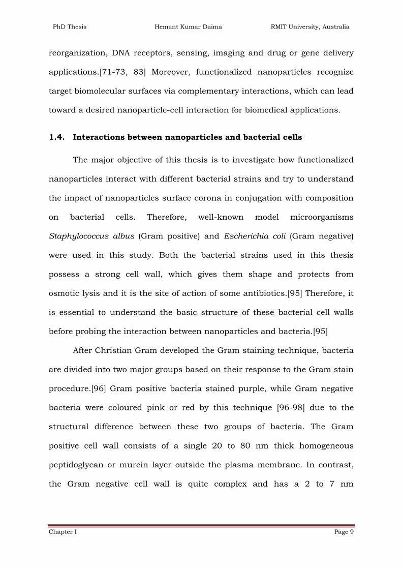
1.4. Interactions between nanoparticles and bacterial cells
The major objective of this thesis is to investigate how functionalized
nanoparticles interact with different bacterial strains and try to understand
the impact of nanoparticles surface corona in conjugation with composition
on bacterial cells. Therefore, well-known model microorganisms
Staphylococcus albus (Gram positive) and Escherichia coli (Gram negative)
were used in this study. Both the bacterial strains used in this thesis
possess a strong cell wall, which gives them shape and protects from
osmotic lysis and it is the site of action of some antibiotics.[95] Therefore, it
is essential to understand the basic structure of these bacterial cell walls
before probing the interaction between nanoparticles and bacteria.[95]
After Christian Gram developed the Gram staining technique, bacteria
are divided into two major groups based on their response to the Gram stain
procedure.[96] Gram positive bacteria stained purple, while Gram negative
bacteria were coloured pink or red by this technique [96-98] due to the
structural difference between these two groups of bacteria. The Gram
positive cell wall consists of a single 20 to 80 nm thick homogeneous
peptidoglycan or murein layer outside the plasma membrane. In contrast,
the Gram negative cell wall is quite complex and has a 2 to 7 nm

PhD Thesis Hemant Kumar Daima RMIT University, Australia
Chapter I Page 10
peptidoglycan layer surrounded by a 7 to 8 nm thick outer membrane.[99]
1.4.1. Structure of Gram positive and negative bacterial cell wall
Peptidoglycan is a polymer composed of two sugar derivatives (N-
acetylglucosamine [NAG] and N-acetylmuramic acid [NAM]) and several
different amino acids, especially three of which viz. D-glutamic acid, D-
alanine and meso-diaminopimelic acid, are not found in proteins. The
backbone of this polymer is composed of alternating NAG and NAM residues
as presented in Figure 1.4.[95] A peptide chain of four alternating D- and L-
amino acids is connected to the carboxyl group of NAM. Chains of linked
peptidoglycan subunits are joined by cross linkers between the peptides.
Often the carboxylic group of the terminal D-alanine is connected directly to
the amino group of diaminopimelic acid, but a peptide inter-bridge may be
used instead. Most Gram negative cell wall peptidoglycan lacks the peptide
inter-bridge.
N-Acetylmuramic acid
N-Acetylglucosamine
Peptide chain
Pentaglycine interbridge
Figure.1.4. Schematic representation of peptidoglycan structure
As illustrated in Figure 1.5 a Gram-positive bacterium has thick and
homogeneous cell wall which is composed of peptidoglycan and contains a
peptide inter-bridge and large amount of teichoic acids (polymers of glycerol

PhD Thesis Hemant Kumar Daima RMIT University, Australia
Chapter I Page 11
or ribitol joined by phosphate groups).[99] Amino acids such as D-alanine or
sugars like glucose are attached to the glycerol and ribitol groups. The
teichoic acids are connected to either peptidoglycan itself by a covalent bond
with the six hydroxyl of NAM or to plasma membrane lipids (called
lipoteichoic acids). Teichoic acid extends to the surface of the peptidoglycan,
and because of its negative charge it gives the Gram-positive cell wall its
negative charge. Teichoic acid is not present in Gram negative bacteria.
Phospholipid
PeptidoglycanLipoteichoic acid
Teichoic acid
Periplasmic space
ProteinPlasma membrane
Figure.1.5. Schematic diagram of the structure of Gram positive bacterial
cell wall
Phospholipid
PeptidoglycanPeriplasmic
space
Protein Inner plasma
membrane
Outer
plasma
membrane
Porins
Lipoprotein
Lipopolysachharide
Figure.1.6. Schematic representation of Gram negative bacterial cell wall
In Escherichia coli, thin peptidoglycan layer next to the plasma
membrane constitute 5-10 % of the wall weight and it is about 2-7 nm thick.

PhD Thesis Hemant Kumar Daima RMIT University, Australia
Chapter I Page 12
The outer membrane lies outside the thin peptidoglycan layer as shown in
Figure 1.6. The outer membrane of Gram negative bacteria is more
permeable than the plasma membrane and permits the passage of small
molecules like glucose and other monosaccharide. This is due to the
presence of special porin proteins. Three porin molecules clusters together
and span the outer membrane to form a narrow channel through which
molecules smaller than about 600-700 Daltons can pass. Large and complex
lipopolysaccharides (LPSs) contain both lipids and carbohydrates and
considered unusual constituent of the outer membrane. The most abundant
membrane proteins in Gram negative bacteria are Braun’s lipoprotein, a
small lipoprotein covalently joined to the underlying peptidoglycan and
embedded in the outer membrane by its hydrophobic end. The outer
membrane and peptidoglycan are so firmly linked by this lipoprotein such
that they can be isolated as one unit.
1.4.2. Antibiotics, antimicrobial peptides and nanomaterial as
antibacterial agents
In bacterial cells, the most important function of cell wall is to serve
as a protective barrier as discussed. It prevents or slows the entry of salts,
antibiotics and other toxic substances that are lethal / toxic to the
bacterium. Antibiotics and antimicrobial peptides interact with bacterial
cells through a variety of mechanism to kill or control bacterial growth.
Conventional antibiotics such as ciprofloxacin, doxycycline and
ceftazidime penetrate into bacteria and act on a particular sub-cellular
target to inhibit bacterial growth. Rather than causing physical damage to

PhD Thesis Hemant Kumar Daima RMIT University, Australia
Chapter I Page 13
the cell wall, these antibiotics typically interact chemically with bacterial
genetic material, block cell-division and sometimes trigger autolysis in target
microorganism. In this manner, bacterial morphology is preserved and
therefore, bacterial species may develop resistance.
On the other hand, antimicrobial peptides are an abundant and
diverse group of molecules which have potential to damage and kill
microorganism by pore formation. The composition of amino acids,
amphipathicity, cationic charge and small size of these antimicrobial
peptides make them suitable class of antibacterial agents.[100] Due to
cationic changes these interact with the cell wall through an electrostatic
interaction which causes physical damage to the bacterial cells by forming
pores. This physical action prevents microbes from developing resistance
and indeed it has been demonstrated that cationic antimicrobial peptides
and nanomaterials can overcome bacterial resistance.[101-103]
Moreover, silver (Ag) has long been known for its antibacterial activity
and applications due to its considerably lower toxicity toward human cells
then to bacteria.[86, 104-108] Additionally, antibacterial nanoparticles have
been synthesised from copper, iron, cerium oxide, zinc oxide, titanium oxide
and silicon dioxide.[16, 109-120] In general, it has been demonstrated that
nanoparticles cause disruption of cell wall, cell lysis, release Ag+ ions,
denature bacterial surface proteins, interact with genetic material and
generate reactive oxygen species (ROS) and thereby exert antimicrobial
activities as illustrated in Figure 1.7.[16, 104, 111, 120-128]

PhD Thesis Hemant Kumar Daima RMIT University, Australia
Chapter I Page 14
Release of ions
Protein oxidation
Disruption of
membrane/wall
DNA
damage
+
Interruption of
e- transport
ROS
e-
e-
Figure.1.7. Schematic representation of mechanism of antibacterial
activities exerted by nanoparticles
In this perspective of antibacterial applications of nanoparticles, gold
nanoparticles are considered relatively inactive.[82] Interestingly, the inert
Au nanoparticles were found to exhibit antibacterial activities after their
surface functionalization with antibiotics and ionic surfactant.[74, 82, 129-
131] In this thesis, Au and Ag nanoparticles were chosen to tailor their
surface properties in conjugation with Au-Ag composition to formulate
controlled antibacterial nanoparticles and effects of Au-Ag composition and
surface functionalization was studied at the interface of nanoparticles and
bacterial cells. Furthermore, since the presence of peptidoglycan layer is a
specific membrane feature of bacterial species (not mammalian cells),
therefore it was predicted that if the antibacterial effect of nanoparticles is
associated with the cell wall; it may be more appropriate to use such
nanoparticles as an antibacterial agent under in-vivo environment.[132]

PhD Thesis Hemant Kumar Daima RMIT University, Australia
Chapter I Page 15
1.5. Other applications of nanobiotechnology
In addition to develop antimicrobial agents for infectious disease,
nanobiotechnology has many other biomedical applications.[133-137] For
instance, nanomaterials are being used for electrochemical bioassays, DNA
and protein detection (Au and Ag metal based), quantum dots (QDs)
bioconjugates in cell and tissue imaging, immunological assays, nanoscale
localized surface plasmon resonance biosensors, catalysis, in-vitro diagnosis
of cancer and other disease etc.[8, 13, 14, 29, 135, 138-154]
In addition to the above mentioned applications, nanobiotechnology
has prototypical applications in bacterial transformation. In molecular
biology, the study of bacterial transformation is a routine practice, which
involves introduction of foreign genetic material to other bacteria for a
variety of applications. Moreover, gene therapy is one of the most frequently
used approach in medicine, wherein foreign DNA is introduced into cells of
patients to express the pharmaceutical proteins and nucleic acid based
drugs to control gene expression. Likewise, antisense therapy involves
transfer of oligonucleotides, which can be used to suppress the expression
of the disease causing genes. Therefore, these therapies can be regarded as
promising and ultimate cure for many life threatening and debilitating
diseases including cancer.[155, 156] Therefore, it is necessary to develop
efficient DNA delivery vectors and to understand their interaction with DNA
in order to realize the full potential of these therapies.
Conventionally, viral vectors have been used for introduction of
genetic material from one species to other but due to their possible side

PhD Thesis Hemant Kumar Daima RMIT University, Australia
Chapter I Page 16
effects [157] and development in nanobiotechnology have influenced design
of a variety of functional nanomaterials for such application, which includes
functional Au nanoparticles, silica materials, QDs, plant based vesicles and
polymers.[71, 155, 158-170] Although, the major objective of this thesis is to
explore interaction of functionalized metal nanoparticles with different
bacterial cells at nano-bio interface, employing the same nanoparticles for
DNA delivery is limited because it is difficult to remove these inorganic
nanomaterials from the biological systems and the metal based
nanoparticles may exhibit toxicity. Hence, it is imperative to utilize organic
materials based nanostructures for such biological applications because
compared to inorganic nanostructures, organic nanostructures can be
broken down within the biological system. Therefore, polymer based
nanostructures were additionally employed to understand the interactions
governing DNA-polymer self-assembly and furthermore for transformation
studies.
The applications of nanobiotechnology described here may have
significant potential but based on the theme of this thesis a variety of Au
and Ag nanoparticles are fabricated to understand the influence of surface
functionalization and metal composition on bacterial cells and their enzyme-
like activities. Therefore, the next part of this chapter discusses about the
available nanoparticles synthesise methodologies, their stability, special
surface plasmon resonance property, etc.

PhD Thesis Hemant Kumar Daima RMIT University, Australia
Chapter I Page 17
1.6. Production of nanoparticles by top-down or bottom-up approach
In general, nanomaterials synthesis can be done by employing either a
‘top-down’ or ‘bottom-up’ approach as illustrated in Figure 1.8. Initially,
these terms were introduced by the Foresight Institute (1989) in the field of
nanotechnology.[171] In the top-down strategy, traditional methods such as
mechanical grinding, erosion or controlled tools are used to mill, cut and
shape materials in to a desired structure.[137, 172] Conversely, in the
bottom-up approach chemical methods are applied to assemble useful
nanostructures based on the concept of molecular self-assembly or
molecular recognition.
Top Down
Bottom Up
Bulk
Powder
Nanoparticles
Clusters
Atoms
Top Down Methods
Erosion
Mechanical Grinding
Bottom up Methods
Self Assembly
Aerosol technique
Chemical precipitation
Figure.1.8. Schematic representation of the ‘bottom-up’ and ‘top-down’
synthesis processes of nanomaterials with the popular techniques

PhD Thesis Hemant Kumar Daima RMIT University, Australia
Chapter I Page 18
In this thesis, the bottom-up strategy is applied to synthesise different
nanoparticles. Among the different kinds of nanomaterials, noble metal
nanoparticles such as mainly Au and Ag are used in the thesis work,
therefore the following section describes the synthetic methodologies of
these nanoparticles, their stability and important properties.
1.7. Metal nanoparticles formation and their stability
Size and shape of the metal nanoparticles need to be uniform for any
of their applications, thus it is important to follow synthetic methods that
control the size and shape of the particles. Physical, chemical and biological
methods have been developed for the synthesis of various inorganic
nanoparticles.[173-193] Among these methods, wet chemical methods are
the most promising because it is relatively easier to control the particles size
during synthesis. Chemical methods of nanoparticles formation involve the
reduction of metal ions by a suitable reducing agent in the presence of a
capping agent. It is similar to the conventional colloids preparation, where a
precipitating agent is added to induce the colloid formation. In 1875,
Faraday published a report on chemical reduction of gold salt in the
presence of stabilizing agent to produce zerovalent gold colloids.[194] Later,
Turkevich established standard protocol for the synthesis of metal colloids
such as 20 nm Au nanoparticles by the reduction of [AuCl4]- with sodium
citrate.[192, 195, 196]
Stability of synthesized nanoparticles against aggregation is always an
important concern before these nanoparticles can be used for any biological
or medicinal applications. Stability of these nanoparticles typically depends

PhD Thesis Hemant Kumar Daima RMIT University, Australia
Chapter I Page 19
on the pH of the medium in which the nanoparticles are dispersed and the
electrolyte concentration in the solvent.[197] Nanoparticles synthesized in
solvents are inherently unstable and tend to aggregate due to their high
surface free energy as a result of their small size.[197-199] Therefore,
stabilization of metal nanoparticles in the solution can be achieved by
adding shielding, stabilizing or protecting agents which are necessary to
prevent agglomeration. The two basic modes of nanoparticles stabilization
are electrostatic and steric stabilization, as illustrated in Figure 1.9 (A and
B, respectively).
+
+
+++
++
+
++
+
+
++
+
+
+
+
+
++
Electrostatic
repulsion
Steric
repulsion
B
A
+
+
+++
++
+
+
+
+
+
++
+
++
_
_
___
_
__
__
_
_
___
_
__
__
_++
+
+
+
Figure.1.9. Scheme illustrating the stabilization of metal nanoparticles by (A)
electrostatic and (B) steric interactions
An electrical double layer is formed by the adsorption of precursor
ions and the corresponding counter ions surrounding nanoparticles surface
when nanoparticles are synthesized in aqueous medium as shown in Figure
1.9 (A). For example, Au nanoparticles are prepared by the reduction of

PhD Thesis Hemant Kumar Daima RMIT University, Australia
Chapter I Page 20
[AuCl4]- with sodium citrate.[192, 195, 196] These nanoparticles have
unreduced [AuCl4]- ions and citrate ions on their surface, which renders
these nanoparticles surface negative charge. If these nanoparticles come
close to each other, due to the same surface charge of each nanoparticle,
Coulombic repulsive force would prevent aggregation. Under solution
conditions of low ionic strength and moderate surface potentials the
electrostatic repulsion between particles is normally sufficient to avert the
attractive forces from causing the particle to aggregate.
Conversely, in the case of organic medium where electrostatic forces
are less effective, stability of nanoparticles comes from steric
interactions.[200] Steric stabilization is achieved by the formation of a
protective shield on the metallic nanoparticles surface using sterically
demanding organic molecules. In general, polymers, block co-polymers,
surfactants and ligands are used as shielding or protecting agents to avoid
aggregation using adsorption of these molecules on the surface of
nanoparticles by physical or chemical adsorption methods.[198, 199]
1.8. Surface plasmon of metallic Au and Ag nanoparticles
Au and Ag nanoparticles dispersed in solvents exhibit wine red and
yellow colours respectively and these materials have very strong intense
absorption bands around 520 nm and 400 nm.[201-204] These bands arise
from the collective oscillation of the conduction electrons due to the
resonant excitation by the incident photos as shown in Figure 1.10 and this
phenomenon is known as ‘Surface Plasmon Resonance’ (SPR). In a bulk
metal, presence of free electrons and the cationic cores form a plasma state.

PhD Thesis Hemant Kumar Daima RMIT University, Australia
Chapter I Page 21
These free electrons can set into oscillations relative to cationic lattice when
it interacts with light or any kind of electromagnetic radiation. In this
manner a dipolar oscillation of electrons is created (called plasma
oscillations) with a certain frequency called bulk plasma frequency that
usually falls in the range of ultraviolet frequencies.[205]
Figure.1.10. Schematic representation of the interaction of light with metal
nanoparticles (Adapted from the reference[206])
When the physical dimension of the metal is smaller than the mean
free path of the electron, these bulk plasma frequencies are quantized
especially in the case of gold, silver and copper and now these frequencies
are called Surface Plasmon Resonant (SPR) frequencies. These absorptions
are quite strong in the visible region of the electromagnetic spectrum, which
becomes the characteristic property of these metal nanoparticles.
Furthermore, size, shape, solvent, surface ligand, core charge and
temperature also have significant influence on the SPR bands of
nanoparticles.[37, 205-208] Significant red-shift of SPR frequency,
broadening in SPR band and change in the solution colour from wine red to
blue is indication of aggregation due to the inter-particle plasmon
coupling.[209]

PhD Thesis Hemant Kumar Daima RMIT University, Australia
Chapter I Page 22
1.9. Objective of the thesis
Using nanomaterials for a variety of biological applications has
become an emerging interdisciplinary area of science; however that raises
the need to understand the nanomaterials interaction with different
biological components.[29, 140, 210] Understanding of biologically inspired
and naturally occurring materials can lead us to develop nanomaterials,
nanodevices and processes with desirable functionality, as nature has
evolved with desired biological and chemical functionality using commonly
found materials.[211] Understanding of biology at molecular level can
provide directions in construction and design of nanomaterials with novel
applications. The four major fundamental bio-macromolecules are nucleic
acid, proteins, lipids and polysaccharides, which are made up of nucleotide,
amino acids, fatty acids and sugars respectively.[212]
Therefore, amino acid functionalized nanoparticles have been chosen
in the current PhD thesis because their synthetic routes don’t involve
noxious chemicals, they involve simple purification procedures, available
binding sites for different chemical functionalization, multi-valent
interactions and stable form of nanoparticles in a wide range of pH. The side
functional groups present in the amino acids reduce either single metal ions
or two different types of metal ions to form monometallic or bimetallic
nanoparticles, respectively, while leaving the amine and carboxylic acid
groups intact. Thus, nanomaterials fabricated in this thesis can be
considered novel hybrid nanobiomaterials consisting of an inorganic core
and amino acid shell/surface, which may be viewed as supported
proteins/enzymes.

PhD Thesis Hemant Kumar Daima RMIT University, Australia
Chapter I Page 23
Moreover, the physicochemical properties of nanoparticles and the
biochemical properties of amino acids can be synergistically combined and it
will be interesting to evaluate for their enzyme-like behaviour and
interaction with bacterial cells because such basic understanding are
required for the development of nanomaterial based diagnosis, therapy and
medicine. Majority of recent work have focussed on that how the size, shape,
surface charge, stability and composition of inorganic nanomaterials control
their interaction with the biological systems, while they often overlook the
other parameters and synergistic effects of above-mention parameters.
Surface chemistry and all the above discussed parameters (Figure1.3) drive
the initial interaction and diffusion across the biological world, help in
binding with biomolecules, mode of entry and interaction within the cells.
Furthermore, the potential of nanomaterials to mimic naturally occurring
enzymes has been demonstrated and these intrinsic biocatalyst-like
activities can be tuned.[213-218] Still, the influence of amino acid surface
functionalized nanoparticles and in combination with different metal
composition on their enzymes-like activity has not been studied.
Therefore, the major focus of the thesis is how the variation in surface
functionalization in conjunction with nanoparticles composition imparts
enzyme-like activity, non-specific or selective toxicity, and total
biocompatibility toward biological entities such as bacteria. Furthermore,
what happens to the bacterial system after their interaction with
functionalized nanoparticles has been attempted to be answered.

PhD Thesis Hemant Kumar Daima RMIT University, Australia
Chapter I Page 24
1.10. Outline of thesis
This thesis consists of six chapters.
Chapter one provides a brief account of nanosciences,
nanobiotechnology and nanotoxicology along with a detailed description
about the cell-nanomaterials interaction at nano-bio interface. This chapter
demonstrates various dimension and physicochemical properties of
nanomaterials involved at the nano-bio interface based on the available
literature. Morphology of Gram positive and negative bacterial cells is briefly
discussed. Furthermore, in the context of antimicrobial application of
nanobiotechnology, mechanisms exerted by nanomaterials to exhibit
antibacterial activities are discussed and additional biology relevant
applications are illustrated. Towards the end of this chapter, formation of
metallic nanoparticles and their important physicochemical properties have
been discussed to gain insight toward designing nanomaterials to achieve a
control over the nano-bio interface.
Chapter two covers the experimental details on the instruments used
in this research work. The underlying physical principles of UV-Visible
Spectroscopy (UV-Vis), Fourier Transform Infrared Spectroscopy (FTIR),
Transmission Electron Microscopy (TEM), Scanning Electron Microscopy
(SEM), X-ray Photoelectron Spectroscopy (XPS), Zeta potential and Cyclic
Voltametric methods and their use in the present thesis work are discussed
in detail. Additionally, this chapter describes significance of these
techniques to understand correlation between various physicochemical
properties of synthesized nanoparticles and observed biological applications

PhD Thesis Hemant Kumar Daima RMIT University, Australia
Chapter I Page 25
(antibacterial, enzyme like activities and gene delivery) presented in the
thesis.
Chapter three focuses on development of synthetic methodologies in
controlling surface functionality and composition of nanoparticles in a single
step using green and eco-friendly synthetic routes. Two different amino
acids were employed as reducing and stabilizing agents to synthesize Au, Ag
and Au-Ag alloy nanoparticles in a highly controlled manner. Second part of
the chapter deals how the individual and synergistic role of the surface
bound amino acids and nanoparticles composition enables their action as
synthetic nanozymes and antimicrobial active materials. Finally at the
interface of nanoparticles-bacterial cells, the correlation between the surface
corona and composition of nanoparticles and the observed biological
applications has been established.
Chapter four deals with functionalizing the metal nanoparticles
surface with polyoxometalates (POMs) and the cationic amino acid lysine to
enhance their interaction with bacteria and render them antimicrobial
active. Tyrosine reduced Au nanoparticles were non-toxic to Gram negative
bacteria and after their sequential surface functionalization with POMs and
lysine, due to the enhanced interaction at the nano-bio interface these
functionalized nanoparticles turns out to be a potential antibacterial agent.
The same sequential surface functionalization approach was extended to Ag
nanoparticles, wherein these nanoparticles showed very high antibacterial
activity even at smaller concentrations.
Chapter five focuses on constructing self-assembled soft
nanostructures of plasmid DNA and biocompatible tri-block copolymer (P-

PhD Thesis Hemant Kumar Daima RMIT University, Australia
Chapter I Page 26
123) and to understand the nature of interaction between pDNA and tri-
block copolymer. Further, transformation studies were performed in cellular
environment using Escherichia coli as a model microorganism and the
integrity of the pDNA in self-assembled soft nanostructures was
demonstrated. Additionally, optimum weight ratio of pDNA and P-123 has
been screened to achieve highest level of bacterial transformation.
Chapter six provides a summary of the research work presented in
the thesis and offers a scope for future work in the area studied.
1.11. References
1. Feynman, R.P., There's plenty of room at the bottom Journal of
Microelectromechanical Systems, 1992. 1(1): p. 60-66.
2. Suh, W.H., et al., Nanotechnology, nanotoxicology, and neuroscience.
Progress in Neurobiology, 2009. 87(3): p. 133-170.
3. Nirmal, M. and L. Brus, Luminescence photophysics in semiconductor
nanocrystals. Accounts of Chemical Research, 1999. 32(5): p. 407-
414.
4. Alivisatos, A.P., Semiconductor clusters, nanocrystals, and quantum
dots. Science, 1996. 271(5251): p. 933-937.
5. Chan, W.C.W. and S. Nie, Quantum Dot Bioconjugates for
Ultrasensitive Nonisotopic Detection. Science, 1998. 281(5385): p.
2016-2018.
6. Dujardin, E. and S. Mann, Bio-inspired Materials Chemistry. Advanced
Materials, 2002. 14(11): p. 775-788.

PhD Thesis Hemant Kumar Daima RMIT University, Australia
Chapter I Page 27
7. Hobson, D.W., Commercialization of nanotechnology. Wiley
Interdisciplinary Reviews: Nanomedicine and Nanobiotechnology,
2009. 1(2): p. 189-202.
8. Fortina, P., et al., Nanobiotechnology: the promise and reality of new
approaches to molecular recognition. Trends in Biotechnology, 2005.
23(4): p. 168-173.
9. Ragnaill, M.N., et al., Internal benchmarking of a human blood-brain
barrier cell model for screening of nanoparticle uptake and transcytosis.
European Journal of Pharmaceutics and Biopharmaceutics, 2011.
77(3): p. 360-367.
10. Mahmoudi, M., et al., Protein-Nanoparticle Interactions: Opportunities
and Challenges. Chemical Reviews, 2011. 111(9): p. 5610-5637.
11. Mahmoudi, M., et al., Superparamagnetic Iron Oxide Nanoparticles:
Promises for Diagnosis and Treatment of Multiple Sclerosis. ACS
Chemical Neuroscience, 2011. 2(3): p. 118-140.
12. AMIN, R., S. HWANG, and S.H. PARK, Nanobiotechnology: An interface
between nanotechnology and biotechnology Nano, 2011. 06(02): p.
101-111.
13. Niemeyer, C.M. and C.A. Mirkin, eds. Nanobiotechnology: Concept,
Application and perspectives. 2004, Wiley-VCH verlag GmbH & Co.
KGaA.
14. Mirkin, C.A. and C.M. Niemeyer, eds. Nanobiotechnology II: More
concepts and applications 2007, Wiley-VCH verlag GmbH & Co. KGaA.

PhD Thesis Hemant Kumar Daima RMIT University, Australia
Chapter I Page 28
15. Rai, M., A. Yadav, and A. Gade, Silver nanoparticles as a new
generation of antimicrobials. Biotechnology Advances, 2009. 27(1): p.
76-83.
16. Li, Q., et al., Antimicrobial nanomaterials for water disinfection and
microbial control: Potential applications and implications. Water
Research, 2008. 42(18): p. 4591-4602.
17. Furno, F., et al., Silver nanoparticles and polymeric medical devices: A
new approach to prevention of infection? Journal of Antimicrobial
Chemotherapy, 2004. 54(6): p. 1019-1024.
18. Gu, H., et al., Using Biofunctional Magnetic Nanoparticles to Capture
Vancomycin-Resistant Enterococci and Other Gram-Positive Bacteria at
Ultralow Concentration. Journal of the American Chemical Society,
2003. 125(51): p. 15702-15703.
19. Cao, H. and X. Liu, Silver nanoparticles-modified films versus
biomedical device-associated infections. Wiley Interdisciplinary
Reviews: Nanomedicine and Nanobiotechnology, 2010. 2(6): p. 670-
684.
20. Whitesides, G.M., The 'right' size in nanobiotechnology. Nat Biotech,
2003. 21(10): p. 1161-1165.
21. Whitesides, G.M., Nanoscience, nanotechnology, and chemistry. Small,
2005. 1(2): p. 172-179.
22. Alivisatos, A.P., et al., From molecules to materials: Current trends and
future directions. Advanced Materials, 1998. 10(16): p. 1297-1336.

PhD Thesis Hemant Kumar Daima RMIT University, Australia
Chapter I Page 29
23. Alivisatos, A.P., Perspectives on the physical chemistry of
semiconductor nanocrystals. The Journal of Physical Chemistry, 1996.
100(31): p. 13226-13239.
24. Bansal, V., et al., Galvanic replacement reaction on metal films: A one-
step approach to create nanoporous surfaces for catalysis. Advanced
Materials, 2008. 20(4): p. 717-723.
25. Bansal, V., et al., Shape dependent electrocatalytic behaviour of silver
nanoparticles. CrystEngComm, 2010. 12(12): p. 4280-4286.
26. Chrimes, A.F., et al., Active Control of Silver Nanoparticles Spacing
Using Dielectrophoresis for SERS. Analytical Chemistry, 2012.
27. Plowman, B., et al., Gold nanospikes formed through a simple
electrochemical route with high electrocatalytic and surface enhanced
Raman scattering activity. Chemical Communications, 2009(33): p.
5039-5041.
28. Jin, R., Super Robust Nanoparticles for Biology and Biomedicine.
Angewandte Chemie International Edition, 2008. 47(36): p. 6750-
6753.
29. Salata, O., Applications of nanoparticles in biology and medicine.
Journal of Nanobiotechnology, 2004. 2(1): p. 3.
30. Emerich, D.F. and C.G. Thanos, Nanotechnology and medicine. Expert
Opinion on Biological Therapy, 2003. 3(4): p. 655-663.
31. Mahmoudi, M., et al., Effect of Nanoparticles on the Cell Life Cycle.
Chemical Reviews, 2011. 111(5): p. 3407-3432.

PhD Thesis Hemant Kumar Daima RMIT University, Australia
Chapter I Page 30
32. Mahmoudi, M., V. Serpooshan, and S. Laurent, Engineered
nanoparticles for biomolecular imaging. Nanoscale, 2011. 3(8): p. 3007-
3026.
33. Albanese, A., P.S. Tang, and W.C.W. Chan, The effect of nanoparticle
size, shape, and surface chemistry on biological systems, in Annual
Review of Biomedical Engineering2012. p. 1-16.
34. Daniel, M.C. and D. Astruc, Gold Nanoparticles: Assembly,
Supramolecular Chemistry, Quantum-Size-Related Properties, and
Applications Toward Biology, Catalysis, and Nanotechnology. Chemical
Reviews, 2004. 104(1): p. 293-346.
35. Kelly, K.L., et al., The optical properties of metal nanoparticles: The
influence of size, shape, and dielectric environment. Journal of Physical
Chemistry B, 2003. 107(3): p. 668-677.
36. Sun, Y. and Y. Xia, Shape-controlled synthesis of gold and silver
nanoparticles. Science, 2002. 298(5601): p. 2176-2179.
37. Link, S. and M.A. El-Sayed, Shape and size dependence of radiative,
non-radiative and photothermal properties of gold nanocrystals.
International Reviews in Physical Chemistry, 2000. 19(3): p. 409-453.
38. Narayanan, R. and M.A. El-Sayed, Shape-dependent catalytic activity
of platinum nanoparticles in colloidal solution. Nano Letters, 2004. 4(7):
p. 1343-1348.
39. Chithrani, B.D., A.A. Ghazani, and W.C.W. Chan, Determining the size
and shape dependence of gold nanoparticle uptake into mammalian
cells. Nano Letters, 2006. 6(4): p. 662-668.

PhD Thesis Hemant Kumar Daima RMIT University, Australia
Chapter I Page 31
40. Pal, S., Y.K. Tak, and J.M. Song, Does the antibacterial activity of silver
nanoparticles depend on the shape of the nanoparticle? A study of the
gram-negative bacterium Escherichia coli. Applied and Environmental
Microbiology, 2007. 73(6): p. 1712-1720.
41. Schaeublin, N.M., et al., Does shape matter? Bioeffects of gold
nanomaterials in a human skin cell model. Langmuir, 2012. 28(6): p.
3248-3258.
42. Yang, H., et al., Comparative study of cytotoxicity, oxidative stress and
genotoxicity induced by four typical nanomaterials: The role of particle
size, shape and composition. Journal of Applied Toxicology, 2009.
29(1): p. 69-78.
43. Mehta, M.D., Nanoscience and Nanotechnology: Assessing the Nature
of Innovation in These Fields. Bulletin of Science, Technology &
Society, 2002. 22(4): p. 269-273.
44. Balzani, V., Nanoscience and Nanotechnology: A Personal View of a
Chemist. Small, 2005. 1(3): p. 278-283.
45. Klaine, S.J., et al., Nanomaterials in the environment: Behavior, fate,
bioavailability, and effects. Environmental Toxicology and Chemistry,
2008. 27(9): p. 1825-1851.
46. Wang, Y., et al., Templated synthesis of single-component polymer
capsules and their application in drug delivery. Nano Letters, 2008.
8(6): p. 1741-1745.
47. Soni, S.K., et al., Self-assembled enzyme capsules in ionic liquid
[BMIM][BF 4] as templating nanoreactors for hollow silica
nanocontainers. Langmuir, 2010. 26(20): p. 16020-16024.

PhD Thesis Hemant Kumar Daima RMIT University, Australia
Chapter I Page 32
48. Sivakumar, S., et al., Degradable, surfactant-free, monodisperse
polymer-encapsulated emulsions as anticancer drug carriers. Advanced
Materials, 2009. 21(18): p. 1820-1824.
49. Farokhzad, O.C. and R. Langer, Impact of Nanotechnology on Drug
Delivery. ACS Nano, 2009. 3(1): p. 16-20.
50. Curtis, A. and C. Wilkinson, Nantotechniques and approaches in
biotechnology. Trends in Biotechnology, 2001. 19(3): p. 97-101.
51. Schrand, A.M., et al., Metal-based nanoparticles and their toxicity
assessment. Wiley Interdisciplinary Reviews: Nanomedicine and
Nanobiotechnology, 2010. 2(5): p. 544-568.
52. Colvin, V.L., The potential environmental impact of engineered
nanomaterials. Nature Biotechnology, 2003. 21(10): p. 1166-1170.
53. Lewinski, N., V. Colvin, and R. Drezek, Cytotoxicity of Nanoparticles.
Small, 2008. 4(1): p. 26-49.
54. Nel, A.E., et al., Understanding biophysicochemical interactions at the
nano-bio interface. Nature Materials, 2009. 8(7): p. 543-557.
55. Ashkarran, A.A., et al., Bacterial Effects and Protein Corona
Evaluations: Crucial Ignored Factors in the Prediction of Bio-Efficacy of
Various Forms of Silver Nanoparticles. Chemical Research in
Toxicology, 2012. 25(6): p. 1231-1242.
56. Arruebo, M., Drug delivery from structured porous inorganic materials.
Wiley Interdisciplinary Reviews: Nanomedicine and
Nanobiotechnology, 2012. 4(1): p. 16-30.
57. Schaeublin, N.M., et al., Surface charge of gold nanoparticles mediates
mechanism of toxicity. Nanoscale, 2011. 3(2): p. 410-420.

PhD Thesis Hemant Kumar Daima RMIT University, Australia
Chapter I Page 33
58. Oberdorster, G., E. Oberdorster, and J. Oberdorster, Nanotoxicology:
An emerging discipline evolving from studies of ultrafine particles.
Environmental Health Perspectives, 2005. 113(7): p. 823-839.
59. Nel, A., et al., Toxic potential of materials at the nanolevel. Science,
2006. 311(5761): p. 622-627.
60. Borm, P.J.A. and W. Kreyling, Toxicological hazards of inhaled
nanoparticles - Potential implications for drug delivery. Journal of
nanoscience and nanotechnology, 2004. 4(5): p. 521-531.
61. Borm, P., et al., Research Strategies for Safety Evaluation of
Nanomaterials, Part V: Role of Dissolution in Biological Fate and Effects
of Nanoscale Particles. Toxicological Sciences, 2006. 90(1): p. 23-32.
62. Yamamoto, A., et al., Cytotoxicity evaluation of ceramic particles of
different sizes and shapes. Journal of Biomedical Materials Research
Part A, 2004. 68A(2): p. 244-256.
63. Karakoti, A.S., L.L. Hench, and S. Seal, The potential toxicity of
nanomaterials-The role of surfaces. JOM, 2006. 58(7): p. 77-82.
64. Kostarelos, K., et al., Cellular uptake of functionalized carbon
nanotubes is independent of functional group and cell type. Nat Nano,
2007. 2(2): p. 108-113.
65. Yoshida, K., M. Morita, and H. Mishina, Cytotoxicity of metal and
ceramic particles in different sizes. JSME International Journal, Series
C: Mechanical Systems, Machine Elements and Manufacturing, 2003.
46(4): p. 1284-1289.

PhD Thesis Hemant Kumar Daima RMIT University, Australia
Chapter I Page 34
66. Geng, Y., et al., Shape effects of filaments versus spherical particles in
flow and drug delivery. Nature Nanotechnology, 2007. 2(4): p. 249-
255.
67. Lundqvist, M., et al., Nanoparticle size and surface properties
determine the protein corona with possible implications for biological
impacts. Proceedings of the National Academy of Sciences, 2008.
105(38): p. 14265-14270.
68. Verma, A. and F. Stellacci, Effect of Surface Properties on Nanoparticle–
Cell Interactions. Small, 2010. 6(1): p. 12-21.
69. Cedervall, T., et al., Detailed Identification of Plasma Proteins Adsorbed
on Copolymer Nanoparticles. Angewandte Chemie International
Edition, 2007. 46(30): p. 5754-5756.
70. Lynch, I., A. Salvati, and K.A. Dawson, Protein-nanoparticle
interactions: What does the cell see? Nat Nano, 2009. 4(9): p. 546-547.
71. Ghosh, P.S., et al., Efficient gene delivery vectors by tuning the surface
charge density of amino acid-functionalized gold nanoparticles. ACS
Nano, 2008. 2(11): p. 2213-2218.
72. Ghosh, P.S., et al., Binding of nanoparticle receptors to peptide α-
helices using amino acid-functionalized nanoparticles. Journal of
Peptide Science, 2008. 14(2): p. 134-138.
73. Ghosh, P.S., et al., Nanoparticles Featuring Amino Acid-functionalized
Side Chains as DNA Receptors. Chemical Biology & Drug Design,
2007. 70(1): p. 13-18.

PhD Thesis Hemant Kumar Daima RMIT University, Australia
Chapter I Page 35
74. Goodman, C.M., et al., Toxicity of Gold Nanoparticles Functionalized
with Cationic and Anionic Side Chains. Bioconjugate Chemistry, 2004.
15(4): p. 897-900.
75. Han, G., P. Ghosh, and V.M. Rotello, Functionalized gold nanoparticles
for drug delivery. Nanomedicine, 2007. 2(1): p. 113-123.
76. Hong, R., et al., Control of Protein Structure and Function through
Surface Recognition by Tailored Nanoparticle Scaffolds. Journal of the
American Chemical Society, 2004. 126(3): p. 739-743.
77. Leroueil, P.R., et al., Wide varieties of cationic nanoparticles induce
defects in supported lipid bilayers. Nano Letters, 2008. 8(2): p. 420-
424.
78. Mout, R., et al., Surface functionalization of nanoparticles for
nanomedicine. Chemical Society Reviews, 2012. 41(7): p. 2539-2544.
79. Moyano, D.F. and V.M. Rotello, Nano Meets Biology: Structure and
Function at the Nanoparticle Interface. Langmuir, 2011. 27(17): p.
10376-10385.
80. Verma, A. and V.M. Rotello, Surface recognition of biomacromolecules
using nanoparticle receptors. Chemical Communications, 2005(3): p.
303-312.
81. You, C.C., et al., Tunable inhibition and denaturation of alpha-
chymotrypsin with amino acid-functionalized gold nanoparticles.
Journal of the American Chemical Society, 2005. 127(37): p. 12873-
12881.

PhD Thesis Hemant Kumar Daima RMIT University, Australia
Chapter I Page 36
82. Shukla, R., et al., Biocompatibility of gold nanoparticles and their
endocytotic fate inside the cellular compartment: A microscopic
overview. Langmuir, 2005. 21(23): p. 10644-10654.
83. De, M., P.S. Ghosh, and V.M. Rotello, Applications of Nanoparticles in
Biology. Advanced Materials, 2008. 20(22): p. 4225-4241.
84. Shenhar, R. and V.M. Rotello, Nanoparticles: Scaffolds and building
blocks. Accounts of Chemical Research, 2003. 36(7): p. 549-561.
85. You, C.-C., A. Verma, and V.M. Rotello, Engineering the nanoparticle-
biomacromolecule interface. Soft Matter, 2006. 2(3): p. 190-204.
86. Irwin, P., et al., Antimicrobial activity of spherical silver nanoparticles
prepared using a biocompatible macromolecular capping agent:
evidence for induction of a greatly prolonged bacterial lag phase.
Journal of Nanobiotechnology, 2010. 8(1): p. 34.
87. Leszczynski, J., Bionanoscience: Nano meets bio at the interface. Nat
Nano, 2010. 5(9): p. 633-634.
88. Fischer, H.C. and W.C.W. Chan, Nanotoxicity: the growing need for in
vivo study. Current Opinion in Biotechnology, 2007. 18(6): p. 565-
571.
89. Holl, M.M.B., Nanotoxicology: a personal perspective. Wiley
Interdisciplinary Reviews: Nanomedicine and Nanobiotechnology,
2009. 1(4): p. 353-359.
90. Walczyk, D., et al., What the Cell Sees in Bionanoscience. Journal of
the American Chemical Society, 2010. 132(16): p. 5761-5768.
91. Lynch, I., K.A. Dawson, and S. Linse, Detecting Cryptic Epitopes
Created by Nanoparticles. Sci. STKE, 2006. 2006(327): p. pe14-.

PhD Thesis Hemant Kumar Daima RMIT University, Australia
Chapter I Page 37
92. Lundqvist, M., et al., Nanoparticle size and surface properties
determine the protein corona with possible implications for biological
impacts. Proceedings of the National Academy of Sciences of the
United States of America, 2008. 105(38): p. 14265-14270.
93. Lynch, I., A. Salvati, and K.A. Dawson, Protein-nanoparticle
interactions: What does the cell see? Nature Nanotechnology, 2009.
4(9): p. 546-547.
94. Walczyk, D., et al., What the cell "sees" in bionanoscience. Journal of
the American Chemical Society, 2010. 132(16): p. 5761-5768.
95. Prescott, L.M., J.P. Harley, and D.A. Klein, Microbiology, ed. 6. 2005:
McGraw-Hill.
96. Gram, H., Uber die isolierte Farbung der Schizomyceten in Schnitt- und
Trockenpraparaten (in German). Fortschritte der Medizin, 1884. 2: p.
185-189.
97. Bergey, D.H.J.G.H.N.R.K.P.H.A.S., Bergey's Manual of Determinative
Bacteriology ed. 9. 1994: Lippincott Williams & Wilkins.
98. Austrian, R., The Gram stain and the etiology of lobar pneumonia, an
historical note. Bacteriological reviews, 1960. 24(3): p. 261-265.
99. http://www.cehs.siu.edu/fix/medmicro/genmicr.htm.
100. Brogden, K.A., Antimicrobial peptides: pore formers or metabolic
inhibitors in bacteria? Nat Rev Micro, 2005. 3(3): p. 238-250.
101. Nederberg, F., et al., Biodegradable nanostructures with selective lysis
of microbial membranes. Nat Chem, 2011. 3(5): p. 409-414.
102. Zasloff, M., Antimicrobial peptides of multicellular organisms. Nature,
2002. 415(6870): p. 389-395.

PhD Thesis Hemant Kumar Daima RMIT University, Australia
Chapter I Page 38
103. Kuroda, K. and G.A. Caputo, Antimicrobial polymers as synthetic
mimics of host-defense peptides. Wiley Interdisciplinary Reviews:
Nanomedicine and Nanobiotechnology, 2013. 5(1): p. 49-66.
104. Clement, J.L. and P.S. Jarrett, Antibacterial Silver. Metal-Based
Drugs, 1994. 1(5-6): p. 467-482.
105. Salem, H.F., K.A.M. Eid, and M.A. Sharaf, Formulation and evaluation
of silver nanoparticles as antibacterial and antifungal agents with a
minimal cytotoxic effect. International Journal of Drug Delivery, 2011.
1(2): p. 293-304.
106. Amato, E., et al., Synthesis, Characterization and Antibacterial Activity
against Gram Positive and Gram Negative Bacteria of Biomimetically
Coated Silver Nanoparticles. Langmuir, 2011. 27(15): p. 9165-9173.
107. Taglietti, A., et al., Antibacterial Activity of Glutathione-Coated Silver
Nanoparticles against Gram Positive and Gram Negative Bacteria.
Langmuir, 2012. 28(21): p. 8140-8148.
108. Zhou, Y., et al., Antibacterial activities of gold and silver nanoparticles
against Escherichia coli and bacillus Calmette-Guerin. Journal of
Nanobiotechnology, 2012. 10(1): p. 19.
109. Fabrega, J., et al., Silver nanoparticle impact on bacterial growth: Effect
of pH, concentration, and organic matter. Environmental Science and
Technology, 2009. 43(19): p. 7285-7290.
110. Kim, J.S., et al., Antimicrobial effects of silver nanoparticles.
Nanomedicine: Nanotechnology, Biology, and Medicine, 2007. 3(1): p.
95-101.

PhD Thesis Hemant Kumar Daima RMIT University, Australia
Chapter I Page 39
111. Sondi, I. and B. Salopek-Sondi, Silver nanoparticles as antimicrobial
agent: A case study on E. coli as a model for Gram-negative bacteria.
Journal of Colloid and Interface Science, 2004. 275(1): p. 177-182.
112. Heinlaan, M., et al., Toxicity of nanosized and bulk ZnO, CuO and TiO2
to bacteria Vibrio fischeri and crustaceans Daphnia magna and
Thamnocephalus platyurus. Chemosphere, 2008. 71(7): p. 1308-1316.
113. Jones, N., et al., Antibacterial activity of ZnO nanoparticle suspensions
on a broad spectrum of microorganisms. FEMS Microbiology Letters,
2008. 279(1): p. 71-76.
114. Kim, Y.H., et al., Preparation and characterization of the antibacterial
Cu nanoparticle formed on the surface of SiO2 nanoparticles. Journal of
Physical Chemistry B, 2006. 110(49): p. 24923-24928.
115. Kasemets, K., et al., Toxicity of nanoparticles of ZnO, CuO and TiO 2 to
yeast Saccharomyces cerevisiae. Toxicology in Vitro, 2009. 23(6): p.
1116-1122.
116. Zhang, L., et al., Investigation into the antibacterial behaviour of
suspensions of ZnO nanoparticles (ZnO nanofluids). Journal of
Nanoparticle Research, 2007. 9(3): p. 479-489.
117. Diao, M. and M. Yao, Use of zero-valent iron nanoparticles in
inactivating microbes. Water Research, 2009. 43(20): p. 5243-5251.
118. Gong, P., et al., Preparation and antibacterial activity of Fe3O4@Ag
nanoparticles. Nanotechnology, 2007. 18(28).
119. Lee, C., et al., Bactericidal effect of zero-valent iron nanoparticles on
Escherichia coli. Environmental Science and Technology, 2008. 42(13):
p. 4927-4933.

PhD Thesis Hemant Kumar Daima RMIT University, Australia
Chapter I Page 40
120. Hajipour, M.J., et al., Antibacterial properties of nanoparticles. Trends
in Biotechnology, 2012. 30(10): p. 499-511.
121. Feng, Q.L., et al., A Mechanistic Study of the Antibacterial Effect of
Silver Ions on Escherichia Coli and Staphylococcus Aureus. Journal of
Biomedical Materials Research 2000. 52(4): p. 662-668.
122. Li, W.R., et al., Antibacterial activity and mechanism of silver
nanoparticles on Escherichia coli. Applied Microbiology and
Biotechnology, 2010. 85(4): p. 1115-1122.
123. Liau, S.Y., et al., Interaction of silver nitrate with readily identifiable
groups: Relationship to the antibacterial action of silver ions. Letters in
Applied Microbiology, 1997. 25(4): p. 279-283.
124. Lok, C.N., et al., Proteomic analysis of the mode of antibacterial action
of silver nanoparticles. Journal of Proteome Research, 2006. 5(4): p.
916-924.
125. Lok, C.N., et al., Silver nanoparticles: Partial oxidation and
antibacterial activities. Journal of Biological Inorganic Chemistry,
2007. 12(4): p. 527-534.
126. Panacek, A., et al., Silver colloid nanoparticles: Synthesis,
characterization, and their antibacterial activity. Journal of Physical
Chemistry B, 2006. 110(33): p. 16248-16253.
127. http://www.snanos.co.th/index.php/how-silver-nanoparticles-kill-
bacteria.html.
128. Hirst, S.M., et al., Bio-distribution and in vivo antioxidant effects of
cerium oxide nanoparticles in mice. Environmental Toxicology, 2013.
28(2): p. 107-118.

PhD Thesis Hemant Kumar Daima RMIT University, Australia
Chapter I Page 41
129. Amin, R.M., et al., Rapid and sensitive microplate assay for screening
the effect of silver and gold nanoparticles on bacteria. Nanomedicine,
2009. 4(6): p. 637-643.
130. Connor, E.E., et al., Gold Nanoparticles Are Taken Up by Human Cells
but Do Not Cause Acute Cytotoxicity. Small, 2005. 1(3): p. 325-327.
131. Gu, H., et al., Presenting Vancomycin on Nanoparticles to Enhance
Antimicrobial Activities. Nano Letters, 2003. 3(9): p. 1261-1263.
132. Kim, J.S., et al., Antimicrobial effects of silver nanoparticles.
Nanomedicine: Nanotechnology, Biology and Medicine, 2007. 3(1): p.
95-101.
133. http://www.understandingnano.com/medicine.html.
134. Hughes, A.D. and M.R. King, Nanobiotechnology for the capture and
manipulation of circulating tumor cells. Wiley Interdisciplinary Reviews:
Nanomedicine and Nanobiotechnology, 2012. 4(3): p. 291-309.
135. Cliffel, D.E., B.N. Turner, and B.J. Huffman, Nanoparticle-based
biologic mimetics. Wiley Interdisciplinary Reviews: Nanomedicine and
Nanobiotechnology, 2009. 1(1): p. 47-59.
136. Clavijo-Jordan, V., et al., Principles and emerging applications of
nanomagnetic materials in medicine. Wiley Interdisciplinary Reviews:
Nanomedicine and Nanobiotechnology, 2012. 4(4): p. 345-365.
137. Canelas, D.A., K.P. Herlihy, and J.M. DeSimone, Top-down particle
fabrication: control of size and shape for diagnostic imaging and drug
delivery. Wiley Interdisciplinary Reviews: Nanomedicine and
Nanobiotechnology, 2009. 1(4): p. 391-404.

PhD Thesis Hemant Kumar Daima RMIT University, Australia
Chapter I Page 42
138. Katz, E. and I. Willner, Integrated Nanoparticle–Biomolecule Hybrid
Systems: Synthesis, Properties, and Applications. Angewandte Chemie
International Edition, 2004. 43(45): p. 6042-6108.
139. Wang, F., et al., Luminescent nanomaterials for biological labelling.
Nanotechnology, 2006. 17(1): p. R1-R13.
140. Mailander, V. and K. Landfester, Interaction of Nanoparticles with
Cells. Biomacromolecules, 2009. 10(9): p. 2379-2400.
141. Lu, Y. and J. Liu, Functional DNA nanotechnology: emerging
applications of DNAzymes and aptamers. Current Opinion in
Biotechnology, 2006. 17(6): p. 580-588.
142. Pradeep, T. and Anshup, Noble metal nanoparticles for water
purification: A critical review. Thin Solid Films, 2009. 517(24): p.
6441-6478.
143. Bellan, L.M., D. Wu, and R.S. Langer, Current trends in nanobiosensor
technology. Wiley Interdisciplinary Reviews: Nanomedicine and
Nanobiotechnology, 2011. 3(3): p. 229-246.
144. Raha, S., T. Paunesku, and G. Woloschak, Peptide-mediated cancer
targeting of nanoconjugates. Wiley Interdisciplinary Reviews:
Nanomedicine and Nanobiotechnology, 2011. 3(3): p. 269-281.
145. Strong, L.E. and J.L. West, Thermally responsive polymer–nanoparticle
composites for biomedical applications. Wiley Interdisciplinary Reviews:
Nanomedicine and Nanobiotechnology, 2011. 3(3): p. 307-317.
146. Chen, W., et al., Nanoparticles as magnetic resonance imaging contrast
agents for vascular and cardiac diseases. Wiley Interdisciplinary

PhD Thesis Hemant Kumar Daima RMIT University, Australia
Chapter I Page 43
Reviews: Nanomedicine and Nanobiotechnology, 2011. 3(2): p. 146-
161.
147. Hu, W. and C.M. Li, Nanomaterial-based advanced immunoassays.
Wiley Interdisciplinary Reviews: Nanomedicine and
Nanobiotechnology, 2011. 3(2): p. 119-133.
148. Lei, J. and H. Ju, Nanotubes in biosensing. Wiley Interdisciplinary
Reviews: Nanomedicine and Nanobiotechnology, 2010. 2(5): p. 496-
509.
149. Chang, T.M.S., Blood replacement with nanobiotechnologically
engineered hemoglobin and hemoglobin nanocapsules. Wiley
Interdisciplinary Reviews: Nanomedicine and Nanobiotechnology,
2010. 2(4): p. 418-430.
150. Dave, S.R. and X. Gao, Monodisperse magnetic nanoparticles for
biodetection, imaging, and drug delivery: a versatile and evolving
technology. Wiley Interdisciplinary Reviews: Nanomedicine and
Nanobiotechnology, 2009. 1(6): p. 583-609.
151. Hu, Y., et al., Nanodevices in diagnostics. Wiley Interdisciplinary
Reviews: Nanomedicine and Nanobiotechnology, 2011. 3(1): p. 11-32.
152. Tong, R. and D.S. Kohane, Shedding light on nanomedicine. Wiley
Interdisciplinary Reviews: Nanomedicine and Nanobiotechnology,
2012. 4(6): p. 638-662.
153. Singh, V., et al., A facile synthesis of PLGA encapsulated cerium oxide
nanoparticles: release kinetics and biological activity. Nanoscale, 2012.
4(8): p. 2597-2605.

PhD Thesis Hemant Kumar Daima RMIT University, Australia
Chapter I Page 44
154. Das, S., et al., The induction of angiogenesis by cerium oxide
nanoparticles through the modulation of oxygen in intracellular
environments. Biomaterials, 2012. 33(31): p. 7746-7755.
155. Putnam, D., Polymers for gene delivery across length scales. Nat
Mater, 2006. 5(6): p. 439-451.
156. Pridgen, E.M., R. Langer, and O.C. Farokhzad, Biodegradable,
polymeric nanoparticle delivery systems for cancer therapy.
Nanomedicine, 2007. 2(5): p. 669(12).
157. Check, E., Gene therapy: A tragic setback. Nature, 2002. 420(6912): p.
116-118.
158. Roy, I., et al., Optical tracking of organically modified silica
nanoparticles as DNA carriers: A nonviral, nanomedicine approach for
gene delivery. Proceedings of the National Academy of Sciences of the
United States of America, 2005. 102(2): p. 279-284.
159. Sokolova, V. and M. Epple, Inorganic Nanoparticles as Carriers of
Nucleic Acids into Cells. Angewandte Chemie International Edition,
2008. 47(8): p. 1382-1395.
160. Wiesman, Z., et al., Novel cationic vesicle platform derived from
vernonia oil for efficient delivery of DNA through plant cuticle
membranes. Journal of Biotechnology, 2007. 130(1): p. 85-94.
161. Fusco, S., A. Borzacchiello, and P.A. Netti, Perspectives on: PEO-PPO-
PEO Triblock Copolymers and their Biomedical Applications. Journal of
Bioactive and Compatible Polymers, 2006. 21(2): p. 149-164.

PhD Thesis Hemant Kumar Daima RMIT University, Australia
Chapter I Page 45
162. Kakizawa, Y. and K. Kataoka, Block copolymer micelles for delivery of
gene and related compounds. Advanced Drug Delivery Reviews, 2002.
54(2): p. 203-222.
163. Moriguchi, R., et al., A multifunctional envelope-type nano device for
novel gene delivery of siRNA plasmids. International Journal of
Pharmaceutics, 2005. 301(1-2): p. 277-285.
164. Nishiyama, N. and K. Kataoka, Current state, achievements, and future
prospects of polymeric micelles as nanocarriers for drug and gene
delivery. Pharmacology and Therapeutics, 2006. 112(3): p. 630-648.
165. Luu, Y.K., et al., Development of a nanostructured DNA delivery
scaffold via electrospinning of PLGA and PLA-PEG block copolymers.
Journal of Controlled Release, 2003. 89(2): p. 341-353.
166. Jeong, J.H. and T.G. Park, Poly(L-lysine)-g-poly(D,L-lactic-co-glycolic
acid) micelles for low cytotoxic biodegradable gene delivery carriers.
Journal of Controlled Release, 2002. 82(1): p. 159-166.
167. Ghosh, P., et al., Gold nanoparticles in delivery applications. Advanced
Drug Delivery Reviews, 2008. 60(11): p. 1307-1315.
168. Zhang, X.-Q., et al., Polymer-Functionalized Nanodiamond Platforms as
Vehicles for Gene Delivery. ACS Nano, 2009. 3(9): p. 2609-2616.
169. Luo, D., et al., Poly(ethylene glycol)-Conjugated PAMAM Dendrimer for
Biocompatible, High-Efficiency DNA Delivery. Macromolecules, 2002.
35(9): p. 3456-3462.
170. Kwak, M. and A. Herrmann, Nucleic acid/organic polymer hybrid
materials: Synthesis, superstructures, and applications. Angewandte
Chemie - International Edition, 2010. 49(46): p. 8574-8587.

PhD Thesis Hemant Kumar Daima RMIT University, Australia
Chapter I Page 46
171. Drexler, K.E. and C. Peterson, Nanotechnology and Enabling
Technologies. Foresight Briefing, 1989. 2.
172. Gaffet, E., et al., Nanostructural materials formation by mechanical
alloying: Morphologic analysis based on transmission and scanning
electron microscopic observations. Materials Characterization, 1996.
36(4–5): p. 185-190.
173. Bhattacharya, D. and R.K. Gupta, Nanotechnology and potential of
microorganisms. Critical Reviews in Biotechnology, 2005. 25(4): p.
199-204.
174. Ramanathan, R., et al., Bacterial kinetics-controlled shape-directed
biosynthesis of silver nanoplates using morganella psychrotolerans.
Langmuir, 2011. 27(2): p. 714-719.
175. Chen, X. and S.S. Mao, Titanium dioxide nanomaterials: Synthesis,
properties, modifications and applications. Chemical Reviews, 2007.
107(7): p. 2891-2959.
176. Gedanken, A., Using sonochemistry for the fabrication of
nanomaterials. Ultrasonics Sonochemistry, 2004. 11(2): p. 47-55.
177. Hulteen, J.C., A general template-based method for the preparation of
nanomaterials. Journal of Materials Chemistry, 1997. 7(7): p. 1075-
1087.
178. Martin, C.R., Nanomaterials: A membrane-based synthetic approach.
Science, 1994. 266(5193): p. 1961-1966.
179. Martin, C.R., Membrane-based synthesis of nanomaterials. Chemistry
of Materials, 1996. 8(8): p. 1739-1746.

PhD Thesis Hemant Kumar Daima RMIT University, Australia
Chapter I Page 47
180. Nam, K.T., et al., Virus-enabled synthesis and assembly of nanowires
for lithium ion battery electrodes. Science, 2006. 312(5775): p. 885-
888.
181. Wang, X., et al., A general strategy for nanocrystal synthesis. Nature,
2005. 437(7055): p. 121-124.
182. West, J.L. and N.J. Halas, Engineered nanomaterials for biophotonics
applications: Improving sensing, imaging, and therapeutics, in Annual
Review of Biomedical Engineering2003. p. 285-292.
183. Jain, D., et al., Synthesis of plant-mediated silver nanoparticles using
papaya fruit extract and evaluation of their anti microbial activities.
Digest Journal of Nanomaterials and Biostructures, 2009. 4(3): p.
557-563.
184. Bansal, V., et al., Biosynthesis of zirconia nanoparticles using the
fungus Fusarium oxysporum. Journal of Materials Chemistry, 2004.
14(22): p. 3303-3305.
185. Bansal, V., et al., Fungus-mediated biosynthesis of silica and titania
particles. Journal of Materials Chemistry, 2005. 15(26): p. 2583-2589.
186. Ahmadi, T.S., et al., Shape-controlled synthesis of colloidal platinum
nanoparticles. Science, 1996. 272(5270): p. 1924-1926.
187. Huang, X., S. Neretina, and M.A. El-Sayed, Gold nanorods: From
synthesis and properties to biological and biomedical applications.
Advanced Materials, 2009. 21(48): p. 4880-4910.
188. Dahl, J.A., B.L.S. Maddux, and J.E. Hutchison, Toward greener
nanosynthesis. Chemical Reviews, 2007. 107(6): p. 2228-2269.

PhD Thesis Hemant Kumar Daima RMIT University, Australia
Chapter I Page 48
189. Selvakannan, P.R., et al., A new method for the synthesis of
hydrophobic gold nanotapes. Journal of Nanoscience and
Nanotechnology, 2003. 3(5): p. 372-374.
190. Selvakannan, P.R., et al., One-step synthesis of hydrophobized gold
nanoparticles of controllable size by the reduction of aqueous
chloroaurate ions by hexadecylaniline at the liquid-liquid interface.
Chemical Communications, 2002(13): p. 1334-1335.
191. Selvakannan, P.R., et al., Synthesis of aqueous Au core-Ag shell
nanoparticles using tyrosine as a pH-dependent reducing agent and
assembling phase-transferred silver nanoparticles at the air-water
interface. Langmuir, 2004. 20(18): p. 7825-7836.
192. Turkevich, J., P.C. Stevenson, and J. Hillier, A study of the nucleation
and growth processes in the synthesis of colloidal gold. Discussions of
the Faraday Society, 1951. 11: p. 55-75.
193. Parikh, R.Y., et al., Extracellular Synthesis of Crystalline Silver
Nanoparticles and Molecular Evidence of Silver Resistance from
Morganella sp.: Towards Understanding Biochemical Synthesis
Mechanism. ChemBioChem, 2008. 9(9): p. 1415-1422.
194. Faraday, M., The Bakerian Lecture: Experimental Relations of Gold (and
Other Metals) to Light. Philosophical Transactions of the Royal Society
of London, 1857. 147: p. 145-181.
195. Turkevich, J. and G. Kim, Palladium: Preparation and catalytic
properties of particles of uniform size. Science, 1970. 169(3948): p.
873-879.

PhD Thesis Hemant Kumar Daima RMIT University, Australia
Chapter I Page 49
196. Turkevich, J., Colloidal gold. Part I. Historical and preparative aspects,
morphology and structure. Gold Bulletin, 1985. 18: p. 86-91.
197. Kallay, N. and S. Zalac, Stability of Nanodispersions: A Model for
Kinetics of Aggregation of Nanoparticles. Journal of Colloid and
Interface Science, 2002. 253(1): p. 70-76.
198. Bonnemann, H. and R.M. Richards, Nanoscopic metal particles -
Synthetic methods and potential applications. European Journal of
Inorganic Chemistry, 2001(10): p. 2455-2480.
199. Richards, R. and H. Bönnemann, Synthetic Approaches to Metallic
Nanomaterials, in Nanofabrication Towards Biomedical Applications.
2005, Wiley-VCH Verlag GmbH & Co. KGaA. p. 1-32.
200. Templeton, A.C., W.P. Wuelfing, and R.W. Murray, Monolayer-
Protected Cluster Molecules. Accounts of Chemical Research, 2000.
33(1): p. 27-36.
201. Jain, P.K., et al., Calculated absorption and scattering properties of
gold nanoparticles of different size, shape, and composition:
Applications in biological imaging and biomedicine. Journal of Physical
Chemistry B, 2006. 110(14): p. 7238-7248.
202. Eustis, S. and M.A. El-Sayed, Why gold nanoparticles are more
precious than pretty gold: Noble metal surface plasmon resonance and
its enhancement of the radiative and nonradiative properties of
nanocrystals of different shapes. Chemical Society Reviews, 2006.
35(3): p. 209-217.

PhD Thesis Hemant Kumar Daima RMIT University, Australia
Chapter I Page 50
203. Link, S. and M.A. El-Sayed, Size and temperature dependence of the
plasmon absorption of colloidal gold nanoparticles. Journal of Physical
Chemistry B, 1999. 103(21): p. 4212-4217.
204. Sun, Y. and Y. Xia, Gold and silver nanoparticles: A class of
chromophores with colors tunable in the range from 400 to 750 nm.
Analyst, 2003. 128(6): p. 686-691.
205. Kelly, K.L., et al., The Optical Properties of Metal Nanoparticles: The
Influence of Size, Shape, and Dielectric Environment. The Journal of
Physical Chemistry B, 2002. 107(3): p. 668-677.
206. Yeh, Y.C., B. Creran, and V.M. Rotello, Gold nanoparticles:
Preparation, properties, and applications in bionanotechnology.
Nanoscale, 2012. 4(6): p. 1871-1880.
207. Templeton, A.C., et al., Solvent Refractive Index and Core Charge
Influences on the Surface Plasmon Absorbance of Alkanethiolate
Monolayer-Protected Gold Clusters. Journal of Physical Chemistry B,
2000. 104(3): p. 564-570.
208. Lee, J.-H., J.-H. Hwang, and J.-M. Nam, DNA-tailored plasmonic
nanoparticles for biosensing applications. Wiley Interdisciplinary
Reviews: Nanomedicine and Nanobiotechnology, 2013. 5(1): p. 96-109.
209. Su, K.H., et al., Interparticle Coupling Effects on Plasmon Resonances
of Nanogold Particles. Nano Letters, 2003. 3(8): p. 1087-1090.
210. McNeil, S.E., Nanotechnology for the biologist. Journal of Leukocyte
Biology, 2005. 78(3): p. 585-594.
211. Bhushan, B., Bioinspired Structured Surfaces. Langmuir, 2012. 28(3):
p. 1698-1714.

PhD Thesis Hemant Kumar Daima RMIT University, Australia
Chapter I Page 51
212. Luo, D., The road from biology to materials. Materials Today, 2003.
6(11): p. 38-43.
213. Gao, L., et al., Intrinsic peroxidase-like activity of ferromagnetic
nanoparticles. Nature Nanotechnology, 2007. 2(9): p. 577-583.
214. Wei, H. and E. Wang, Fe 3O 4 magnetic nanoparticles as peroxidase
mimetics and their applications in H 2O 2 and glucose detection.
Analytical Chemistry, 2008. 80(6): p. 2250-2254.
215. He, W., et al., Au@Pt nanostructures as oxidase and peroxidase
mimetics for use in immunoassays. Biomaterials, 2011. 32(4): p. 1139-
1147.
216. Zhang, K., et al., Formation of PdPt Alloy Nanodots on Gold Nanorods:
Tuning Oxidase-like Activities via Composition. Langmuir, 2011. 27(6):
p. 2796-2803.
217. Fan, J., et al., Direct evidence for catalase and peroxidase activities of
ferritin-platinum nanoparticles. Biomaterials, 2011. 32(6): p. 1611-
1618.
218. He, W., et al., Design of AgM Bimetallic Alloy Nanostructures (M = Au,
Pd, Pt) with Tunable Morphology and Peroxidase-Like Activity.
Chemistry of Materials, 2010. 22(9): p. 2988-2994.

PhD Thesis Hemant Kumar Daima RMIT University, Australia
Chapter II Page 52
Chapter II
Characterization techniques
Spectroscopic, microscopic and other analytical characterization of
nanomaterials are indispensable to correlate their structure, their properties
and their interaction with biological systems. Thus the physical principles
and instrumentation of different characterization techniques consisting
various spectroscopic, electron microscopic and other physicochemical
techniques used during the course of the present work are discussed in this
chapter.

PhD Thesis Hemant Kumar Daima RMIT University, Australia
Chapter II Page 53
2.1. Introduction
Nanotechnology and nanoscience are considered as the most
important technologies of modern era and the basic of their development lies
in the fact that properties of bulk materials changes extensively when their
size is reduced to nano-meter scale. However, there are many challenges in
the research and development of nano-scale based products and efforts are
being made around the globe to create smart, intelligent and multifunctional
nanomaterials to exhibit extraordinary applications in the field of catalysis,
biology and medicine.[1-5]
It is imperative to understand fundamental key characteristics that
contribute to the uniqueness of nanomaterials and ultimately defines their
fate in biological entity.[3, 6-8] Size, shape, surface charge, surface area,
surface functionality, composition, hydrophilic/hydrophobic nature and
structure are the main characteristics on nanomaterials and their for all the
synthesised and functionalized nanomaterials prepared in this thesis have
been characterized by microscopic and spectroscopic techniques to
understand their size, size distribution, shape, physical, chemical and other
properties at nano-meter level.
The main emphasis of the thesis is the formation of inorganic and
organic nanostructures and investigation of their biological applications.
Prior to biological applications, these all the nanomaterials were
characterized by UV-Visible, Fourier Transform Infrared (FTIR), X-Ray
Photoelectron Spectroscopy (XPS), Transmission Electron Microscopy (TEM),
Scanning Electron Microscopy (SEM), Atomic Absorption Spectroscopy
(AAS), Inductively Coupled Plasma-Mass Spectroscopy (ICP-MS), Dynamic

PhD Thesis Hemant Kumar Daima RMIT University, Australia
Chapter II Page 54
Light Scattering (DLS) Zeta Potential and Cyclic Voltammetry (CV)
techniques. This chapter is devoted to physical principles and
instrumentation of the various characterization techniques employed in this
thesis, to characterize nanomaterials and to correlate with observed
biological applications.
2.2. Microscopy techniques
Micron sized materials can be observed using optical microscopes with
a reasonable resolution but due to lens aberrations and higher wavelength
of visible light, higher magnification to image sub-micron materials cannot
be achieved or rather difficult. Therefore, powerful imaging techniques like
TEM was used in this thesis to characterize nanostructures and surface
functionalized nanomaterials. Wherein, SEM imaging was carried out to
study morphological changes in bacteria after their treatments with various
nanoparticles. Basic principles and applications of these imaging techniques
used in this thesis are described below.
2.2.1. Transmission electron microscopy (TEM)
The TEM is used extensively both in materials science and biological
sciences. In both the cases, the samples must be very thin and able to bear-
up the high vacuum (HV) present inside the instrument. Nanostructures
prepared in this thesis for assorted biological applications at nano-bio
interface have structural and morphological diversity and the use of TEM
enabled the direct imaging of nanostructures. Primary, information about
the particles size, shape and surface layers (coatings) were collected from
TEM.

PhD Thesis Hemant Kumar Daima RMIT University, Australia
Chapter II Page 55
Lamp
Collimator
Specimen
Objective
Eyepiece
Glass screen
Camera
Light
Microscope
Light
Microscope
Electron gun
Condenser
Thin specimen
Objective
Projector
Fluorescent screen
Plates or film
Electron
Microscope
Electron
Microscope
Figure.2.1. Schematic representation of the major components of the optical
systems of a TEM (adopted from the reference [9]) (equivalent terms of light
and electron microscopy are also shown)
Typically in TEM, a beam of electrons emitted from a cathode (usually
tungsten or occasionally lanthanum hexaboride) are accelerated using
accelerating voltage (≈100 kV) and directed to a thin sample through the use
of magnetic condenser lenses. The sample (specimen) must be prepared as
thin as possible so that the electron beam can penetrate through the
sample. After the interaction with the thin sample, the incident electron
beam have several possible fates such as (i) the electrons may pass thought
the sample and remain un-deflected; (ii) it may be elastically scattered

PhD Thesis Hemant Kumar Daima RMIT University, Australia
Chapter II Page 56
(conserve all energy of incident electron beam) or (iii) it may be in-elastically
scattered (a portion of energy is lost of the incident electron beam). If all
these three interactions contribute to the obtained image, all the regions
would look identical or there would be no contrast, but if the scattered
electrons are removed through an aperture after interaction the image
formed will therefore be a direct correlation to the thickness and type of the
material that the electron beam has passes through.[9] This image, formed
by the transmitted electrons (through the specimen), magnified and focused
by an objective lens and appears on an imaging screen as shown in Figure
2.1.
All the TEM images presented in this thesis were carried out using a
JEOL 1010 TEM instrument operated at an accelerating voltage at 100 KV
and samples were prepared by drop caste on to strong carbon coated 200
mesh copper grid and the solvent was allowed to evaporate at room
temperature.
2.2.2. Scanning electron microscopy (SEM)
To a certain extent SEM is considered as a limited tool to characterize
nanoparticles because sometimes it is not possible to clearly distinguish the
nanoparticles from the substrate and this becomes more critical when
nanomaterials, which are under study have tendency to adhere strongly to
each other, forming agglomerates. Further, in contrast to TEM, SEM cannot
determine the inner structure of these domains. In this thesis,
microorganism (Escherichia coli and Staphylococcus albus) were analysed
under SEM before and after their treatments with various nanoparticles to

PhD Thesis Hemant Kumar Daima RMIT University, Australia
Chapter II Page 57
obtain information related to their surface structure or morphological
changes with great clarity.
Backscattered
electrons (SEM)Secondary
electrons (SEM)
Electrons
Beam
Transmitted
electrons (TEM)
Information on internal structure by
dark and bright field imaging
Information on
surface structureInformation on
surface structure
Specimen
Figure.2.2. Schematic representation of electron-sample interaction in SEM
Similar to TEM, SEM uses a beam of electrons to image surface of the
sample. As similar to TEM, which generates electrons through thermionic
emission, the SEM also generates electrons through thermionic or using
Field Emission Gun (FEG) which operated by holding a sharp pointed
tungsten emitter at several kV negative potential relative to a nearby counter
electrode. A FEG is able to produce an electron beam that has much greater
current density, is smaller in diameter and is more than conventional
thermionic emitters. When the beam of electrons strikes on the specimen or
sample, signals are generated in the form of secondary electrons (SE), back
scattered electrons (BSE) and characteristic X-rays that contains important

PhD Thesis Hemant Kumar Daima RMIT University, Australia
Chapter II Page 58
information of the specimen’s topography and composition. The SEM
produces high resolution image of specimen surface, revealing details ≈1-5
nm in size in its primary detection mode for instance SE imaging.
Characteristic X-rays in SEM are used to recognize the elemental
composition of the specimen by energy dispersive X-ray (EDX) technique.
BSE from the specimen may also be used to form an image and this image
often used in analytical SEM along with the spectra made from the
characteristic X-ray as the confirmation of the elemental composition of the
specimen.[10]
Electron gun
Anode
Magnetic lens
Secondary
electron detector
Scanning
coils
Electron
bean
Specimen
Backscattered
electron detector
Stage
To TV
scanner
Figure.2.3. Schematic representation of scanning electron microscope
In this thesis, SEM analysis of bacterial cells without any treatment
(control) and after their interaction with nanoparticles were performed on a
FEI Nova-SEM, operated at an accelerating voltage of 15 kV, 3.5 nm spot

PhD Thesis Hemant Kumar Daima RMIT University, Australia
Chapter II Page 59
size and 5 mm working distance. The samples were drop casted on glass
cover slips, which were mounted on aluminium SEM stub using double-
sided carbon tape. SEM requires that the specimen should be conductive for
the electron beam to scan the surface and that the electrons have a path to
ground for conventional imaging. Therefore, before SEM imaging, all the
samples were coated with a 20 Ǻ thick platinum film using precision etching
coating system (Gatan model 682). The coatings were done at rock angle
20°, speed 40°/sec, rotation 25 rpm and beam current 5 KeV to avoid
sample charging issues.
2.3. Spectroscopy techniques
Electronic, Vibrational and photoelectron spectroscopic techniques are
mainly used to study the surface plasmon resonant features of
nanoparticles, functional groups bound to the surface of nanoparticles and
the chemical state of elements respectively.
2.3.1. Ultraviolet-visible (UV-vis) spectroscopy
UV-vis spectrophotometer is consists of a light source, reference and
sample beams, a monochromator and a detector. In the field of
nanobiotechnology, UV-vis spectroscopy is being used to characterize the
metal nanoparticles based on their distinct SPR features. Primarily, the
broadening and peak shift in the absorption spectra of the nanoparticles,
how they depend on their size, shape, state of aggregation and the local
dielectric environment. Compared to other methods, this is faster, more
flexible and less complicated technique, but it is not conclusive and needs to

PhD Thesis Hemant Kumar Daima RMIT University, Australia
Chapter II Page 60
be performed in association with other complementary spectroscopic and
structural investigations.
The magnitude, peak wavelength and spectral bandwidth of the
plasmon resonance associated with a nanoparticle are dependent on the
particles size, shape, and materials composition as well as the local
environment.[11, 12] According to Mie’s theory, only a single SPR band is
expected in the absorption spectra of spherical metal nanoparticles, whereas
anisotropic metal particles could give rise to two or more SPR bands
depending on the shape of the particles.
All the UV-visible (UV-vis) spectra presented in this thesis were
obtained using Varian Cary 50 Bio Spectrophotometer operated at a
resolution of 2 nm over a wavelength of 200-800 nm. In this thesis, we have
designed mono and bimetallic alloy nanoparticles of gold and silver and UV-
vis spectroscopy was used primary technique to confirm the formation of
nanoparticles and nano-alloys.
2.3.2. Fourier transform infrared (FTIR) spectroscopy
In this technique, materials are analysed by observing the atomic
vibrations of molecules when exposed to electromagnetic radiation typically
in the wavelength ranging from 2.5 to 25 µm, equivalent to 4000-400 cm-1
are employed, an order of magnitude greater than those used in UV-vis
spectroscopy. Both, RAMAN and FTIR spectroscopy techniques investigate
vibrational modes and provides complementary information which can
assist molecular interaction studies such as hydrogen bonding and give
molecular orientation information for surface studies.[13]

PhD Thesis Hemant Kumar Daima RMIT University, Australia
Chapter II Page 61
In the field of nano-biotechnology, these spectroscopic methods not
only used to detect conformational changes, they also confirm the amino
acid functionalization onto nanoparticles through the appearance of
additional characteristic bands. In fact, both the techniques have different
selection rules and specific advantages and disadvantages for biomedical
applications. FTIR spectroscopy has been used to monitor the structure of
amino acids bound to the nanoparticles bound protein and also confirm the
secondary structure of the proteins on the basis of the absorption of amide
bonds. Side chains present in amino acids are fundamentally important in
stabilising protein structure and in catalysing enzyme reactions.[14]
In this thesis, FTIR spectroscopy was used to obtain information
about the surface properties of amino acid reduced nanoparticles and to
detect amino acid binding onto the nanoparticles surface. Also, FTIR
analysis was carried out to understand the nature of interaction between
plasmid DNA and tri-block copolymer. All the FTIR spectra presented in
different chapters of this thesis were recorded in diffuse reflectance
spectroscopy (DRS) mode using Perkin-Elmer D100 spectrophotometer with
a resolution of 4 cm-1.
2.3.3. Other spectroscopic techniques
2.3.3.1. Atomic absorption spectroscope (AAS)
AAS is an analytical method for the qualitative detection and
quantitative determination of elements through the absorption of optical
radiation by free atoms in the gas phase. This follows the Beer Lambert Law,
which states that absorbance (denoted as A, negative logarithm of the

PhD Thesis Hemant Kumar Daima RMIT University, Australia
Chapter II Page 62
transmission factor) is proportional to the concentration c of the absorbing
substance and the thickness d of the absorbing layer:
Where Øt is the power of transmitted radiation and Øi is the power of
incident radiation.
AAS analysis of nanoparticles (concentrations of gold and silver,
present in nanoparticles) discussed in this thesis were analysed using a
Varian PerkinElmer atomic absorption spectrometer after dissolution of
samples in freshly prepared aqua regia. Aqua regia is a yellow-reddish
coloured corrosive mixture of concentrated nitric acid and hydrochloric acid
(HNO3 + HCl) in a volume ratio of 1:3.
2.3.3.2. Inductively coupled plasma mass spectroscopy (ICP-MS)
ICP-MS is a special type of mass spectroscopy technique, which is
capable to determine metals and some non-metals up to the concentration
of part per trillion (one part of 10-12). This is accomplished by the ionizing
the sample with inductively couple plasma and using a mass spectrometer
to separate and quantify those ions. In compare to AAS, ICP-MS has better
speed, precision and sensitivity.
In this thesis, ICP-MS analysis was carried out to detect the presence
of molybdenum (Mo) and tungsten (W) concentrations qualitatively and
quantitatively in surface functionalized gold and silver nanomaterials using
Agilent Technologies machine (7700 series ICP-MS) after dissolution of
samples in freshly prepared aqua regia.

PhD Thesis Hemant Kumar Daima RMIT University, Australia
Chapter II Page 63
2.4. X-ray based characterization techniques
2.4.1. X-ray photoemission spectroscopy (XPS)
XPS is also known as electron spectroscopy for chemical analysis
(ESCA) and PES (photo emission spectroscopy). This is a quantitative
spectroscopic surface chemical analysis technique to estimate the elemental
composition, empirical formula and chemical or electronic state (oxidation
state) of the element on the surface of a material (up to 10 nm).[15] It also
detects the contamination, if any, exist on the surface or the bulk of the
sample.
K
L1
L2,3
Fermi valence
band
Vacuum
Ejected K electron (1s electron)
Incident
X-ray
Figure.2.4. Schematic representation of the XPS process, showing photo-
ionization of an atom by the ejection of a 1s electron[15]
Under ultra-high vacuum (UHV), X-ray irradiation on a material leads
to the emission of electrons from the core orbital of the surface elements.
Kinetic energy (KE) measurement and the number of electrons escaping
from the surface of the material give the XPS spectrum. From the KE of the
electrons, the binding energy (BE) of the electrons can be calculated. This
BE (of electrons) reflects the oxidation state of the specific element present

PhD Thesis Hemant Kumar Daima RMIT University, Australia
Chapter II Page 64
on the surface. The number of electrons reflects the proportion of the
specific elements on the surface. As the energy of a particular X-ray
wavelength used to excite the electron from a core orbital is a known
quantity, electron BE of each of the emitted electrons can be determined
using the following equation:
Ebinding = Ephoton – Ekinetic – Ø
This equation is based on the work of Ernest Rutherford (1914), where
Ebinding is the energy of the electron emitted from one electron configuration
within the atom; Ephoton is the energy of the X-ray photons being used; Ekinetic
is the kinetic energy of the emitted electrons as measured be the
instrument; and Ø is the work function of the spectrometer (not the
material).
All the XPS analysis presented in this thesis were performed using a
THERMO K-Alpha XPS machine, at a pressure better than 1 x 10⁻⁹ Torr (1
Torr = 1.333 × 10² Pa). The core level spectra for all the samples studied in
this thesis were recorded with un-monochromatized Mg Kα radiation
(photon energy of 1253.6 eV) at pass energy of 50 eV and electron take off
angle of 90°. The overall resolution was 0.1 eV for XPS measurements. The
core level spectra were background corrected using Shirley algorithm,[16]
binding energies (BEs) were aligned with adventitious carbon binding energy
of 285 eV and chemically distinct species were resolved using a nonlinear
least squares fitting procedure.

PhD Thesis Hemant Kumar Daima RMIT University, Australia
Chapter II Page 65
2.4.2. Energy dispersive X-ray analysis (EDX)
EDX is a useful technique to analyse near surface elements and to
estimate their proportion at different positions of the specimen. In general,
this technique is employed in the conjugation with SEM and it provides an
overall mapping of the specimen. In this technique, an electron beam
(typically in the range of 10-20 keV) strikes or interacts with the surface of a
specimen and causes X-ray to be emitted from the material. Energy of these
emitted X-ray depends on the type of element present in the material.
Actually, the X-rays are generated in a region about 2 microns (µm) in depth
and therefore EDX is not a true surface technique. By moving the electron
beam across the specimen an image of each element in the sample can be
obtained. The composition or the amount of nanoparticles near and on the
surface can be estimated using the EDX. Elements of low atomic number
are difficult to be detected by EDX.
2.5. Particles size analyser
Size and size distribution of the nanomaterials have influence on their
biological activities and therefore, size and size distribution study of the
nanomaterials in the dispersion as well as suspension is important before
applying for any biological or medical application. Particles size and particles
size distribution (PSD) can be measured using different techniques like
acoustic spectroscopy, laser diffraction methods, electro resistance
counting, optical counting, sedimentation techniques or dynamic light
scattering (DLS). Although, UV-vis spectroscopy gives an idea about the
particle size, while electron microscopy provides direct and visual indication

PhD Thesis Hemant Kumar Daima RMIT University, Australia
Chapter II Page 66
about the particles size and morphology but DLS is the most commonly
used technique to measure particles size and PSD. Therefore, this technique
was employed as an additional characterization tool in this thesis for
obtaining size distribution of nanomaterials prior to investigate their
biological uses.
2.5.1. Dynamic light scattering (DLS)
DLS is an invasive and well-established characterization technique to
determine the size distribution profile of colloidal particles. This is also
known as photon correlation spectroscopy (PCS) or quasi-elastic light
scattering (QELS). Due to the thermal energy of solvent molecules,
suspension of particles, emulsions or molecules undergo Brownian motion.
If these colloidal particles or molecules are illuminated with a laser light, the
intensity of the scattered light fluctuates which is dependent upon the size
of the particles (smaller particles move more rapidly than larger particles).
Analysis of this intensity fluctuations yield the velocity of the Brownian
motion and hence the particles size (hydrodynamic radius) using the stokes-
Einstein relationship, as shown below:
rΚ = Κ Т / 6 π ŋ D
Where Κ is the Boltzmann’s constant; Т, the temperature in K; ŋ, the solvent
viscosity and D, the diffusion coefficient.
However, it is important to notice that there is often a concentration of
ions, or any surface structure around the colloidal particles that can affect
how the particles diffuse within the fluid, the diameter that is measured in

PhD Thesis Hemant Kumar Daima RMIT University, Australia
Chapter II Page 67
DLS is known as the hydrodynamic diameter and can often be larger that
measured by electron microscopy.
2.6. Zeta potential measurement (particles charge analyser)
Many nanoparticles in solution bear an electrostatic charge on their
surface that hinders the inter particle interaction/aggregation by
electrostatic repulsion. However, ions and ionic surfactants in the solution
can adsorb onto the nanoparticles surface, modifying the overall surface
charge and forming an electrical double layer around each particle. This
consists of an inner layer, called Stern layer, more strongly bound to the
surface and an outer diffuse layer less firmly bound to the nanoparticles
surface. Within the diffuse layer, there is a boundary, the slipping plane,
inside which the ions and particles from a stable entity (and travel together
under an electric field), and the zeta (ζ) potential measures the electric
potential at the slipping plane.
++
+++
++
+++++
+++++ +
++++++
-
-
+
+
+
+
-
+
Electric double
layer
Slipping layer
Particle with
negative
surface charge
Stern layer Diffuse layer
Figure.2.5. Schematic representation of the electrical double layer on the
surface of nanoparticle

PhD Thesis Hemant Kumar Daima RMIT University, Australia
Chapter II Page 68
Generally, nanoparticles characterized by a larger ζ potential repel
each other and do not flocculate, thereby forming a stable dispersion. Thus,
the ζ potential gives information on the surface charge of the nanoparticles
and can be used to detect protein binding to the nanoparticles surface as
this will change the overall surface charge. Therefore, the surface charge of
the nanomaterials is a standard parameter to be characterized and reported
as it is one of the most basic and crucial properties influencing the
environmental interactions of nanoparticulate matter.
The initial electrostatic interaction of nanomaterials with biological
entity critically depends on the surface charge of nanomaterials and cationic
surface charged nanoparticles are reported to have an influence on toxicity
on living organisms and the barrier integrity.[17] Also, this is recognized for
the stability of the nanoparticles suspension, which is achieved by
electrostatic repulsion (or sometimes by the steric hindrance). Therefore, in
the field of nano-biotechnology, surface charge analysis of the nanoparticles
is essential to achieve assorted and controlled biological applications.
Surface charge of nanomaterials developed during this thesis work were
determined by the Zeta potential measurements performed in deionized
water using a Malvern 2000 Zetasizer.
2.7. Electrochemical studies using cyclic voltammetry (CV)
In analytical chemistry, CV is used to study the electrochemical
properties of an analyte in solution. CV is a type of potentiodynamic
electrochemical measurement.[18] For CV characterization, analyte has to
be redox active and must display reversible wave. Once an analyte is

PhD Thesis Hemant Kumar Daima RMIT University, Australia
Chapter II Page 69
reduced/oxidized on a forward scan and then reoxidized / rereduced in
expected manner on return scan, is known as reversible wave.
In the field of nanobiotechnology, CV studies are relatively new and in
this thesis, CV measurements were performed to understand
electrochemical behaviour of fabricated nanomaterials to correlate their
redox behaviour to the observed biological activities. For CV analysis, 5µL of
the relevant nanoparticle solution was drop caste onto an ITO-coated glass
electrode surface and allowed to dry at room temperature. CV experiments
were conducted using a CH Instruments (CHI 760C) electrochemical
analyser in a three electrode setup. A modified 3mm GC (BAS) with
nanoparticles was used as the working electrode, which in each case prior to
modification was polished with an aqueous 0.3μm alumina slurry on a
polishing cloth (Microcloth, Buehler), sonicated in deionized water for 5 min
and dried with a flow of nitrogen gas. A Ag/AgCl (3 M NaCl) reference and a
Pt wire counter electrode were used. The electrolyte solutions were degassed
with nitrogen for 10 min prior to any electrochemical measurement.
2.8. References
1. Sanvicens, N. and M.P. Marco, Multifunctional nanoparticles -
properties and prospects for their use in human medicine. Trends in
Biotechnology, 2008. 26(8): p. 425-433.
2. Bhattacharya, R. and P. Mukherjee, Biological properties of "naked"
metal nanoparticles. Advanced Drug Delivery Reviews, 2008. 60(11): p.
1289-1306.

PhD Thesis Hemant Kumar Daima RMIT University, Australia
Chapter II Page 70
3. Suh, W.H., et al., Nanotechnology, nanotoxicology, and neuroscience.
Progress in Neurobiology, 2009. 87(3): p. 133-170.
4. Niemeyer, C.M. and C.A. Mirkin, eds. Nanobiotechnology: Concept,
Application and perspectives. 2004, Wiley-VCH verlag GmbH & Co.
KGaA.
5. Mirkin, C.A. and C.M. Niemeyer, eds. Nanobiotechnology II: More
concepts and applications 2007, Wiley-VCH verlag GmbH & Co. KGaA.
6. Nel, A.E., et al., Understanding biophysicochemical interactions at the
nano-bio interface. Nature Materials, 2009. 8(7): p. 543-557.
7. Mahmoudi, M., V. Serpooshan, and S. Laurent, Engineered
nanoparticles for biomolecular imaging. Nanoscale, 2011. 3(8): p. 3007-
3026.
8. Arvizo, R.R., et al., Intrinsic therapeutic applications of noble metal
nanoparticles: past, present and future. Chemical Society Reviews,
2012. 41(7): p. 2943-2970.
9. Dawn Chescoe and P.J. Goodhew, The operation of the transmission
electron microscope. 1984: Oxford University Press.
10. in http://www.purdue.edu/rem/rs/sem.htm.
11. Mulvaney, P., Surface Plasmon Spectroscopy of Nanosized Metal
Particles. Langmuir, 1996. 12(3): p. 788-800.
12. Jain, P.K., et al., Noble Metals on the Nanoscale: Optical and
Photothermal Properties and Some Applications in Imaging, Sensing,
Biology, and Medicine. Accounts of Chemical Research, 2008. 41(12):
p. 1578-1586.

PhD Thesis Hemant Kumar Daima RMIT University, Australia
Chapter II Page 71
13. Neil W Barnett, et al., Industrial Analysis with Vibrational
Spectroscopy. 1997: RSC Publishing
14. Barth, A., The infrared absorption of amino acid side chains. Progress
in Biophysics and Molecular Biology, 2000. 74: p. 141-173.
15. John F. Watts and J. Wolstenholme, An Introduction to Surface
Analysis by XPS and AES. 2003: John Wiley & Sons, Ltd.
16. Shirley, D.A., High-Resolution X-Ray Photoemission Spectrum of the
Valence Bands of Gold. Physical Review B, 1972. 5(12): p. 4709-4714.
17. Goodman, C.M., et al., Toxicity of Gold Nanoparticles Functionalized
with Cationic and Anionic Side Chains. Bioconjugate Chemistry, 2004.
15(4): p. 897-900.
18. Heinze, J., Cyclic Voltammetry—“Electrochemical Spectroscopy”. New
Analytical Methods (25). Angewandte Chemie International Edition in
English, 1984. 23(11): p. 831-847.

PhD Thesis Hemant Kumar Daima RMIT University, Australia
Chapter III Page 72
Chapter III
Influence of composition and surface functionalization of nanomaterials towards their biological activities
This chapter first focuses on the development of synthetic methodologies in
controlling surface functionality and composition of metal nanoparticles in a
single step using green and eco-friendly synthetic routes. Tryptophan and
tyrosine amino acids were employed as reducing and stabilizing agents to
synthesize gold, silver and their bimetallic alloy nanoparticles in a highly
controlled manner. Second part of the chapter shows influence of the
individual and synergistic role of the surface bound amino acids and
nanoparticle composition on intrinsic enzyme-like and antimicrobial
activities. Finally the correlation between the surface corona and metal
composition of nanoparticles and the observed biological applications has
been established.

PhD Thesis Hemant Kumar Daima RMIT University, Australia
Chapter III Page 73
3.1. Introduction
Nanomaterials illustrate dimensional similarities with biomolecules
such as enzymes, antigens, antibodies and DNA therefore nanomaterial are
emerging for myriads of biological applications. Understanding the interface
of nanomaterials and biomolecules is imperative because functionalization
of nanomaterials with biomolecules can impart unique recognition, catalytic
and other properties along with their well-known physicochemical properties
(electronic, photonic, optical etc.). Moreover, these hybrid nanomaterials can
be employed for several applications in the field of materials science,
chemistry, biology and nanomedicine.[1-4] In the context of biology and
nanomedicine, designing inorganic nanomaterials requires a deep
understanding of interaction between these materials and complex biological
systems at the nanometer level to achieve the desired type of selective
biological action.[5-9]
In living organisms, nanomaterial interaction with the cell wall, cell
membrane, cell organelles, cytoplasm, proteins and genetic material
critically depends on many parameters including the size, shape, surface
charge, surface functionality, composition, hydrophobicity or hydrophilicity
and the surface roughness of nanomaterials as illustrated in Figure 1.3.[10-
17] Moreover, either individually or cooperatively all these parameters
influence the initial electrostatic adhesion of particles on the cell surface,
their cellular uptake or direct penetration inside the cells, and their
interaction with cellular components, which ultimately translates into the

PhD Thesis Hemant Kumar Daima RMIT University, Australia
Chapter III Page 74
level of toxicity posed by inorganic materials towards a specific living
organism.[11, 18, 19]
Recently it has been reported that nanomaterials have the potential to
mimic natural enzymes as they possess intrinsic biocatalyst-like properties
[20-25] and these enzyme-like activities can be tuned in a controlled
manner.[23, 26] Especially, nanomaterials are found to function similar to
peroxidase- or catalase-like enzymes. Furthermore, it has been documented
that nanomaterials which have peroxidase-like activity possess considerable
ability to induce production of reactive oxygen species (ROS) thereby
exhibiting assorted biological activities. Presence of higher level of ROS in
any biological system is harmful due to the significant changes in
homeostasis and it may induce cell damage irrevocably while a lesser
amount of ROS can be effectively neutralized by natural antioxidants.[27-35]
Therefore, to achieve specific applications in the field of biology and
nanomedicine, the intrinsic properties, surface chemistry and composition
of nanomaterials must be tailored carefully and there is a need to
understand their interaction at the nano-bio interface.[36-38]
In this perspective, a range of nanomaterials have been evaluated for
antibacterial applications wherein the majority of focus is on the toxicity of
the inorganic component [39-47] while relatively few reports discuss the
surface charge induced toxicity.[48-51] As highlighted in a recent review,
most of the previous studies have utilized as-synthesized metal nanoparticle
solutions for antimicrobial studies, without taking into account the
contribution of antimicrobial activities originating from unreduced metal ion
component in metal nanoparticle solutions; due to this combination it is

PhD Thesis Hemant Kumar Daima RMIT University, Australia
Chapter III Page 75
rather difficult to unequivocally assign the antimicrobial effect to metal
nanoparticles.[39]
In general, toxicity of metal ions or nanoparticles is believed to be
arisen through cation induced cell wall rupturing,[9, 52-54] uncoupling of
the respiratory chain from oxidative phosphorylation, denaturation of thiol
containing membrane proteins and DNA damage.[40, 55-59] In the context
of nanoparticles composition, silver nanoparticles (Ag nanoparticles) are
typically found to exhibit significantly higher level of toxicity, [40, 44, 55, 57,
58, 60-69] whereas gold nanoparticles (Au nanoparticles) are proven to be
biocompatible.[40, 70, 71] The toxicity of Ag nanoparticles is attributed to
the partial oxidation of Ag nanoparticles into Ag+ cations, which leads to
rupturing of the negatively charged bacterial cell wall.[55] Further, in an
interesting study it was confirmed that cationic Au nanoparticles may also
be made moderately toxic causing cell lysis, whereas anionic Au
nanoparticles were found nontoxic. This clearly indicates that even the most
biocompatible nanomaterials such as Au nanoparticles can be made toxic by
tuning its surface charge.[72] Also, functionalization of Au nanoparticles
surface by small molecules can render their biocompatible nature to target
bacterial cells.[73]
Additionally, it must be noted that most of the nanoparticle-based
antimicrobial agents reported in the literature have shown profound toxic
effect on Gram negative bacteria, [41, 43, 45, 56, 58, 73-75] whereas those
nanomaterials didn’t show much antibacterial activity against Gram positive
bacteria.[40, 76] This is predominantly because Gram negative bacteria
such as Escherichia coli containing a negatively charged cell membrane can

PhD Thesis Hemant Kumar Daima RMIT University, Australia
Chapter III Page 76
be ruptured by interaction with positively charged nanoparticles or
cations,[9, 53, 72, 77] whereas Gram positive bacteria such as
Staphylococcus have stronger defence mechanism due to the thick
peptidoglycan cell wall, which is difficult to be decomposed by
nanoparticles.[78] However, nature has developed its own defence
mechanism against Gram positive bacteria using lysozymes and
antimicrobial peptides that are known to hydrolyse the peptidoglycan cell
wall.[79, 80]
Lysozymes and antibacterial peptides consisting of simple amino acids
are capable of hydrolysing the polymeric linkage between NAG and NAM
present in peptidoglycan layer. The structural features, conformation and
selectivity of these proteins help in their binding with the bacterial cell wall,
thereby enabling amino acids to cooperatively hydrolyse the glycosidic bond
to break the cell walls of Gram positive bacteria. Therefore, metal
nanoparticles enveloped within a protein or amino acid corona may offer
opportunities for the initial interaction of nanomaterials with the
peptidoglycan cell wall, followed by hydrolysis and bacterial cell death. This
concept is also supported by some of the previous reports which suggest
that metal nanoparticulate systems surrounded by a protein corona may
mimic natural enzymatic catalysis processes, in which metal nanoparticles
act as support to render stability, limited mobility and conformation
constraint to the enzymes.[81, 82]
From the above discussion, it is apparent that the guiding principles
toward designing an efficient metal nanoparticle-based antibacterial agent
for Gram positive bacteria are not clearly understood. Even though

PhD Thesis Hemant Kumar Daima RMIT University, Australia
Chapter III Page 77
nanoparticle composition and surface charge are considered two major
factors influencing antibacterial activity, the additional role of surface
functionality in tailoring antibacterial profile of metal nanoparticles has
further scope of the study. Furthermore, it is important to use metal ion free
nanoparticle solutions for antibacterial studies to unequivocally assign the
antibacterial effect to metal nanoparticles. Also, the effect of Au-Ag alloy
nanoparticles in comparison with pristine Au or Ag nanoparticles on
antibacterial activity has hitherto not been studied. Varying the proportions
of two metal components in different compositions of Au-Ag alloy
nanoparticles may be a uniquely interesting aspect for controlling intrinsic
enzyme-like activity and metal toxicity, since the interaction of these
systems is expected to be different from the monometallic Au or Ag
nanoparticles.
The major focus of this chapter is to address the aforementioned
issues by developing Au, Ag and their bimetallic Au-Ag alloy nanoparticles in
a highly facile and controllable manner using amino acids as reducing and
functionalizing agents. Although, amino acid based synthesis of
nanomaterials is available in literature, [83-86] their application at the
nano-bio interface has not been explored widely. Moreover, their capability
to produce bimetallic Au-Ag alloy nanoparticles provides an opportunity to
investigate their biological applications with different composition and
surface functionality.
The amino acids were chosen over conventional reducing agents for
metal nanoparticles synthesis because amino acids-mediated synthesis of
nanoparticles offers advantages of making alloy nanoparticles of different

PhD Thesis Hemant Kumar Daima RMIT University, Australia
Chapter III Page 78
compositions in a single step without using any additional stabilizers or
toxic chemical reducing agents in contrast to using other reducing agents
[87-89] and this is a major step towards greener synthesis of
nanomaterials.[90-92] Further, amino acids such as tryptophan and
tyrosine have the potential to reduce gold and silver ions into their
nanoparticulate forms through their indole and phenol groups, respectively,
[84, 85] while their amine and carboxylic acid groups remain intact and may
essentially behave the similar manner as amino acids in a protein.
Therefore, amino acid-modified nanomaterials may resemble the naturally
occurring Gram positive antibacterial agents such as lysozymes and
antimicrobial peptides, thereby enabling the formulation of efficient
antimicrobial agents against Gram positive bacteria.
Additionally, the availability of amine and carboxyl groups of the
amino acids on the nanoparticle surface may provide further opportunities
and biological identity to anchor amino acid-functionalized metal
nanoparticles to biological surfaces through polyvalent interactions, similar
to that in enzymatic interaction with any substrate. Moreover, amino acids
are zwitterionic in nature and therefore surface charge of amino acid-
functionalized nanoparticles can be easily tuned by varying the solution pH.
These properties of amino acids, when combined with the nanoparticle
composition in a cooperative manner, amino acid-functionalized
nanomaterials have significant potential to be used as generic materials for
enzyme-like or antibacterial applications.

PhD Thesis Hemant Kumar Daima RMIT University, Australia
Chapter III Page 79
3.2. Scheme of the work
In first part of this chapter, a green and eco-friendly synthetic method
using amino acid mediated synthesis of Au, Ag and their Au-Ag alloy
nanoparticles is described. Tryptophan (Trp or W) and tyrosine (Tyr or Y)
amino acids were selected as reducing agent to produce different surface
functionalization on nanoparticulate systems due to their similar isoelectric
points (tryptophan: 5.89, tyrosine: 5.66) and hydropathy indexes
(tryptophan: -0.9, tyrosine: -1.3,), however both the amino acids have
different side functional groups (tyrosine: phenol group, tryptophan: indole
ring) and these side groups are responsible for the reduction of metal ions.
Structures of tryptophan and tyrosine amino acids are illustrated in Figure
3.1.
Tryptophan Tyrosine
O
NH2NH
OH
O
NH2OH
OH
O
NH2
NH2OH
O
NH2NH
OH
O
NH2OH
OH
O
NH2
NH2OH
Figure.3.1. Chemical structures of tryptophan and tyrosine amino acids
To achieve control over the composition of bimetallic Au-Ag alloy
nanoparticles, three different ratios of precursor gold and silver ions such as
75:25 (in Au rich alloy), 50:50 (in Au=Ag alloy) and 25:75 (in Ag rich alloy)
respectively, were introduced in the solution containing amino acids as
represented in Figure 3.2. In the next step, all these nanoparticles were
subjected to dialysis to remove potentially unbound amino acids and free
metal ions, if any. Further, all these mono and bimetallic nanoparticles were

PhD Thesis Hemant Kumar Daima RMIT University, Australia
Chapter III Page 80
thoroughly characterized using various spectroscopic, microscopic and other
analytical techniques.
AuNPs AgNPs
Au rich alloy Ag rich alloy
Au=Ag alloy
Ph-OHCOOHNH2
Amino acidAg+ ionsAu+3 ions
Au+3 (100%) Ag+ (100%)
Au+3
(50
%)
Ag+
(50%
)
Figure.3.2. Schematic representation of synthesis of Au, Ag and their alloy
nanoparticles with different compositions and surface functionality
Next part of the chapter focuses to investigate the biocatalytic
properties of amino acid shell present on the surface of nanoparticles and to
study how it get affected by the varying metal composition. In this context,
intrinsic peroxidase-like activities of these nanoparticles were investigated
by catalysing the oxidation of 3,3´,5,5´-tetramethylbenzidine (TMB)
substrate. Finally, antibacterial potential of all these nanoparticles were
evaluated against model Gram positive bacterium Staphylococcus albus and
Gram negative bacterium Escherichia coli. Nano-SEM imaging was carried
out to understand the influence of nanoparticle composition and amino acid
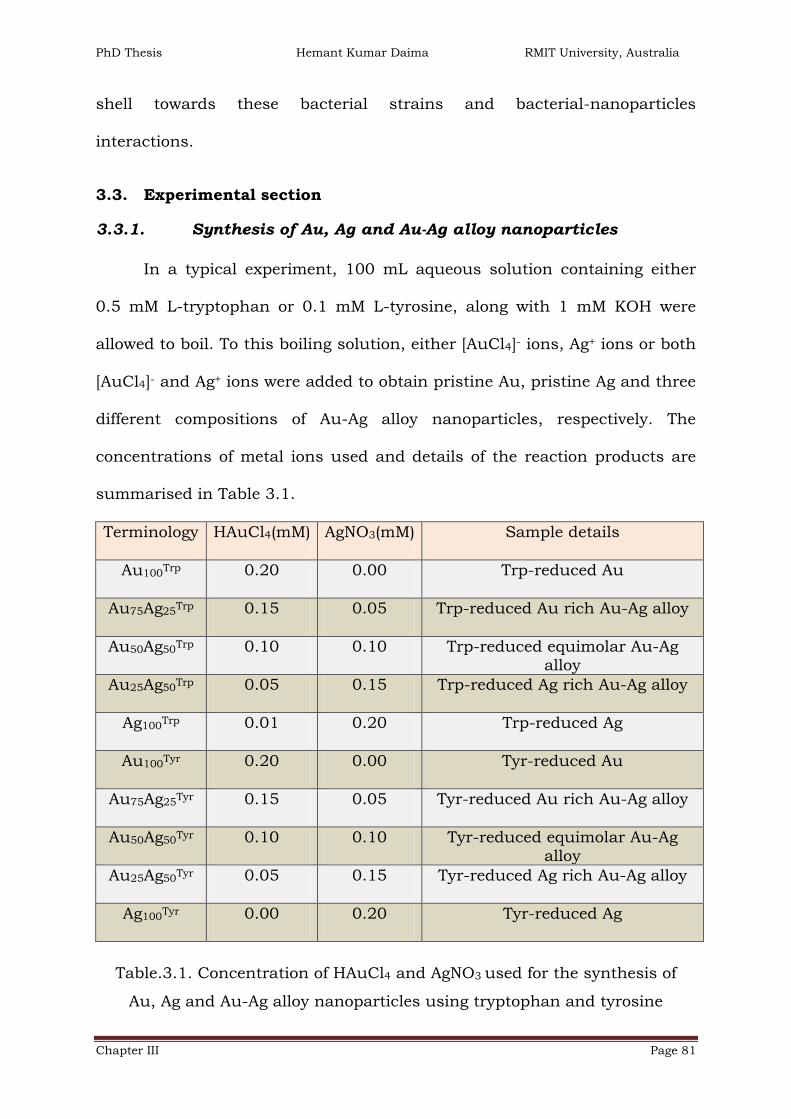
PhD Thesis Hemant Kumar Daima RMIT University, Australia
Chapter III Page 81
shell towards these bacterial strains and bacterial-nanoparticles
interactions.
3.3. Experimental section
3.3.1. Synthesis of Au, Ag and Au-Ag alloy nanoparticles
In a typical experiment, 100 mL aqueous solution containing either
0.5 mM L-tryptophan or 0.1 mM L-tyrosine, along with 1 mM KOH were
allowed to boil. To this boiling solution, either [AuCl4]- ions, Ag+ ions or both
[AuCl4]- and Ag+ ions were added to obtain pristine Au, pristine Ag and three
different compositions of Au-Ag alloy nanoparticles, respectively. The
concentrations of metal ions used and details of the reaction products are
summarised in Table 3.1.
Terminology HAuCl4(mM) AgNO3(mM) Sample details
Au100Trp 0.20 0.00 Trp-reduced Au
Au75Ag25Trp 0.15 0.05 Trp-reduced Au rich Au-Ag alloy
Au50Ag50Trp 0.10 0.10 Trp-reduced equimolar Au-Ag
alloy
Au25Ag50Trp 0.05 0.15 Trp-reduced Ag rich Au-Ag alloy
Ag100Trp 0.01 0.20 Trp-reduced Ag
Au100Tyr 0.20 0.00 Tyr-reduced Au
Au75Ag25Tyr 0.15 0.05 Tyr-reduced Au rich Au-Ag alloy
Au50Ag50Tyr 0.10 0.10 Tyr-reduced equimolar Au-Ag
alloy
Au25Ag50Tyr 0.05 0.15 Tyr-reduced Ag rich Au-Ag alloy
Ag100Tyr 0.00 0.20 Tyr-reduced Ag
Table.3.1. Concentration of HAuCl4 and AgNO3 used for the synthesis of
Au, Ag and Au-Ag alloy nanoparticles using tryptophan and tyrosine

PhD Thesis Hemant Kumar Daima RMIT University, Australia
Chapter III Page 82
The total concentrations of metal ions were kept constant at 0.2 mM
in all the reactions. Here, it must be noted that although tyrosine amino
acid could efficiently synthesize pristine Ag nanoparticles (Ag100Tyr), despite
repeated efforts, tryptophan amino acid failed to reduce Ag+ ions into Ag
nanoparticles. Therefore, a minute amount of [AuCl4]- ions (10 µM) was
added to obtain Ag nanoparticles by tryptophan-mediated reduction
(Ag100Trp).
3.3.2. Processing of nanoparticles by concentration and dialysis
All of the ten nanoparticle solutions were heated in water bath to
concentrate the nanoparticle solutions (20 times). These solutions were
overnight dialysed to remove free metal ions and unbound amino acids from
the nanoparticle solutions against deionized water using 12 kDa dialysis
membranes. After concentration and dialysis, all the metal nanoparticles
solutions remained stable, indicating that these nanoparticles were strongly
capped with amino acids. The solutions thus obtained were found stable
under standard laboratory storage conditions at room temperature over 6
months, and used as such for materials characterisation and biological
studies.
3.3.3. Peroxidase-like activity of Au, Ag and Au-Ag nanoparticles
The peroxidase enzyme-like behaviour of monometallic Au, Ag and
bimetallic Au-Ag alloy nanoparticles was examined using 3,3’,5,5’-
tetramethylbenzidine (TMB) as a chromogenic substrate by catalysing the
oxidation of the substrate. Different metal concentrations of mono- and bi-
metallic nanoparticles were incubated with TMB substrate in the presence of

PhD Thesis Hemant Kumar Daima RMIT University, Australia
Chapter III Page 83
H2O2 to evaluate effects of nanoparticle concentration and composition on
their peroxidise-like activity, in dark.
In dark experimental conditions, the blue colour developed as the
reaction proceeded was monitored kinetically using a multimode
spectrophotometer. TMB conversion was measured at 650 nm. All these
experiments were performed at three different temperatures, 20ºC, 37ºC and
55ºC to investigate effects of temperature on peroxidise-like activities along
with surface functionalization and metal composition. Pure tryptophan,
tyrosine and precursor metal ions were also evaluated for their peroxidase
enzyme-like activities under same experimental conditions for comparison
purposes.
3.3.4. Antibacterial assays of Au, Ag and Au-Ag alloy
nanoparticles
Quantitative assessment of antimicrobial performance of monometallic
Au, Ag and bimetallic Au-Ag alloy nanoparticles was made using colony
count methods. Gram positive bacterium S. albus and Gram negative
bacterium E. coli were used as model microorganism for antibacterial
studies. Before performing microbiological experiments, all the culture
media, glass-wares, containers and micro tips were sterilized by autoclaving
at 121°C for 15 min.
To assess antimicrobial activity by colony counting method, 104
colony forming units (CFU) of bacteria were incubated with different
nanoparticle solutions for 24 hours at 4°C in LB broth, followed by spread-
plating of 100 μl aliquots onto nutrient agar (NA) plates. The NA plates were
PhD Thesis Hemant Kumar Daima RMIT University, Australia
Chapter III Page 84
incubated for 24 hours (E. coli) and 48 hours (S. albus) at 37 °C and colonies
formed after incubation were counted. These colonies correspond to the
number of live (viable) bacteria in each suspension at the time of aliquot
withdrawal. Plot of the bacterial viability versus the dose of nanoparticles
were constructed to visualize the dose dependant bacterial activity. All the
bacterial toxicity tests were performed in duplicates and repeated at 3
different times to ensure reproducibility. Further, Nano-SEM was employed
to reveal the morphological changes in treated bacterial cells after bacteria-
nanoparticles interaction.
3.4. Results and discussion
Tryptophan and tyrosine amino acids can be employed as a reducing
agents for gold and silver ions for the synthesis of stable metal nanoparticles
in aqueous medium, wherein indole group in tryptophan was found to
participate in the metal ions reduction, whereas tyrosine reduces the metal
ions through its phenol group.[84, 85]
In this thesis the capability of these amino acids was further explored
to form Au-Ag alloy metal nanoparticles of varying composition and surface
functionalization. This demonstrated that natural biomolecules such as
amino acids can act as green and eco-friendly reducing agents to achieve
control over the synthesis of metal alloys in a single step without any
requirement of extra additives or special reaction conditions.

PhD Thesis Hemant Kumar Daima RMIT University, Australia
Chapter III Page 85
3.4.1. UV-visible spectral studies of Au, Ag and Au-Ag alloy
nanoparticles
SPR absorption bands are strong characteristic signatures for the
noble metal and their alloy nanoparticles.[87] Usually, bimetallic
nanoparticles can be either core-shell or alloy type and UV-visible
spectrophotometer analysis can differentiate between these two systems.
The core-shell nanoparticles give rise to two different SPR bands and the
intensities of these individual bands will depend on the relative percentage
of each metal present in the core-shell system [93-95] and the similar
condition will occur from a dispersion containing separate Au nanoparticles
and Ag nanoparticles. Conversely, the single SPR feature of metal alloys will
lie between those of two independent metal nanoparticles. Therefore, the
formation of alloy nanoparticles can be confirmed using UV-visible
absorption spectroscopy.
250 400 550 700 850
Ab
so
rban
ce (
a.u
.)
Wavelength (nm)
Au100
Trp
Au75
Ag25
Trp
Au50
Ag50
Trp
Au25
Ag75
Trp
Ag100
Trp
250 400 550 700 850
Ab
so
rban
ce (
a.u
.)
Wavelength (nm)
Au100
Tyr
Au75
Ag25
Tyr
Au50
Ag50
Tyr
Au25
Ag75
Tyr
Ag100
Tyr
200 240 280 320
Ab
so
rban
ce (
a.u
.)
Wavelength (nm)
Trp
Tyr CBA
Figure.3.3. UV-visible absorbance spectra of (A) tryptophan (Trp) and
tyrosine (Tyr), (B) tryptophan- and (C) tyrosine- reduced Au, Ag and Au-Ag
alloy nanoparticles, respectively

PhD Thesis Hemant Kumar Daima RMIT University, Australia
Chapter III Page 86
In order to examine whether the nanoparticles synthesized using
tryptophan and tyrosine amino acids were alloys or core shell particles, UV-
visible absorption spectroscopic studies were carried out and presented in
Figure 3.3 (B and C). Illustrated in Figures 3.3 A is UV-vis absorbance
spectra of pure tryptophan and tyrosine amino acids. Since tryptophan and
tyrosine has indole and phenol groups, they have absorbance at 280 and
275 nm, respectively.
A single absorption band was obtained in UV-vis absorbance spectra
of Au, Au75Ag25 (gold-rich alloy), Au50Ag50 (equimolar Au and Ag alloy),
Au25Ag75 (silver-rich alloy) and Ag nanoparticles obtained from tryptophan
and tyrosine mediated reduction of metal ions (Figures 3.3 B and C,
respectively). Figures 3.3 B illustrates that tryptophan reduced Au100Trp and
Ag100Trp nanoparticles that showed their respective SPR bands at ca. 530 nm
and 420 nm, whereas the three alloy nanoparticles showed SPR features at
ca. 515 nm (Au75Ag25Trp), 495 (Au50Ag50
Trp) and 425 nm (Au75Ag25Trp),
respectively. Likewise, tyrosine reduced Au100Tyr, Au75Ag25
Tyr, Au50Ag50Tyr,
Au75Ag25Tyr and Ag100
Tyr nanoparticles show their respective SPR bands at ca.
520, 510, 485, 425 and 415 nm, respectively (Figures 3.3 C). A clear blue
shift of Au SPR features with an increase in Ag content, along with absence
of any noticeable additional features in the spectra of alloy nanoparticles
provides an evidence towards the formation of a Au-Ag alloy phase, without
forming any heterogeneous population of Au and Ag, or a core-shell type
structure.[87, 96] Additionally, the positions of the SPR absorption bands of
metal and metal alloy nanoparticles observed in this study are very similar
to those reported previously.[87]

PhD Thesis Hemant Kumar Daima RMIT University, Australia
Chapter III Page 87
A B
Au
10
0T
rp
Ag
10
0T
rp
Au
75A
g2
5T
rp
Au
50A
g5
0T
rp
Au
25A
g7
5T
rp
Au
10
0T
yr
Ag
10
0T
yr
Au
75A
g2
5T
yr
Au
50A
g5
0T
yr
Au
25A
g7
5T
yr
Figure.3.4. Digital photographs of metal nanoparticle solutions synthesised
using (A) tryptophan and (B) tyrosine
The digital photographs of metal nanoparticle solutions obtained by
tryptophan and tyrosine mediated reduction are illustrated in Figure 3.4 (A
and B, respectively), which clearly show a transition in solution colour in
both the cases from ruby red (Au) to yellow (Ag), respectively. The
intermediate compositions resulted in colours that were varying between
ruby red and yellow as can be seen from the digital photographs in Figure
3.4 demonstrating the alloy forming capability of tryptophan and tyrosine
amino acids. This is interesting that simple biomolecules such as amino
acids can provide a facile handle to control the composition of metal alloys
because synthesis of alloys almost always involve a reducing agent along
with a capping agent.
Additionally, UV-visible absorbance spectroscopy was employed to
compare nanoparticles as-obtained (post-synthesis), and after dialysis as
discussed earlier and shown in Appendix C.1. Interestingly, no considerable
changes in the position of the SPR plasmon of nanoparticles was observed
post- dialysis, except that reduction of п-п* absorption band due to the

PhD Thesis Hemant Kumar Daima RMIT University, Australia
Chapter III Page 88
aromatic rings of tryptophan and tyrosine at ca. 280 or 275 nm, suggesting
that the final nanoparticle solutions are free from unbound amino acids.
Since removal of free metal ions and capping agents from metal
nanoparticle solution typically results in nanoparticle aggregation, absence
of any such behaviour after dialysis in amino-acid reduced metal
nanoparticles indicates that amino acids strongly cap these nanoparticles
during synthesis. The stability of amino acid-reduced metal nanoparticles
was also evident when these particles showed no evidence of aggregation on
storage under standard laboratory conditions in deionized water for over six
months. Therefore, synthesis of highly stable, metal ion free, monometallic
and alloy nanoparticles is an important advantage of amino acids-mediated
synthesis of these nanomaterials.
3.4.2. TEM studies of amino acid reduced Au, Ag and Au-Ag alloy
nanoparticles
Illustrated in Figure 3.5 are TEM images, mean particle size and size
distribution histogram of tryptophan reduced Au100 (A1); Au75Ag25 (A2);
Au50Ag50 (A3); Au25Ag75 (A4) and Ag100 (A5) nanoparticles, respectively.
As shown in Figure 3.5 (A1-A5), all these nanoparticles are quasi-
spherical in shape. The mean particle size of Au100Trp is around 8.2 nm with
a standard deviation of 4.4 nm, while Ag100Trp nanoparticles are ~10.8 nm in
size with a standard deviation of 5.5 nm. The mean particles size for alloy
nanoparticles is ranging from 5 to 6 nm in size.

PhD Thesis Hemant Kumar Daima RMIT University, Australia
Chapter III Page 89
A1 A2 A3
A4 A5 Sample
Mean particle
size (nm) ± SD
A1 8.2 ± 4.0
A2 5.8 ± 1.9
A3 4.9 ± 2.2
A4 5.3 ± 2.8
A5 10.8 ± 5.5
Sample
Mean particle
size (nm) ± SD
A1 8.2 ± 4.0
A2 5.8 ± 1.9
A3 4.9 ± 2.2
A4 5.3 ± 2.8
A5 10.8 ± 5.5
A6
4 6 8 10
10
20
30
40
50
60
70 A2
Nu
mb
er
De
ns
ity
Particle Diameter (nm)
4 6 8
10
20
30
40
50
60
70
80 A3
Nu
mb
er
De
ns
ity
Particle Diameter (nm)
3 6 9 12 15
10
20
30
40
50
60
70 A1
Nu
mb
er
De
ns
ity
Particle Diameter (nm)
4 8 12 16 20
10
20
30
40
50
60
70
80
90 A5
Nu
mb
er
De
ns
ity
Particle Diameter (nm)
3 6 9 12
15
30
45
60
75
90
105
120 A4
Nu
mb
er
De
ns
ity
Particle Diameter (nm)
Fre
qu
en
cy (
%)
Particles Diameter (nm)
Figure.3.5. TEM images and particle size distribution histogram of
tryptophan reduced Au, Ag and their alloy nanoparticles. Scale bars
correspond to 100 nm. Average particle sizes along with standard deviations
are also shown (A6)
Figure 3.6 shows TEM images, mean particle size and particle size
distribution histogram of tyrosine synthesised Au, Ag and their Au-Ag alloy
nanoparticles. All these nanoparticles are spherical in shape. As shown in
Figure 3.6 (B1-B4), the mean particles size of Au100Tyr and Au containing all
the three alloys are ~ 5.5 to 6.5 nm in size with a standard deviation of ~2.5
nm. Conversely, Figure 3.6 (B5) shows that Ag100Tyr are comparatively bigger

PhD Thesis Hemant Kumar Daima RMIT University, Australia
Chapter III Page 90
in size then all the other nanoparticles synthesised either by tryptophan or
tyrosine and the mean particles size of Ag100Tyr is around 26.8 nm with a
standard deviation of 5.8 nm.
B2 B3B1
B4 B5Sample Mean particle
size (nm) ± SD
B1 6.5 ± 2.0
B2 6.6 ± 2.4
B3 6.3 ± 2.1
B4 5.5 ± 2.8
B5 26.8 ± 5.8
Sample Mean particle
size (nm) ± SD
B1 6.5 ± 2.0
B2 6.6 ± 2.4
B3 6.3 ± 2.1
B4 5.5 ± 2.8
B5 26.8 ± 5.8
B6
4 6 8 10
10
20
30
40
50
60
70
80 B1
Nu
mb
er
De
ns
ity
Particle Diameter (nm)
5 6 7 8 9
10
20
30
40
50
60
70
Particle Diameter (nm)
B3
Nu
mb
er
De
ns
ity
Particle Diameter (nm)
4 6 8 10 12
15
30
45
60
75
90
105 B2
Particle Diameter (nm)
3 6 9 12 15
15
30
45
60
75
90
105 B4
Particle Diameter (nm)
6 12 18 24 30 36 42
5
10
15
20
25
30
35 B5
Nu
mb
er
De
ns
ity
Particle Diameter (nm)
Fre
qu
en
cy (
%)
Particles Diameter (nm)
Figure.3.6. TEM images and particle size distribution histogram of tyrosine
reduced Au, Ag and Au-Ag alloy nanoparticles. Scale bars correspond to 100
nm. Corresponding average particle sizes along with standard deviations are
also shown (B6)
Shape and size of nanoparticles has considerable effects on their
biological activities.[31, 74, 97-100] All the nanoparticle synthesised with
amino acids are spherical or quasi-spherical in shape and 5 to 10 nm in size

PhD Thesis Hemant Kumar Daima RMIT University, Australia
Chapter III Page 91
(except Ag100Tyr). This is the size domain where size-dependent toxicity of
metal nanoparticles was typically not observed. Hence, shape and size of
these nanoparticles does not have significant influence on the biological
effects and therefore biological effects of composition and different surface
functionalization can be compared confidently.
3.4.3. Estimation of the metal content present in dialysed
nanoparticle solutions
Atomic absorption spectroscopy (AAS) was employed to measure the
metal content in each sample after nanoparticles synthesis because it is
likely that not all of the metal ions employed in the reaction are reduced
during metal nanoparticles synthesis, as discussed earlier. Therefore, after
nanoparticle formation and purification by dialysis, AAS studies were
performed on aqua regia (corrosive mixture of 1HNO3 + 3HCl) digested
nanoparticle solutions.
AAS results indicated that Au:Ag ratios in finally obtained
nanoparticle samples follow a distinct trend as shown in Table 3.2, which is
very close to the initial ratios of metal ions used during the synthesis of
metal nanoparticles (Table 3.1). Further, AAS results indicated that in
different alloy compositions, amino acids have a tendency to form a Ag-rich
alloy on the cost of gold ions used during the synthesis. For instance, the
initial molar ratios of [AuCl4]- and Ag+ ions used to form Au75Ag25Trp alloy
was 75:25, however in the nanoparticles, Au and Ag metal was found to be
in 68:32 ratio. Similarly, in the case of Au75Ag25Tyr it was found to be 66:34
for Au:Ag. This is most likely because it is easier to reduce Ag+ ions in

PhD Thesis Hemant Kumar Daima RMIT University, Australia
Chapter III Page 92
comparison to [AuCl4]- ions, and therefore when both [AuCl4]- and Ag+ ions
coexist in a single system, a Ag-rich Au-Ag alloy is obtained.
Sample % Molar ratios Zeta potential
(mV) Au Ag
Au100Trp 100 0 -34.6
Au75Ag25Trp 68 32 -41.0
Au50Ag50Trp 46 54 -21.1
Au25Ag50Trp 26 74 -41.6
Ag100Trp 8 92 -36.8
Au100Tyr 100 0 -42.4
Au75Ag25Tyr 66 34 -44.4
Au50Ag50Tyr 43 57 -42.1
Au25Ag50Tyr 23 77 -43.8
Ag100Tyr 0 100 -42.9
Table.3.2: Molar ratios (%) of Au and Ag estimated from AAS and zeta
potential measurements of nanoparticle solutions
AAS results further emphasize that by finely controlling the molar
ratios of metal ions during amino acid-mediated reduction, Au-Ag alloy
composition can be delicately tuned. It is noteworthy that the ratio of Au
and Ag in both tyrosine and tryptophan-reduced alloys were very similar,
which makes them suitable for comparison during peroxidase-like and
antibacterial studies.
3.4.4. Zeta potential measurements of Au, Ag and Au-Ag alloy
nanoparticles
Illustrated in Table 3.2 are zeta potential values of different metal
nanoparticles. The ζ potential reflects the charge on the surface of

PhD Thesis Hemant Kumar Daima RMIT University, Australia
Chapter III Page 93
nanomaterials. Interestingly, all the monometallic Au or Ag and bimetallic
Au-Ag nanoparticles possess high negative surface charge at physiological
pH. The pH of the nanoparticle solutions can indicate about the possible
surface charge of the nanoparticles due to the presence of tryptophan and
tyrosine amino acids on their surfaces. In principle, if pH of solution is
above the isoelectric point of tryptophan or tyrosine (5.89 or 5.66,
respectively), relevant nanoparticles must bear a negative surface charge.
Conversely, if the pH of the nanoparticles solution is below the isoelectric
point of amino acid the nanoparticles must bear a positive surface charge.
The negative zeta potential values were expected since these
nanoparticles were stabilized with tryptophan or tyrosine, thus making them
negatively charged at physiological pH. A high negative zeta potential value
also indicated that these particles have good stability, which is required to
prevent these nanoparticles from aggregation in bacterial growth media
because inorganic salts and other nutrients may induce aggregation.
Further, as all these nanoparticles have similar type of surface charge, it
excludes any possibility of surface charge dependent biological activities. It
is also imperative to add that charge on the surface of amino acids-capped
nanoparticles can be easily controlled by changing the solution pH due to
the zwitterionic nature of tryptophan and tyrosine.
3.4.5. X-ray photoemission spectroscopy analysis of Au, Ag and
Au-Ag alloy nanoparticles
XPS analysis was carried out to confirm zerovalent state of gold (Au0)
and silver (Ag0) in mono and bimetallic alloy nanoparticles after reduction of

PhD Thesis Hemant Kumar Daima RMIT University, Australia
Chapter III Page 94
respective [AuCl4]- and Ag+ ions with tryptophan and tyrosine amino acids.
XPS is regarded as a highly surface sensitive technique, therefore presence
of amino acid shell on the surface of dialysed nanoparticles samples were
also confirmed using XPS. All the spectra discussed in this thesis have been
background corrected using Shirley algorithm[101] and their core level
binding energies were aligned with respect to the adventitious C1s binding
energy (BE) of 285 eV.
84 86 88 90
Inte
ns
ity (
a.u
.)
Binding Energy (eV)
84 86 88 90
Inte
ns
ity (
a.u
.)
Binding Energy (eV)
84 86 88 90
Inte
nsit
y (
a.u
.)
Binding Energy (eV)
Au4f
Au100Trp
Au4f
Au50Ag50Trp
Au4f
Au100Tyr
368 371 374 377
Inte
ns
ity (
a.u
.)
Binding Energy (eV)
Ag3d
Au50Ag50Trp
368 371 374 377
Inte
nsit
y (
a.u
.)
Binding Energy (eV)
Ag3d
Ag100Tyr
368 371 374 377
Inte
ns
ity (
a.u
.)
Binding Energy (eV)
Counts_Original
Ag3d
Ag100Trp
Inte
nsit
y (
a.u
.)
Binding Energy (eV)
A
B D
C E
F
Figure.3.7. Core level Au4f and Ag3d spectra of tryptophan and tyrosine
synthesised Au and Ag nanoparticles (A-D) and core level Au4f (E) and Ag3d
(F) spectra arising from Au50Ag50Trp alloy nanoparticles, respectively

PhD Thesis Hemant Kumar Daima RMIT University, Australia
Chapter III Page 95
XPS studies were undertaken for all the 10 different compositions of
tryptophan and tyrosine reduced metal and metal alloy nanoparticles.
However, selected representative spectra have been shown for brevity. These
include Au100Trp, Au100
Tyr and Au50Ag50Trp. All the Au4f core level spectra
presented in Figure 3.7 (A, B and E for Au100Trp, Au100
Tyr and Au50Ag50Trp,
respectively) could be decomposed into two chemically distinct spin-orbit
pairs. The chemically distinct components are observed ca. 83.5 eV and
85.8 eV, respectively. The Au4f signal at the lower BE (~83.5 eV) Au4f7/2 is
attributed to fully reduced metallic Au0. On the other hand, it is interesting
to detect the presence of higher BE component (at ~85.8 eV) in all the
samples, which indicated the presence of a small amount of unreduced
[AuCl4]- ions absorbed on the surface of nanoparticles and even extensive
dialysis could not remove these ions due to strong electrostatic complexation
of absorbed gold ions with amino acids. Similar Au4f spectra were observed
in all the samples containing Au.
The core level XPS spectrum of Ag3d originating from Ag100Trp, Ag100
Tyr
and Au50Ag50Trp are illustrated in Figure 3.7 C, D and F, respectively. Similar
to Au4f core level spectra, the Ag3d spectra also could be decomposed into
two chemically distinctive spin-orbit pairs. Two chemically distinct BEs for
Ag3d appeared at 368.2 and 371.6 eV, respectively. Here, the low BE
component is attributed to metallic silver (Ag0), whereas the high BE
component occurs from the unreduced ions, demonstrating some Ag+ ions
bound on the surface of Ag nanoparticles.
Figure 3.8 (A-B) illustrates spectra of C1s core level recorded for
pristine tryptophan and tyrosine amino acids, respectively. Figure 3.8 (C-D)

PhD Thesis Hemant Kumar Daima RMIT University, Australia
Chapter III Page 96
shows the core level C1s, spectra taken from tryptophan and tyrosine
synthesised Au nanoparticles, respectively. N1s and K2p spectra taken from
tryptophan synthesised Au nanoparticles (Au100Trp) are presented in Figure
3.8 E and F, respectively.
285 288 291 294
Inte
nsit
y (
a.u
.)
Binding Energy (eV)
Counts_Original
285 288 291 294
Inte
nsit
y (
a.u
.)
Binding Energy (eV)
Counts_Original
C1s Trp
Inte
nsit
y (
a.u
.)
Binding Energy (eV)
284 286 288 290
Inte
ns
ity (
a.u
.)
Binding Energy (eV)
284 286 288 290
Inte
ns
ity (
a.u
.)
Binding Energy (eV)
398 400 402In
ten
sit
y (
a.u
.)
Binding Energy (eV)
294 296
Inte
ns
ity
(a
.u.)
Binding Energy (eV)
C1s Tyr
C1s Au100Trp
C1s Au100Tyr
N1s
Au100Trp
K2p
Au100Trp
A
B
C
D
E
F
Figure.3.8. Core level XPS spectra of C1s in pristine tryptophan (A), tyrosine
(B), Au100Trp(C) and Au100
Tyr (D) respectively. Core level N1s (E) and K2p (F)
spectra of tryptophan synthesised Au100Trp, respectively
C1s core level spectra recorded for pure tryptophan and tyrosine can
be decomposed into four energy levels (Figure 3.8 A-B). BE at 285.0 eV is
assigned to alkyl chain and aromatic carbon chain, shoulder of higher BE at

PhD Thesis Hemant Kumar Daima RMIT University, Australia
Chapter III Page 97
286.4 could be assigned to carbon attached with oxygen and nitrogen (C-O
and C-N) species. The high BE at 289.1 eV can be attributed to the
corboxylic group present in amino acids. Whereas, additional shaken-up
satellite feature at 291.9 eV in both the amino acids arises from the benzyl
side chain.[102] Likewise, the C1s core level spectra recorded from the
Au100Trp and Au100
Tyr Figure 3.8 (C and D, respectively) could be decomposed
into three major chemically distinct components at ca. 285, 286.4 and 288.3
eV. The higher BE component observed at 288.3 eV can be assigned to the
carboxylate carbon (–COO–) from nanoparticles-bound amino acid molecules
(tryptophan or tyrosine), another evidence suggesting why these
nanoparticles have very high negative zeta potential. Whereas 286.4 eV BE
peak is attributed to the C-N and C-O species again coming from respective
amino acid shell present on nanoparticles.
The existence of N1s signals is indicative of surface bound amine
groups of amino acids. The representative XPS spectrum of N1s is illustrated
in Figure 3.8-E. Furthermore, it was interesting to detect the presence of
K2p3/2 core levels at ca. 293 eV in the all the samples. Representative, XPS
spectrum of K2p recorded for Au100Trp is shown in Figure 3.8 (F). The source
of potassium ions in these samples was mainly due to the usage of KOH
solution during the synthesis of nanoparticles. Presence of K+ in
nanoparticles even after extensive dialysis of samples suggests that K+ ions
bind strongly to negatively charged nanoparticle to counter balance their
surface charges and to maintain electro-neutrality,[10, 11] which leads to
highly stable metal nanoparticles in aqueous solutions.
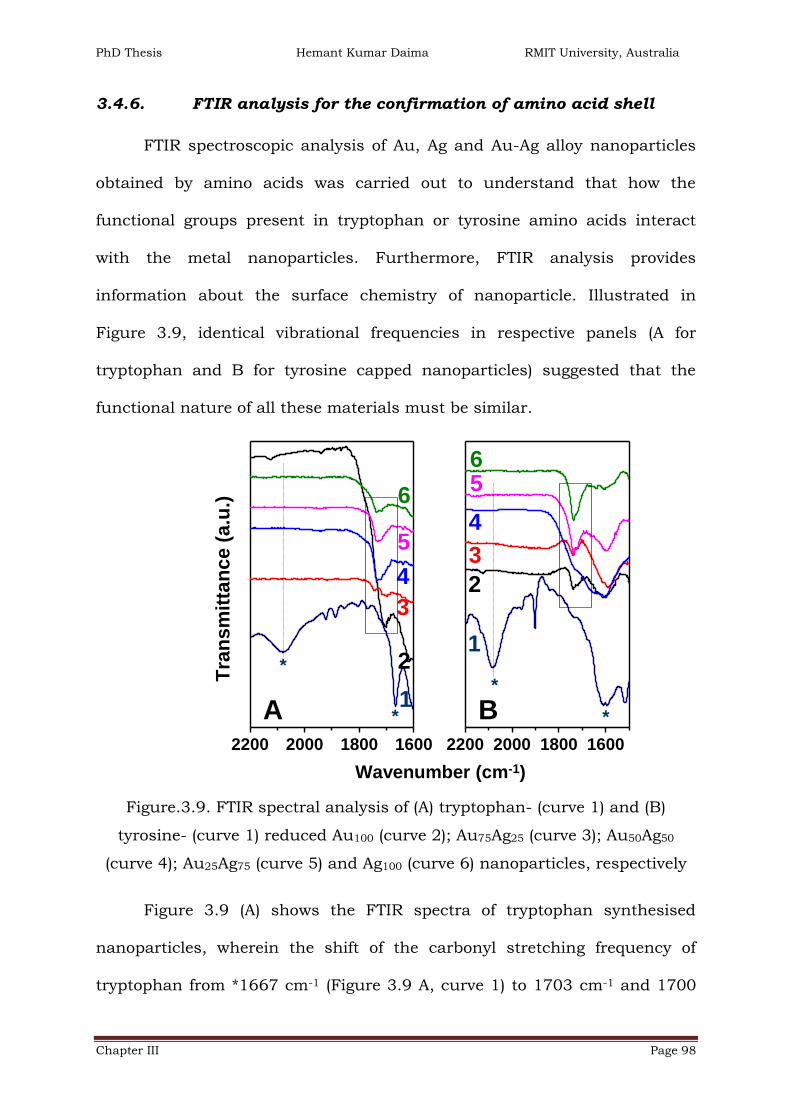
PhD Thesis Hemant Kumar Daima RMIT University, Australia
Chapter III Page 98
3.4.6. FTIR analysis for the confirmation of amino acid shell
FTIR spectroscopic analysis of Au, Ag and Au-Ag alloy nanoparticles
obtained by amino acids was carried out to understand that how the
functional groups present in tryptophan or tyrosine amino acids interact
with the metal nanoparticles. Furthermore, FTIR analysis provides
information about the surface chemistry of nanoparticle. Illustrated in
Figure 3.9, identical vibrational frequencies in respective panels (A for
tryptophan and B for tyrosine capped nanoparticles) suggested that the
functional nature of all these materials must be similar.
2200 2000 1800 1600
B1
WN
curve translate:
curve translate:
curve translate:
curve translate:
curve translate:
curve translate:
2200 2000 1800 1600
A1
WN
curve translate:
curve translate:
curve translate:
curve translate:
curve translate:
C11
*
1
2
3
5
4
6
B*
*
1
2
3
5
4
6
A *
Wavenumber (cm-1)
Tra
ns
mit
tan
ce
(a
.u.)
Figure.3.9. FTIR spectral analysis of (A) tryptophan- (curve 1) and (B)
tyrosine- (curve 1) reduced Au100 (curve 2); Au75Ag25 (curve 3); Au50Ag50
(curve 4); Au25Ag75 (curve 5) and Ag100 (curve 6) nanoparticles, respectively
Figure 3.9 (A) shows the FTIR spectra of tryptophan synthesised
nanoparticles, wherein the shift of the carbonyl stretching frequency of
tryptophan from *1667 cm-1 (Figure 3.9 A, curve 1) to 1703 cm-1 and 1700

PhD Thesis Hemant Kumar Daima RMIT University, Australia
Chapter III Page 99
cm-1 were observed for Au100Trp and Au rich alloy, as shown in Figure 3.9 A
curve 2 and 3, respectively. While in the case of Ag100Trp (Figure 3.9 A curve
6) and Ag rich alloys (Figure 3.9 A, curve 4 and 5) it was observed at 1736
cm-1 which can be attributed to the formation of carboxylate ions during the
reaction.[103] The difference in the value of shifts observed in different
samples correspond to varying degree of carboxylate ion formation in
different samples.
As shown in Figure 3.9 B, tyrosine has a carbonyl stretching
frequency around *1609 cm-1 (Figure 3.9 B, curve 1) that shifted to 1739
cm-1 in case of Au100Tyr (Figure 3.9 B, curve 2) or 1737 cm-1 in case of Ag100
Tyr
(Figure 3.9 B, curve 3) after nanoparticles synthesis, due to the carboxylate
ion formation. Hence, it was evident from the FTIR spectroscopy that
nanoparticles have surface carboxylate groups, which was in support of XPS
analysis. Further, the presence of K+ ions observed in the dialysed samples
during XPS analysis supported that carboxylic group of amino acid bound
onto metal nanoparticle surface was deprotonated into carboxylate ions.
Additionally, presence of surface carboxylate groups also supported the
negative surface charge determined by zeta potential measurements.
Moreover, the mechanism of reduction of [AuCl4]- ions and Ag+ ions by
tryptophan or tyrosine molecules is clearly understood by comparing FTIR
spectra of pristine amino acids and after reduction of metal ions.
Heterocyclic amine such as pyrrole and indole undergo oxidative
polymerization in the presence of an oxidizing agent and pyrrole is reported
to reduce [AuCl4]- ions to produce Au nanoparticles.[104] On the other hand,
electrochemical methods are documented for oxidative polymerization of

PhD Thesis Hemant Kumar Daima RMIT University, Australia
Chapter III Page 100
-C of the secondary amine.[105]
The amino acid tryptophan has a benzopyrrole (indole) group, which can
undergo oxidative polymerization in a similar manner to reduce [AuCl4]-
ions and Ag+ ions to create Au and Ag nanoparticles, respectively. Both
tryptophan and tyrosine molecules show a peak at *2080 cm-1 due to the
asymmetrical NH3+ bending vibration. The absence of this peak after
nanoparticles formation indicates that deprotonation of amine groups
during the reduction of metal ions. Further, the similar vibrational features
of all these nanoparticles suggest they all have similar amino acid shells on
the surface and the symmetry in the vibrational bands indicating that these
amino acids were accommodated on the surface of metal in a structured
fashion.
3.4.7. Electrochemical behaviour of amino acid capped Au, Ag and
their alloy nanoparticles
The binding capacity of amino acids on the surface of metal
nanoparticles will have considerable impact on the redox behaviour of Au,
Ag and Au-Ag alloy nanoparticles. This kind of fundamental understanding
of capping behaviour of tryptophan and tyrosine amino acids on
monometallic Au or Ag nanoparticles and on the surface of bimetallic Au-Ag
alloy nanoparticles is imperative to design effective nanomaterials for
biological activities. Furthermore, it is important to study the distribution of
Au and Ag atoms in the bimetallic alloy nanoparticles, which might be
affected by the strength of interactions occurring between metallic surface
and respective amino acids. Therefore, electrochemical behaviour of Au, Ag

PhD Thesis Hemant Kumar Daima RMIT University, Australia
Chapter III Page 101
nanoparticles and Au-Ag alloy nanoparticles was studied by employing
cyclic voltammetry (CV). CV is an excellent electrochemical technique to
study the aforementioned properties and cyclic voltammograms of these
nanoparticles were carried out by drop-casting these nanoparticles onto GC
substrates and the results are presented in Figure 3.10 (A and B).
0.0 0.2 0.4 0.6 0.8 1.0 1.2 1.4
-1
0
1
2
3
Cu
rren
t [
A]
Potential (V)
Au100
Trp
Au75
Ag25
Trp
Au50
Ag50
Trp
Au25
Ag75
Trp
Ag100
Trp
0.0 0.2 0.4 0.6 0.8 1.0 1.2 1.4
-2
0
2
4
6
8
Cu
rren
t [
A]
Potential (V)
Au100
Tyr
Au75
Ag25
Tyr
Au50
Ag50
Tyr
Au25
Ag75
Tyr
Ag100
Tyr
A B
Figure.3.10. Cyclic voltametric profiles of tryptophan- (A) and tyrosine (B)
synthesised nanoparticles recorded at 5 mV s-1 in 1 M H2SO4
In the case of tryptophan-reduced nanoparticles, the CV profile was
found to be remarkably similar for the Au100Trp, Au75Ag25
Trp, Au50Ag50Trp and
Au25Ag75Trp samples, all of which contained gold in different proportions. No
indication of the CV profile typically observed for gold in acidic solution was
seen (Figure 3.10 A), where typical gold-oxide formation and reduction is
observed in Figure 3.11 (A). The complete absence of a gold oxide reduction
process suggests that the capping by the tryptophan amino acid effectively
blocked the underlying metallic surface. However, another broad oxidation
process was reflected in Au100Trp, Au75Ag25
Trp, Au50Ag50Trp and Au25Ag75
Trp
samples from 0.80 to 1.40 V, which is similar to that recorded for a 1 mM
tryptophan solution in 1 M H2SO4 recorded at a GC electrode (Figure 3.11-

PhD Thesis Hemant Kumar Daima RMIT University, Australia
Chapter III Page 102
B). This broad response is most likely due to the oxidation of the tryptophan
capping agent on the nanoparticle surface, leading to formation of poly-
tryptophan.[83]
0.0 0.3 0.6 0.9 1.2 1.5
0
5
10
15
20
25
30
35
Cu
rren
t [
A]
Potential (V)
0.0 0.4 0.8 1.2 1.6
-30
-25
-20
-15
-10
-5
0
5
10
15
Cu
rren
t [
A]
Potential (V)
0.0 0.3 0.6 0.9 1.2
0
1
2
3
4
5
6
7
Cu
rren
t [
A]
Potential (V)
A B C
Cu
rren
t (µ
A)
Potential (V)
Figure.3.11. Cyclic voltametric profiles of (A) typical gold oxide formation
and reduction (B) CV profile of 1 mM tryptophan in 1 M H2SO4 and (C) CV
profile of tyrosine oxidation
Interestingly, a distinctly different profile is seen for Ag100
Trp sample
that showed a characteristic Ag oxidative stripping peak at 0.30 V (Figure
3.10-A).[106, 107] This suggests that during tryptophan-mediated synthesis
of pristine Ag nanoparticles, a complete compact coverage of the
nanoparticle surface with a capping layer does not occur as effectively as in
the presence of Au in the sample. Notably, upon increasing the silver
content within the alloy, a feature in the range of 0.30 to 0.80 V does begin
to emerge, which shifts to more negative potential as the Ag content is
increased (highlighted by arrow in Figure 3.10-A). This illustrates the ease
with which the nanomaterial can be oxidised, thus indicating a decrease in
the blocking ability of the capping agent with increase in Ag content.
In the case of tyrosine-reduced nanoparticles, evidence of the gold-
oxide reduction process at 0.90 V was seen in all the alloys containing Au,

PhD Thesis Hemant Kumar Daima RMIT University, Australia
Chapter III Page 103
including Au75Ag25Tyr, Au50Ag50
Tyr and Au25Ag75Tyr samples as shown in
Figure 3.10 B. However such feature was found absent in Au100Tyr sample,
suggesting again a blocking capping layer to electrochemical surface oxide
formation. Interestingly, as the Ag fraction is increased across the samples
from Au75Ag25Tyr, Au50Ag50
Tyr and Au25Ag75Tyr to Ag100
Tyr, the feature from
0.30 to 0.70 V increases and shifts to a more negative potential indicating a
material which is more readily oxidised until sample Ag100Tyr. The broad
process recorded from 0.90 to 1.40 V in all the tyrosine-reduced samples
most likely consists also of some contribution from tyrosine oxidation, that
recorded for a 1 mM tyrosine solution in 1 M H2SO4 recorded at a GC
electrode (Figure 3.11 C).
Thus, CV analysis suggested that in the case of tryptophan-reduced
nanoparticles get very strongly capped due to the formation of poly-
tryptophan during synthesis, which interferes with oxidation of Ag present
in tryptophan-reduced alloy nanoparticles. Conversely, in the case of
tyrosine-reduced nanoparticles they are not as strongly capped, and Ag
present in these alloy nanoparticles can be feasibly oxidized during CV
experiments. These observations may have significant impact on biological
profile of amino acid-reduced nanoparticles due to diverse surface
functionality. Moreover, when different metal nanoparticles are exposed to
biological system, the ease in oxidation of metal into metal ions may
determine the ultimate biological action (toxicity) of these materials.

PhD Thesis Hemant Kumar Daima RMIT University, Australia
Chapter III Page 104
3.5. Rational behind employing these nanomaterials for biological
applications
Tryptophan and tyrosine amino acids utilize their side functional
groups to reduce metal ions to form nanoparticles, while retaining their
amine and carboxylic acid groups intact on the nanoparticle surface. Hence
these tryptophan and tyrosine capped nanoparticles can be viewed as amino
acids supported on an inorganic nanoparticle, which can provide biological
identity to inorganic nanoparticles. Further, amino acids are the basic
building blocks for proteins and enzymes, and they become the active sites
of many enzymes for adsorption of selective substrates. The selectivity is
induced by the geometrical constraints specified by the structure of enzyme.
Therefore, amino acids supported on the surface of an inorganic
nanoparticle may function like a synthetic enzyme and it is essential to
study the enzymatic-behaviour of these systems in order to use these
materials for any other biological application.
In addition to the various properties of amino acids, when they
combine with the suitable metal composition of nanoparticles in a
cooperative manner, this will modulate their biophysical and biochemical
properties. Therefore, these carefully tailored nanoparticles were subjected
to investigate their enzyme-like behaviour and antimicrobial applications.
3.5.1. In vitro peroxidase-like behaviour of amino acids capped Au, Ag
and Au-Ag alloy nanoparticles
A variety of nanomaterials have been designed to exhibit biological
activities which depend on their considerable ability to provoke production

PhD Thesis Hemant Kumar Daima RMIT University, Australia
Chapter III Page 105
of reactive oxygen species (ROS). High level of ROS present in any biological
system is harmful due to the significant changes in homeostasis, which can
induce cell damage irrevocably while the lesser amount of ROS can be
effectively neutralized by natural antioxidants.[27-34, 108-112] Recently,
Ferrum-based (Fe) materials such as iron oxide nanoparticles have been
discovered to execute peroxidise enzyme-like activity. This peroxidise-like
activity of iron oxide nanoparticles has been attributed due to their close
resemblance with Fe present in the heme unit of natural peroxidases.[20,
21, 24, 113] Other nanomaterials have also been employed to explore
peroxidases enzyme-like activity or to mimic naturally occurring enzymes
besides Fe-based nanoparticles.[22, 26, 114, 115]
Tryptophan and tyrosine amino acids are two important building
blocks of natural enzymes and their capping on the surface of Au, Ag and
Au-Ag alloy nanoparticles has potential to act as synthetic ‘nanozymes’.
Therefore, in vitro study of peroxidases enzymes-like activity has been
explored to understand the significance of amino acids shell on inorganic
nanoparticles support in combination with metal composition in alloy
nanoparticles.
Au, Ag and Au-Ag alloy nanoparticles capped with tryptophan and
tyrosine amino acids exhibited peroxidise-like activity by catalysing the
oxidation of TMB substrate in the presence of H2O2 to produce a blue
coloured reaction product as shown in Figure 3.12. Prior to peroxidase-like
activity, various solutions of tryptophan and tyrosine synthesised
nanoparticles showed ruby red (Au nanoparticles) to yellow colour (Ag
nanoparticles) (Figure 3.13-A) based on the composition of nanoparticles, as

PhD Thesis Hemant Kumar Daima RMIT University, Australia
Chapter III Page 106
discussed previously. Intensities of these colours varied because the
concentrations employed to investigate peroxidase-like activity were different
such as 20, 10 and 5 ppm.
H2O2 +O
O
H
H
O.
H
H
O.
O.
H
H
O.+ + +2H2O
+
NPs NPs NPs
NPs NPsH2N
H3C
H3C
CH3
NH2
CH3
2 H2N
H3C
H3C
CH3
NH
CH3
2
H2N
H3C
H3C
CH3
NH
CH3
2 H2N
H3C
H3C
CH3
NH2
CH3
Figure.3.12. Schematic representation of oxidation of TMB by H2O2
catalysed by nanoparticles
Au100
Trp Au50
Ag50
Trp Ag100
Trp Au100
Tyr Ag100
TyrAu50
Ag50
Tyr
Au75
Ag25
Trp Au25
Ag75
Trp Au75
Ag25
Tyr Au25
Ag75
Tyr
20 ppm
10 ppm
05 ppm
20 ppm
10 ppm
05 ppm
A
BAu
100Trp Au
50Ag
50Trp Ag
100Trp Au
100Tyr Ag
100TyrAu
50Ag
50Tyr
Au75
Ag25
Trp Au25
Ag75
Trp Au75
Ag25
Tyr Au25
Ag75
Tyr
Figure.3.13. Peroxidase-like behaviour of tryptophan- and tyrosine- capped
Au, Ag and Au-Ag alloy nanoparticles with different concentrations

PhD Thesis Hemant Kumar Daima RMIT University, Australia
Chapter III Page 107
Illustrated in Figure 3.13 B is the optical photograph showing blue
colour response after peroxidase-like activity of amino acid capped
nanoparticles. It was visualized in Figure 3.13 that the peroxidase-like
activity of amino acid capped metal nanoparticles were modulated by the
composition and the type of amino acid that forms the surface corona
around the nanoparticles. Further, quantitative potential of nanoparticles
synthesised by amino acids were measured by absorption as a function of
reaction time for peroxidase-like behaviour and correlated with temperature,
composition and surface functionalization.
20°C 37°C 55°C
0 1 2 3 4 5 60.0
0.1
0.2
0.3
0.4
0.5
0.6
0.7
Ab
so
rba
nc
e (
65
0 n
m)
Time (Hours)
A1
A5
B1
B5
0 1 2 3 4 5 60.0
0.1
0.2
0.3
0.4
0.5
0.6
0.7
Ab
so
rba
nc
e (
65
0 n
m)
Time (Hours)
A1
A5
B1
B5
0 1 2 3 4 5 60.0
0.1
0.2
0.3
0.4
0.5
0.6
0.7
Ab
so
rba
nc
e (
65
0 n
m)
Time (Hours)
A1
A5
B1
B5
1
2
3
4
1
2
34
1
2
3
4
A B C
Figure.3.14. Temperature dependent peroxidase-like activity of tryptophan-
and tyrosine- capped Au and Ag nanoparticles In all the panels, curve 1, 2,
3 and 4 correspond to Au100Trp, Au100
Tyr, Ag100Trp and Ag100
Tyr, respectively
Figure.3.14 illustrates the kinetics of peroxidase-like behaviour of
monometallic Au and Ag nanoparticles at 5 ppm metal concentration on
20ºC, 37ºC and 55ºC temperatures in panel A, B and C respectively. In all
the panels of Figure.3.14, curve 1 and 2 shows peroxidase-like behaviour of
tryptophan and tyrosine synthesised Au nanoparticles, whereas, curves 3
and 4 correspond to respective amino acids synthesised Ag nanoparticles.

PhD Thesis Hemant Kumar Daima RMIT University, Australia
Chapter III Page 108
Relatively, tyrosine capped Au nanoparticles demonstrated higher catalysing
potential toward TMB substrate, indicating that tyrosine capping of Au
nanoparticles have comparatively higher peroxidase-like activity then
tryptophan capped Au nanoparticles. These results show that amino acid
shell/corona present on the surface of nanoparticles also had some
influence on the peroxidase-like behaviour.
At all the examined temperatures (panels A, B and C), it was seen that
the activity of Ag based nanoparticles was significantly higher than Au
nanoparticles in both tryptophan and tyrosine capping, while there were
slight difference in the catalysing potential of Ag100Trp and Ag100
Tyr. This
phenomenon suggests that the peroxidase-like behaviour of amino acid
capped metal nanoparticles predominantly depends on the core metal,
whereas amino acid surface corona has slight influence of peroxidase-like
activity. For example, at 20ºC (Figure.3.14, panel A) after 4 hour, Ag100Trp
illustrated over six time higher oxidation of TMB substrate then Au100Trp in
the form of absorbance at 650 nm, signifying impact of core material.
Moreover, temperature has significant influence on the peroxidase-like
activity of Ag nanoparticles. As illustrated, at 37ºC (Figure.3.14-B) the
activity was optimal, whereas at 55ºC (Figure.3.14-C) their activity was
found to be lowest while moderate activity was reported at 37ºC
(Figure.3.14-A). Conversely for Au nanoparticles, a slight reduction in
peroxidase-like activity was observed with increase in temperature from 20
to 37 to 55ºC. There observations demonstrate that temperature plays an
influential role in peroxidase-like activity of Ag nanoparticles. These
observations revealed the importance of inorganic core metal content and

PhD Thesis Hemant Kumar Daima RMIT University, Australia
Chapter III Page 109
amino acid surface corona present around the nanoparticles and shows that
the peroxidase-like behaviour of nanomaterials depends on both the
composition and surface functionalization. These observations, further
suggest that the composition and surface chemistry of nanomaterials may
have cooperative effects on any biological activity, which is in general
overlooked. Therefore, to further understand the role of surface functionality
and metal composition along with temperature on peroxidase-like
behaviour, tryptophan and tyrosine capped alloys nanoparticles were
subjected for investigation under similar experimental conditions. Illustrated
in Figure.3.15 is peroxidase-like behaviour of Au-Ag nanoalloys synthesised
by tryptophan (Panel A, B and C) and tyrosine (Panel D, E and F) amino
acids at 20ºC (Panel A and D), 37ºC (Panel B and E) and 55ºC (Panel C and
F) respectively. In all panels, curve 1, 2 and 3 correspond to Au75Ag25,
Au50Ag50 and Au25Ag75 composition either prepared by tryptophan or
tyrosine, respectively.
Furthermore, Figure.3.15 revealed the significant effect of amino acid
corona on peroxidase-like activity. Comparison of same stoichiometric alloys
with different amino acids showed different peroxidase-like activity in all the
temperatures. Assessment between different panels (such as A with D, B
with E and C with F) captivatingly revealed that the presence of surface
corona has considerable influence on peroxidise-like behaviour along with
temperature and composition. As it could be seen that the core material
employed to construct curves 1, 2 and 3 during peroxidise-like activity in all
panels are similar but the absorbance is considerably different revealing
different catalysing power of these nanomaterials towards TMB. It was

PhD Thesis Hemant Kumar Daima RMIT University, Australia
Chapter III Page 110
interesting to detect that tyrosine capped alloy nanoparticles have
significantly higher peroxidise-like activity then tryptophan capped
nanoparticles. For instance, Figure 3.15.panel A, in the case of tryptophan
reduced Au75Ag25Trp (curve 1), Au50Ag50
Trp (curve 2), Au25Ag75Trp (curve 3) the
absorbance was found to be less than 0.2 after 6 hours in all the cases. On
the contrary, assessment of similar composition and temperature with
different surface functionalization (in case of tyrosine Figure 3.15.panel D),
Au75Ag25Tyr (curve 1), Au50Ag50
Tyr (curve 2), Au25Ag75Tyr (curve 3) showed 0.3,
0.6 and 0.9 absorbance revealing differential catalysing potential of alloy
nanoparticles, respectively.
Peroxidise-like behaviour of amino acid capped alloy nanoparticles
was found to be metal composition dependent and with respective increment
in the Ag fraction of alloy the peroxidise-like behaviour shows enhancement
by catalysing TMB substrate as it shows increment in absorbance at 650
nm. This tendency is widespread in all tested temperatures irrespective to
surface functionalization, although the effects were more prominent in the
case of tyrosine capped Au-Ag alloy nanoparticles. These results are in good
agreement with observations obtained during peroxidise-like behaviour of
monometallic Au and Ag nanoparticles. Thus, validating the hypothesis that
the peroxidise-like activity of inorganic nanoparticles depends on the core
material or composition used to prepare nanoparticles.
Moreover, to validate these results and observations, control
experiments were performed in parallel, wherein pure tryptophan, tyrosine
and free metal ions were used under same experimental conditions to
investigate their effects on peroxidase-like activity. It was interesting to

PhD Thesis Hemant Kumar Daima RMIT University, Australia
Chapter III Page 111
discover that free amino acids and metal ions alone do not have any
peroxidase-like activity as they could not catalyse oxidation of TMB to
produce blue coloured reaction product. These control experiments further
confirmed that the peroxidase-like activity of amino acid capped
nanoparticles were intrinsic nature of nanoparticulate systems which was
introduced during their synthesis and controlling nanomaterials
composition and surface functionalization has potential to fine tune their
biological activity.
0 1 2 3 4 5 60.0
0.2
0.4
0.6
0.8
1.0
1.2
Ab
so
rba
nc
e (
65
0 n
m)
Time (Hours)
B2
B3
B4
0 1 2 3 4 5 60.0
0.2
0.4
0.6
0.8
1.0
1.2
Ab
so
rba
nc
e (
65
0 n
m)
Time (Hours)
B2
B3
B4
0 1 2 3 4 5 60.0
0.2
0.4
0.6
0.8
1.0
1.2
Ab
so
rban
ce
(650 n
m)
Time (Hours)
B2
B3
B4
0 1 2 3 4 5 60.0
0.2
0.4
0.6
0.8
1.0
1.2
Ab
so
rba
nc
e (
65
0 n
m)
Time (Hours)
A2
A3
A4
194 pts SG smooth of A2
0 1 2 3 4 5 60.0
0.2
0.4
0.6
0.8
1.0
1.2
Ab
so
rban
ce
(650 n
m)
Time (Hours)
A2
A3
A4
104 pts SG smooth of A2
0 1 2 3 4 5 60.0
0.2
0.4
0.6
0.8
1.0
1.2
Ab
so
rba
nc
e (
65
0 n
m)
Time (Hours)
A2
A3
A4
77 pts SG smooth of A2
12
3
1
2
3
1
2
3
1
2
3
1
2
3
1
2
3
20°C 37°C 55°C
20°C 37°C 55°C
A B C
D E F
Figure.3.15. Temperature dependent peroxidase-like activity of tryptophan
(Panel A, B and C) and tyrosine (Panel D, E and F) capped Au-Ag nanoalloys.
In all the panels, curve 1, 2 and 3 correspond to Au75Ag25, Au50Ag50 and
Au25Ag75 respectively

PhD Thesis Hemant Kumar Daima RMIT University, Australia
Chapter III Page 112
These observations on composition and surface-functionality
dependent peroxidase-like activity of Au, Ag and Au-Ag alloy nanoparticles
are particularly interesting. As discussed in cyclic voltametric studies, due
to the presence of poly tryptophan, Ag stripping of alloy nanoparticles was
not possible, while in the case of tyrosine capped alloys, Ag stripping was
observed. Peroxidase-like activity of alloy nanoparticles was considerably
influenced by the Ag stripping from the alloy nanoparticles and therefore
tyrosine capped alloy nanomaterials exhibited higher activity. These results
suggest that choice of an appropriate biological capping agent can play a
major role in cooperative manner with composition in controlling the
biological action of nanomaterials. Similar influence of composition,
temperature and amino acid corona were obtained when the metal
concentrations of nanoparticles were kept 10 and 20 ppm to evaluate
peroxidase-like activity. Appendix C (Figure C.2 and C.3) illustrates
temperature dependent peroxidase-like activity of tryptophan- and tyrosine-
capped Au, Ag and Au-Ag alloy nanoparticles at 10 ppm.
3.5.2. Antibacterial assays of amino acid capped nanoparticles
Peroxidase-like activity of amino acid-functionalized Au, Ag and Au-Ag
alloy nanoparticles was found to be composition and surface
functionalization dependent. Due to the high intrinsic peroxidase-like
behaviour, these amino acid-functionalized nanoparticles are expected to
have other biological applications as well. Therefore, composition and
surface functionality dependent antibacterial activities of these amino acid-
functionalized nanoparticles were investigated against model Gram positive

PhD Thesis Hemant Kumar Daima RMIT University, Australia
Chapter III Page 113
bacterium Staphylococcus albus and Gram negative bacterium Escherichia
coli. Two different bacterial strains are preferred because these bacterial
strains have chemically dissimilar cell walls, which provide first line of
defence for these bacterial strains. Hence, the cooperative effect of metal
composition and surface functionalization may be entirely different toward
different bacterial strains as it was observed in the case of peroxidase-like
behaviour.
3.5.2.1. Antibacterial effects of nanoparticles and influence of
surface functionalization
A colony counting method was adopted to investigate the antibacterial
potential of amino acid functionalized Au and Ag nanoparticles. To evaluate
influence of surface functionalization on different bacterial strains, constant
metal concentration (25 ppm) was introduced to 104 colony forming units
(CFU) of bacteria and allowed to interact for 24 hours at 4°C, followed by
spread-plating onto nutrient agar plates. Colonies formed after incubation
were counted and curves of the bacterial viability versus nanoparticles dose
effect were constructed and shown in Figurer 3.16 A and B.
Illustrated in Figurer 3.16 A is antibacterial effect of tryptophan and
tyrosine functionalized Au and Ag nanoparticles against Gram negative
bacterium E. coli. At 25 ppm metal concentration, similar antibacterial
activity was observed by both tryptophan and tyrosine capped
nanoparticles. In the case of Au nanoparticles, both Au100Trp and Au100
Tyr
caused minimal cell death as the bacterial viability was found to be over
85% and 95% respectively. Relatively, tryptophan capped Au100Trp

PhD Thesis Hemant Kumar Daima RMIT University, Australia
Chapter III Page 114
nanoparticles have more toxic effects than tyrosine capped Au100Tyr
nanoparticles. Comparative difference in the cell viability shows that amino
acid surface functionalization has slight influence on the antibacterial
activity against E. coli as the core material is identical. Whereas, in the case
of amino acid functionalized Ag nanoparticles at the same 25 ppm
concentration both Ag100Trp and Ag100
Tyr nanoparticles exhibited significantly
higher level of toxicity against E. coli. In both cases cell viability was found
to be less than 5% (Figure 3.16 A). This is expected as Ag nanoparticles are
considered as highly efficient Gram negative antibacterial agents and
biocompatible nature of Au nanoparticles is well established.[41, 56, 58]
These results demonstrates that in the case of Gram negative bacteria (E.
coli), although the surface functionalization has some influence (shown by
Au nanoparticles) on antibacterial activity but predominantly it depends on
the metal fraction of nanoparticles (shown by Ag nanoparticles).
Furthermore, shown in Figurer 3.16 B is antibacterial potential of
amino acids functionalized Au and Ag nanoparticles against Gram positive
bacterium S. albus. It was interesting to observe that both tryptophan and
tyrosine capped Au and Ag nanoparticles demonstrate significantly higher
toxicity (>90%) against Gram positive bacteria. In the case of Ag+ ions it is
expected to perform such a high level of toxicity due to their known
interaction with building elements of bacterial cells, DNA and proteins.[77]
The ease of Ag+ ions stripping in both Ag100Trp and Ag100
Tyr nanoparticles
provide strong rational behind the toxicity of these tryptophan and tyrosine
functionalized Ag nanoparticles as discussed during cyclic voltammetry
analysis.

PhD Thesis Hemant Kumar Daima RMIT University, Australia
Chapter III Page 115
0
20
40
60
80
100
120Control
Ag100
Tyr
Ba
cte
rial V
iab
ilit
y (
%)
Treatments with Au and Ag nanoparticles
Ag100
Trp
Au100
Trp Au100
Tyr
0
20
40
60
80
100
120
Ba
cte
rial V
iab
ilit
y (
%)
Treatments with Au and Ag nanoparticles
Control
Au100
Trp Au100
TyrAg
100
Trp
Ag100
Tyr
A
B
Figure.3.16. Assessment of antibacterial potential of amino acid capped Au
and Ag nanoparticles against (A) E. coli and (B) S. albus, respectively
Recently, Dawson and co-workers established that during interaction,
a cell sees the surface corona or other biomolecules present on the surface
of nanoparticles rather than bare nanoparticles itself, [36, 116, 117] which
gives a rationale behind the unusual toxicity of Au100Trp and Au100
Tyr toward
S. albus that the toxicity is originating through the amino acid surface
functionalization toward Gram positive bacteria. Therefore, in the case of
Gram positive bacteria, it is believed that the toxicity arises due to the
amino acid shell because there was no considerable change in toxicity
whether Au or Ag nanoparticles were employed for antibacterial studies.
These interesting results suggest that there is a strong interaction between
the amino acid shell and the Gram positive bacterial cell wall that causes

PhD Thesis Hemant Kumar Daima RMIT University, Australia
Chapter III Page 116
disruption of the cell wall. These observations are further confirmed by
nano-SEM imaging where the amino acids present on metal nanoparticles
act cooperatively to destroy the integrity of Gram positive bacterial cell wall
and is discussed in the later part of this chapter.
Additionally, in order to validate this mechanism, control experiments
were performed in which pure amino acids were directly added in the
bacterial culture, where these amino acids were found to have comparatively
higher toxicity towards Gram positive bacteria than Gram negative bacteria.
For instant, after treatments with tryptophan amino acid (1mM), E. coli
bacteria showed 75% cell viability while at the similar concentration, S.
albus illustrated only 20% cell viability. Likewise, at the similar
concentration in the case of tyrosine, E. coli and S. albus exhibited 68% and
16% bacterial viability, which was in support of the proposed hypothesis.
The exact mechanism how amino acids are capable of hydrolysing Gram
positive bacterial cells in more effective manner is not clear at this stage and
requires further investigations. Therefore, amino acid-reduced metal
nanoparticles enveloped within an amino acid corona may offer
opportunities against controlling Gram positive bacteria by enabling initial
interaction of these nanomaterials with the peptidoglycan cell wall, followed
by cell wall lysis and finally bacterial cell death.
3.5.2.2. Effect of metal composition on antibacterial activity
employing Au-Ag alloy nanoparticles
Additionally, to evaluate the antibacterial effects of tryptophan and
tyrosine synthesised Au-Ag alloy nanoparticles, dilutions were made in such
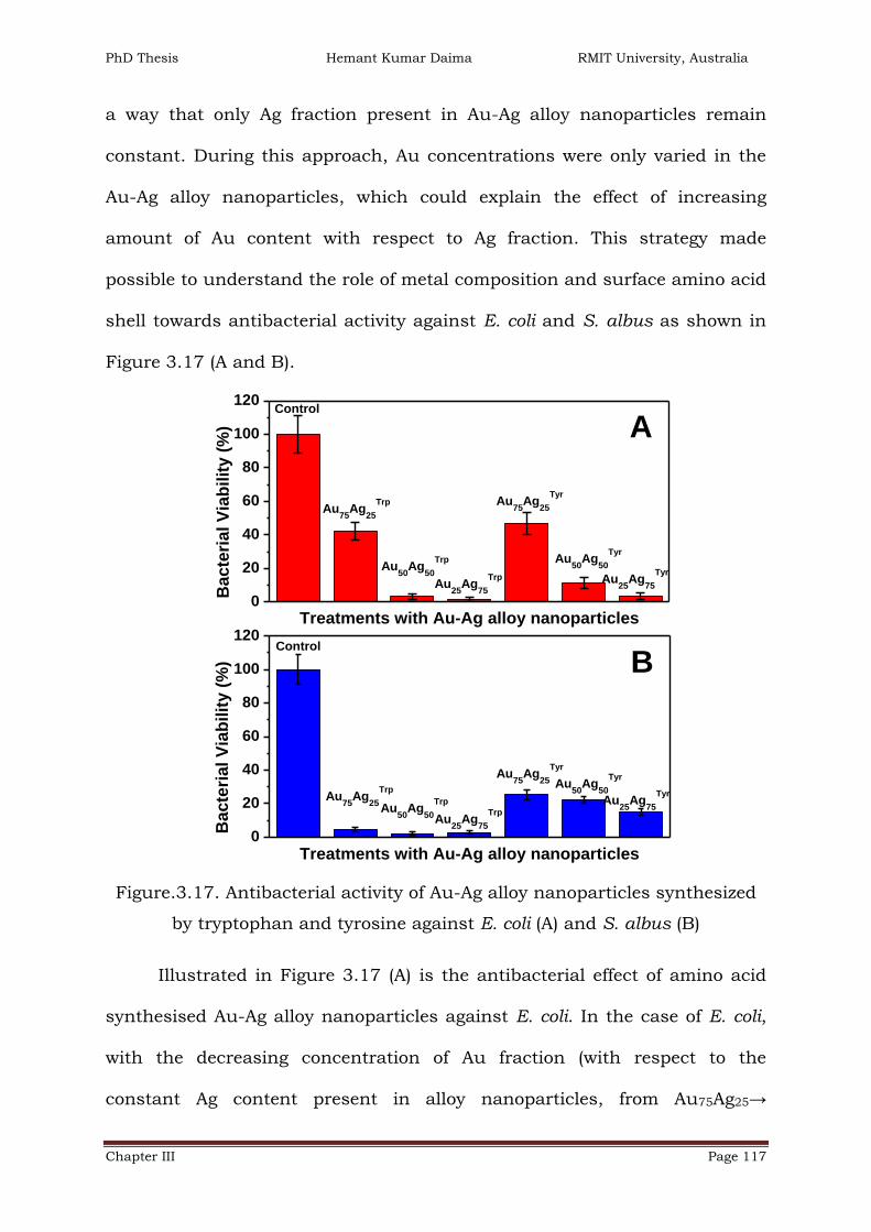
PhD Thesis Hemant Kumar Daima RMIT University, Australia
Chapter III Page 117
a way that only Ag fraction present in Au-Ag alloy nanoparticles remain
constant. During this approach, Au concentrations were only varied in the
Au-Ag alloy nanoparticles, which could explain the effect of increasing
amount of Au content with respect to Ag fraction. This strategy made
possible to understand the role of metal composition and surface amino acid
shell towards antibacterial activity against E. coli and S. albus as shown in
Figure 3.17 (A and B).
0
20
40
60
80
100
120
Au25
Ag75
TrpAu
50Ag
50
Trp
Bacte
rial V
iab
ilit
y (
%)
Treatments with Au-Ag alloy nanoparticles
Control
Au75
Ag25
Trp
Au50
Ag50
Tyr
Au75
Ag25
Tyr
Au25
Ag75
Tyr
0
20
40
60
80
100
120
Bacte
rial V
iab
ilit
y (
%)
Treatments with Au-Ag alloy nanoparticles
Control
Au25
Ag75
Trp
Au50
Ag50
TyrAu75
Ag25
Tyr
Au25
Ag75
TyrAu75
Ag25
Trp
Au50
Ag50
Trp
A
B
Figure.3.17. Antibacterial activity of Au-Ag alloy nanoparticles synthesized
by tryptophan and tyrosine against E. coli (A) and S. albus (B)
Illustrated in Figure 3.17 (A) is the antibacterial effect of amino acid
synthesised Au-Ag alloy nanoparticles against E. coli. In the case of E. coli,
with the decreasing concentration of Au fraction (with respect to the
constant Ag content present in alloy nanoparticles, from Au75Ag25→

PhD Thesis Hemant Kumar Daima RMIT University, Australia
Chapter III Page 118
Au50Ag50→ Au25Ag75), bacterial cell viability decreases significantly and this
trend was observed for both tryptophan and tyrosine amino acid capped Au-
Ag alloy nanoparticles (Figure 3.17.A). These observations are in strong
support that antibacterial activity against Gram negative bacteria arises
from the Ag fraction of the nanoparticles, as discussed in previous section.
Furthermore, tryptophan capped alloy nanoparticles were found to have
relatively more toxic effects than tyrosine capped alloy nanoparticles as
reflected in pristine Au100Trp and Au100
Tyr nanoparticles as well (Figure
3.16.A). For instance, 40% cell viability was observed in Au75Ag25Trp, which
increased up to 10% in the case of Au75Ag25Tyr wherein the central material
is identical, confirming the influence of surface functionalization.
Comparative variation in the cell viability of E. coli shows that amino acid
surface functionalization has control on the antibacterial activity and
validating results discussed in the case of pristine Au and Ag nanoparticles.
Conversely, in the case of S. albus (Figure 3.17.B) there were no
considerable changes observed in bacterial viability with the decreasing
amount of Au fraction present in Au-Ag alloy nanoparticles (from Au75Ag25→
Au50Ag50→ Au25Ag75). However, there is a slight reduction in bacterial cell
viability in the case of tyrosine capped alloy nanoparticles, but overall their
toxicity was higher than toxicity observed in E. coli. These results are also in
support that in the case of Gram positive bacteria, composition of Au-Ag
alloy nanoparticles do not have significant influence. Additionally, Figure
3.17.B demonstrated that in the case of S. albus also, tryptophan capped
alloy nanoparticles have significantly higher toxicity compared to tyrosine
capped alloy nanoparticles. For instance, Au75Ag25Trp has less than 5%

PhD Thesis Hemant Kumar Daima RMIT University, Australia
Chapter III Page 119
bacterial viability whereas Au75Ag25Tyr has over 25% cell viability, which
clearly explains the effect of surface corona present on nanoparticles in the
form of the amino acid.
3.5.2.3. Morphological studies of Au and Ag nanoparticles treated
E. coli and S. albus bacterial cells
Towards antimicrobial activities, it is hypothesized that nanomaterials
and cations such as Ag+ ions primarily affect the function of membrane-
bound enzymes including enzymes of the respiratory chain. The inhibitory
mechanism of nanoparticles on microorganisms shows that after treatment,
DNA loses its replication ability; expression of ribosomal subunits proteins
and other cellular proteins and enzymes essential for ATP production
becomes inactive.[45, 53, 56, 58, 59, 74, 77] However, the mechanism of
antibacterial action of nanomaterials is still not clearly understood but
reported literature could highlight that bacterial morphology and its
membrane integrity is one of the key factors, which is crucial for bacterial
survival. Therefore, to understand the effects of amino acid surface
functionalized metal nanoparticles on bacterial morphology nano-scanning
electron microscopy (Nano-SEM) was employed to visualize changes in
bacterial cell wall and morphology after their treatments.
SEM micrographs of E. coli bacterial cells before and after their
treatments with nanoparticles are illustrated in Figure 3.18. E. coli bacterial
cells without any treatment (control cells) showed intact cell architecture
and the cells are around 3 μm Χ 1 μm size and appeared as small rod
shaped as illustrated in Figure 3.18 A. Bacterial cells exposed to amino

PhD Thesis Hemant Kumar Daima RMIT University, Australia
Chapter III Page 120
acids capped Au nanoparticles (Au100Trp or Au100
Tyr) are shown in Figure 3.18
B and D respectively. These cells revealed that there were no significant
changes in their morphology and they had intact cell architecture as
untreated E. coli cells. Although, these bacterial cells appeared smaller in
size compare to untreated cells. Conversely, illustrated in Figure 3.18 C and
E are bacterial cells treated with Ag100Trp or Ag100
Tyr nanoparticles,
respectively. In contrast to untreated or Au nanoparticles treated bacterial
cells, Ag nanoparticles treated bacterial cells revealed distinct changes in
cell morphology. In lower magnification micrographs of Figure 3.18 C and E
it could be seen that treated E. coli cells were dissolved and their cellular
components were disintegrated, wherein higher magnification images
exposed that Ag100Trp (Figure 3.18 c) treated bacterial cell were entirely
disintegrated and severely damaged. While in the case of Ag100Tyr (Figure
3.18 e), it could be seen that the bacterial cell were broken in small pieces
along with distinct morphology and confirmed patchy bacterial cells.
The components of E. coli bacterial cell wall became disorganised and
scattered from their original order and close arrangement after their
exposure to amino acid coated Ag nanoparticles. Thus confirming that
amino acid coated Ag nanoparticles had significant influence on the E. coli
bacterial cell morphology, while similar surface functionality on Au
nanoparticles did not show any influential toxic effect on bacterial cells.
These observations revealed that in the case of Gram negative bacterium E.
coli the toxicity depends on the metal fraction of nanoparticles and presence
of either amino acid on the surface of nanoparticles did not exhibited any
toxic effect.

PhD Thesis Hemant Kumar Daima RMIT University, Australia
Chapter III Page 121
RMIT University©2011
B
C
D
E
A
E. coli
E. coli_Au100W
E. coli_Ag100Y
E. coli_Au100Y
E. coli_Ag100W
E.coli_Au100Trp
E.coli_Ag100Trp
E.coli_Au100Tyr
E.coli_Ag100Tyr
Figure.3.18. SEM micrographs of E. coli before and after their treatments
with tryptophan and tyrosine synthesised nanoparticles (Scale bar 5 and 1
µM, respectively)

PhD Thesis Hemant Kumar Daima RMIT University, Australia
Chapter III Page 122
B
C
D
E
A
S. albus
S. albus_Au100W
S. albus_Ag100Y
S. albus_Au100Y
S. albus_Ag100W
S.albus_Au100Trp
S.albus_Ag100Trp
S.albus_Au100Tyr
S.albus_Ag100Tyr
Figure.3.19. SEM micrographs of S. albus before and after their treatments
with tryptophan and tyrosine synthesised nanoparticles (Scale bar 5 and 1
µM, respectively)

PhD Thesis Hemant Kumar Daima RMIT University, Australia
Chapter III Page 123
Illustrated in Figure 3.19 (A) are S. albus bacterial cells without any
treatment showing spherical shape and in lower magnification micrograph
the bacterial cluster resembles grapes along with integral cell morphology.
Wherein, the higher magnification image shows that these bacterial cells
have slightly rough surface. Figure 3.19 B and C, treated with tryptophan
coated Au and Ag nanoparticles shows that after treatment S. albus cells are
dissolved and dispersed. At higher magnification, it could be seen that the
bacterial cell wall is peeling out from the bacteria after their exposure to
Au100Trp (Figure 3.19 b); while the dispersion effect is clearly evident in
bacterial cells treated with Ag100Trp (Figure 3.19 c). Similar, experiments were
performed on the tyrosine synthesised nanoparticles, which revealed that
these nanomaterials have even more severe morphological changes and
along with dissolving bacterial cells tyrosine capped nanoparticles shows
significantly higher level of roughness on the bacterial surface (Figure 3.19
D and E). After treatments, the cell wall components became disorganised
and scattered from their original ordered and close arrangement. In this sort
of morphology and disintegration, it is believed that cell wall possibly lost
their natural function which ultimately leads to bacterial cell death. These
observations revealed that even the most biocompatible Au nanoparticles
may exhibit significant toxicity toward Gram positive bacteria (S. albus) by
rupturing and collapsing the bacterial cell wall.
The rationale behind this is that in the case of E. coli only Ag
nanoparticles exhibited significant toxicity compare to the Au nanoparticles
because the metal fraction of the nanoparticles was responsible for
antibacterial studies as reported in previous studies. Conversely, in case of

PhD Thesis Hemant Kumar Daima RMIT University, Australia
Chapter III Page 124
S. albus, due to the presence of the amino acids on the surface of
nanoparticles these materials caused structural changes which finally lead
to cell death. Based on the observed antibacterial activity and existing
literature, Figure 3.20 demonstrates various mechanisms by which Au and
Ag nanoparticles exhibit antibacterial activities toward Gram positive and
Gram negative bacteria.
e-
e-
E. coli
S. albus
Non-toxic
AgNPs
AuNPs
Disruption of membrane/wall
Protein oxidation
Interruption of e- transport
DNA damage
Release of ions
Production of ROS
e-
e-
e-
Figure.3.20. Schematic illustration of antibacterial activity of Au and Ag
nanoparticles toward E. coli and S. albus

PhD Thesis Hemant Kumar Daima RMIT University, Australia
Chapter III Page 125
3.6. Conclusions
In existing literature, stabilizer or toxic chemical reducing agents have
been employed to synthesis bimetallic nanoparticles, whereas the method
adopted in this chapter provides highly stable, water dispersible metal
nanoparticles with different metal composition and surface functionalization
without using any additional stabilizer or noxious chemical. [87, 89, 92] In
this chapter, a green and eco-friendly method has been developed to prepare
Au, Ag and Au-Ag alloy nanoparticles with different metal composition and
functionalization using tryptophan and tyrosine amino acids. Indole and
phenols groups present in tryptophan and tyrosine are found to reduce gold
and silver ions into their respective mono and bimetallic nanoparticulate
forms. Thus this is a major step towards greener synthesis of nanomaterials.
Moreover, presence of amino acid corona may provide biological
identity to these nanoparticles and these nanomaterials may have
significant potential for biological applications. In general, Ag nanoparticles
are regarded as toxic by their nature whereas Au nanoparticles are
considered as biocompatible.[66, 70, 118] Co-existence of both the metals in
Au-Ag alloy nanoparticles in different ratios may have differential biological
activity. Therefore, prior to investigate biological activities of these amino
acid-functionalized nanoparticles, these nanomaterials were thoroughly
characterized by UV-vis, AAS, FTIR, XPS, Zeta potential, TEM and cyclic
voltammetry studies to understand their physicochemical nature.
The amino acid-functionalized nanoparticles exhibited intrinsic
peroxidase enzyme-like activity, which was found to be metal composition,
surface functionalization and temperature dependent. Ag nanoparticles

PhD Thesis Hemant Kumar Daima RMIT University, Australia
Chapter III Page 126
exhibited higher peroxidase-like activity compared to Au nanoparticles and
in the case of alloy nanoparticles; this activity was dependant on the Ag
fraction. Presence of tyrosine corona on nanoparticles surface had
significant influence on peroxidase-like behaviour. Further, antibacterial
potential of these amino acid-functionalized Au, Ag and Au-Ag nanoparticles
were investigated on Gram positive (S. albus) and Gram negative bacteria (E.
coli) bacteria. Both, tryptophan and tyrosine synthesised Au, Ag and Au-Ag
alloy nanoparticles have significant activity towards S. albus, whereas in E.
coli, only Ag nanoparticles or Ag rich alloy nanoparticles were considerably
active. This study indicates that even biocompatible Au nanoparticles can be
made toxic. Moreover, it was revealed that toxic effect towards S. albus is
due to the presence of amino acid shell, wherein the toxicity originates due
to the Ag fraction of nanoparticles against E. coli. Nano-SEM validated
observed antibacterial results and confirmed morphological changes and
level of damage in treated bacterial cells.
3.7. References
1. Katz, E. and I. Willner, Integrated Nanoparticle–Biomolecule Hybrid
Systems: Synthesis, Properties, and Applications. Angewandte Chemie
International Edition, 2004. 43(45): p. 6042-6108.
2. Jain, P.K., et al., Noble Metals on the Nanoscale: Optical and
Photothermal Properties and Some Applications in Imaging, Sensing,
Biology, and Medicine. Accounts of Chemical Research, 2008. 41(12):
p. 1578-1586.

PhD Thesis Hemant Kumar Daima RMIT University, Australia
Chapter III Page 127
3. Shim, M., et al., Functionalization of Carbon Nanotubes for
Biocompatibility and Biomolecular Recognition. Nano Letters, 2002.
2(4): p. 285-288.
4. Liong, M., et al., Multifunctional inorganic nanoparticles for imaging,
targeting, and drug delivery. ACS Nano, 2008. 2(5): p. 889-896.
5. Dobrovolskaia, M.A. and S.E. McNeil, Immunological properties of
engineered nanomaterials. Nature Nanotechnology, 2007. 2(8): p. 469-
478.
6. Colvin, V.L., The potential environmental impact of engineered
nanomaterials. Nature Biotechnology, 2003. 21(10): p. 1166-1170.
7. Lewinski, N., V. Colvin, and R. Drezek, Cytotoxicity of Nanoparticles.
Small, 2008. 4(1): p. 26-49.
8. Hall, J.B., et al., Characterization of nanoparticles for therapeutics.
Nanomedicine, 2007. 2(6): p. 789(15).
9. Leroueil, P.R., et al., Nanoparticle Interaction with Biological
Membranes: Does Nanotechnology Present a Janus Face? Accounts of
Chemical Research, 2007. 40(5): p. 335-342.
10. Nel, A., et al., Toxic potential of materials at the nanolevel. Science,
2006. 311(5761): p. 622-627.
11. Nel, A.E., et al., Understanding biophysicochemical interactions at the
nano-bio interface. Nature Materials, 2009. 8(7): p. 543-557.
12. Xia, T., et al., Cationic polystyrene nanosphere toxicity depends on cell-
specific endocytic and mitochondrial injury pathways. ACS Nano, 2008.
2(1): p. 85-96.

PhD Thesis Hemant Kumar Daima RMIT University, Australia
Chapter III Page 128
13. Weissleder, R., et al., Cell-specific targeting of nanoparticles by
multivalent attachment of small molecules. Nature Biotechnology,
2005. 23(11): p. 1418-1423.
14. Brunetti, V., et al., InP/ZnS as a safer alternative to CdSe/ZnS
core/shell quantum dots: In vitro and in vivo toxicity assessment.
Nanoscale, 2013. 5(1): p. 307-317.
15. Mout, R., et al., Surface functionalization of nanoparticles for
nanomedicine. Chemical Society Reviews, 2012. 41(7): p. 2539-2544.
16. Doane, T.L. and C. Burda, The unique role of nanoparticles in
nanomedicine: imaging, drug delivery and therapy. Chemical Society
Reviews, 2012. 41(7): p. 2885-2911.
17. Schaeublin, N.M., et al., Does shape matter? Bioeffects of gold
nanomaterials in a human skin cell model. Langmuir, 2012. 28(6): p.
3248-3258.
18. Walkey, C.D., et al., Nanoparticle size and surface chemistry determine
serum protein adsorption and macrophage uptake. Journal of the
American Chemical Society, 2012. 134(4): p. 2139-2147.
19. Goethals, E.C., et al., Decoupling the Effects of the Size, Wall
Thickness, and Porosity of Curcumin-Loaded Chitosan Nanocapsules on
Their Anticancer Efficacy: Size Is the Winner. Langmuir, 2013. 29(2): p.
658-666.
20. Gao, L., et al., Intrinsic peroxidase-like activity of ferromagnetic
nanoparticles. Nature Nanotechnology, 2007. 2(9): p. 577-583.

PhD Thesis Hemant Kumar Daima RMIT University, Australia
Chapter III Page 129
21. Wei, H. and E. Wang, Fe 3O 4 magnetic nanoparticles as peroxidase
mimetics and their applications in H 2O 2 and glucose detection.
Analytical Chemistry, 2008. 80(6): p. 2250-2254.
22. He, W., et al., Au@Pt nanostructures as oxidase and peroxidase
mimetics for use in immunoassays. Biomaterials, 2011. 32(4): p. 1139-
1147.
23. Zhang, K., et al., Formation of PdPt Alloy Nanodots on Gold Nanorods:
Tuning Oxidase-like Activities via Composition. Langmuir, 2011. 27(6):
p. 2796-2803.
24. Fan, J., et al., Direct evidence for catalase and peroxidase activities of
ferritin-platinum nanoparticles. Biomaterials, 2011. 32(6): p. 1611-
1618.
25. Pirmohamed, T., et al., Nanoceria exhibit redox state-dependent
catalase mimetic activity. Chemical Communications, 2010. 46(16): p.
2736-2738.
26. He, W., et al., Design of AgM Bimetallic Alloy Nanostructures (M = Au,
Pd, Pt) with Tunable Morphology and Peroxidase-Like Activity.
Chemistry of Materials, 2010. 22(9): p. 2988-2994.
27. Banerjee, M., et al., Heightened Reactive Oxygen Species Generation in
the Antimicrobial Activity of a Three Component Iodinated Chitosan-
Silver Nanoparticle Composite. Langmuir, 2010. 26(8): p. 5901-5908.
28. Premanathan, M., et al., Selective toxicity of ZnO nanoparticles toward
Gram-positive bacteria and cancer cells by apoptosis through lipid
peroxidation. Nanomedicine: Nanotechnology, Biology and Medicine,
2011. 7(2): p. 184-192.

PhD Thesis Hemant Kumar Daima RMIT University, Australia
Chapter III Page 130
29. AshaRani, P.V., et al., Cytotoxicity and genotoxicity of silver
nanoparticles in human cells. ACS Nano, 2009. 3(2): p. 279-290.
30. Carlson, C., et al., Unique cellular interaction of silver nanoparticles:
Size-dependent generation of reactive oxygen species. Journal of
Physical Chemistry B, 2008. 112(43): p. 13608-13619.
31. Choi, O. and Z. Hu, Size dependent and reactive oxygen species
related nanosilver toxicity to nitrifying bacteria. Environmental Science
and Technology, 2008. 42(12): p. 4583-4588.
32. Limbach, L.K., et al., Exposure of engineered nanoparticles to human
lung epithelial cells: Influence of chemical composition and catalytic
activity on oxidative stress. Environmental Science and Technology,
2007. 41(11): p. 4158-4163.
33. Long, T.C., et al., Titanium dioxide (P25) produces reactive oxygen
species in immortalized brain microglia (BV2): Implications for
nanoparticle neurotoxicity. Environmental Science and Technology,
2006. 40(14): p. 4346-4352.
34. Xia, T., et al., Comparison of the Abilities of Ambient and Manufactured
Nanoparticles To Induce Cellular Toxicity According to an Oxidative
Stress Paradigm. Nano Letters, 2006. 6(8): p. 1794-1807.
35. Hirst, S.M., et al., Bio-distribution and in vivo antioxidant effects of
cerium oxide nanoparticles in mice. Environmental Toxicology, 2013.
28(2): p. 107-118.
36. Walczyk, D., et al., What the Cell Sees in Bionanoscience. Journal of
the American Chemical Society, 2010. 132(16): p. 5761-5768.

PhD Thesis Hemant Kumar Daima RMIT University, Australia
Chapter III Page 131
37. Walkey, C.D. and W.C.W. Chan, Understanding and controlling the
interaction of nanomaterials with proteins in a physiological
environment. Chemical Society Reviews, 2012. 41(7): p. 2780-2799.
38. Arvizo, R.R., et al., Intrinsic therapeutic applications of noble metal
nanoparticles: past, present and future. Chemical Society Reviews,
2012. 41(7): p. 2943-2970.
39. Neal, A., What can be inferred from bacterium–nanoparticle interactions
about the potential consequences of environmental exposure to
nanoparticles? Ecotoxicology, 2008. 17(5): p. 362-371.
40. Amin, R.M., et al., Rapid and sensitive microplate assay for screening
the effect of silver and gold nanoparticles on bacteria. Nanomedicine,
2009. 4(6): p. 637-643.
41. Baker, C., et al., Synthesis and antibacterial properties of silver
nanoparticles. Journal of Nanoscience and Nanotechnology, 2005.
5(2): p. 244-249.
42. Chen, X. and H.J. Schluesener, Nanosilver: A nanoproduct in medical
application. Toxicology Letters, 2008. 176(1): p. 1-12.
43. Dror-Ehre, A., et al., Silver nanoparticle-E. coli colloidal interaction in
water and effect on E. coli survival. Journal of Colloid and Interface
Science, 2009. 339(2): p. 521-526.
44. Jain, D., et al., Synthesis of plant-mediated silver nanoparticles using
papaya fruit extract and evaluation of their anti microbial activities.
Digest Journal of Nanomaterials and Biostructures, 2009. 4(3): p.
557-563.

PhD Thesis Hemant Kumar Daima RMIT University, Australia
Chapter III Page 132
45. Cui, Y., et al., The molecular mechanism of action of bactericidal gold
nanoparticles on Escherichia coli. Biomaterials, 2012. 33(7): p. 2327-
2333.
46. Amato, E., et al., Synthesis, Characterization and Antibacterial Activity
against Gram Positive and Gram Negative Bacteria of Biomimetically
Coated Silver Nanoparticles. Langmuir, 2011. 27(15): p. 9165-9173.
47. Zhou, Y., et al., Antibacterial activities of gold and silver nanoparticles
against Escherichia coli and bacillus Calmette-Guerin. Journal of
Nanobiotechnology, 2012. 10(1): p. 19.
48. Sadiq, I.M., et al., Antimicrobial sensitivity of Escherichia coli to
alumina nanoparticles. Nanomedicine: Nanotechnology, Biology, and
Medicine, 2009. 5(3): p. 282-286.
49. Suresh, A.K., et al., Cytotoxicity Induced by Engineered Silver
Nanocrystallites Is Dependent on Surface Coatings and Cell Types.
Langmuir, 2012. 28(5): p. 2727-2735.
50. Shai, Y., Mode of action of membrane active antimicrobial peptides.
Biopolymers - Peptide Science Section, 2002. 66(4): p. 236-248.
51. Gottenbos, B., et al., Antimicrobial effects of positively charged
surfaces on adhering Gram-positive and Gram-negative bacteria.
Journal of Antimicrobial Chemotherapy, 2001. 48(1): p. 7-13.
52. Leroueil, P.R., et al., Wide varieties of cationic nanoparticles induce
defects in supported lipid bilayers. Nano Letters, 2008. 8(2): p. 420-
424.

PhD Thesis Hemant Kumar Daima RMIT University, Australia
Chapter III Page 133
53. Woo, K.J., et al., Antibacterial activity and mechanism of action of the
silver ion in Staphylococcus aureus and Escherichia coli. Applied and
Environmental Microbiology, 2008. 74(7): p. 2171-2178.
54. El Badawy, A.M., et al., Surface Charge-Dependent Toxicity of Silver
Nanoparticles. Environmental Science & Technology, 2011. 45(1): p.
283-287.
55. Lok, C.N., et al., Silver nanoparticles: Partial oxidation and
antibacterial activities. Journal of Biological Inorganic Chemistry,
2007. 12(4): p. 527-534.
56. Sondi, I. and B. Salopek-Sondi, Silver nanoparticles as antimicrobial
agent: A case study on E. coli as a model for Gram-negative bacteria.
Journal of Colloid and Interface Science, 2004. 275(1): p. 177-182.
57. Lok, C.N., et al., Proteomic analysis of the mode of antibacterial action
of silver nanoparticles. Journal of Proteome Research, 2006. 5(4): p.
916-924.
58. Li, W.R., et al., Antibacterial activity and mechanism of silver
nanoparticles on Escherichia coli. Applied Microbiology and
Biotechnology, 2010. 85(4): p. 1115-1122.
59. Holt, K.B. and A.J. Bard, Interaction of Silver(I) Ions with the
Respiratory Chain of Escherichia coli: An Electrochemical and Scanning
Electrochemical Microscopy Study of the Antimicrobial Mechanism of
Micromolar Ag+ Biochemistry, 2005. 44(39): p. 13214-13223.
60. Shrivastava, S., et al., Characterization of enhanced antibacterial
effects of novel silver nanoparticles. Nanotechnology, 2007. 18(22).

PhD Thesis Hemant Kumar Daima RMIT University, Australia
Chapter III Page 134
61. Panacek, A., et al., Silver colloid nanoparticles: Synthesis,
characterization, and their antibacterial activity. Journal of Physical
Chemistry B, 2006. 110(33): p. 16248-16253.
62. Sharma, V.K., R.A. Yngard, and Y. Lin, Silver nanoparticles: Green
synthesis and their antimicrobial activities. Advances in Colloid and
Interface Science, 2009. 145(1-2): p. 83-96.
63. Russell, A.D. and W.B. Hugo, Antimicrobial Activity and Action of
Silver, in Progress in Medicinal Chemistry1994. p. 351-370.
64. Nair, L.S. and C.T. Laurencin, Silver nanoparticles: Synthesis and
therapeutic applications. Journal of Biomedical Nanotechnology, 2007.
3(4): p. 301-316.
65. Morones, J.R., et al., The bactericidal effect of silver nanoparticles.
Nanotechnology, 2005. 16(10): p. 2346-2353.
66. Kim, J.S., et al., Antimicrobial effects of silver nanoparticles.
Nanomedicine: Nanotechnology, Biology and Medicine, 2007. 3(1): p.
95-101.
67. Kazachenko, A., et al., Synthesis and antimicrobial activity of silver
complexes with histidine and tryptophan. Pharmaceutical Chemistry
Journal, 2000. 34(5): p. 257-258.
68. Ashkarran, A.A., et al., Bacterial Effects and Protein Corona
Evaluations: Crucial Ignored Factors in the Prediction of Bio-Efficacy of
Various Forms of Silver Nanoparticles. Chemical Research in
Toxicology, 2012. 25(6): p. 1231-1242.
69. Irwin, P., et al., Antimicrobial activity of spherical silver nanoparticles
prepared using a biocompatible macromolecular capping agent:

PhD Thesis Hemant Kumar Daima RMIT University, Australia
Chapter III Page 135
evidence for induction of a greatly prolonged bacterial lag phase.
Journal of Nanobiotechnology, 2010. 8(1): p. 34.
70. Shukla, R., et al., Biocompatibility of gold nanoparticles and their
endocytotic fate inside the cellular compartment: A microscopic
overview. Langmuir, 2005. 21(23): p. 10644-10654.
71. Connor, E.E., et al., Gold Nanoparticles Are Taken Up by Human Cells
but Do Not Cause Acute Cytotoxicity. Small, 2005. 1(3): p. 325-327.
72. Goodman, C.M., et al., Toxicity of Gold Nanoparticles Functionalized
with Cationic and Anionic Side Chains. Bioconjugate Chemistry, 2004.
15(4): p. 897-900.
73. Zhao, Y., et al., Small molecule-capped gold nanoparticles as potent
antibacterial agents that target gram-negative bacteria. Journal of the
American Chemical Society, 2010. 132(35): p. 12349-12356.
74. Pal, S., Y.K. Tak, and J.M. Song, Does the antibacterial activity of silver
nanoparticles depend on the shape of the nanoparticle? A study of the
gram-negative bacterium Escherichia coli. Applied and Environmental
Microbiology, 2007. 73(6): p. 1712-1720.
75. Taglietti, A., et al., Antibacterial Activity of Glutathione-Coated Silver
Nanoparticles against Gram Positive and Gram Negative Bacteria.
Langmuir, 2012. 28(21): p. 8140-8148.
76. Zhang, Y., et al., Facile preparation and characterization of highly
antimicrobial colloid Ag or Au nanoparticles. Journal of Colloid and
Interface Science, 2008. 325(2): p. 371-376.

PhD Thesis Hemant Kumar Daima RMIT University, Australia
Chapter III Page 136
77. Feng, Q.L., et al., A Mechanistic Study of the Antibacterial Effect of
Silver Ions on Escherichia Coli and Staphylococcus Aureus. Journal of
Biomedical Materials Research 2000. 52(4): p. 662-668.
78. Kim, J.S., et al., Antimicrobial effects of silver nanoparticles.
Nanomedicine: Nanotechnology, Biology, and Medicine, 2007. 3(1): p.
95-101.
79. Zasloff, M., Antimicrobial peptides of multicellular organisms. Nature,
2002. 415(6870): p. 389-395.
80. Prescott, L.M., J.P. Harley, and D.A. Klein, Microbiology, ed. 6. 2005:
McGraw-Hill.
81. Kisailus, D., et al., Functionalized gold nanoparticles mimic catalytic
activity of a polysiloxane-synthesizing enzyme. Advanced Materials,
2005. 17(10): p. 1234-1239.
82. Manea, F., et al., Nanozymes: Gold-nanoparticle-based
transphosphorylation catalysts. Angewandte Chemie - International
Edition, 2004. 43(45): p. 6165-6169.
83. Selvakannan, P.R., et al., Water-dispersible tryptophan-protected gold
nanoparticles prepared by the spontaneous reduction of aqueous
chloroaurate ions by the amino acid. Journal of Colloid and Interface
Science, 2004. 269(1): p. 97-102.
84. Si, S. and T.K. Mandal, Tryptophan-based peptides to synthesize gold
and silver nanoparticles: A mechanistic and kinetic study. Chemistry -
A European Journal, 2007. 13(11): p. 3160-3168.
85. Selvakannan, P.R., et al., Synthesis of aqueous Au core-Ag shell
nanoparticles using tyrosine as a pH-dependent reducing agent and

PhD Thesis Hemant Kumar Daima RMIT University, Australia
Chapter III Page 137
assembling phase-transferred silver nanoparticles at the air-water
interface. Langmuir, 2004. 20(18): p. 7825-7836.
86. Si, S., et al., A Mechanistic and Kinetic Study of the Formation of Metal
Nanoparticles by Using Synthetic Tyrosine-Based Oligopeptides.
Chemistry – A European Journal, 2006. 12(4): p. 1256-1265.
87. Link, S., Z.L. Wang, and M.A. El-Sayed, Alloy formation of gold-silver
nanoparticles and the dependence of the plasmon absorption on their
composition. Journal of Physical Chemistry B, 1999. 103(18): p. 3529-
3533.
88. Srnova-Sloufova, I., et al., Bimetallic (Ag)Au Nanoparticles Prepared by
the Seed Growth Method: Two-Dimensional Assembling,
Characterization by Energy Dispersive X-ray Analysis, X-ray
Photoelectron Spectroscopy, and Surface Enhanced Raman
Spectroscopy, and Proposed Mechanism of Growth. Langmuir, 2004.
20(8): p. 3407-3415.
89. Li, Z.Y., et al., Structures and optical properties of 4-5 nm bimetallic
AgAu nanoparticles. Faraday Discussions, 2008. 138: p. 363-373.
90. Dickerson, M.B., K.H. Sandhage, and R.R. Naik, Protein- and Peptide-
Directed Syntheses of Inorganic Materials. Chemical Reviews, 2008.
108(11): p. 4935-4978.
91. Dahl, J.A., B.L.S. Maddux, and J.E. Hutchison, Toward greener
nanosynthesis. Chemical Reviews, 2007. 107(6): p. 2228-2269.
92. Bhargava, S.K., et al., Gold nanoparticle formation during bromoaurate
reduction by amino acids. Langmuir, 2005. 21(13): p. 5949-5956.

PhD Thesis Hemant Kumar Daima RMIT University, Australia
Chapter III Page 138
93. Hodak, J.H., et al., Laser-Induced Inter-Diffusion in AuAg Core-Shell
Nanoparticles. The Journal of Physical Chemistry B, 2000. 104(49): p.
11708-11718.
94. Sun, Y. and Y. Xia, Gold and silver nanoparticles: A class of
chromophores with colors tunable in the range from 400 to 750 nm.
Analyst, 2003. 128(6): p. 686-691.
95. Chen, D.-H. and C.-J. Chen, Formation and characterization of Au-Ag
bimetallic nanoparticles in water-in-oil microemulsions. Journal of
Materials Chemistry, 2002. 12(5): p. 1557-1562.
96. Mulvaney, P., Surface Plasmon Spectroscopy of Nanosized Metal
Particles. Langmuir, 1996. 12(3): p. 788-800.
97. Yen, H.J., S.H. Hsu, and C.L. Tsai, Cytotoxicity and immunological
response of gold and silver nanoparticles of different sizes. Small,
2009. 5(13): p. 1553-1561.
98. Martinez-Castanon, G.A., et al., Synthesis and antibacterial activity of
silver nanoparticles with different sizes. Journal of Nanoparticle
Research, 2008. 10(8): p. 1343-1348.
99. Mao, Y. and S.S. Wong, Size- and shape-dependent transformation of
nanosized titanate into analogous anatase titania nanostructures.
Journal of the American Chemical Society, 2006. 128(25): p. 8217-
8226.
100. Narayanan, R. and M.A. El-Sayed, Shape-dependent catalytic activity
of platinum nanoparticles in colloidal solution. Nano Letters, 2004. 4(7):
p. 1343-1348.

PhD Thesis Hemant Kumar Daima RMIT University, Australia
Chapter III Page 139
101. Shirley, D.A., High-Resolution X-Ray Photoemission Spectrum of the
Valence Bands of Gold. Physical Review B, 1972. 5(12): p. 4709-4714.
102. Petoral Jr, R.M. and K. Uvdal, XPS and NEXAFS study of tyrosine-
terminated propanethiol assembled on gold. Journal of Electron
Spectroscopy and Related Phenomena, 2003. 128(2-3): p. 159-164.
103. Silverstein R.B. and B. G.C., Spectrometric identification of organic
compounds. second ed. 1967, New York: John Wiley & Sons. 88.
104. Tamil Selvan, S., Novel nanostructures of gold-polypyrrole composites.
Chemical Communications, 1998(3): p. 351-352.
105. Waltman, R.J., A.F. Diaz, and J. Bargon, Substituent effects in the
electropolymerization of aromatic heterocyclic compounds. The Journal
of Physical Chemistry, 1984. 88(19): p. 4343-4346.
106. Ivanova, O.S. and F.P. Zamborini, Size-dependent electrochemical
oxidation of silver nanoparticles. Journal of the American Chemical
Society, 2010. 132(1): p. 70-72.
107. Tominaga, M., et al., Composition-activity relationships of carbon
electrode-supported bimetallic gold-silver nanoparticles in
electrocatalytic oxidation of glucose. Journal of Electroanalytical
Chemistry, 2008. 615(1): p. 51-61.
108. Li, Q., et al., Antimicrobial nanomaterials for water disinfection and
microbial control: Potential applications and implications. Water
Research, 2008. 42(18): p. 4591-4602.
109. Pulskamp, K., S. Diabate, and H.F. Krug, Carbon nanotubes show no
sign of acute toxicity but induce intracellular reactive oxygen species in

PhD Thesis Hemant Kumar Daima RMIT University, Australia
Chapter III Page 140
dependence on contaminants. Toxicology Letters, 2007. 168(1): p. 58-
74.
110. Ratnam, D.V., et al., Role of antioxidants in prophylaxis and therapy: A
pharmaceutical perspective. Journal of Controlled Release, 2006.
113(3): p. 189-207.
111. Xia, T., et al., Comparison of the mechanism of toxicity of zinc oxide
and cerium oxide nanoparticles based on dissolution and oxidative
stress properties. ACS Nano, 2008. 2(10): p. 2121-2134.
112. Lovric, J., et al., Unmodified cadmium telluride quantum dots induce
reactive oxygen species formation leading to multiple organelle damage
and cell death. Chemistry and Biology, 2005. 12(11): p. 1227-1234.
113. Yu, F., et al., The artificial peroxidase activity of magnetic iron oxide
nanoparticles and its application to glucose detection. Biomaterials,
2009. 30(27): p. 4716-4722.
114. Chen, H., et al., Graphene supported Au-Pd bimetallic nanoparticles
with core-shell structures and superior peroxidase-like activities.
Journal of Materials Chemistry, 2011. 21(44): p. 17658-17661.
115. Ma, M., Y. Zhang, and N. Gu, Peroxidase-like catalytic activity of cubic
Pt nanocrystals. Colloids and Surfaces A: Physicochemical and
Engineering Aspects, 2011. 373(1-3): p. 6-10.
116. Monopoli, M.P., et al., Physical-Chemical Aspects of Protein Corona:
Relevance to in Vitro and in Vivo Biological Impacts of Nanoparticles.
Journal of the American Chemical Society, 2011. 133(8): p. 2525-
2534.

PhD Thesis Hemant Kumar Daima RMIT University, Australia
Chapter III Page 141
117. Lynch, I., A. Salvati, and K.A. Dawson, Protein-nanoparticle
interactions: What does the cell see? Nat Nano, 2009. 4(9): p. 546-547.
118. Sondi, I. and B. Salopek-Sondi, Silver nanoparticles as antimicrobial
agent: a case study on E. coli as a model for Gram-negative bacteria. J
Colloid Interface Sci, 2004. 275: p. 177 - 182.

PhD Thesis Hemant Kumar Daima RMIT University, Australia
Chapter IV Page 142
Chapter IV
Sequential surface functionalization of metal nanoparticles using polyoxometalates and lysine to controllably enhance their antibacterial potential
Sequential surface functionalization of metal nanoparticles to improve their
antibacterial potential is the major focus of this chapter. In the previous
chapter, tyrosine reduced gold nanoparticles were found to be non-toxic
toward Gram negative bacteria. Step-wise surface functionalization of
biocompatible gold nanoparticles by polyoxometalates (POMs) and the
cationic amino acid lysine turned them strong antimicrobial active agent in
a sequential manner. The similar strategy was extended to tyrosine capped
silver nanoparticles to enhance their antibacterial potential and as a result,
these nanoparticles exhibited very high antibacterial activity even at smaller
concentrations. This chapter revealed that suitable surface functionalization
of nanomaterials either enable them antibacterial active or improve their
antibacterial potential.

PhD Thesis Hemant Kumar Daima RMIT University, Australia
Chapter IV Page 143
4.1. Introduction
In the previous chapter, it has been shown that how different amino
acid capped metal nanoparticles exhibit antibacterial activities against Gram
negative and Gram positive bacteria via different mechanisms. In the case of
Gram positive bacteria, it was shown that the surface bound amino acids
are responsible for the observed antibacterial activity, whereas composition
of nanoparticle played a significant role in rendering them as antibacterial
active against Gram negative bacteria. In particular, tyrosine functionalized
silver nanoparticles (AgNPsTyr) were found to be toxic; while the same
tyrosine functionalized gold nanoparticles (AuNPsTyr) were found to be non-
toxic against E. coli. Since both the nanoparticles (AuNPsTyr and AgNPsTyr)
have similar surface bound tyrosine ligand shell and similar type of surface
charge, these nanoparticles are expected to interact and taken up by the E.
coli bacterium in a similar manner, though the antibacterial activity was
observed only in the case of AgNPsTyr.
Silver (Ag) has long been known for its antimicrobial activities and it
has been used in many related applications.[1] Among different
nanomaterials, Ag nanoparticles are found to exhibit a significantly higher
level of antibacterial activity, [2-4] as they are known to initiate denaturation
of proteins and DNA, which instigate toxicity.[3-13] Ag binds to many
cellular and membrane components and it is not clearly understood that
how the strong binding of Ag to different biological sub-components controls
its toxicity.[14] Moreover, antibacterial activity of Ag nanoparticles is size
and shape dependent and direct interaction between nanoparticles and

PhD Thesis Hemant Kumar Daima RMIT University, Australia
Chapter IV Page 144
bacterial cells are important to demonstrate antimicrobial activities.[10, 11,
15, 16]
Conversely, AuNPsTyr didn’t demonstrate any appreciable toxicity,
which shows that these nanoparticles did not affect bacterial growth.
Though, in contrast to Ag nanoparticles, Au nanoparticles are considered as
biocompatible,[2, 17, 18] interestingly it was shown that cationic Au
nanoparticles may exhibit toxic effects, whereas anionic Au nanoparticles
were found to be nontoxic.[19] Furthermore, it has been demonstrated that
Au nanoparticles capped with small molecules may work as potential
antibacterial agents.[20] In this perspective, Au nanoparticles were
chemically surface functionalized with amino-substituted pyrimidine and
other drug molecules and it was revealed that these functionalized Au
nanoparticles became effective antimicrobial and anticancer agents due to
their tailored surface properties.[20-23] Aforementioned arguments are clear
evidence of the importance of surface modification / functionalization of
nanomaterials for their biological applications and states that even the most
biocompatible nanomaterials may be rendered toxic by carefully tuning their
surface chemistry.
Therefore, it is believed that appropriate surface functionalization of
both tyrosine synthesised Au and Ag nanoparticles may improve their
antibacterial activities due to the additional toxic effects created by the
surface bound molecules and ions.[24-27] This concept stems from the fact
that conventional antibiotics such as ciprofloxacin, doxycycline and
ceftazidime selectively penetrate into the bacterial membrane and act on
specific target to inhibit bacterial growth. Rather than causing physical

PhD Thesis Hemant Kumar Daima RMIT University, Australia
Chapter IV Page 145
damage to bacterial cell wall, these antibiotics chemically interact with
genetic material of bacteria, block cell-division and sometimes trigger
autolysins in targeted pathogens. In this manner, bacterial morphology is
preserved and therefore, bacterial species have possibility to develop
resistance.[28] After discovery of bacterial-resistance through genetic
mutations or physical integrity, it was realized that it is a critical issue
related to public health[29, 30] and rapid development in the field of
nanomedicine may provide an excellent opportunity to control pathogenic
microorganisms and bacterial strains.[31-37]
In this context, it has been revealed that many cationic antimicrobial
peptides do not have a specific target in bacteria and usually they interact
with the bacterial cell wall through an electrostatic interaction which causes
physical damage to bacterial cells by forming pores.[38, 39] This physical
action prevents microbes from developing resistance against antimicrobial
peptides and in fact it is proven that cationic antimicrobial peptides and
nanomaterials have potential to overcome bacterial resistance.[13, 28, 38-
40] In addition to this, it has been reported that surface coatings of
nanoparticles have a significant effect on antimicrobial profiles of
functionalized nanoparticles [20, 41-49] and predominantly, polymers and
poly-electrolytes are the most commonly used surface coatings to tailor the
surface properties of antibacterial nanomaterials. These peripheral coatings
can impart charge to the particles (for example positive or negative surface
charge) and can stabilize them against aggregation. Furthermore, toxic
effects from positively charged nanoparticles have been explored, but these
effects were not observed when the same particles were coated with
PhD Thesis Hemant Kumar Daima RMIT University, Australia
Chapter IV Page 146
negatively charged functional groups.[50, 51] The rationale behind this is
that if the antimicrobial composition had the same surface charge as the
bacterial cells, this induces repulsion and prevents direct contact. Therefore,
controlling the surface charge of formulated nanomaterials using surface
coatings have considerably possibilities to improve the activity of the
formulation[52] because surface charge of antibacterial nanomaterials may
be crucial for initial electrostatic attraction and their activity.
Thus, antibacterial activity of nanomaterials involves direct contact
between cellular (bacterial) surface and nanoparticles; this suggests that the
surface chemistry / functionalization, size and morphology of nanomaterials
could be very influential for toxicity against bacteria.[53] Additionally, as
shown in previous chapters, biologically inspired understanding of naturally
occurring materials can lead to the development of nanomaterials,
nanodevices and processes with desirable functionality, as nature has
evolved with desired biological and chemical functionality using commonly
found materials.[54] Moreover to improve efficiency, nanomaterials can be
modified to facilitate their application in different fields such as bacteriology
and nanomedicine. Therefore, the major focus of this chapter is design
surface functionalized Au and Ag nanoparticles for their step wise improved
antibacterial potential, which causes toxicity by physical damage. To achieve
this, metal nanoparticles were successively surface functionalized with
polyoxometalates (POMs) and the cationic amino acid lysine as illustrated in
Figure 4.1 and their antibacterial potential was investigated against model
Gram negative bacterium E. coli. Inorganic Au nanoparticles were used as
inert support to stabilize POMs ions on their surface since Au nanoparticles

PhD Thesis Hemant Kumar Daima RMIT University, Australia
Chapter IV Page 147
are biologically inert and they do not have any direct biological effect.
Therefore, Au nanoparticles are the most suitable material for nanomedicine
applications. Conversely, Ag nanoparticles are already known for their
antibacterial activity, hence their surface functionalization with other
molecules such as POMs may enhance their antibacterial potential in
synergistic manner, whereas these Ag nanoparticles can also work as active
core and support.
To control the surface functionalization of metal nanoparticles, POMs
and the cationic amino acid (lysine or lys) were used as functional
molecules. POMs are inorganic anionic clusters of early transition metals
such as tungsten, molybdenum or vanadium etc, and oxygen, formed by the
self-assembly processes.[55-57] Furthermore, these POMs are known to
induce ROS and bacterial resistance against inorganic systems are relatively
difficult. POMs have molecular diversity which makes them a versatile class
of inorganic clusters for surface coating applications.[58] In the field of
biomedicine, these inorganic compounds are known for their antibacterial,
antiviral and anticancer activities.[55, 58, 59] However, most of the POMs
are unstable in aqueous solution at physiological pH and degrade in to
mixture of inorganic products, which limits their medicinal and biological
applications.[55] Since they are anionic in nature, they can electrostatically
bind with any cationic molecules, which can provide additional stability to
POMs and at the same time the cationic system may elevate additional
toxicity. Therefore, in this regard tyrosine synthesized Au and Ag
nanoparticles are employed as discussed earlier. Additionally, lysine is used
because it has two amine groups, so it is believed to bind to POMs at one

PhD Thesis Hemant Kumar Daima RMIT University, Australia
Chapter IV Page 148
end and while the other terminal amine group can be used to tether to the
negatively charged bacterial wall or membrane faster.
4.2. Scheme of the work
The major part of this chapter focuses on the development of the
sequential surface functionalization strategy of tyrosine synthesised metal
nanoparticles. As discussed in chapter 3, AuNPsTyr were synthesised
employing tyrosine amino acid and sequentially surface functionalized with
POMs and lysine, which resulted in functional AuNPsTyr@POMs and
AuNPsTyr@POMs-Lys nanoparticles, respectively (Figure 4.1). The similar
strategy was also employed toward tyrosine reduced silver nanoparticles
(AgNPsTyr) to develop highly active Ag nanoparticles base antibacterial active
agents.
Two different POMs such as 12-phosphotungstic acid (PTA) and 12-
phosphomolybdic acid (PMA) were employed for surface functionalization
due to their well-known biomedical applications,[58, 60-62] which could be
imparted on metal nanoparticles, whereas inorganic metal nanoparticles
anchor POMs onto their surface. In this surface functionalization strategy,
lysine amino acid was used due to its cationic nature, which can work as
guiding molecule toward negatively charged bacterial cells to initiate
electrostatic attraction. Physicochemical characterization of these Au and Ag
based functional nanoparticles was carried out by UV vis, FTIR, XPS, Zeta
potential, AAS, ICPMA and TEM studies. Gram negative bacterium E. coli
was used as model microorganism to understand the effects of surface
functionalization

PhD Thesis Hemant Kumar Daima RMIT University, Australia
Chapter IV Page 149
Chloroaurate ions
Tyrosine
AuNPsTyr
PTA PMA
Lysine Lysine
AuNPsTyr@PTA
AuNPsTyr@PTA-Lys
AuNPsTyr@PMA
AuNPsTyr@PMA-Lys
Figure.4.1. Schematic representation of sequential functionalization of
tyrosine capped gold nanoparticles by polyoxometalates and lysine
4.3. Experimental section
4.3.1. Synthesis of gold and silver nanoparticles
Tyrosine capped Au and Ag nanoparticles were synthesized as
discussed in chapter 3, with slight modifications. Briefly, 300 ml aqueous

PhD Thesis Hemant Kumar Daima RMIT University, Australia
Chapter IV Page 150
solution consisting of 0.1 mM L-tyrosine and 0.1 mM KOH were allowed to
boil. Under alkaline boiling conditions, 0.2 mM equivalent of [AuCl4]- or Ag+
ions were added to the above solution. These solutions were further boiled
for 5 minutes, which resulted in a ruby-red or yellow coloured solution
consisting of tyrosine-reduced AuNPsTyr or AgNPsTyr nanoparticles,
respectively.
4.3.2. Processing and dialysis of tyrosine capped nanoparticles
To increase the metal concentration of these solutions by the factor of
three, these solutions were further allowed to boil to reduce the volume to
100 mL. These colloidal solutions were found to be highly stable even after
concentration, signifying that both Au and Ag nanoparticles were strongly
capped by tyrosine amino acid. Further, concentrated solutions of AuNPsTyr
and AgNPsTyr were dialyzed three times against deionized MilliQ water using
cellulose dialysis membrane to remove the excess amount of KOH,
unreduced metal ions and unbound amino acid, if any.
4.3.3. Sequential surface functionalization with POMs and lysine
After processing and purification of Au and Ag nanoparticles, these
materials were successively surface functionalized with POMs and lysine.
For surface functionalization of Au and Ag nanoparticles with POMs viz. PTA
and PMA, concentrated Au and Ag nanoparticles were separately mixed with
aqueous solution of either PTA or PMA to achieve 0.1 mM POMs
concentration in these solutions, and incubated for 24 hours under vigorous
stirring. Following incubation, these solutions were again subjected to
dialysis to remove uncoordinated PTA or PMA molecules; thereby resulting

PhD Thesis Hemant Kumar Daima RMIT University, Australia
Chapter IV Page 151
in POM functionalized Au or Ag nanoparticles (AuNPsTyr@PTA or AgNPsTyr@PTA
and AuNPsTyr@PMA or AgNPsTyr@PMA). Subsequently, these POMs functionalized
Au and Ag nanoparticles were further modified with a cationic amino acid
lysine by adding aqueous solution of L-lysine amino acid to these solutions
(final lysine concentration 0.1 mM), incubating for 24 hours under vigorous
stirring, followed by dialysis to remove uncoordinated lysine molecules,
which resulted in lysine-functionalized nanomaterials viz. AuNPsTyr@PTA-Lys or
AgNPsTyr@PTA-Lys and AuNPsTyr@PMA-Lys or AgNPsTyr@PMA-Lys as represented in
Figure 4.1.
4.3.4. Antibacterial activity of POMs and lysine functionalized
nanoparticles
Two diverse strategies were employed to investigate antibacterial
activities of POMs and lysine functionalized Au and Ag nanoparticles
because Au nanoparticles are non-toxic by their nature, while Ag
nanoparticles are known to have antibacterial activities. In the case of Au
nanoparticles, parent solutions of POMs and lysine functionalized
nanomaterials were diluted to obtain stock solutions containing the overall
molybdenum (Mo) or tungsten (W) concentrations of 25 μM in solutions to
evaluate dose or concentration dependent antibacterial activity. Conversely,
in the case of Ag nanoparticles, functionalized parental solutions were
diluted to acquire overall silver (Ag) concentration of 25 μM in all the
solutions. All the bacterial toxicity tests were performed in duplicate and
were performed three times to ensure reproducibility. Before performing
antibacterial assay, at each step, dialysis was considered essential to ensure

PhD Thesis Hemant Kumar Daima RMIT University, Australia
Chapter IV Page 152
that the observed antibacterial action is due to nanomaterials, and not from
unreduced metal ions, free amino acids and free POMs (PTA or PMA).
To perform antibacterial assessment, different concentrations of Mo or
W (present in functionalized Au nanoparticles) or Ag (present in
functionalized Ag nanoparticles) were introduced to the bacterial culture
containing approximately 104 CFU/ml of E. coli. Four different
concentrations equivalent of 1 μM, 2 μM, 5 μM and 10 μM of W/Mo/Ag
were introduced to the bacterial culture (in 1 ml total reaction) and
incubated for 15 minutes followed by 100 μl aliquots being spread onto
nutrient agar plates. Colonies formed after overnight incubation at 37ºC
were counted. These colonies correspond to the number of live bacteria in
each suspension at the time of aliquot withdrawal. Plot of the bacterial cell
viability versus dose of the nanoparticles (concentrations of W or Mo or Ag
present on the surface of functionalized nanoparticles) were constructed to
visualize the dose effect on cell viability.
4.4. Results and Discussion
Illustrated in Figure 4.1 are different steps involved in sequential
surface functionalization strategy to formulate antibacterial nanoparticles in
a highly controlled manner. Initially, [AuCl4]- and Ag+ ions were reduced
using tyrosine amino acid under alkaline reaction conditions to form
tyrosine-capped AuNPsTyr and AgNPsTyr, respectively. Under alkaline
conditions, phenolic group of tyrosine acts as a reducing functional
group,[63] which assists in reduction of metal ions to form tyrosine-capped
nanoparticles, and during this reduction process oxidized tyrosine molecules

PhD Thesis Hemant Kumar Daima RMIT University, Australia
Chapter IV Page 153
act as a capping agent to stabilize nanoparticles in the aqueous solution as
discussed in chapter 3. These solutions were concentrated and extensively
dialyzed followed by their sequential surface functionalization with POMs
and lysine to control and enhance their antibacterial performance.
4.4.1. Surface charge measurements of POMs and lysine
functionalized Au and Ag nanoparticles using zeta potential
The pH of these AuNPsTyr and AgNPsTyr nanoparticles can indicate
about the possible surface charge of these nanoparticles due to the presence
of tyrosine amino acid on their surfaces. In principle, if the pH of the
solution is above the isoelectric point of tyrosine (pI~5.66), respective
nanoparticle must bear a negative surface charge and conversely, if the
solution pH is below the isoelectric point of amino acid the nanoparticles
must bear a positive surface charge. The pH values of the extensively
dialyzed solutions containing AuNPsTyr and AgNPsTyr nanoparticles were
found to be 8.4 and 8.6 respectively, which is well above the isoelectric point
of tyrosine amino acid. Therefore, in principle, both the AuNPsTyr and
AgNPsTyr nanoparticles should bear a negative surface charge, which was
further confirmed by the negative zetapotential values of -29 mV and -38.8
mV for AuNPsTyr and AgNPsTyr, respectively. These high negative
zetapotential values further indicated the stability of AuNPsTyr and AgNPsTyr
nanoparticles, as discussed in pervious chapter.
In the next step of surface functionalization, AuNPsTyr and AgNPsTyr
were separately surface functionalized with PTA or PMA molecules to obtain
AuNPsTyr@PTA or AgNPsTyr@PTA and AuNPsTyr@PMA or AgNPsTyr@PMA respectively.

PhD Thesis Hemant Kumar Daima RMIT University, Australia
Chapter IV Page 154
Since POMs are highly acidic in nature, when PTA and PMA molecules were
added to AuNPsTyr and AgNPsTyr nanoparticles solutions, the pH of solutions
immediately dropped significantly to ca. 1.5-2.0 (AuNPsTyr) and 2.0-2.5
(AgNPsTyr), which remained 3.7 and 4.2, for AuNPsTyr@PTA and AuNPsTyr@PMA
and 3.6 and 4.5, for AgNPsTyr@PTA and AgNPsTyr@PMA, respectively even after
extensive dialysis. Since these pH values are well below the pI value of
tyrosine, it is expected that as soon as POMs (PTA or PMA) were added to
the solutions containing AuNPsTyr or AgNPsTyr nanoparticles, the protonation
of amine groups in the surface bound tyrosine molecules occurred and this
enabled highly negatively charged POM molecules to bind electrostatically to
AuNPsTyr and AgNPsTyr nanoparticles, resulting in AuNPsTyr@PTA or
AgNPsTyr@PTA and AuNPsTyr@PMA or AgNPsTyr@PMA with zetapotential values of -
35.5 and -46.3 (AuNPsTyr@PTA and AuNPsTyr@PMA), or -35.0 and -38.8
(AgNPsTyr@PTA and AgNPsTyr@PMA) respectively.
In the last step of surface functionalization (of AuNPsTyr@PTA or
AuNPsTyr@PMA and AgNPsTyr@PTA or AgNPsTyr@PMA), when lysine amino acid (pI –
9.74) was introduced to the solutions, the pH of the solutions returned back
to 6.1 and 6.4 (AuNPsTyr@PTA and AuNPsTyr@PMA), or 4.5 and 5.2 (AgNPsTyr@PTA
and AgNPsTyr@PMA) respectively after extensive dialysis. These pH values are
significantly lower than the pI of lysine; therefore amine groups of lysine will
be protonated at these pH values, which will enable lysine molecules to
efficiently functionalize negatively charged AuNPsTyr@PTA and AuNPsTyr@PMA to
form AuNPsTyr@PTA-Lys and AuNPsTyr@PMA-Lys, or AgNPsTyr@PTA and AgNPsTyr@PMA
to form AgNPsTyr@PTA-Lys and AgNPsTyr@PMA-Lys respectively. This is evident from
a reduction in negative zetapotential values from -35.5 mV to -30.9 mV in

PhD Thesis Hemant Kumar Daima RMIT University, Australia
Chapter IV Page 155
case of AuNPsTyr@PTA-Lys and from -46.3 mV to -17.9 mV in case of
AuNPsTyr@PMA-Lys. However, it should also be noted that the solution pH of
lysine-functionalized materials is closer to the pI of tyrosine, which is likely
to result in both the negatively and positively charged functional groups of
tyrosine residues (zwitterionic state) activated at this pH. This may also
potentially result in the rearrangement of the POM and lysine molecules,
wherein due to the highly cationic nature of lysine molecules, it is likely that
some of the lysine molecules are sequestered between tyrosine and POMs.
4.4.2. Estimation of metal content using AAS and ICP-MS studies
AAS and inductively coupled plasma mass spectroscopy (ICP-MS)
analysis were employed to quantify metal content present in dialyzed
nanoparticle solutions after their digestion with aqua regia. AAS analysis
was carried out to estimate Au or Ag concentrations while ICP-MS was used
to quantify tungsten (W) or molybdenum (Mo) concentrations present in
functionalized nanoparticles samples. Existence of W and Mo in dialyzed
nanoparticle solutions was in support of effective surface functionalization
of Au and Ag nanoparticles since unbound POMs (W or Mo) have been
removed during the dialysis process.
Furthermore, AAS and ICP-MS studies supported the possibility of
molecular rearrangement in the nanoparticle surface corona from the
quantification of tungsten (W from PTA) and molybdenum (Mo from PMA) in
different samples for fixed amount of Au, which indicates that 27% w/w W
(PTA molecules) and 56% w/w Mo (PMA molecules) were lost through lysine
functionalization. Similarly, 60% w/w PTA molecules and 31% w/w PMA

PhD Thesis Hemant Kumar Daima RMIT University, Australia
Chapter IV Page 156
molecules were lost during lysine functionalization of AgNPsTyr@PTA to
AgNPsTyr@PTA-Lys and AgNPsTyr@PMA to AgNPsTyr@PMA-Lys, respectively. Therefore,
although it may appear from the zetapotential measurements that these
sequentially functionalized nanomaterials bear an overall negative surface
charge at the physiological pH (at which antimicrobial activities were
assessed), the surfaces of these nanomaterials are rather complex and it is
most likely that the amine groups of lysine will be protonated within this
complex surface environment and may rather act as guiding molecules to
facilitate their interaction with negatively charged bacterial cells.
4.4.3. UV-visible spectroscopic analysis of sequential surface
functionalized nanoparticles
UV-visible absorption analysis of AuNPsTyr and AgNPsTyr
nanoparticles, after their sequential surface functionalization with POMs
and lysine, were carried out before and after dialysis. Further, it is
noteworthy to mention that all nanoparticle absorbance spectra (Figure 4.2
and 4.3) are normalized against nanoparticle SPR absorbance maxima to
have a fair assessment of surface functionalization.
Illustrated in Figure 4.2 are UV-visible absorbance spectra of AuNPsTyr
before and after their surface functionalization with PTA/lysine Figure 4.2
(A) and PMA/lysine Figure 4.2 (B), respectively. In UV-vis, there were no
considerable changes in the position of the AuNPsTyr or AgNPsTyr plasmon
absorption even after extensive dialysis, except the absence of pi-pi*
absorption band around 280 nm due to the aromatic rings of tyrosine as
discussed in chapter 3. Pristine AuNPsTyr showed a SPR band with

PhD Thesis Hemant Kumar Daima RMIT University, Australia
Chapter IV Page 157
maximum at 525 nm, which red shifted to 535 nm after their surface
functionalization with PTA in AuNPsTyr@PTA, whereas no considerable
changes were observed after lysine binding on the surface of AuNPsTyr@PTA to
form AuNPsTyr@PTA-Lys Figure 4.2 (A). 10 nm shift in SPR band maxima
position after PTA functionalization indicated binding of electron-rich PTA
molecules to AuNPsTyr surface. Additionally, after surface functionalization
of AuNPsTyr with PTA, AuNPsTyr@PTA and AuNPsTyr@PTA-Lys showed blue shifts in
the absorbance maximum of PTA. For instance, pristine PTA molecules
exhibited their absorbance maxima at 255* nm, which shifted to 250* nm in
AuNPsTyr@PTA and AuNPsTyr@PTA-Lys.
200 400 600 800
Ab
so
rba
nc
e (
a.u
.)
Wavelenght (nm)
PTA
AuNPsTyr
AuNPsTyr@PTA
AuNPsTyr@PTA-Lys
200 400 600 800
Ab
so
rba
nc
e (
a.u
.)
Wavelenght (nm)
PMA
AuNPsTyr
AuNPsTyr@PMA
AuNPsTyr@PMA-Lys
A B
*
*
* *
Wavelength (nm) Wavelength (nm)
Figure.4.2. UV-visible absorbance spectra of AuNPsTyr and after their surface
functionalization with (A) PTA/lysine and (B) PMA/lysine molecules
Illustrated in Figure 4.2 (B) are UV-visible absorbance spectra of
pristine PMA, AuNPsTyr, AuNPsTyr@PMA and AuNPsTyr@PMA-Lys, respectively.
After surface functionalization of AuNPsTyr with PMA molecules, a 5 nm red

PhD Thesis Hemant Kumar Daima RMIT University, Australia
Chapter IV Page 158
shift in the SPR band maxima position was observed and it showed an SPR
band at 530 nm for both AuNPsTyr@PMA and AuNPsTyr@PMA-Lys. This shift
indicates surface modification of AuNPsTyr with electron-rich PMA molecules.
The similar blue shift, as discussed in the case of PTA functionalization, was
observed in the case of PMA which shifted to 215* nm wherein the original
position was found at 205* nm in Pristine PMA. The blue shift in the
absorbance maximum of PTA and PMA molecules concomitant with the red
shift of AuNPsTyr SPR affirms the strong association of PTA and PMA
molecules with the AuNPsTyr surface.
200 400 600 800
Ab
so
rba
nc
e (
a.u
.)
Wavelenght (nm)
PTA
AgNPsTyr
AgNPsTyr@PTA
AgNPsTyr@PTA-Lys
A B
*
*
*
*
200 400 600 800
Ab
so
rba
nc
e (
a.u
.)
Wavelenght (nm)
PMA
AgNPsTyr
AgNPsTyr@PMA
AgNPsTyr@PMA-Lys
Wavelength (nm) Wavelength (nm)
Figure.4.3. UV-visible absorbance spectra of AgNPsTyr and after their surface
functionalization with (A) PTA/lysine and (B) PMA/lysine molecules
UV-visible absorption spectroscopic analysis of AgNPsTyr before and
after dialysis was carried out and dialyzed spectra are presented in Figure
4.3 (A and B). Figure 4.3 (A) illustrates the UV-visible absorption spectra of
AgNPsTyr at different stages of surface functionalization with PTA and lysine

PhD Thesis Hemant Kumar Daima RMIT University, Australia
Chapter IV Page 159
whereas Figure 4.3 (B) shows the similar stages of surface functionalization
with PMA and lysine molecules.
Pristine AgNPsTyr showed their SPR band with maximum at 420 nm,
and modification of these AgNPsTyr with POMs and lysine did not result in
any significant spectral shifts in the SPR absorbance maxima. Although,
there were no considerable changes in the position of SPR band maxima of
AgNPsTyr before and after their surface functionalization, post-POMs
functionalization, all the functionalized nanoparticles displayed 5 to 10 nm
blue shifts in the absorbance maximum of POMs. For instance, pristine PTA
showed 5 nm shift from 255* nm to 250* nm in AgNPsTyr@PTA and
AgNPsTyr@PTA-Lys Figure 4.3 (A), whereas pristine PMA exhibited 10 nm shift
from 215* nm to 205* nm in AgNPsTyr@PMA and AgNPsTyr@PMA-Lys Figure 4.3 (B),
respectively. Occurrence of additional absorbance bands of PTA and PMA
along with the observed blue shift is an indication of the binding of electron-
rich POMs molecules on the surface of tyrosine-capped AgNPsTyr.
It is believed that these POMs molecules (PTA or PMA) are strongly
associated with the Au and Ag nanoparticle surface even after dialysis as
discussed in the previous section during AAS and ICP-MS analysis. This was
further confirmed employing FTIR and XPS studies and discussed in the
later part of this chapter.
4.4.4. TEM and DLS measurements of functionalized Au and Ag
nanoparticles
TEM images corresponding to sequentially surface functionalized
AuNPsTyr and AgNPsTyr nanoparticles are illustrated in Figure 4.4 and 4.5,

PhD Thesis Hemant Kumar Daima RMIT University, Australia
Chapter IV Page 160
respectively. Corresponding particle size histograms of respective
nanoparticles are also illustrated in Figure 4.4 and 4.5, whereas inserted
tables showe mean particles size and standard deviation for surface
functionalized nanoparticles calculated using ImageJ software.
A B C
D E Sample Mean particle
size SD
A 6.001 .852
B 6.157 .868
C 6.145 .986
D 6.152 .869
E 6.068 .926
Particles Diameter (nm)4 5 6 7 8
10
20
30
40
50
60
Pa
rtic
les
De
ns
ity
Particles Size
4 5 6 7 8
10
20
30
40
50
Pa
rtic
les
De
ns
ity
Particles Size
4 5 6 7 8
10
20
30
40
50
60
Fre
qu
en
cy
(%
)
Particles Diameter
4 5 6 7
10
20
30
40
50
60
Pa
rtic
les
De
ns
ity
Particles Size
4 5 6 7 8
10
20
30
40
50
60F
req
ue
nc
y (
%)
Particles Diameter
Fre
qu
en
cy (
%)
A B C D E
Figure.4.4. TEM images and particle size distribution histograms of (A)
AuNPsTyr, (B) AuNPsTyr@PTA, (C) AuNPsTyr@PTA-Lys, (D) AuNPsTyr@PMA and (E)
AuNPsTyr@PMA-Lys. Scale bar correspond to 50 nm (Inset scale bar 5 nm)
TEM images revealed that AuNPsTyr obtained using tyrosine amino
acid as a reducing and capping agent are spherical in shape and these
nanoparticles are highly monodispersed. These nanoparticles showed
narrow size distribution and average diameter of these AuNPsTyr is round of
6 nm with 0.85 standard deviation (Figure.4.4, A). It was interesting to

PhD Thesis Hemant Kumar Daima RMIT University, Australia
Chapter IV Page 161
observe that AuNPsTyr maintained their monodispersity after their surface
functionalization with POMs and lysine molecules. Even the size of
sequential surface functionalized Au nanoparticles did not exhibit any
significant changes in AuNPsTyr@PTA, AuNPsTyr@PTA-Lys, AuNPsTyr@PMA and
AuNPsTyr@PMA-Lys, respectively (Figure.4.4, B-E).
Furthermore, the TEM analysis of all these nanoparticles revealed that
there was no aggregation, even after their surface functionalization with
lysine, which is in good agreement with UV-vis spectroscopic analysis. The
particle size distribution histograms are shown at the bottom of TEM images
in Figure 4.4 A-E and average particle diameters are listed in table. It is
reported that the shape and size of nanomaterials has considerable effect on
nanomaterials biological activities.[16, 64-66] The spherical shape and good
monodispersity in the size of all these functionalized Au nanoparticles
excluded any such possibility of shape or size dependant antibacterial
activity.
Dynamic light scattering (DLS) is an intensive and well-established
technique to determine the size distribution profile of nanoparticles and DLS
provides information about the hydrodynamic radii of nanoparticles in
solution. Therefore, to confirm the hydrodynamic radii and surface
functionalization of nanoparticles in solution, DLS analysis was carried out
on all the functional Au nanoparticles. Interestingly, DLS measurements
revealed that the average hydrodynamic radii of these nanoparticles
corresponded to ca. 12-15 nm. Since DLS provides information about the
hydrodynamic radii of nanoparticles in solution, an apparent increase in

PhD Thesis Hemant Kumar Daima RMIT University, Australia
Chapter IV Page 162
AuNPs size from DLS over TEM measurements further confirmed successful
functionalization of AuNPs with POMs and lysine.
A
D E
B C
Sample Mean particle
size SD
A 18.2 1.9
B 32.5 5.5
C 32.5 5.3
D 30.4 5.5
E 30.6 5.2
12 15 18 21 240
5
10
15
20
25
30
35
40
45
Fre
qu
en
cy
(%
)
Particles Diameter
15 21 27 33 39 450
5
10
15
20
25
30
35
40
45
50
Fre
qu
en
cy
(%
)
Particles Diameter
A B C
15 21 27 33 39 450
5
10
15
20
25
30
35
40
Fre
qu
en
cy
(%
)
Particles Diameter
15 21 27 33 39 45 510
5
10
15
20
25
30
35
40
45
Fre
qu
en
cy
(%
)
Particles Diameter
15 21 27 33 39 45 510
5
10
15
20
25
30
35
40
Fre
qu
en
cy
(%
)
Particles Diameter
D E
Particles Diameter (nm)
Fre
qu
en
cy (
%)
Figure.4.5. TEM images and corresponding particle size distribution
histograms of (A) AgNPsTyr, (B) AgNPsTyr@PTA, (C) AgNPsTyr@PTA-Lys, (D)
AgNPsTyr@PMA and (E) AgNPsTyr@PMA-Lys. Scale bar correspond to 50 nm
TEM images corresponding to step-wise surface functionalization of
tyrosine capped AgNPsTyr are presented in Figure 4.5 (A-E) along with their
particle size histograms. The tyrosine reduced AgNPsTyr are spherical in
shape with an average diameter of around 18.2 nm with 1.9 nm standard
deviation as illustrated in Figure.4.5, A. After surface functionalization of
these AgNPsTyr with PTA and PMA molecules average diameter of resulted

PhD Thesis Hemant Kumar Daima RMIT University, Australia
Chapter IV Page 163
nanoparticles increase in AgNPsTyr@PTA and AgNPsTyr@PMA to 32.5 ± 5.5 and
30.4 ± 5.5 nm for respectively. The increase in the average particles size
suggests effective surface functionalization of Ag nanoparticles with PTA and
PMA molecules, thus confirming UV-vis absorption spectroscopy results.
After lysine functionalization of AgNPsTyr@PTA and AgNPsTyr@PMA the average
particle size in AgNPsTyr@PTA-Lys and AgNPsTyr@PMA-Lys were found to be 32.5 ±
5.3 and 30.6 ± 5.2 nm, respectively.
Furthermore, it can be clearly seen in TEM images that there were no
signs of aggregation after POMs and lysine binding on the surface of
AgNPsTyr. Additionally, DLS analysis was carried out on AgNPsTyr,
AgNPsTyr@PTA, AgNPsTyr@PMA, AgNPsTyr@PTA-Lys and AgNPsTyr@PMA-Lys to validate
TEM results. Interestingly, DLS measurements revealed that the average
hydrodynamic radii at the initial stage of functionalization was 19.9 nm
(hydrodynamic diameter ≈40 nm) in AgNPsTyr, which increased to 28.2 and
21.1 nm in AgNPsTyr@PTA-Lys and AgNPsTyr@PMA-Lys respectively, at the final
stage of surface functionalization. Thus, increase in the hydrodynamic radii
confirms surface functionalization of AgNPsTyr.
4.4.5. FTIR spectral analysis to understand surface corona of
functionalized nanoparticles
FTIR spectroscopy was employed to elucidate the surface
functionalization of different species onto AuNPsTyr and AgNPsTyr.
Additionally, FTIR spectroscopy provides experimental evidence that the
amino acids and POMs used for sequential functionalization of Au and Ag

PhD Thesis Hemant Kumar Daima RMIT University, Australia
Chapter IV Page 164
nanoparticles constructs a stable corona that remains present on the
surface of nanoparticles even after extensive dialysis.
1200 1100 1000 900 800
PTA
AuNPsTyrPTA
AuNPsTyrPTALys
AgNPsTyrPTA
AgNPsTyrPTALys
Tra
nsm
itta
nc
e (
a.u
.)
Wavenumber (cm-1)
1200 1100 1000 900 800
Tra
nsm
itta
nc
e (
a.u
.)
Wavenumber (cm-1)
PMA
AuPMA
AuPMALys
AgPMA
AgPMALys
1700 1650 1600 1550
Tra
nsm
itta
nce (
a.u
.)
Wavenumber (cm-1)
Tyrosine
AuNPsTyr
AgNPsTyr
Wavenumber (cm-1)
Tra
ns
mit
tan
ce
(a
.u.)
A B C
1 1
2
3
2
3
4
5
1
2
3
4
5
Figure.4.6. FTIR spectra in Panel A: tyrosine, AuNPsTyr and AgNPsTyr; in
Panel B: PTA, AuNPsTyr@PTA, AuNPsTyr@PTA-Lys, AgNPsTyr@PTA and AgNPsTyr@PTA-
Lys; and in Panel C: PMA, AuNPsTyr@PMA, AuNPsTyr@PMA-Lys, AgNPsTyr@PMA and
AgNPsTyr@PMA-Lys, respectively
Illustrated in Figure 4.6 are FTIR spectra of pure tyrosine amino acid
and tyrosine capped Au and Ag nanoparticles (Panel A), pristine PTA and
PTA surface functionalized Au or Ag nanoparticles (Panel B) and pristine
PMA and PMA surface functionalized Au or Ag nanoparticles (Panel C). FTIR
analysis presents clear evidence that the carbonyl stretching vibration from
the carboxylate ion in tyrosine shifts from 1608 cm-1 (in the case of pristine
tyrosine, Figure 4.6-A, curve 1) to 1632 cm-1 in AuNPsTyr (Figure 4.6-A,
curve 2) and 1656 cm-1 AgNPsTyr (Figure 4.6-A, curve 3). This shift may be
attributed to formation of a quinone type structure on the surface of Au and
Ag nanoparticles due to oxidation of the phenolic group in tyrosine while
tyrosine molecules act as reducing agent for [AuCl4]- and Ag+ ions, as shown
in the Figure 4.7.[63, 67]

PhD Thesis Hemant Kumar Daima RMIT University, Australia
Chapter IV Page 165
Further, binding modes of PTA and PMA molecules on the surface of
tyrosine capped Au and Ag nanoparticles were analysed by comparing the
FTIR spectra originating from pristine POMs molecules (PTA or PMA
respectively) and spectra coming from the POMs functionalized Au and Ag
nanoparticles as shown in Figure 4.6 (Panel B and C). The Keggin structures
of POMs (PTA – H3PW12O40 and PMA – H3PMo12O40) consist of a cage of
either tungsten (W) or Molybdenum (Mo) atoms linked by oxygen atoms with
the phosphorus atom at the centre of the tetrahedra.[68-71]
OH
COO
NH2
O
COO
NH2
O
COO
NH2
2KOH AgNO3
2 2 2 2Ag+
Figure.4.7. Schematic representation of formation of quinone type structure
during reduction of Ag+ ions
Oxygen atoms form distinct bonds both in case of PTA (P–O, W–O–W,
and W=O) and PMA (P–O, Mo–O–Mo, and Mo=O) within their Keggin
structure, which have distinguishable infrared vibrational frequencies.
Wherein, P–O (1075 cm-1 and 1058 cm-1 for PTA and PMA, respectively)
corresponds to an asymmetric stretching vibrational mode between
phosphorus and oxygen atoms at the center of the Keggin structure; W–O–W
or Mo–O–Mo (874 cm-1 or 864 cm-1 for PTA and PMA, respectively)
corresponds to bending vibrational modes of oxygen atoms that form a
bridge between the two W or Mo atoms within the Keggin structure; and

PhD Thesis Hemant Kumar Daima RMIT University, Australia
Chapter IV Page 166
W=O or Mo=O (972 cm-1 or 950 cm-1 for PTA and PMA, respectively)
corresponds to the asymmetric stretching of terminal oxygen atoms.
The binding of PTA and PMA molecules on the surface of tyrosine
capped Au and Ag nanoparticles is evident from the shifts observed in the
vibrational modes of PTA and PMA molecules originating from POMs surface
functionalized Au and Ag nanoparticles. For instance, FTIR spectral
signature of pristine PTA for P-O, W-O and W-O-W appeared at 1075, 972
and 874 cm-1, respectively (Figure 4.6-B, curve 1), whereas the FTIR spectral
signatures appeared at 1097, 966 and 861 cm-1 for AuNPsTyr@PTA (Figure 4.6-
B, curve 2) and at 1097, 971 and 860 cm-1 for AgNPsTyr@PTA, respectively
(Figure 4.6-B, curve 4). Likewise, notable FTIR spectral signature of pristine
PMA for P-O, Mo-O and Mo-O-Mo appeared at 1058, 950 and 864 cm-1
(Figure 4.6-C curve 1) whereas the similar FTIR spectral signatures were
found at 1078, 944 and 849 cm-1 for AuNPsTyr@PMA (Figure 4.6-C curve 2) and
at 1085, 958 and 857 cm-1 for AgNPsTyr@PMA, respectively (Figure 4.6-C curve
4).
Subsequently, lysine functionalization of AuNPsTyr@PTA and
AgNPsTyr@PTA leading to form AuNPsTyr@PTA-Lys and AgNPsTyr@PTA-Lys was also
established by observed shifts in comparing the FTIR spectra. In case of
AuNPsTyr@PTA-Lys distinguished FTIR spectral signature of PTA molecules
showed additional changes and originated at 1062, 938 and 850 cm-1
(Figure 4.6-B curve 3), while corresponding signature of AgNPsTyr@PTA-Lys
instigated at 1089, 951 and 852 cm-1, respectively (Figure 4.6-B curve 5).
Further functionalization of AuNPsTyr@PMA with lysine leading to form
AuNPsTyr@PMA-Lys was confirmed by FTIR spectra of these samples since

PhD Thesis Hemant Kumar Daima RMIT University, Australia
Chapter IV Page 167
distinguished features emerged at 1062, 938 and 850 cm-1,
respectively(Figure 4.6-C curve 3). These additional shifts observed after
lysine functionalization provide evidence that these nanoparticles were
effectively surface functionalized with lysine amino acid.
These vibrational shifts were in agreement to those previously
reported, indicating the complexation of tyrosine and lysine amino acids
with POMs, leading to an amino acid-POM salt-like structure.[72] Therefore,
FTIR spectroscopy provides strong evidence that the amino acids and POMs
used for sequential functionalization of Au and Ag nanoparticles provide a
stable surface corona around nanoparticles.
4.4.6. XPS analysis of POMs and lysine functionalized
nanoparticles
XPS analysis was carried out to understand surface chemistry of
these functionalized Au and Ag nanoparticles. Furthermore, the presence of
zerovalent colloidal gold (Au0) and silver (Ag0) in all the samples as well as
existence of tungsten (W) or molybdenum (Mo) containing PTA or PMA
molecules, were confirmed by XPS analysis. The Au4f, C1s, W4f and Mo4f
core levels recorded from the XPS and representative spectra are illustrated
in Figure 4.8. All these spectra have been background corrected using
Shirley algorithm [73] prior to curve resolution and the core levels are
aligned with respect to the adventitious C1s BE of 285 eV.[63, 74-77]
The Au4f core level spectra of all the Au based samples are illustrated
in Figure4.8 (A-E) and all these spectra could be stripped into two
chemically distinct spin-orbit pairs. The chemically distinct components are

PhD Thesis Hemant Kumar Daima RMIT University, Australia
Chapter IV Page 168
observed at 83.5 eV and 85.7 eV. The Au4f signal at the lower BE Au4f7/2
(83.5) is attributed to fully reduced metallic AuNPsTyr. Conversely, is was
interesting to detect the presence of a smaller higher BE component (at 85.7
eV) in all the samples, which indicated the presence of small amount of
unreduced [AuCl4]- ions absorbed on the surface of metallic AuNPsTyr and
even extensive dialysis could not remove these ions due to strong
electrostatic complexation with amino acids.[78]
83 85 87 89
Inte
ns
ity
(a
.u.)
Binding Energy (eV)83 85 87 89
Inte
ns
ity
(a
.u.)
Binding Energy (eV)
83 85 87 89
Inte
nsit
y (
a.u
.)
Binding Energy (eV)
83 85 87 89
Inte
nsit
y (
a.u
.)
Binding Energy (eV)83 85 87 89 91
Inte
ns
ity
(a
.u.)
Binding Energy (eV)
284 286 288 290
Inte
ns
ity
(a
.u.)
Binding Energy (eV)
34 36 38
Inte
ns
ity
(a.u
.)
Binding Energy (eV)
35 36 37 38 39
Inte
nsit
y (
a.u
.)
Binding Energy (eV)
232 234 236
Inte
ns
ity (
a.u
.)
Binding Energy (eV)
232 234 236
Inte
ns
ity
(a
.u.)
Binding Energy (eV)
A B C D E
Inte
nsit
y (
a.u
.)
Binding Energy (eV)
F G H I J
Figure.4.8. Core level spectra of Au4f (A-E), recorded in AuNPsTyr,
AuNPsTyr@PTA, AuNPsTyr@PTA-Lys, AuNPsTyr@PMA and AuNPsTyr@PMA-Lys samples,
respectively. Core level spectra of C1s (F), W4f (G-H), Mo4f (I-J) recorded in
AuNPsTyr, AuNPsTyr@PTA, AuNPsTyr@PTA-Lys, AuNPsTyr@PMA and AuNPsTyr@PMA-Lys,
respectively
The C1s core level spectra recorded from the AuNPsTyr could be
decomposed into three main chemically distinct components at 285, 286.6
and 288.8 eV (Figure 4.8-F). The higher BE component observed at 288.8 eV
can be assigned to the carboxylate carbon and 286.6 eV BE peak is

PhD Thesis Hemant Kumar Daima RMIT University, Australia
Chapter IV Page 169
attributed to the C-N and C-O species present in tyrosine amino acid bound
to nanoparticle surface. Furthermore, as shown in Figure 4.8 (G-H), W4f
signals obtained from PTA functionalized AuNPsTyr@PTA and AuNPsTyr@PTA-Lys
nanoparticles, respectively. Similarly, Figure 4.8-I and J illustrate Mo4f
signals acquired from PMA surface functionalized AuNPsTyr@PMA and
AuNPsTyr@PMA-Lys, respectively. Presence of additional signals of W4f and Mo4f
in successively functionalized nanoparticles confirmed that tyrosine-capped
Au nanoparticles are effectively coated with PTA and PMA molecules in their
respective samples.
368 371 374 377
Inte
nsit
y (
a.u
.)
Binding Energy (eV)
Counts_Original
34 36 38 40
Inte
nsit
y (
a.u
.)
Binding Energy (eV)
Counts_Original
34 36 38
Inte
nsit
y (
a.u
.)
Binding Energy (eV)
Counts_Original
232 234 236 238
Inte
nsit
y (
a.u
.)
Binding Energy (eV)
Counts_Original
232 234 236
Inte
ns
ity
(a
.u.)
Binding Energy (eV)
Counts_Original
A B C D E
Inte
ns
ity (
a.u
.)
Binding Energy (eV)
Figure.4.9. Core level spectra of Ag3d (A), W4f (B-C), Mo4f (D-E) recorded in
AgNPsTyr, AgNPsTyr@PTA, AgNPsTyr@PTA-Lys, AgNPsTyr@PMA and AgNPsTyr@PMA-Lys
samples, respectively
The core level XPS spectrum of Ag3d originated from the tyrosine
capped Ag nanoparticles is illustrated in Figure 4.9 (A). The Ag3d spectrum
could be decomposed into two chemically distinctive spin-orbit pairs. Two
chemically distinct BEs for Ag3d appeared at 368 and 371.5 eV,
respectively. Here, the low BE component is attributed to metallic silver
nanocore, whereas the high BE component arises from the unreduced ions,
indicating that small fraction of silver ions remain bound to the surface of
Ag nanoparticles. Similar to AuNPsTyr, C1s core level spectra recorded from

PhD Thesis Hemant Kumar Daima RMIT University, Australia
Chapter IV Page 170
the surface of AgNPsTyr could be decomposed into three chemically distinct
components, as discussed earlier. Moreover, Figure 4.9 (B-E) showed W4f
and Mo4f signals obtained from PTA and PMA functionalized Ag
nanoparticles confirming the presence of W and Mo in respective samples
which verifies that this functionalized strategy produced effectively and
stable surface corona around nanoparticles.
4.5. Antibacterial applications of functionalized nanoparticles
Presence and stability of functional molecules (POMs and lysine) on
the surface of Au and Ag nanoparticles were confirmed by various
physicochemical characterisation techniques. Once, it was established that
strategy employed in sequentially surface functionalization, resulted in well-
controlled functional nanoparticulate systems with different surface
functionalities, the antibacterial potential of these functionalized
nanomaterials were evaluated against Gram negative bacterium E. coli.
4.5.1. Assessment of antibacterial activities of gold, silver and
their POMs and lysine functionalized nanoparticles
Metallic Au nanoparticles are predominantly considered highly
biocompatible as discussed,[17] whereas POMs such as PTA or PMA
molecules are considered toxic to the cells due to the presence of toxic heavy
metals in their structure such as tungsten (W) and molybdenum (Mo),
respectively.[58, 79] It has been reported that in combination with β-lactam,
polyoxotungstates, enhance the antibiotic efficiency against otherwise
resistance strains of bacteria.[61] Moreover, due to their specific three-
dimensional structure and high negative charge, POMs might selectively

PhD Thesis Hemant Kumar Daima RMIT University, Australia
Chapter IV Page 171
bind to the positive patches of proteins. Whereas, presence of cationic lysine
molecules in the corona, may guide these nanoparticles toward negatively
charged bacterial cells, which may result in higher antibacterial activity.
Therefore, it is interesting to investigate the toxicity of sequentially surface
functionalized materials in a concentration-dependent manner. Since,
AuNPsTyr did not exhibit any significant toxicity toward E. coli, as established
in previous chapter (III), the amount of W or Mo were kept constant in
different samples to investigate toxicity of surface functionalized Au
nanoparticles. Furthermore, since AuNPsTyr did not contain any W or Mo,
the amount of AuNPsTyr used as respective controls was equivalent to the
highest amount of Au present as shown in Table 4.1 below.
Sample Name
Metal concentrations (μM) used for antibacterial studies
Au W or Mo
Au W or Mo
Au W or Mo
Au W or Mo
AuNPsTyr 16.25 0 32.50 0 81.25 0 162.5 0
AuNPsTyr@PTA 1.86 1 3.72 2 9.3 5 18.6 10
AuNPsTyr@PTA-Lys 2.55 1 5.10 2 12.75 5 25.5 10
AuNPsTyr@PMA 7.24 1 14.48 2 36.2 5 72.4 10
AuNPsTyr@PMA-Lys 16.04 1 32.08 2 80.2 5 160.4 10
Table.4.1. Relative concentrations of Au and W or Mo present on the surface
of functionalized Au nanoparticles, as used for antibacterial studies
Illustrated in Figure 4.10-A and B are antibacterial activities of
nanoparticles at each step of surface functionalization involving PTA or PMA
and lysine molecules. AuNPsTyr were found to be non-toxic to the E. coli cells
even at the highest concentration (162.5 µM) used for antibacterial studies
and bacterial cell viability was found over 93%, indicating that surface
capping of AuNPs with oxidized tyrosine amino acid residues during their

PhD Thesis Hemant Kumar Daima RMIT University, Australia
Chapter IV Page 172
synthesis does not significantly manipulate their biocompatible nature.
However, surface functionalization of these nanoparticles with POMs
rendered them antibacterial active and further surface modification with
cationic amino acid lysine enhanced their antibacterial potential
significantly (Figure 4.10-A and B). This is evident from comparing the
antibacterial activity of different materials at a particular POMs
concentration. E. coli (Control) AuNPs
Tyr AuNPs
Tyr@PMA AuNPs
Tyr@PMA-Lys
2 4 6 8 10
20
40
60
80
100B
ac
teri
al v
iab
ilit
y (
%)
Doses (M)
12
3
4
2 4 6 8 10
20
40
60
80
100 12
3
4
Ba
cte
ria
l v
iab
ilit
y (
%)
Doses (M)
A B
Figure.4.10. Dose dependent antibacterial activity of PTA (Panels A), PMA
(Panels B) and lysine functionalized Au nanoparticles
For instance, as shown in Figure 4.10-A at a fixed W concentration of
10 µM, AuNPsTyr cause ca. 7% bacterial cell death (Figure 4.10-A Curve 2),
which increases to 43% in the case of AuNPsTyr@PTA (Figure 4.10-A Curve 3)
with a further increase to more than 75% cell death by AuNPsTyr@PTA-Lys
(Figure 4.10-A Curve 4). In comparison to PTA, equivalent amount of PMA
(10 µM) exhibited higher antibacterial activity as shown in Figure 4.10-B.

PhD Thesis Hemant Kumar Daima RMIT University, Australia
Chapter IV Page 173
AuNPsTyr@PMA (Figure 4.10-B Curve 3) caused ca. 49% bacterial cell death
which increased to 96% after treatments with AuNPsTyr@PMA-Lys (Figure 4.10-
B Curve 4). It is remarkable that at the highest concentration of 10 µM PMA
used, the same concentration of lysine functionalization (AuNPsTyr@PMA-Lys)
resulted in almost doubling the antibacterial potential of AuNPsTyr@PMA.
Captivatingly, the difference in the antibacterial profile of AuNPsTyr@PTA and
AuNPsTyr@PTA-Lys (Figure 4.10-A) at lower concentration of W (e.g. 2 µM) is
even more remarkable suggesting that lysine functionalization may enhance
the antibacterial efficiency by almost seven times. Furthermore, to
investigate whether this increase in toxicity arises from the cationic nature
of lysine which might assist in improving the interaction of nanomaterials
with negatively charged bacterial cell wall, control experiments were
performed under same experimental conditions wherein bacterial cells were
incubated directly with lysine amino acid; however no significant toxicity
was observed.
Since POMs and lysine functionalization have capability to modify the
biocompatible nature of Au nanoparticles, these molecules may also provide
an opportunity to work with Ag nanoparticles in a synergistic manner to
enhance their intrinsic antibacterial activity. As discussed, Ag nanoparticles
are regarded as antibacterial agent by their nature due to their interaction
with cellular proteins, enzymes and nucleic acid. Therefore, it is fascinating
to explore their antibacterial potential after their surface functionalization
with POMs and lysine molecules. Therefore, toxicity of sequentially surface
functionalized Ag nanoparticles has been investigated in a dose-dependent
manner. To evaluate antibacterial performance of functionalized Ag

PhD Thesis Hemant Kumar Daima RMIT University, Australia
Chapter IV Page 174
nanoparticles, a different strategy has been adopted compared to
functionalized Au nanomaterials. Since AgNPsTyr are antibacterial active, in
this approach amount of Ag was kept constant in all the nanoparticles
samples and effect of surface functionalization and stable corona was
observed comparatively. The amount of metals present in respective
nanoparticles samples used for antibacterial activities are illustrated below
in Table 4.2.
Sample Name
Metal concentrations (μM) used for antibacterial studies
Ag W or Mo
Ag W or Mo
Ag W or Mo
Ag W or Mo
AgNPsTyr 1 0 2 0 5 0 0 0
AgNPsTyr@PTA 1 2.38 2 4.76 5 11.9 10 23.8
AgNPsTyr@PTA-Lys 1 1.63 2 3.26 5 8.15 10 16.3
AgNPsTyr@PMA 1 0.51 2 1.02 5 2.55 10 5.10
AgNPsTyr@PMA-Lys 1 0.16 2 0.32 5 0.80 10 3.20
Table.4.2. Relative concentrations of Ag and W or Mo present on the surface
of functionalized Ag nanoparticles, as used for antibacterial studies
Illustrated in Figure 4.11-A is antibacterial performance of different Ag
nanoparticles involving PTA in one of the functionalization steps while
Figure 4.11-B compares those involving PMA in surface functionalization
steps. It is not ideal to evaluate the effects of surface functionalization at 5
or 10 µM Ag concentrations since at higher doses AgNPTyr exhibited
significantly high level of antibacterial activity. Whereas, at lower amount of
doses (1 or 2 µM) antibacterial performances are significantly different and
reveals the effect of surface functionalization. This is evident from comparing
the antibacterial activity of different nanoparticles at a particular Ag
concentration. For instance, at a fixed Ag dose of 1 µM, AgNPsTyr cause ca.

PhD Thesis Hemant Kumar Daima RMIT University, Australia
Chapter IV Page 175
36 % bacterial cell death, which increases up to 85 % in the case of
AgNPsTyr@PTA with further slight increase after lysine funtionalization. The
similar trend was observed for 2 and 5 µM as shown in Figure 4.11-A.
E. coli (Control) AgNPsTyr
AgNPsTyr@PMA
AgNPsTyr@PMA-Lys
2 4 6 8 10
20
40
60
80
100
Ba
cte
ria
l v
iab
ilit
y (
%)
Doses (M)
1
2
34
E. coli (Control) AgNPsTyr
AgNPsTyr@PTA
AgNPsTyr@PTA-Lys
2 4 6 8 10
20
40
60
80
100
43
2
Ba
cte
ria
l v
iab
ilit
y (
%)
Doses (M)
1A B
Figure.4.11. Dose dependent antibacterial activity of PTA (Panels A), PMA
(Panels B) and lysine functionalized Ag nanoparticles
In comparison to PTA functionalized Ag nanoparticles at constant Ag
concentration PMA functionalized Ag nanoparticles exhibited less
antibacterial activity, ca. 66 % and 70 % bacterial cell death after treatments
with AgNPsTyr@PMA and AgNPsTyr@PMA-Lys, respectively. Here, it must be taken
in account that loading of PMA molecules on the surface of AgNPsTyr
compared to PTA molecules is comparatively less as illustrated in Table 4.2,
hence the amount of PMA present on the identical dose of Ag nanoparticles
(ca.1 µM) was comparatively less. Furthermore, the effect of lysine
functionalization was not as significant as was observed in the case of
functionalized Au nanoparticles. Although, a slight influence of lysine

PhD Thesis Hemant Kumar Daima RMIT University, Australia
Chapter IV Page 176
modification was observed and it was prominent at 2 and 5 µM
concentrations in PMA functionalized system as illustrated in Figure.4.11
(B).
4.5.2. Morphological changes in bacterial cell after their
treatments with gold, silver and functionalized nanoparticles
Physical integrity of bacterial cell is imperative for its survival and to
overcome environmental changes and stress. Therefore, the morphological
changes occurring in bacterial cells after their interaction with Au, Ag
nanoparticles and their POMs and lysine functionalized nanoparticles, were
visualized under Nano-SEM as shown in Figure 4.12 and 4.13.
The SEM images of bacteria without treatment (Figure 4.12-A) and
those treated with AuNPsTyr (Figure 4.12-B) were almost similar, showing an
intact cell architecture with an oval morphology, thereby supporting the fact
that AuNPsTyr were non-toxic to bacteria. However treatment of bacteria with
POMs- (Figure 4.12 C and E) and lysine-modified nanoparticles (Figure 4.12
D and F) for 20 min reveal distinct morphological changes indicative of
significant damage to the bacterial cell integrity. The higher level of damage
caused to the bacterial cells by lysine-functionalized materials in
comparison to those functionalized only with POMs (PTA or PMA) is also
evident.
For example, AuNPsTyr@PTA (Figure 4.12C) or AuNPsTyr@PMA (Figure 4.12
E) treatment of bacterial cells resulted in reduction in the bacterial cell
thickness (from original ca. 1 µm to ca. 500 nm) along with significant
roughening of the bacterial cell, which clearly suggests the disruption of

PhD Thesis Hemant Kumar Daima RMIT University, Australia
Chapter IV Page 177
bacterial cell wall and membrane. In comparison, AuNPsTyr@PTA-Lys (Figure
4.12 D) and AuNPsTyr@PMA-Lys (Figure 4.12 F) treatment resulted in further
damage to bacteria, wherein sub-cellular components of bacteria discharged
due to complete disintegration of the bacterial and bacterial cells become
indiscernible. Further, the most prominent effect of AuNPsTyr@PMA-Lys against
E. coli in comparison to other nanoparticle systems is also evident from the
comparison of these Nano-SEM micrographs. From antimicrobial
experiments and Nano-SEM imaging of bacterial cells, it is clear that Au
nanoparticles sequentially surface functionalized with POMs and lysine
cause irreversible bacterial cell damage and ultimate cell death by
disrupting the integrity of bacterial cell wall and membrane.
A B
C D
E F
Figure.4.12. SEM micrographs of Escherichia coli cells (A) untreated and
after their treatments with (B) AuNPsTyr, (C) AuNPsTyr@PTA, (D) AuNPsTyr@PTA-
Lys, (E) AuNPsTyr@PMA, and (F) AuNPsTyr@PMA-Lys. (Scale bars 1 μm)

PhD Thesis Hemant Kumar Daima RMIT University, Australia
Chapter IV Page 178
A B
C D
E F
Figure.4.13. SEM micrographs of Escherichia coli cells (A) untreated and
after treatments with (B) AgNPsTyr, (C) AgNPsTyr@PTA, (D) AgNPsTyr@PTA-Lys, (E)
AgNPsTyr@PMA, and (F) AgNPsTyr@PMA-Lys. (Scale bars correspond to 2 μm)
Illustrated in Figure 4.13 are Nano-SEM micrographs of bacterial cells
before and after their treatments with different Ag based nanoparticles
revealing mode of interaction of different nanomaterials with bacterial cells.
AgNPsTyr treated bacterial cells (Figure 4.13 B) reveal distinct morphological
changes, in contrast to untreated bacterial cells (Figure 4.13 A). After
treatments with AgNPsTyr, the cell wall of bacteria was dissolved and
significant damage was observed to the bacterial cell integrity. Furthermore,
broken bacterial cell are visible with distinct morphology and patchy cells.
Functionalization of AgNPsTyr with PTA molecules created big holes in

PhD Thesis Hemant Kumar Daima RMIT University, Australia
Chapter IV Page 179
bacterial cells (Figure 4.13 C). Additional functionalization of these
AgNPsTyr@PTA nanoparticles with cationic lysine, enhanced level of damage
and along with big holes, small broken pieces of bacterial cells and patchy
morphology are clearly detectable in nano-SEM micrograph (Figure 4.13 D)
Similarly, Figure 4.13 E and F demonstrate bacterial images of AgNPsTyr@PMA
AgNPsTyr@PMA-Lys treated bacterial cells.
Disruption of membrane/wall
Protein
DNA
Release of ions
Production of ROS
Nanoparticles
POMs
LysineDeactivated DNA and protein
Leaked bacterial
cytoplasm
Au
Ag
Figure.4.14. Interaction of Au, Ag and their POMs and lysine surface
functionalized nanoparticles with E. coli bacterial cells

PhD Thesis Hemant Kumar Daima RMIT University, Australia
Chapter IV Page 180
Treatment of bacterial cells resulted in reduction in the bacterial cell
thickness along with significant roughening of the bacterial cell, which
clearly shows the disruption of bacterial cell wall and membrane, wherein
sub-cellular components discharged from bacteria due to complete
disintegration of the bacterial cells. Figure 4.14 illustrates interaction of Au,
Ag and sequentially surface functionalized nanoparticles with bacterial cells.
Since functionalized Au nanoparticles, Ag nanoparticles and functionalized
Ag nanoparticles, utilize physical mode of action against bacteria by causing
pore formation, cell wall cleavage and cell lysis, it is likely that such
nanomaterials may offer significant opportunities to control pathogenic
bacteria by preventing them to develop resistance.
4.6. Conclusions
It has been reported that molecules or ions present on the surface of
nanomaterials may control or create additional toxic effects because surface
functionalization or peripheral coating can impart charge on the surface of
nanoparticles and may stabilize them against aggregation.[24, 27, 46] Thus,
appropriate formulation of surface corona of nanoparticle is essential to
improve their biological activities.[52] In this context, present chapter
demonstrated the sequential surface functionalization strategy to control
surface corona and surface chemistry of Au and Ag nanoparticles and
revealed noteworthy influence of surface bound ions or molecules on
antibacterial activities.
Polyoxometalates are known for their biological applications and they
have shown antibacterial, anticancer and antiviral activities.[58] Therefore,

PhD Thesis Hemant Kumar Daima RMIT University, Australia
Chapter IV Page 181
two different POMs (PTA and PMA) were employed as functional molecules to
impart antibacterial potential on the surface of tyrosine synthesized Au
nanoparticles. Sequentially, the cationic amino acid lysine was used as a
guiding molecule to target negatively charged bacterial cells for antibacterial
activities. Antibacterial studies against E. coli showed that after surface
functionalization due to the presence of surface bound POMs, biocompatible
Au nanoparticles exhibited antibacterial activities and the effect was
enhanced in the presence of lysine modified nanoparticles. The similar
strategy was extended to tyrosine synthesized Ag nanoparticles and it was
found that after surface functionalization with POMs and lysine,
antibacterial potential of Ag nanoparticles enhanced significantly and these
nanoparticles exhibited antibacterial activity even at smaller concentrations.
Moreover, this chapter provide strong evidence that since Au and Ag
nanoparticle-based functional antibacterial agents employ a physical mode
of action against bacteria by causing pore formation, cell wall cleavage and
cell lysis, it is likely that such designer made functional nanomaterials may
offer significant opportunities to control pathogenic microorganisms by
preventing them to develop resistance in contrast to most of the
conventional antibiotics.
4.7. References
1. Jain, J., et al., Silver nanoparticles in therapeutics: Development of an
antimicrobial gel formulation for topical use. Molecular Pharmaceutics,
2009. 6(5): p. 1388-1401.

PhD Thesis Hemant Kumar Daima RMIT University, Australia
Chapter IV Page 182
2. Amin, R.M., et al., Rapid and sensitive microplate assay for screening
the effect of silver and gold nanoparticles on bacteria. Nanomedicine,
2009. 4(6): p. 637-643.
3. Lok, C.N., et al., Silver nanoparticles: Partial oxidation and
antibacterial activities. Journal of Biological Inorganic Chemistry,
2007. 12(4): p. 527-534.
4. Li, W.R., et al., Antibacterial activity and mechanism of silver
nanoparticles on Escherichia coli. Applied Microbiology and
Biotechnology, 2010. 85(4): p. 1115-1122.
5. Sondi, I. and B. Salopek-Sondi, Silver nanoparticles as antimicrobial
agent: A case study on E. coli as a model for Gram-negative bacteria.
Journal of Colloid and Interface Science, 2004. 275(1): p. 177-182.
6. Feng, Q.L., et al., A Mechanistic Study of the Antibacterial Effect of
Silver Ions on Escherichia Coli and Staphylococcus Aureus. Journal of
Biomedical Materials Research 2000. 52(4): p. 662-668.
7. Baker, C., et al., Synthesis and antibacterial properties of silver
nanoparticles. Journal of Nanoscience and Nanotechnology, 2005.
5(2): p. 244-249.
8. Choi, O., et al., The inhibitory effects of silver nanoparticles, silver ions,
and silver chloride colloids on microbial growth. Water Research, 2008.
42(12): p. 3066-3074.
9. Kim, J.S., et al., Antimicrobial effects of silver nanoparticles.
Nanomedicine: Nanotechnology, Biology, and Medicine, 2007. 3(1): p.
95-101.

PhD Thesis Hemant Kumar Daima RMIT University, Australia
Chapter IV Page 183
10. Martinez-Castanon, G.A., et al., Synthesis and antibacterial activity of
silver nanoparticles with different sizes. Journal of Nanoparticle
Research, 2008. 10(8): p. 1343-1348.
11. Morones, J.R., et al., The bactericidal effect of silver nanoparticles.
Nanotechnology, 2005. 16(10): p. 2346-2353.
12. Nair, L.S. and C.T. Laurencin, Silver nanoparticles: Synthesis and
therapeutic applications. Journal of Biomedical Nanotechnology, 2007.
3(4): p. 301-316.
13. Li, Q., et al., Antimicrobial nanomaterials for water disinfection and
microbial control: Potential applications and implications. Water
Research, 2008. 42(18): p. 4591-4602.
14. Clement, J.L. and P.S. Jarrett, Antibacterial Silver. Metal-Based
Drugs, 1994. 1(5-6): p. 467-482.
15. Panacek, A., et al., Silver colloid nanoparticles: Synthesis,
characterization, and their antibacterial activity. Journal of Physical
Chemistry B, 2006. 110(33): p. 16248-16253.
16. Pal, S., Y.K. Tak, and J.M. Song, Does the antibacterial activity of silver
nanoparticles depend on the shape of the nanoparticle? A study of the
gram-negative bacterium Escherichia coli. Applied and Environmental
Microbiology, 2007. 73(6): p. 1712-1720.
17. Shukla, R., et al., Biocompatibility of Gold Nanoparticles and Their
Endocytotic Fate Inside the Cellular Compartment: A Microscopic
Overview. Langmuir, 2005. 21(23): p. 10644-10654.
18. Connor, E.E., et al., Gold Nanoparticles Are Taken Up by Human Cells
but Do Not Cause Acute Cytotoxicity. Small, 2005. 1(3): p. 325-327.

PhD Thesis Hemant Kumar Daima RMIT University, Australia
Chapter IV Page 184
19. Goodman, C.M., et al., Toxicity of Gold Nanoparticles Functionalized
with Cationic and Anionic Side Chains. Bioconjugate Chemistry, 2004.
15(4): p. 897-900.
20. Zhao, Y., et al., Small molecule-capped gold nanoparticles as potent
antibacterial agents that target gram-negative bacteria. Journal of the
American Chemical Society, 2010. 132(35): p. 12349-12356.
21. Selvaraj, V., et al., Antimicrobial and anticancer efficacy of
antineoplastic agent capped gold nanoparticles. Journal of Biomedical
Nanotechnology, 2010. 6(2): p. 129-137.
22. Nirmala Grace, A. and K. Pandian, Antibacterial efficacy of
aminoglycosidic antibiotics protected gold nanoparticles-A brief study.
Colloids and Surfaces A: Physicochemical and Engineering Aspects,
2007. 297(1-3): p. 63-70.
23. Nath, S., et al., Dextran-Coated Gold Nanoparticles for the Assessment
of Antimicrobial Susceptibility. Analytical Chemistry, 2008. 80(4): p.
1033-1038.
24. Zhang, Z., et al., Mussel-inspired functionalization of graphene for
synthesizing Ag-polydopamine-graphene nanosheets as antibacterial
materials. Nanoscale, 2013. 5(1): p. 118-123.
25. Neelgund, G.M. and A. Oki, Deposition of silver nanoparticles on
dendrimer functionalized multiwalled carbon nanotubes: synthesis,
characterization and antimicrobial activity. Journal of nanoscience and
nanotechnology, 2011. 11(4): p. 3621-3629.
26. Li, Z., et al., Two-level antibacterial coating with both release-killing
and contact-killing capabilities. Langmuir, 2006. 22(24): p. 9820-9823.

PhD Thesis Hemant Kumar Daima RMIT University, Australia
Chapter IV Page 185
27. Song, J., H. Kong, and J. Jang, Enhanced antibacterial performance of
cationic polymer modified silica nanoparticles. Chemical
Communications, 2009. 0(36): p. 5418-5420.
28. Nederberg, F., et al., Biodegradable nanostructures with selective lysis
of microbial membranes. Nat Chem, 2011. 3(5): p. 409-414.
29. Leeb, M., Antibiotics: A shot in the arm. Nature, 2004. 431(7011): p.
892-893.
30. Norrby, S.R., C.E. Nord, and R. Finch, Lack of development of new
antimicrobial drugs: a potential serious threat to public health. The
Lancet Infectious Diseases, 2005. 5(2): p. 115-119.
31. Chen, X. and H.J. Schluesener, Nanosilver: A nanoproduct in medical
application. Toxicology Letters, 2008. 176(1): p. 1-12.
32. Lee, H.Y., et al., A practical procedure for producing silver nanocoated
fabric and its antibacterial evaluation for biomedical applications.
Chemical Communications, 2007(28): p. 2959-2961.
33. Yacoby, I., H. Bar, and I. Benhar, Targeted Drug-Carrying
Bacteriophages as Antibacterial Nanomedicines. Antimicrobial Agents
and Chemotherapy, 2007. 51(6): p. 2156-2163.
34. Wong, K.K.Y. and X. Liu, Silver nanoparticles-the real "silver bullet" in
clinical medicine? MedChemComm, 2010. 1(2): p. 125-131.
35. Yacoby, I. and I. Benhar, Antibacterial nanomedicine. Nanomedicine,
2008. 3(3): p. 329(13).
36. Hamouda, I.M., Current perspectives of nanoparticles in medical and
dental biomaterials. Journal of Biomedical Research, 2012. 26(3): p.
143-151.

PhD Thesis Hemant Kumar Daima RMIT University, Australia
Chapter IV Page 186
37. Amato, E., et al., Synthesis, Characterization and Antibacterial Activity
against Gram Positive and Gram Negative Bacteria of Biomimetically
Coated Silver Nanoparticles. Langmuir, 2011. 27(15): p. 9165-9173.
38. Brogden, K.A., Antimicrobial peptides: pore formers or metabolic
inhibitors in bacteria? Nat Rev Micro, 2005. 3(3): p. 238-250.
39. Shai, Y., Mode of action of membrane active antimicrobial peptides.
Biopolymers - Peptide Science Section, 2002. 66(4): p. 236-248.
40. Hancock, R.E.W. and R. Lehrer, Cationic peptides: A new source of
antibiotics. Trends in Biotechnology, 1998. 16(2): p. 82-88.
41. Allahverdiyev, A.M., et al., Coping with antibiotic resistance: Combining
nanoparticles with antibiotics and other antimicrobial agents. Expert
Review of Anti-Infective Therapy, 2011. 9(11): p. 1035-1052.
42. Patil, S.S., et al., Synthesis and antibacterial studies of
chloramphenicol loaded nano-silver against salmonella typhi. Synthesis
and Reactivity in Inorganic, Metal-Organic and Nano-Metal Chemistry,
2009. 39(2): p. 65-72.
43. Gu, H., et al., Presenting Vancomycin on Nanoparticles to Enhance
Antimicrobial Activities. Nano Letters, 2003. 3(9): p. 1261-1263.
44. Huang, W.C., P.J. Tsai, and Y.C. Chen, Functional gold nanoparticles
as photothermal agents for selective-killing of pathogenic bacteria.
Nanomedicine, 2007. 2(6): p. 777-787.
45. Subash, A.A., et al., Preparation, characterization, and functional
analysis of zinc oxide nanoparticle-coated cotton fabric for antibacterial
efficacy. Journal of the Textile Institute, 2012. 103(3): p. 298-303.

PhD Thesis Hemant Kumar Daima RMIT University, Australia
Chapter IV Page 187
46. Taglietti, A., et al., Antibacterial Activity of Glutathione-Coated Silver
Nanoparticles against Gram Positive and Gram Negative Bacteria.
Langmuir, 2012. 28(21): p. 8140-8148.
47. Ashkarran, A.A., et al., Bacterial Effects and Protein Corona
Evaluations: Crucial Ignored Factors in the Prediction of Bio-Efficacy of
Various Forms of Silver Nanoparticles. Chemical Research in
Toxicology, 2012. 25(6): p. 1231-1242.
48. Zhou, Y., et al., Antibacterial activities of gold and silver nanoparticles
against Escherichia coli and bacillus Calmette-Guerin. Journal of
Nanobiotechnology, 2012. 10(1): p. 19.
49. Irwin, P., et al., Antimicrobial activity of spherical silver nanoparticles
prepared using a biocompatible macromolecular capping agent:
evidence for induction of a greatly prolonged bacterial lag phase.
Journal of Nanobiotechnology, 2010. 8(1): p. 34.
50. Wiesner, M.R., et al., Assessing the risks of manufactured
nanomaterials. Environmental Science and Technology, 2006. 40(14):
p. 4336-4345.
51. Ozay, O., et al., P(4-VP) based nanoparticles and composites with dual
action as antimicrobial materials. Colloids and Surfaces B:
Biointerfaces, 2010. 79(2): p. 460-466.
52. Hamouda, T. and J.R. Baker, Antimicrobial mechanism of action of
surfactant lipid preparations in enteric Gram-negative bacilli. Journal of
Applied Microbiology, 2000. 89(3): p. 397-403.

PhD Thesis Hemant Kumar Daima RMIT University, Australia
Chapter IV Page 188
53. Dror-Ehre, A., et al., Silver nanoparticle-E. coli colloidal interaction in
water and effect on E. coli survival. Journal of Colloid and Interface
Science, 2009. 339(2): p. 521-526.
54. Bhushan, B., Bioinspired Structured Surfaces. Langmuir, 2012. 28(3):
p. 1698-1714.
55. Rhule, J.T., et al., Polyoxometalates in medicine. Chemical Reviews,
1998. 98(1): p. 327-357.
56. Pope, M.T. and A. Muller, Polyoxometalate chemistry: An old field with
new dimensions in several disciplines. Angewandte Chemie
(International Edition in English), 1991. 30(1): p. 34-48.
57. Hill, C.L. and C.M. Prosser-McCartha, Homogeneous catalysis by
transition metal oxygen anion clusters. Coordination Chemistry
Reviews, 1995. 143: p. 407-455.
58. Yamase, T., Anti-tumor, -viral, and -bacterial activities of
polyoxometalates for realizing an inorganic drug. Journal of Materials
Chemistry, 2005. 15(45): p. 4773-4782.
59. Hasenknopf, B., Polyoxometalates: Introduction to a class of inorganic
compounds and their biomedical applications. Frontiers in Bioscience,
2005. 10(1): p. 275-287.
60. Yamase, T., H. Fujita, and K. Fukushima, Medical chemistry of
polyoxometalates. Part 1. Potent antitumor activity of
polyoxomolybdates on animal transplantable tumors and human cancer
xenograft. Inorganica Chimica Acta, 1988. 151(1): p. 15-18.
61. Yamase, T., N. Fukuda, and Y. Tajima, Synergistic effect of
polyoxotungstates in combination with β-lactam antibiotics on

PhD Thesis Hemant Kumar Daima RMIT University, Australia
Chapter IV Page 189
antibacterial activity against methicillin-resistant Staphylococcus
aureus. Biological and Pharmaceutical Bulletin, 1996. 19(3): p. 459-
465.
62. Yanagie, H., et al., Anticancer activity of polyoxomolybdate.
Biomedicine and Pharmacotherapy, 2006. 60(7): p. 349-352.
63. Selvakannan, P.R., et al., Synthesis of aqueous Au core-Ag shell
nanoparticles using tyrosine as a pH-dependent reducing agent and
assembling phase-transferred silver nanoparticles at the air-water
interface. Langmuir, 2004. 20(18): p. 7825-7836.
64. Albanese, A., P.S. Tang, and W.C.W. Chan, The effect of nanoparticle
size, shape, and surface chemistry on biological systems, in Annual
Review of Biomedical Engineering2012. p. 1-16.
65. Chithrani, B.D., A.A. Ghazani, and W.C.W. Chan, Determining the size
and shape dependence of gold nanoparticle uptake into mammalian
cells. Nano Letters, 2006. 6(4): p. 662-668.
66. Yang, H., et al., Comparative study of cytotoxicity, oxidative stress and
genotoxicity induced by four typical nanomaterials: The role of particle
size, shape and composition. Journal of Applied Toxicology, 2009.
29(1): p. 69-78.
67. Silverstein R.B. and B. G.C., Spectrometric identification of organic
compounds. second ed. 1967, New York: John Wiley & Sons. 88.
68. Iliev, V., et al., Influence of the size of gold nanoparticles deposited on
TiO2 upon the photocatalytic destruction of oxalic acid. Journal of
Molecular Catalysis A: Chemical, 2007. 263(1-2): p. 32-38.

PhD Thesis Hemant Kumar Daima RMIT University, Australia
Chapter IV Page 190
69. Linsebigler, A.L., G. Lu, and J.T. Yates Jr, Photocatalysis on TiO2
surfaces: Principles, mechanisms, and selected results. Chemical
Reviews, 1995. 95(3): p. 735-758.
70. Pearson, A., S.K. Bhargava, and V. Bansal, UV-Switchable
Polyoxometalate Sandwiched between TiO2 and Metal Nanoparticles for
Enhanced Visible and Solar Light Photococatalysis. Langmuir, 2011.
27(15): p. 9245-9252.
71. Pearson, A., et al., Gold Nanoparticle-Decorated Keggin Ions/TiO2
Photococatalyst for Improved Solar Light Photocatalysis. Langmuir,
2011. 27(11): p. 6661-6667.
72. Sanyal, A., S. Mandal, and M. Sastry, Synthesis and Assembly of Gold
Nanoparticles in Quasi-Linear Lysine–Keggin-Ion Colloidal Particles.
Advanced Functional Materials, 2005. 15(2): p. 273-280.
73. Shirley, D.A., High-Resolution X-Ray Photoemission Spectrum of the
Valence Bands of Gold. Physical Review B, 1972. 5(12): p. 4709-4714.
74. Mandal, S., A.B. Mandale, and M. Sastry, Keggin ion-mediated
synthesis of aqueous phase-pure Au@Pd and Au@Pt core-shell
nanoparticles. Journal of Materials Chemistry, 2004. 14(19): p. 2868-
2871.
75. Bansal, V., et al., Fungus-mediated biosynthesis of silica and titania
particles. Journal of Materials Chemistry, 2005. 15(26): p. 2583-2589.
76. Joshi, H., et al., Isothermal titration calorimetry studies on the binding
of amino acids to gold nanoparticles. Journal of Physical Chemistry B,
2004. 108(31): p. 11535-11540.

PhD Thesis Hemant Kumar Daima RMIT University, Australia
Chapter IV Page 191
77. Ramanathan, R., et al., Cationic amino acids specific biomimetic
silicification in ionic liquid: A quest to understand the formation of 3-D
structures in diatoms. PLoS ONE, 2011. 6(3).
78. Kumar, A., et al., Investigation into the interaction between surface-
bound alkylamines and gold nanoparticles. Langmuir, 2003. 19(15): p.
6277-6282.
79. Kong, Y., et al., Preparation and antibacterial activity of nanorod-amino
acid polyoxometalates. Materials Letters, 2007. 61(11-12): p. 2393-
2397.
PhD Thesis Hemant Kumar Daima RMIT University, Australia
Chapter V Page 192
Chapter V
Self-assembled soft nanostructures of plasmid DNA and tri-block copolymer as non-viral DNA delivery vehicle
The major focus of this chapter is to construct self-assembled soft
nanostructures of plasmid DNA (pDNA) and biocompatible tri-block
copolymer P-123 (PEO20-PPO69-PEO20). A variety of characterization
techniques have been discussed, which were employed to understand the
nature of interaction between pDNA and PEO20-PPO69-PEO20 and how this
interaction governed the self-assembly process. Further, these
nanostructures were subjected to screen optimum weight ratio of pDNA and
PEO20-PPO69-PEO20 to achieve highest level of bacterial transformation.
Transformation studies of these structures were performed in cellular
environment using Escherichia coli DH5α as model. During transformation,
the integrity and functionality of the pDNA in nanostructures were also
demonstrated within the cellular environment by the expression of green
fluorescent protein (GFP) gene.

PhD Thesis Hemant Kumar Daima RMIT University, Australia
Chapter V Page 193
5.1. Introduction
In previous chapters, significance of inorganic metal nanoparticles has
been explored for their peroxidase-like behavior and antibacterial activities.
Small size of these nanoparticles and easy tailoring of their surface
properties provide an opportunity to employ them for a variety of other
applications including drug, proteins and even nucleic acids (DNA or RNA)
delivery for medicinal purposes.[1-5] In the context of nucleic acid delivery,
conventional viral vectors have been used and still considered favorably
because of their small particle size, protective core (viral capsid), presence of
receptor specific moieties and membrane fusogenic elements. Beside all
these important features, viral vectors have potential side effects which limit
their application.[6, 7] Therefore, advanced nanomaterials have gained
significant attraction within the scientific community for such applications
and functional gold nanoparticles, silica materials, quantum dots (QDs),
plant based cationic vesicles and cationic polymers are some of the
advanced materials currently in use for DNA delivery.[2, 5, 8-23]
Among these advanced nanomaterials due to nanotoxicological
properties, inorganic nanomaterials are typically not suitable for DNA
delivery applications and moreover, it is not easy to clear inorganic
nanomaterials from the living organism due to their strong association with
different cellular components. Therefore, it is imperative to utilize organic
materials based nanostructures for drug or DNA delivery applications
because compared to inorganic nanostructures, organic nanostructures can
be easily broken down within the biological system due to enzymatic

PhD Thesis Hemant Kumar Daima RMIT University, Australia
Chapter V Page 194
reactions. However, applications of such soft organic nanostructures in the
field of biology and medicine has not been fully explored and has
predominantly been restricted in imaging, screening and sensing [24] while
cellular environment and cell itself is a soft object which is composed of
organic molecules in complex and functional configurations.[25, 26]
In this perspective, self-assembled vesicles or micelles are at the
forefront with possible uses ranging from biomedicine to nano reactors.[27-
31] In biomedicine, it is essential to realize that numerous human diseases
originate form defective genetic material [11] and gene or antisense therapies
are the most frequently used approaches to treat such genetic disorders. In
gene therapy, pDNA is introduced into cells of patients to express the
pharmaceutical proteins and nucleic acid based drugs and these have
emerged as potential new drugs, which could control gene expression.
Conversely, in antisense therapy an oligonucleotide could be used to
suppress the expression of the disease causing genes. These therapies have
remarkable possibilities in cancer treatment because of genetic differences
between cancer and normal cells.[32, 33] Therefore, it is necessary to
develop efficient non-viral DNA delivery vectors in order to realize the full
potential of these therapies and to overcome the safety issues related to viral
delivery systems.[34]
In this context, ‘polyplexes’ have been designed which are typically
complex structures of nucleic acids and synthetic polymers formed by self-
assembly processes through entropically driven interactions in aqueous
solutions. Formation of polyplexes is possible due to the electrostatic
interactions between negatively charged DNA and the positively charged

PhD Thesis Hemant Kumar Daima RMIT University, Australia
Chapter V Page 195
groups of polymers (poly-cations).[7, 35-37] Conventional cationic polymer
vehicles very strongly condense genetic material within the polymer vesicle,
which result in low transformation efficiency and sometimes cationic
polymers illustrate toxicity.[38] Therefore, additionally amphiphilic block
copolymers also have been screened to enhance DNA vaccination for
therapeutic needs and it has been discovered that appropriate formulation
of block copolymer and DNA can reduce the dose of antigen-encoding
plasmid.[39-43] Moreover, block copolymer systems have shown thermo
reversible gelation around body temperature (37ºC) and stimuli responsive
(temperature, pH, redox condition etc.) delivery. Therefore, they are
particularly attractive for biomedical applications such as drug or DNA
delivery and tissue engineering.[12, 13, 41, 44-47]
In addition to aforementioned properties, block copolymer systems
have a range of designer made polymers, wherein especially tri-block
copolymers have been extensively used in chemical and pharmaceutical
industries as detergents, colloidal dispersion stabilizers and even in
cosmetic products.[48] Furthermore, these block copolymers are biologically
active polymers which can self-assemble into micelle structures and has
spontaneous association with poly-anionic DNA to form block copolymer-
DNA micelles.[7, 49]
In the context of tri-block copolymers, it is remarkable to know that
PEO-PPO-PEO are known to have diverse effects on lipid membrane and
activities of these copolymers on bio-membranes mainly depend on the
hydrophobic and hydrophilic component of the copolymers. Hydrophobic
part (PPO chain) of the PEO-PPO-PEO has potential to insert into the lipid

PhD Thesis Hemant Kumar Daima RMIT University, Australia
Chapter V Page 196
bilayers while hydrophilic part (PEO chain) of PEO-PPO-PEO weekly adsorb
at the membrane surface.[49-51] Thus, depending upon the length of the
hydrophobic and hydrophilic chains PEO-PPO-PEO has capability to act as
membrane sealant or as membrane permeabilizer to enhance interaction
with biological membrane.[7, 50] Further, it has been reported that PEO
chain (hydrophilic part) can offer solubility to the block copolymer and mask
DNA-block copolymer complex from immune recognition which can
efficiently protect DNA from degradation from nucleases.[7, 52, 53]
HO CH2CH2O CH2CHO CH2CH2O
CH3
H
n m n
PEO PEOPPO
HO CH2CH2O CH2CHO CH2CH2O
CH3
H
n m n
PEO PEOPPO
Figure.5.1. Chemical structure of tri-block copolymer (PEO-PPO-PEO)
Therefore, in current chapter, organic tri-block copolymers PEO20-
PPO69-PEO20 [poly(ethylene oxide)–poly(propylene oxide)–poly(ethylene oxide)]
(Figure 5.1) was chosen to understand its interaction with pDNA and for
bacterial transformation studies since it has high molecular diversity, well-
known physicochemical characteristics and its physicochemical properties
can be simply modified for biomedical applications. This tri-block copolymer
has basic linear A-B-A type architecture in which the end of one segment is
covalently joined to the head of the other segment as illustrated in Figure
5.1. This polymer is also known as P-123 and it has an average molecular
weight of 5750, in which PEO contributes 30% of total weight. The studied
tri-block copolymer was found capable to self-assemble with pDNA and it
created compact soft nano-conjugate system of pDNA and PEO20-PPO69-

PhD Thesis Hemant Kumar Daima RMIT University, Australia
Chapter V Page 197
PEO20 on heating over 31ºC (melting point of polymer). These soft self-
assembled structures can preserve integrity and functionality of the pDNA
and can improve DNA transformation.
5.2. Scheme of the work
The major aim of this chapter is to prepare soft nanostructures of
pDNA and tri-block copolymer using self-assembly process. A-B-A type tri-
block copolymer (PEO20-PPO69-PEO20) started micellization after heating at
50ºC and formed polymer micelles, which on incubation with pDNA lead to
pDNA/PEO20-PPO69-PEO20 complex as illustrated in Figure 5.2. After
encapsulation of pDNA within the complex nanostructures, since very little
is known about the physicochemical aspects that govern the association of
the negatively charged polyelectrolyte (pDNA) and tri-block copolymer,
different techniques including TEM, FTIR, and XPS were employed to
understand the interaction between pDNA and PEO20-PPO69-PEO20.
pDNA
Polymer micelle Polymer /pDNA complex
50ºC
PEO-PPO-PEO
Figure.5.2. Schematic representation of self-assembly process of A-B-A type
tri-block copolymer and pDNA

PhD Thesis Hemant Kumar Daima RMIT University, Australia
Chapter V Page 198
Moreover, these self-assembled nanostructures of pDNA/PEO20-
PPO69-PEO20 were utilized for bacterial transformation studies in the cellular
environment. Escherichia coli DH5α was used as a model microorganism to
study transformation of these self-assembled complex nanostructures.
During transformation studies, optimum weight ratio of pDNA and PEO20-
PPO69-PEO20 was screened to achieve highest level of transformation. In
addition to transformation, the integrity of pDNA in the studied delivery
system was demonstrated by the expression of green fluorescence protein
(GFP) gene.
5.3. Experimental Section
5.3.1. Isolation of pDNA and purification
pDNA containing green fluorescence protein (GFP) and ampicillin
resistance genes was isolated according to the Shambrook et al. with some
modifications.[54] Briefly, overnight grown fresh culture of E. coli
was inoculated in LB medium and incubated for 3 hours. Cells were
harvested by centrifugation and suspended in chilled Solution I (glucose +
Tris + EDTA) followed by vortex. Then, Solution II (NaOH + SDS) and chilled
Solution III (potassium acetate + glacial acetic acid) were added sequentially
and mixed gently, and supernatant was recovered by centrifugation (c/f) and
treated with 20 μl/ml RNase to remove RNA contamination, if any. After
that, equal volume of cholophenol : isoamyl alcohol (24:1 V/V) was added
and mixed gently and centrifuged. Aqueous phase obtained in this process
was transferred in fresh tubes and 0.7 Volume of chilled isopropanol was
added and kept in deep freezer for 2 hours and centrifuged. Finally,

PhD Thesis Hemant Kumar Daima RMIT University, Australia
Chapter V Page 199
precipitated pDNA was washed with 70% ethanol and dissolved in Tris-
EDTA (TE) buffer solution.
5.3.2. Preparation of pDNA and PEO-PPO-PEO complex
Under vigorous stirring, 10 g of PEO20-PPO69-PEO20 was dissolved in
100 ml deionised miliQ water at 50°C to obtain stock solution of PEO20-
PPO69-PEO20. Different volumes of this solution were incubated with fixed
amount of pDNA for 2 hours at 37°C to form self-assembled nanostructure.
Details of the final weight ratios of pDNA and PEO20-PPO69-PEO20 employed
for complex nanostructure formation in 1 ml reaction solutions are
illustrated in table 5.1. This approach was utilized to screen the optimum
weight ratio of pDNA and PEO20-PPO69-PEO20 to achieve higher level of
bacterial transformation.
S.No. pDNA PEO-PPO-PEO Weight ratio
(pDNA : PEO-PPO-PEO)
1 10 µg - 1:0
2 10 µg 10 µg 1:1
3 10 µg 50 µg 1:5
4 10 µg 100 µg 1:10
5 10 µg 150 µg 1:15
6 10 µg 200 µg 1:20
7 10 µg 500 µg 1:50
8 10 µg 1000 µg 1:100
Table.5.1. Weight ratios of pDNA and PEO20-PPO69-PEO20 employed to
prepare self-assembled nanostructures

PhD Thesis Hemant Kumar Daima RMIT University, Australia
Chapter V Page 200
5.3.3. Preparation of competent cells
E. coli DH5α competent cells were prepared by modified CaCl2.MgCl2
mediated method in the presence of tetracycline as illustrated in flow chart
given below.[54, 55] These competent cells were employed to study
transformation of pDNA and PEO20-PPO69-PEO20 complexes at varying
weight ratios.
In 10 ml LB medium containing (10 µg/ml tetracycline), single colony of E.
coli
↓ Overnight incubation (37°C, 200 rpm)
1 ml fresh inoculum was introduced in 50 ml LB medium
↓ 3 hours incubation at (37° C, 200 rpm)
Kept in ice (1 hour) and transferred into chilled centrifuged tubes
↓ Centrifugation (4,000 rpm, 10 minutes)
Supernatant was discarded; tubes were inverted on paper towel for 1 minute
↓
Cells were suspended in freshly prepared CaCl2/MgCl2 (ice cold) solution
↓ Centrifugation (4,000 rpm, 10 minutes)
Re-suspended bacterial cells in ice cold CaCl2/MgCl2 solution
↓ Centrifugation (4,000 rpm, 10 minutes)
Finally bacterial cells were suspended in 1.5 ml ice cold freshly prepared
CaCl2 (100 mM) solution
More specifically, 1 ml of overnight grown fresh bacterial culture was
used to inoculate LB broth containing 10µg/ml tetracycline for 3 hours at
37ºC or till optical density reached at 0.5-0.6 at 600 nm. This culture was
stored on ice for 1 hour and transferred into sterilized and ice cooled tubes
and centrifuged at 4000 rpm for 10 min at 4ºC. The obtained pellet was re-
suspended in freshly prepared ice cold CaCl2.MgCl2 solution (20 mM CaCl2
and 80 mM MgCl2). The cells were recovered by centrifugation and re-

PhD Thesis Hemant Kumar Daima RMIT University, Australia
Chapter V Page 201
suspended in CaCl2.MgCl2 solution. Finally, recovered cells were suspended
in ice cold and freshly prepared 100 mM CaCl2 solution and used for
transformation studies.
5.3.4. Transformation and gene expression
To perform bacterial transformation, pDNA and PEO20-PPO69-PEO20
complexes were mixed with 200 μl of freshly prepared competent cells (E.
coli) and incubated on ice. After 30 minutes incubation, heat shock was
given for 90 seconds at 42ºC (to expand the pore size of bacterial cell wall to
facilitate DNA uptake) and instantly transferred in ice for 2 minutes (to
narrow down the pore size to keep foreign pDNA within the bacteria)
followed by addition of 800 μl of LB media. Finally, these bacterial cells were
subjected to incubate at 37ºC for 1 hour, followed by spreading 100 μl
aliquots on nutrient agar plates containing 10 μg/ml ampicillin. Colonies
grown after overnight incubation (37ºC) were counted and graph was plotted
between number of transformed colonies (Y axis) and the ratio of pDNA and
PEO20-PPO69-PEO20 complex (X axis), respectively. During these
experiments, 200 μl of competent cells and 800 μl of LB media was used as
negative control, while equal amount of pDNA under same experimental
conditions was employed as positive control. Further, integrity and
functionality of pDNA after transformation was demonstrated in the E. coli
cellular environment through the expression of GFP gene.
5.4. Results and Discussion
In aqueous solution, tri-block copolymer (PEO-PPO-PEO) has a
tendency to self-assemble into soft spherical micelles like structures. The

PhD Thesis Hemant Kumar Daima RMIT University, Australia
Chapter V Page 202
size and shape of these soft structures predominantly depends on the
concentration of PEO-PPO-PEO present in the solution. Since, PEO-PPO-
PEO has the hydrophilic PEO segment, it weakly coordinates to the DNA by
electrostatic interactions and form spherical vesicles wherein DNA is
entrapped inside [38] as illustrated in Figure 5.2. Soft structures, made up
of pDNA and PEO20-PPO69-PEO20 were characterized using various
techniques to understand interaction between pDNA and PEO20-PPO69-
PEO20. Towards the end of this chapter, pDNA and PEO20-PPO69-PEO20
complexes were employed for bacterial transformation studies.
5.4.1. TEM studies of self-assembled structures of pDNA and PEO-
PPO-PEO with varying weight ratios
It is reported that at a sufficient charge ratio of nitrogen to phosphate
(N:P), the polymer can condense DNA to sizes, suitable for cellular uptake
and also provides steric protection from degradation.[56] Therefore, different
weight ratios of pDNA and PEO20-PPO69-PEO20 were prepared to screen
optimum size of the complex hybrid nanostructures of pDNA and PEO20-
PPO69-PEO20 as discussed in section 5.3.2. (Table 5.1).
Figure 5.3 illustrates TEM images of as-formed PEO20-PPO69-PEO20
micelles at 0.1 mg/ml (A), 0.5 mg/ml (B), and 1 mg/ml (C) concentrations,
and pDNA polymer complexes (pDNA : PEO20-PPO69-PEO20) at 1:1 (D), 1:5
(E), and 1:10 (F), respectively. Pure PEO20-PPO69-PEO20 was pseudo-
spherical in shape at lower concentration such as 0.1 mg/ml as shown in
Figure 5.3 (A). With the increasing concentration of PEO20-PPO69-PEO20, it
developed spherical shapes and additionally size of these spherical

PhD Thesis Hemant Kumar Daima RMIT University, Australia
Chapter V Page 203
structures increased significantly from 0.1 mg/ml to 1 mg/ml from 80 nm to
around 3 µm as shown in Figure 5.3 (A and C), which confirmed structural
dependence of size of polymer structures on concentration.
100 nm
500 nm
5000 nm
100 nm
500 nm
5000 nm
A
B
C
D
E
F
Figure.5.3. TEM images of as-formed (A-C) PEO-PPO-PEO micelles at
different concentrations and after their complex formation with pDNA (D-F)
Figure 5.3 (D-F) shows soft nanostructures of pDNA and PEO20-PPO69-
PEO20 at 1:1 (D), 1:5 (E), and 1:10 (F) weight ratios containing 0.01, 0.05
and 0.1 mg/ml equivalent of polymer, respectively. With the increasing
concentration of PEO20-PPO69-PEO20 with respect to pDNA, an increase in
the complex size was noted. Further, it is interesting to observe that in the
complex of pDNA and PEO20-PPO69-PEO20, when samples containing
equivalent amount of polymer (A-F) were compared, the organization became
compact in the presence of pDNA and relative size of the complex

PhD Thesis Hemant Kumar Daima RMIT University, Australia
Chapter V Page 204
significantly increased in contrast to pure PEO20-PPO69-PEO20. This increase
in size (1-2 µm) is compatible to the size of bacteria and therefore it may be
well-suited for parallel bacterial transformation, while further increment in
size may decrease transformation efficiency and this hypothesis was further
validated during bacterial transformation studies.
5.4.2. FTIR spectral analysis to recognize chemical interaction of
self-assembled pDNA and PEO-PPO-PEO complex
FTIR analysis of PEO20-PPO69-PEO20, pDNA and after their complex
formation were carried out to understand chemical interaction during self-
assembly process. Figure 5.4 shows comparative FTIR spectra of pure
PEO20-PPO69-PEO20 (curve 1), pure pDNA (curve 2) and after their complex
formation at 1:1 (curve 3) and 1:10 concentrations (curve 4).
*
*
1800 1600 1400 1200 1000 800
Tra
ns
mit
tan
ce
(a
.u.)
Wavenumber (cm-1)
curve translate:
curve translate:
curve translate:
curve translate:1
2
3
4
Figure.5.4. FTIR spectra of PEO-PPO-PEO (curve 1), pDNA (curve 2), and
after their complexation at 1:1 and 1:10 ratios (curve 3 and 4), respectively

PhD Thesis Hemant Kumar Daima RMIT University, Australia
Chapter V Page 205
FTIR studies of pure pDNA show specific vibrational frequencies
comparable to the nitrogenous bases of DNA in region 1700-1500 cm-1 and
phosphate (PO43-) stretching frequencies in 1250-1050* cm-1. For instance,
illustrated in Figure 5.4 curve 2, the two very strong absorption bands
located at 1230 and 1088 cm-1 (marked with *) are assigned to the
asymmetric and symmetric stretching vibrations of PO43- group, respectively.
Other vibrational frequencies at 1701, 1661, 1610, 1582 (shoulder), 1534
and 1482 cm-1 are assigned to guanine (C7=N stretching), thymine (C2=O
stretching), adenine (C7=N stretching), purine stretching (N7), in-plane
vibration of cytosine and guanine and in-plane vibration of cytosine,
respectively. Additionally, C-C deoxyribose stretching was observed at 969
cm-1 while deoxyribose, B-marker frequency was measured at 875 cm-1. The
observed pDNA vibrational frequencies were in good harmony with reported
literature.[57-60]
Comparative analysis of FTIR spectra provided clear evidence for an
interaction between the PO43- groups of pDNA and PEO20-PPO69-PEO20
during soft complex nanostructure formation. In the case of pristine PEO20-
PPO69-PEO20 (curve 1), there were no vibrational frequencies in the region
between 1700 to 1500 cm-1 but interestingly all these signatures appeared
in the self-assembled soft nanostructure of pDNA and PEO20-PPO69-PEO20
without any significant changes (curve 3 and 4). Further, FTIR spectroscopy
confirmed that PO43- groups present in pDNA interacted with PEO20-PPO69-
PEO20 during self-assembly process to form complex structures of pDNA and
PEO20-PPO69-PEO20. As shown in curve 3 and 4 (pDNA and PEO20-PPO69-
PEO20 ratio 1:1 and 1: 10, respectively) after interaction between pDNA and

PhD Thesis Hemant Kumar Daima RMIT University, Australia
Chapter V Page 206
PEO20-PPO69-PEO20 in the complex, shifts were observed in both the
asymmetric and symmetric stretching vibrations of PO43- from 1230 to 1237
(1:1) and 1242 (1:10) cm-1 and from 1088 to 1118 cm-1, respectively. While
there were no considerable changes in vibrational frequencies comparable to
other groups. Therefore, FTIR spectroscopy provided strong confirmation
that PO43- group of pDNA is chemically involved with PEO20-PPO69-PEO20
during self-organization process to make soft nanostructures of pDNA and
PEO20-PPO69-PEO20.
5.4.3. XPS spectral analysis of self-assembled structures of pDNA
and PEO-PPO-PEO
XPS analysis of pDNA, PEO20-PPO69-PEO20 and after their self-
assembly were carried out to understand surface composition and for
qualitative estimation of the presence of the oxygen, carbon and nitrogen.
Illustrated in Figure 5.5 are core level XPS spectra of C1s, P2p, N1s and
O1s, which are the four basic elements of pDNA. Wherein, Figure 5.6 shows
core level XPS spectra of C1s, O1s present in tri-block copolymer and N1s
(in pDNA:PEO20-PPO69-PEO20 at 1:1 ratio) and C1s (in pDNA:PEO20-PPO69-
PEO20 at 1:10 ratio), respectively. All these spectra have been background
corrected using Shirley algorithm [61] and their core level binding energies
were aligned with respect to the adventitious C1s BE of 285 eV.
As shown in Figure 5.5 core level XPS spectra of C1s recorded for
pDNA can be decomposed into three energy levels. BE at 285.0 eV is
assigned to the aromatic carbon chain whereas the BE at 286.4 eV can be
assigned to carbon attached with O and N species. While the higher BE at
287.9 eV can be specified to C=O, present in nitrogenous bases. The core

PhD Thesis Hemant Kumar Daima RMIT University, Australia
Chapter V Page 207
level XPS spectrum of P2p coming from pDNA shows its BE at 133.4 eV and
it can be assigned to phosphate group (PO43-) of pDNA. The core level XPS
spectra of N1s can be decomposed into two energy levels, in which the lower
BE at 399.2 eV can be assigned to –C=N group whereas the higher BE at
400.4 eV can be denoted to -NH2. Similarly, the O1s recorded for pure pDNA
sample can be decomposed into two energy levels. The BEs were observed at
531.3 and 532.75 eV, which may be assigned to phosphate-oxygen and
deoxyribose sugar-oxygen respectively.
284 286 288
Binding Energy
132 133 134 135
Binding Energy
398 399 400 401 402
Binding Energy
530 532 534
Binding Energy
C1s N1sP2p O1s
Binding Energy (eV)
Inte
nsit
y (
a.u
.)
Figure.5.5. Core level XPS spectra of four principal elements present in
pDNA
Illustrated in Figure 5.6 are core level XPS spectra of C1s and O1s
present in PEO20-PPO69-PEO20. The core level C1s spectrum can be
decomposed into two major energy levels at 285.0 eV and 286.5 eV. The
lower BE at 285.0 eV is assigned to the alkyl chain present in PEO20-PPO69-
PEO20 and the higher BE at 286.5 eV can be assigned to carbon attached
with oxygen. BE at 532.7 eV was recorded for O1s core level spectrum
originating from PEO20-PPO69-PEO20. Since pristine PEO20-PPO69-PEO20 did
not have nitrogen, N signatures were not seen in XPS analysis, while N

PhD Thesis Hemant Kumar Daima RMIT University, Australia
Chapter V Page 208
signatures appeared in pDNA and PEO20-PPO69-PEO20 structures,
suggesting association of pDNA and PEO20-PPO69-PEO20. Representative
core level XPS spectra of N1s (in pDNA: PEO-PPO-PEO at 1:1 ratio) is shown
in Figure 5.6, which illustrated single BE at 399.6 eV. Furthermore, core
level XPS spectra of C1s (in pDNA: PEO-PPO-PEO at 1:10 ratio) is presented
in Figure 5.6. It was interesting to observe that there were no significant
changes in the binding energies of C1s, even after self-assembly between
pDNA: PEO-PPO-PEO at different weight rations. C1s XPS spectra recorded
for pDNA: PEO-PPO-PEO (1:10 ratio) can be decomposed into two energy
levels. The lower BE is observed at 285.0 eV and the higher BE is recorded
at 286.5 eV, are comparable to PEO20-PPO69-PEO20.
531 532 533 534
Binding Energy
284 285 286 287 288
Binding Energy
398 399 400 401 402
Binding Energy
284 285 286 287 288
Binding Energy
Counts_Original
C1s N1sO1s C1s
Binding Energy (eV)
Inte
nsit
y (
a.u
.)
Figure.5.6. XPS spectra of C1s, O1s (tri-block copolymer), N1s and C1s in
pDNA: PEO20-PPO69-PEO20 at 1:1 and 1: 10 ratios, respectively
Illustrated in table 5.2 are the atomic % of O, C and N present in
pDNA, PEO20-PPO69-PEO20 and in the complex structures of pDNA and
PEO20-PPO69-PEO20 (D/P) at different weight ratios, respectively. Coexisting
N and P are indicators of DNA since their presence is typically unaffected by
surface contaminations, if any. Still, in DNA nucleotides, N atoms are

PhD Thesis Hemant Kumar Daima RMIT University, Australia
Chapter V Page 209
present in high amount than P, and N has a higher XPS cross section.
Therefore, between these P and N, N can provide more reliable reference for
composition measurements.[62]
Sample O atomic % C atomic % N atomic % C/N ratio
PEO-PPO-PEO (P) 12.72 37.28 - -
DNA (D) 12.12 33.51 04.38 7.65
D/P 1:1 12.77 36.04 1.16 31.07
D/P 1:5 12.16 37.05 0.78 47.50
D/P 1:10 12.45 37.02 .53 69.85
Table.5.2. Atomic % of oxygen, carbon and nitrogen present in pDNA, PEO-
PPO-PEO and in the nanostructures of pDNA and PEO-PPO-PEO
It is remarkable to observe that in the complex of pDNA and PEO20-
PPO69-PEO20, with the increasing concentration of PEO20-PPO69-PEO20 (with
respect to pDNA), C/N ratio increased significantly. In case of D/P at 1:1,
1:5 and1:10 ratios, C/N ratio was 31%, 47.5 % and 69.8% respectively. The
constant increase in C/N ratio indicates that the pDNA is efficiently
encapsulated in the self-assembled complex structures.
5.5. PEO20-PPO69-PEO20 mediated transformation and expression of
GFP in transformed colonies
Although the major objective of this chapter was to study the chemical
interaction of pDNA and PEO20-PPO69-PEO20 that drives self-assembly
between pDNA and block copolymer, these self-assembled structures were
also prepared at varying weight ratios of pDNA and PEO20-PPO69-PEO20 to
screen the optimum ratio to achieve higher level of transformation. The
pDNA and PEO20-PPO69-PEO20 are held together in water due the physical

PhD Thesis Hemant Kumar Daima RMIT University, Australia
Chapter V Page 210
interactions as discussed in previous sections, when these structures come
into the contact of bacterial cell, they release pDNA. Moreover, integrity of
pDNA during transformation studies is also confirmed and function of pDNA
in the cellular environment was demonstrated using E. coli DH 5α.
1:0 1:1 1:5 1:10 1:15 1:20 1:50 1:1000
100
200
300
400
500
600
700
N
o.
of
tra
ns
form
ed
co
lon
ies
pDNA:Polymer
Figure.5.7. Number of transformed colonies grown on ampicillin plates for
varying ratios of pDNA and PEO20-PPO69-PEO20
Illustrated in Figure 5.7 are number of transformed colonies grown on
ampicillin plates (10 μg/ml) after overnight incubation at 37°C. Ampicillin
antibiotic was used during these experiments because E. coli DH5α cells
originally did not have resistant gene towards ampicillin and therefore, only
those competent bacterial cells which have up taken foreign pDNA (with
ampicillin resistant gene) during transformation could be able to survive on
the antibiotic containing nutrient agar plates. It was interesting to observe
that only few competent bacterial cells were able to uptake freely available

PhD Thesis Hemant Kumar Daima RMIT University, Australia
Chapter V Page 211
pDNA (without employing tri-block copolymer) as some bacterial colonies
were grown on antibiotic containing nutrient agar plates (positive control
experiments, Figure 5.7 at 1:0 ratio). Conversely, competent bacterial cells
themselves could not survive on ampicillin plates due to the lack of the
ampicillin resistant gene and no colonies were developed in negative control
experiments.
A B
C D
Figure.5.8. Representative transformed colonies grown on ampicillin plates
showing expression of GFP gene. pDNA (A), pDNA:PEO20-PPO69-PEO20
complexes (B-D) at 1:1 (B), 1:5 (C), and 1:10 (D) ratios, respectively
Transformation studies with soft structures of pDNA and PEO20-
PPO69-PEO20 revealed that with the increasing ratio of PEO20-PPO69-PEO20
(with respect of pDNA), number of transformed colonies exhibited steep rise
up to 1:10 weight ratios and reached at the maximum. Further, increment
in the PEO20-PPO69-PEO20 concentration, present in complex showed
significant decline at 1:15 ratio and this feature was continued till the
evaluated ratios. It is believed that the number of transformed colonies were
maximum at 1:10 ratio because the size of complex at this ratio was the

PhD Thesis Hemant Kumar Daima RMIT University, Australia
Chapter V Page 212
optimum for parallel transformation and condensation of pDNA. At this
ratio, the size of the complex (pDNA+PEO20-PPO69-PEO20) was found to be
almost half of the bacterial cells. As illustrated in Figure 5.9, the hydrophilic
part of complex (PEO) adsorb on the bacterial cell wall and hydrophobic part
(PPO) can insert into the bacterial cell and efficiently delivery pDNA.[49-51]
Therefore, at 1:10 ratio of pDNA to PEO20-PPO69-PEO20 transformation was
maximum and compared to pure pDNA, it showed over 6 folds higher
transformation.
pDNA/polymer complex
E. coli
pDNATransformed pDNA
Transformed bacteria
under UV light
Figure.5.9. Schematic representation of transformation by polymer/pDNA
complex
In addition to higher transformation, gene expression is also an
important phenomenon to study by which information from a gene (segment
of DNA) can be utilized to synthesize a functional gene product. To achieve
gene expression it is essential to protect DNA from degradation during DNA

PhD Thesis Hemant Kumar Daima RMIT University, Australia
Chapter V Page 213
delivery or transformation.[63] Therefore, to confirm the integrity of pDNA in
the complex of self-assembled structures (pDNA and PEO20-PPO69-PEO20)
and during transformation progression, the capability of transformed pDNA
to function as normal, which can synthesize gene product is also
demonstrated in the cellular environment E. coli DH 5α.
Illustrated in Figure 5.8 is expression of green fluorescence protein
(GFP) pDNA in transformed colonies under UV light. In earlier reports it was
demonstrated that after transformation, if green fluorescence protein (GFP)
pDNA is delivered into cells and stay functional, GFP will be synthesized in
the cells through the expression of GFP gene and it will be fluorescent.
Figure 5.8 (A-D) exhibit fluorescence originating form transformed bacterial
colonies through the expression of GFP gene thus confirming the integrity
and functionality of pDNA in cellular environment. Figure 5.9 illustrates
mechanism by which pDNA/PEO20-PPO69-PEO20 complex release pDNA in to
the bacterial cells and gene expression by transferred GFP genes.
5.6. Conclusions
This chapter demonstrates that tri-block copolymer (PEO20-PPO69-
PEO20) is capable to self-assemble on heating and it can prepare
nanostructures with pDNA. During, self-assembly with pDNA, this polymer
encapsulated most of the pDNA. Physicochemical studies of these self-
assembled nanostructures revealed that the phosphate group (PO43-) of
pDNA is involved in self-assembly process. These self-assembled
nanostructures are spherical in shape and size of these structures depends
on the concentration (or weight ratio) of PEO20-PPO69-PEO20. Moreover, in
PhD Thesis Hemant Kumar Daima RMIT University, Australia
Chapter V Page 214
this process pDNA condenses and due to the presence of polymer corona, it
gets protection from degradation and can efficiently travel and invade
bacterial cells to perform transformation. Integrity of pDNA in these self-
assembled nanostructures and gene expression in cellular environment is
also shown during this chapter. These results revealed that PEO20-PPO69-
PEO20 has potential to become one of the biocompatible and efficient DNA
delivery carriers due to their well-known chemistry and physical
characteristics. Moreover, these polymers have molecular diversity and their
physicochemical properties can be fine-tuned for biomedical applications.
5.7. References
1. Couvreur, P. and C. Vauthier, Nanotechnology: Intelligent design to
treat complex disease. Pharmaceutical Research, 2006. 23(7): p. 1417-
1450.
2. Sokolova, V. and M. Epple, Inorganic Nanoparticles as Carriers of
Nucleic Acids into Cells. Angewandte Chemie International Edition,
2008. 47(8): p. 1382-1395.
3. Verma, A. and F. Stellacci, Effect of Surface Properties on Nanoparticle–
Cell Interactions. Small, 2010. 6(1): p. 12-21.
4. You, C.-C., A. Verma, and V.M. Rotello, Engineering the nanoparticle-
biomacromolecule interface. Soft Matter, 2006. 2(3): p. 190-204.
5. Ghosh, P., et al., Gold nanoparticles in delivery applications. Advanced
Drug Delivery Reviews, 2008. 60(11): p. 1307-1315.
6. Check, E., Gene therapy: A tragic setback. Nature, 2002. 420(6912): p.
116-118.

PhD Thesis Hemant Kumar Daima RMIT University, Australia
Chapter V Page 215
7. Gebhart, C.L. and A.V. Kabanov, Perspectives on polymeric gene
delivery. Journal of Bioactive and Compatible Polymers, 2003. 18(2):
p. 147-166.
8. Ghosh, P.S., et al., Efficient gene delivery vectors by tuning the surface
charge density of amino acid-functionalized gold nanoparticles. ACS
Nano, 2008. 2(11): p. 2213-2218.
9. Roy, I., et al., Optical tracking of organically modified silica
nanoparticles as DNA carriers: A nonviral, nanomedicine approach for
gene delivery. Proceedings of the National Academy of Sciences of the
United States of America, 2005. 102(2): p. 279-284.
10. Wiesman, Z., et al., Novel cationic vesicle platform derived from
vernonia oil for efficient delivery of DNA through plant cuticle
membranes. Journal of Biotechnology, 2007. 130(1): p. 85-94.
11. Putnam, D., Polymers for gene delivery across length scales. Nat
Mater, 2006. 5(6): p. 439-451.
12. Fusco, S., A. Borzacchiello, and P.A. Netti, Perspectives on: PEO-PPO-
PEO Triblock Copolymers and their Biomedical Applications. Journal of
Bioactive and Compatible Polymers, 2006. 21(2): p. 149-164.
13. Kakizawa, Y. and K. Kataoka, Block copolymer micelles for delivery of
gene and related compounds. Advanced Drug Delivery Reviews, 2002.
54(2): p. 203-222.
14. Moriguchi, R., et al., A multifunctional envelope-type nano device for
novel gene delivery of siRNA plasmids. International Journal of
Pharmaceutics, 2005. 301(1-2): p. 277-285.

PhD Thesis Hemant Kumar Daima RMIT University, Australia
Chapter V Page 216
15. Nishiyama, N. and K. Kataoka, Current state, achievements, and future
prospects of polymeric micelles as nanocarriers for drug and gene
delivery. Pharmacology and Therapeutics, 2006. 112(3): p. 630-648.
16. Luu, Y.K., et al., Development of a nanostructured DNA delivery
scaffold via electrospinning of PLGA and PLA-PEG block copolymers.
Journal of Controlled Release, 2003. 89(2): p. 341-353.
17. Jeong, J.H. and T.G. Park, Poly(L-lysine)-g-poly(D,L-lactic-co-glycolic
acid) micelles for low cytotoxic biodegradable gene delivery carriers.
Journal of Controlled Release, 2002. 82(1): p. 159-166.
18. Zhang, X.-Q., et al., Polymer-Functionalized Nanodiamond Platforms as
Vehicles for Gene Delivery. ACS Nano, 2009. 3(9): p. 2609-2616.
19. Luo, D., et al., Poly(ethylene glycol)-Conjugated PAMAM Dendrimer for
Biocompatible, High-Efficiency DNA Delivery. Macromolecules, 2002.
35(9): p. 3456-3462.
20. Kwak, M. and A. Herrmann, Nucleic acid/organic polymer hybrid
materials: Synthesis, superstructures, and applications. Angewandte
Chemie - International Edition, 2010. 49(46): p. 8574-8587.
21. Meng, F., Z. Zhong, and J. Feijen, Stimuli-Responsive Polymersomes
for Programmed Drug Delivery. Biomacromolecules, 2009. 10(2): p.
197-209.
22. Duncan, R., H. Ringsdorf, and R. Satchi-Fainaro, Polymer
Therapeutics: Polymers as Drugs, Drug and Protein Conjugates and
Gene Delivery Systems: Past, Present and Future Opportunities Polymer
Therapeutics I. 2006, Springer Berlin / Heidelberg. p. 1-8.

PhD Thesis Hemant Kumar Daima RMIT University, Australia
Chapter V Page 217
23. Chen, C.J., et al., Self-assembly cationic nanoparticles based on
cholesterol-grafted bioreducible poly(amidoamine) for siRNA delivery.
Biomaterials, 2013. 34(21): p. 5303-5316.
24. Whitesides, G.M. and D.J. Lipomi, Soft nanotechnology: "structure" vs.
"function". Faraday Discussions, 2009. 143: p. 373-384.
25. Colombo, G., P. Soto, and E. Gazit, Peptide self-assembly at the
nanoscale: a challenging target for computational and experimental
biotechnology. Trends in Biotechnology, 2007. 25(5): p. 211-218.
26. Hamley, I.W., Nanotechnology with soft materials. Angewandte Chemie
- International Edition, 2003. 42(15): p. 1692-1712.
27. Blanazs, A., S.P. Armes, and A.J. Ryan, Self-Assembled Block
Copolymer Aggregates: From Micelles to Vesicles and their Biological
Applications. Macromolecular Rapid Communications, 2009. 30(4-5):
p. 267-277.
28. Zhou, L., et al., Multifunctional triblock co-polymer mP3/4HB-b-PEG-b-
lPEI for efficient intracellular siRNA delivery and gene silencing. Acta
Biomaterialia, 2013. 9(4): p. 6019-6031.
29. Xiong, X.B., et al., Amphiphilic block co-polymers: Preparation and
application in nanodrug and gene delivery. Acta Biomaterialia, 2012.
8(6): p. 2017-2033.
30. Pawar, P.V., et al., Functionalized polymersomes for biomedical
applications. Polymer Chemistry, 2013. 4(11): p. 3160-3176.
31. Duncan, R. and M.J. Vicent, Polymer therapeutics-prospects for 21st
century: The end of the beginning. Advanced Drug Delivery Reviews,
2013. 65(1): p. 60-70.

PhD Thesis Hemant Kumar Daima RMIT University, Australia
Chapter V Page 218
32. Pridgen, E.M., R. Langer, and O.C. Farokhzad, Biodegradable,
polymeric nanoparticle delivery systems for cancer therapy.
Nanomedicine, 2007. 2(5): p. 669(12).
33. Sharma, B., et al., Selective biophysical interactions of surface modified
nanoparticles with cancer cell lipids improve tumor targeting and gene
therapy. Cancer Letters, 2013. 334(2): p. 228-236.
34. Huang, S., et al., Tumor targeting and microenvironment-responsive
nanoparticles for gene delivery. Biomaterials, 2013. 34(21): p. 5294-
5302.
35. Boussif, O., et al., A versatile vector for gene and oligonucleotide
transfer into cells in culture and in vivo: Polyethylenimine. Proceedings
of the National Academy of Sciences of the United States of America,
1995. 92(16): p. 7297-7301.
36. Felgner, P.L., et al., Nomenclature for synthetic gene delivery systems.
Human Gene Therapy, 1997. 8(5): p. 511-512.
37. Pack, D.W., et al., Design and development of polymers for gene
delivery. Nat Rev Drug Discov, 2005. 4(7): p. 581-593.
38. Wong, S.Y., J.M. Pelet, and D. Putnam, Polymer systems for gene
delivery-Past, present, and future. Progress in Polymer Science, 2007.
32(8-9): p. 799-837.
39. McIlroy, D., et al., DNA/amphiphilic block copolymer nanospheres
promote low-dose DNA vaccination. Molecular Therapy, 2009. 17(8): p.
1473-1481.
40. Torchilin, V., Micellar Nanocarriers: Pharmaceutical Perspectives.
Pharmaceutical Research, 2007. 24(1): p. 1-16.

PhD Thesis Hemant Kumar Daima RMIT University, Australia
Chapter V Page 219
41. Kataoka, K., A. Harada, and Y. Nagasaki, Block copolymer micelles for
drug delivery: design, characterization and biological significance.
Advanced Drug Delivery Reviews, 2001. 47(1): p. 113-131.
42. Peng, K.Y., S.W. Wang, and R.S. Lee, Amphiphilic diblock copolymers
based on poly(2-ethyl-2-oxazoline) and poly(4-substituted-ε-
caprolactone): Synthesis, characterization, and cellular uptake. Journal
of Polymer Science, Part A: Polymer Chemistry, 2013. 51(13): p. 2769-
2781.
43. Wang, Y. and S.M. Grayson, Approaches for the preparation of non-
linear amphiphilic polymers and their applications to drug delivery.
Advanced Drug Delivery Reviews, 2012. 64(9): p. 852-865.
44. Du, J. and R.K. O'Reilly, Advances and challenges in smart and
functional polymer vesicles. Soft Matter, 2009. 5(19): p. 3544-3561.
45. Onaca, O., et al., Stimuli-Responsive Polymersomes as Nanocarriers for
Drug and Gene Delivery. Macromolecular Bioscience, 2009. 9(2): p.
129-139.
46. Li, W., S. Feng, and Y. Guo, Tailoring polymeric micelles to optimize
delivery to solid tumors. Nanomedicine, 2012. 7(8): p. 1235-1252.
47. Lee, J.S. and J. Feijen, Polymersomes for drug delivery: Design,
formation and characterization. Journal of Controlled Release, 2012.
161(2): p. 473-483.
48. Green, R.J., et al., Adsorption of PEO-PPO-PEO Triblock Copolymers at
the Solid/Liquid Interface: A Surface Plasmon Resonance Study.
Langmuir, 1997. 13(24): p. 6510-6515.

PhD Thesis Hemant Kumar Daima RMIT University, Australia
Chapter V Page 220
49. Batrakova, E.V., et al., Pluronic P85 enhances the delivery of digoxin to
the brain: In vitro and in vivo studies. Journal of Pharmacology and
Experimental Therapeutics, 2001. 296(2): p. 551-557.
50. Wang, J.-Y., J. Marks, and K.Y.C. Lee, Nature of Interactions between
PEO-PPO-PEO Triblock Copolymers and Lipid Membranes: (I) Effect of
Polymer Hydrophobicity on Its Ability to Protect Liposomes from
Peroxidation. Biomacromolecules, 2012. 13(9): p. 2616-2623.
51. Alakhov, V.Y. and A.V. Kabanov, Block copolymeric biotransport
carriers as versatile vehicles for drug delivery. Expert Opinion on
Investigational Drugs, 1998. 7(9): p. 1453-1473.
52. Batrakova, E.V. and A.V. Kabanov, Pluronic block copolymers:
Evolution of drug delivery concept from inert nanocarriers to biological
response modifiers. Journal of Controlled Release, 2008. 130(2): p. 98-
106.
53. Thomas, M. and A.M. Klibanov, Non-viral gene therapy: Polycation-
mediated DNA delivery. Applied Microbiology and Biotechnology, 2003.
62(1): p. 27-34.
54. Sambrook, J. and D. Russell, Molecular Cloning: A Laboratory Manual
Third Edition ed. 2000.
55. Tu, Z., et al., An improved system for competent cell preparation and
high efficiency plasmid transformation using different Escherichia coli
strains. 2005. 2005.
56. Bielinska, A.U., J.F. Kukowska-Latallo, and J.R. Baker Jr, The
interaction of plasmid DNA with polyamidoamine dendrimers:
mechanism of complex formation and analysis of alterations induced in

PhD Thesis Hemant Kumar Daima RMIT University, Australia
Chapter V Page 221
nuclease sensitivity and transcriptional activity of the complexed DNA.
Biochimica et Biophysica Acta (BBA) - Gene Structure and
Expression, 1997. 1353(2): p. 180-190.
57. Ouameur, A.A. and H.-A. Tajmir-Riahi, Structural Analysis of DNA
Interactions with Biogenic Polyamines and Cobalt(III)hexamine Studied
by Fourier Transform Infrared and Capillary Electrophoresis. Journal of
Biological Chemistry, 2004. 279(40): p. 42041-42054.
58. Ruiz-Chica, J., et al., Fourier transform Raman study of the structural
specificities on the interaction between DNA and biogenic polyamines.
Biophysical Journal, 2001. 80(1): p. 443-454.
59. Taillandier, E. and J. Liquier, Infrared spectroscopy of DNA. Methods
in Enzymology, 1992. 211: p. 307-335.
60. Choosakoonkriang, S., et al., Biophysical characterization of PEI/DNA
complexes. Journal of Pharmaceutical Sciences, 2003. 92(8): p. 1710-
1722.
61. Shirley, D.A., High-Resolution X-Ray Photoemission Spectrum of the
Valence Bands of Gold. Physical Review B, 1972. 5(12): p. 4709-4714.
62. Petrovykh, D.Y., et al., Quantitative Analysis and Characterization of
DNA Immobilized on Gold. Journal of the American Chemical Society,
2003. 125(17): p. 5219-5226.
63. Luo, D. and W.M. Saltzman, Synthetic DNA delivery systems. Nat
Biotech, 2000. 18(1): p. 33-37.

PhD Thesis Hemant Kumar Daima RMIT University, Australia
Chapter VI Page 222
Chapter VI
Conclusions and scope for the future research
First part of this chapter summarizes the research work presented in this
thesis and discusses the major conclusions obtained from this research
work. Second part of the chapter emphasizes the scope of the present work
and how it can be extended for the potential avenues for future work.

PhD Thesis Hemant Kumar Daima RMIT University, Australia
Chapter VI Page 223
6.1. Summary and Conclusions
In the current literature, majority of the research work at the interface
of nano-biology has underestimated, if not overlooked, the role of surface
functionalization and its cooperative interaction with the nanoparticle
composition, towards the biological activities. The research work presented
in this thesis is an attempt to establish the correlation between surface
chemical features of nanoparticles with their biological activities. Amino acid
functionalized metal nanoparticles have been chosen as model systems to
understand the influence of surface corona and metal composition on their
peroxidase-like behaviour and antibacterial activity. Amino acids were
chosen since the major focus of the thesis is to study the applications of
nanomaterials in biology. Therefore, it is imperative to use synthetic
methods, which don’t use chemical reducing agents, surfactants, organic
solvents and harsh reaction conditions in order to obtain the nanomaterials
in a green chemical approach.
The synthetic method described here to make mono and bimetallic
nanoparticles employs amino acids as reducing groups for the metal ions,
which are fundamental building blocks of proteins. Side groups present in
amino acids were utilized as reducing agents while retaining amine and
carboxylic acid groups, enabling bio-functionalized nanoparticles synthesis
at ambient conditions. The as-formed nanoparticles were capped
instantaneously by a shell of amino acids, enabling the nanoparticle surface
to appear like a polymer of amino acids pseudo-protein, stabilizing the
nanoparticles against aggregation, providing sites for further

PhD Thesis Hemant Kumar Daima RMIT University, Australia
Chapter VI Page 224
functionalization and pH dependant surface charge control. Importantly, cell
and cellular components appear to approach these nanoparticles in a
similar manner how they approach a supported enzyme. Moreover, presence
of amino acids on the surface of nanoparticles can provide biological identity
to the nanomaterials.
In first part of the thesis, tryptophan and tyrosine amino acids were
used to make gold, silver and their bimetallic alloy nanoparticles, wherein
respective metal ions were reduced by indole (tryptophan) or phenol
(tyrosine) groups. These nanoparticles are highly stable, water dispersible
with controlled metal composition in bimetallic system. All these
nanoparticles were found to have intrinsic peroxidase enzyme-like activities.
Peroxidase-like behaviour of these nanoparticles was found to be
temperature, composition and amino acid surface functionalization
dependant. Tyrosine capped nanoparticles exhibited higher peroxidase-like
activity compare to tryptophan capped nanoparticles. In mono metallic
system, Ag nanoparticles show more peroxidase-like behaviour compared to
Au nanoparticles, irrespective of amino acid coating. In bimetallic alloy
systems, with the increment in Ag fraction (with respect to Au), the
peroxidase-like activity increases and a steep rise in the activity of tyrosine
capped Au-Ag alloy nanoparticles was observed due to the ease of Ag
stripping. Further, to understand interaction of these nanomaterials with
bacteria, assessments were performed on Gram positive (Staphylococcus
albus) and Gram negative (Escherichia coli) bacterial strains. Interestingly,
all the nanoparticles were proven to be highly toxic to S. albus irrespective of
their composition such that even Au nanoparticles, which are typically

PhD Thesis Hemant Kumar Daima RMIT University, Australia
Chapter VI Page 225
considered as biocompatible, were found to be toxic. Conversely, only Ag
and Ag rich alloy nanoparticles were found to be toxic to E. coli, while Au
nanoparticles didn’t illustrate much toxicity. Amino acid surface corona,
present around the nanoparticles raised toxicity towards Gram positive
bacteria similar to lysozymes and other antimicrobial peptides, whereas
composition dependant toxicity was revealed against Gram negative bacteria
and capping of amino acids had little influence. This work is the first
attempt to show that how an amino acid shell on a nanoparticles surface
and their metal composition may function like a selective antibiotic.
Second part of the thesis was focussed on fine-tunning the
antimicrobial profile of non-toxic tyrosine reduced Au nanoparticles by
sequential surface functionalization with appropriate molecules.
Polyoxometalates (POMs) and cationic amino acid lysine were shown to
impart antibacterial activity on the surface of Au nanoparticles to change
their inherent biocompatible nature. In this case, presence of POMs and
lysine renders these Au nanoparticles active by their tendency to create
oxidative stress and attacking the negatively charged bacterial cell
membrane, respectively. Moreover, presence of lysine molecules can initially
guide Au nanoparticles toward negatively charged bacterial cells through
electrostatic forces, which improves the direct contact between bacterial
cells and nanomaterials enabling further interaction, ultimately leading to
cell death due to the cationic nature of lysine. These results clearly
demarcated the effect of surface composition of nanoparticles on the overall
antibacterial activity of these materials. The same sequential
functionalization was extended to silver nanoparticles, wherein these

PhD Thesis Hemant Kumar Daima RMIT University, Australia
Chapter VI Page 226
nanoparticles have shown very high antibacterial activity even at smaller
concentrations due to the synergistic effects of silver and POMs.
All the nanomaterials used in this thesis for antibacterial studies,
employed physical mode of action against both the bacterial strains. These
nanomaterials caused cell wall degradation, cell lysis and created big pores
(holes) within the studied bacterial strains. With such morphological
changes, bacterial cells could not maintain their physical integrity and were
unable to perform their biological reactions, which is important for their
survival. Therefore, the nanomaterials fabricated in this thesis grant an
opportunity to control pathogenic bacteria and moreover these
nanomaterials can prevent bacterial strains to develop resistance.
In the last part of the thesis, formation of complex of plasmid DNA-tri
block copolymer nanostructures has been demonstrated. The electrostatic
interaction between negatively charged phosphate groups present in pDNA
and positively charged groups of block copolymer governs the self-assembly
process to make their combined structures. During self-assembly, the pDNA
encapsulated in the micelles of block copolymer and the size of these
micelles depends on the weight ratio of pDNA and block copolymer. Due to
the encapsulation of pDNA these micelles preserve integrity of pDNA during
transformation, and show high transformation efficiency for a green
fluorescent protein (GFP) expressing plasmid.
6.2. Scope for Future work
Materials utilized for peroxidase-like behaviour and antibacterial
activities in the present research work can be explored further in the

PhD Thesis Hemant Kumar Daima RMIT University, Australia
Chapter VI Page 227
following ways. Utilizing more two amino acids to reduce the metal ions to
form nanoparticles, which contain multiple amino acids on their surface.
This combination of different amino acid on the surface of nanoparticles can
form numerous materials and the potential materials to be considered as
“nanozymes”. Making artificial enzymes is an emerging field of research,
synthesizing these nanozymes and studying their enzyme-like properties will
have significant scope in the future. Moreover, by varying the amino acid
shell composition, toxicity of these materials can be tailored in such a way
to create materials with wide range of controlled toxicities and biomedical
applications. Their interaction with different proteins, enzymes, DNA and
other cell components can be a broad area to explore. Further, uptake of
nanoparticles within bacteria, cancer and mammalian cells can be followed
by where these nanoparticles can be localized within the specific cellular
space.
Au nanoparticles are inherently biocompatible and their biological
profile can be controllably fine-tuned by their surface functionalization with
appropriate molecules, as demonstrated using POMs and lysine. Although
antibacterial activities of these functionalized nanoparticles have been
discussed in this thesis but in near future these nanomaterials can be
employed as potential anticancer and antiviral agents due to the presence of
active POMs. Preliminary, work has been done in this aspect, where these
nanomaterials exhibited similar toxicity pattern against human lung
carcinoma cells (data not included for brevity). Furthermore, the sequential
surface functionalization strategy can be expanded from POMs and lysine to
other biomolecules or chemical moieties to realize the full potential of

PhD Thesis Hemant Kumar Daima RMIT University, Australia
Chapter VI Page 228
surface functionalized nanomaterials, which might go beyond current
expectations.
Tri block copolymer can be used as safe and efficient carrier to deliver
drug and genes. The principle forces and chemical interactions developing
DNA-polymer structures have been established in this thesis. In future, it
will be interesting to investigate the principle forces governing
destabilization of DNA-polymer or drug-polymer structures. Also, it will be of
particular interest to explore the effects of external stimuli including
temperature, pH and redox conditions on drug and gene delivery using
polymer based nanocarriers.

PhD Thesis Hemant Kumar Daima RMIT University, Australia
Appendix Page 229
Appendices
Appendix -A. Author’s achievements and professional activities
Appendix-B. List of publications
Appendix-C.

PhD Thesis Hemant Kumar Daima RMIT University, Australia
Appendix Page 230
Appendix -A. Author’s achievements and professional activities
Profile was added to the Victorian Government website http://www.studymelbourne.vic.gov.au/living_in_melbourne/student-stories/hemant-daima

PhD Thesis Hemant Kumar Daima RMIT University, Australia
Appendix Page 231
Research and Professional Skill Development Training/Courses
2012: Research Commercialisation Queensland University of Technology, Brisbane (Australia)
2012: Project Management
University of Technology, Sydney (Australia) 2012: Entrepreneurship
University of South Australia, Adelaide, (Australia) 2012: Leadership and Communication
Curtin University, Sydney (Australia)
2012: Advanced FTIR and UV-Vis spectroscopic training 2010: Spectroscopic training
Honour, Awards and Fellowships
1. Awarded with DVC (R&I)’s PGRS International Conference fund to attend ICCMPP-2012 held at Lucknow, India (26 October 2012).
2. Recipient of PerkinElmer Pty Ltd Award for the most outstanding contribution to the field of FTIR spectroscopy (14 May 2012).
3. Awarded with DVC (R&I)’s PGRS International Conference fund and
School Conference Fund to attend Nanotech 2012 held at California, USA (4 April 2012, 1 June 2012).
4. Recipient of the PerkinElmer Research Excellence Award for academic achievements in 2011 (28 March 2012).
5. Selected for the Australian Nanotechnology Network (ANN) Bursary to attend ICONN 2012 held at Perth, Australia.
6. Commendation in Vision to reality: Higher Degrees by Research Conference 2011 held at RMIT University, Melbourne, Australia (21 October, 2011).
7. Awarded with RACI Travel Bursary to attend 41st CHEMECA 2011 held at Sydney, Australia (20 September 2011) and
8. Granted with RACI Travel Bursary to attend 40th CHEMECA 2010 held at Adelaide, Australia (27 September 2010).
9. Recipient of National Overseas Scholarship from the Government of
India, New Delhi to pursue doctoral studies abroad in Nanotechnology, Biotechnology & Genetic Engineering (2007-08).

PhD Thesis Hemant Kumar Daima RMIT University, Australia
Appendix Page 232
Appendix-B. List of publications
Referred journal publications
1. PR. Selvakannan, R. Ramanathan, B. J. Plowman, Y. Sabri, H. Daima, A.
O'Mullane, V. Bansal, S. K. Bhargava. Probing the effect of charge transfer
enhancement in off resonance mode SERS via conjugation of the probe dye
between silver nanoparticles and metal substrates. 2013 (Accepted in
Physical Chemistry Chemical Physics, Manuscript ID: CP-ART-04-2013-
051646 (Impact factor-3.573).
2. A. F. Chrimes, K. Khoshmanesh, P. R. Stoddart, A. A. Kayani, A. Mitchell, H.
Daima, V. Bansal, K. Kalantar-zadeh. Active control of silver nanoparticles
spacing using dielectrophoresis for SERS. Analytical Chemistry 84(9), 2012,
pp. 4029-4035. Publisher: American Chemical Society, Print ISSN: 0003-
2700, Web ISSN: 1520-6882 (Impact factor-5.874, Cited>13).
3. D. Jain, H. K. Daima, S. Kachhwala, S. L. Kothari. Synthesis of plant-
mediated silver nanoparticles using papaya fruit extract and evaluation of
their antimicrobial activities. Digest Journal of Nanomaterials and
Biostructures 4 (3), 2009, pp. 557-563. Publisher: National Institute R&D
Materials Physics, ISSN 1842-3582 (Impact factor-2.078, Cited>93).
4. H. K. Daima, J. Anitha, P. Manivachakam, S. Marimuthu, S. Murugesan.
Isolation, analysis and identification of phytochemicals of Cynodon dactylon
(L.) Pers.; Poaceae using AAS, HPLC and GC-M S methods. Pestology 31 (8),
2007, pp. 60-67. Publisher: Scientia Publications, ISSN: 09703012
(Cited>1).
5. H. K. Daima, PR. Selvakannan, R. Shukla, S. K. Bhargava, V. Bansal. Fine-
tuning the antimicrobial profile of biocompatible gold nanoparticles by their
sequential surface functionalization using polyoxometalates and lysine
(Under review in Langmuir, Manuscript ID: la-2013-01391b (Impact factor-
4.186).
6. H. K. Daima, PR. Selvakannan, A. E. Kandjani, S. K. Bhargava, V. Bansal.
Tyrosine-capped, lysine-guided Ag nanoparticle-polyoxometalate composites
as efficient antibacterial agents. (Will be communicated to RSC Advances).
7. H. K. Daima, PR. Selvakannan, R. Shukla, V. Mistry, A. P.O’ Mullane, S. K.
Bhargava, V. Bansal. Amino acids mediated synthesis of gold, silver and
their alloy nanoparticles: Influence of surface functionlization and metal

PhD Thesis Hemant Kumar Daima RMIT University, Australia
Appendix Page 233
composition on antibacterial activity. (Will be communicated to Nature
Nanotechnology).
8. H. Daima, S. Shankar, PR. Selvakannan, S. K Bhargava, V. Bansal. Self-
assembled nanostructures of triblock co-polymer and plasmid DNA and their
applications as non-viral DNA delivery vector. (Under preparation).
Refereed conference publications
1. H. K. Daima, PR. Selvakannan, S. Shankar, Ravi Shukla, S. K. Bhargava, V.
Bansal. Amino acid and gold nanoparticles modified mesoporous silica
materials synthesis and their applications in DNA transformation.
Proceedings of CHEMECA 2012: Quality of Life through Chemical Engineering
(Wellington, New Zealand) 23-26 September 2012, pp. 160-164.
2. H. K. Daima, PR. Selvakannan, R. Shukla, S. K. Bhargava, V. Bansal.
Stabilization of polyoxometalates on gold nanoparticles surface using amino
acid linker to control their toxicity. Proceedings of CHEMECA 2012: Quality
of Life through Chemical Engineering (Wellington, New Zealand) 23-26
September 2012, pp. 1628-1634.
3. H. K. Daima, PR. Selvakannan, S. K. Bhargava, V. Bansal. Sequential
surface functionalization by polyoxometalates and lysine renders non-toxic
gold nanoparticles strong antibacterial agents. Technical Proceedings of
Nanotech 2012 TechConnect World Conference and Expo (Santa Clara, USA)
18-21 June 2012, Vol. 3, pp.154-158.
4. H. K. Daima, S. Shankar PR. Selvakannan, S. K. Bhargava, V. Bansal. Self
assembled nanostructures of triblock co-polymer and plasmid DNA and their
applications as non-viral DNA delivery vector. Technical Proceedings of
Nanotech 2012 TechConnect World Conference and Expo (Santa Clara, USA)
18-21 June 2012, Vol. 3, pp.178-181.
5. H. K. Daima, PR. Selvakannan, S.K. Bhargava, V. Bansal. Rendering the
gold nanoparticles surface antibacterial active by sequential surface-
modification with polyoxometalates and lysine amino acid. Proceedings of
ICONN 2012 (Perth, Australia) 5-9 February 2012, pp. 1-2.
6. H. K. Daima, PR. Selvakannan, Z. Homan, S. Bhargava, V. Bansal. Tyrosine
mediated gold, silver and their alloy nanoparticles synthesis: Antibacterial
activity toward Gram positive and Gram negative bacterial strains.
Proceedings of 2011 International Conference on Nanoscience, Technology and

PhD Thesis Hemant Kumar Daima RMIT University, Australia
Appendix Page 234
Societal Implications (NSTSI) (Bhubaneswar, India) 8-10 December 2011,
pp.1-6.
7. H. K. Daima, PR. Selvakannan, S. K. Bhargava, V. Bansal.
Metal/polyoxometalate/amino acid as functional nanoconjugates for
potential antibacterial activity. Proceedings of CHEMECA 2011: Engineering a
Better World (Sydney, Australia) 18-21 September 2011, pp. 1971-1980.
8. H. K. Daima, PR. Selvakannan, S. K. Bhargava, V. Bansal. Threonine amino
acid mediated photochemical synthesis of Au, Ag and bimetallic Au-Ag
nanoparticles with antibacterial activity. Proceedings of CHEMECA 2011:
Engineering a Better World (Sydney, Australia) 18-21 September 2011, pp.
365-374.
9. H. K. Daima, PR. Selvakannan, V. Bansal, S. K. Bhargava. Surface
functionality driven antimicrobial activity: Case study of amino acid reduced
nanoparticles with different bacterial strains. Proceedings of CHEMECA
2010: Engineering at the Edge (Adelaide, Australia) 26-29 September 2010,
pp. 2934-2943.

PhD Thesis Hemant Kumar Daima RMIT University, Australia
Appendix Page 235
Appendix-C.
200 400 600 800
Ab
so
rba
nc
e (
a.u
.)
Wavelength (nm)
Au100Trp
Au100Tyr
A
200 400 600 800
B
Ab
so
rban
ce
(a
.u.)
Wavelength (nm)
Ag100
w
Ag100
Y
12 12
Wavelength (nm)
Ab
so
rban
ce
(a
.u.)
200 400 600 800 1000
C
2
Ab
so
rba
nc
e (
a.u
.)
Wavelength (nm)
Au75
Ag25
w
Au75
Ag25
Y
1
200 400 600 800 1000
D
Ab
so
rba
nc
e (
a.u
.)
Wavelength (nm)
Au50
Ag50
w
Au50
Ag50
Y
200 400 600 800 1000
E
Wavelength (nm)
Au25
Ag75
w
Au25
Ag75
Y
12 12 12
Au100
Wavelength (nm)
Au100 Ag100
Au75Ag25 Au50Ag50 Au25Ag75
Ab
so
rban
ce
(a
.u.)
Figure.C.1. UV-visible absorbance spectra of as-obtained nanoparticles
(solid lines) and post-dialysis (dotted lines) wherein curve 1 and 2
corresponds to tryptophan and tyrosine reduced nanoparticles, respectively

PhD Thesis Hemant Kumar Daima RMIT University, Australia
Appendix Page 236
0 1 2 3 4 5 60.0
0.2
0.4
0.6
0.8
1.0
1.2
1.4
Ab
so
rban
ce
(650 n
m)
Time (Hours)
A1
A5
B1
B5
% (5)
0 1 2 3 4 5 60.0
0.2
0.4
0.6
0.8
1.0
1.2
1.4
Ab
so
rban
ce
(650 n
m)
Time (Hours)
A1
A5
B1
B5
77 pts SG smooth of A5
0 1 2 3 4 5 60.0
0.2
0.4
0.6
0.8
1.0
1.2
1.4
Ab
so
rban
ce
(6
50 n
m)
Time (Hours)
A1
A5
B1
B5
77 pts SG smooth of A5
20°C 37°C 55°C
1
2
3
4
1
2
3
4
1
2
3
4
Figure.C.2. Temperature dependent peroxidase-like activity of tryptophan-
and tyrosine- capped Au and Ag nanoparticles (10 ppm). Curve 1, 2, 3, and
4 correspond to Au100Trp, Au100
Tyr, Ag100Trp and Ag100
Tyr nanoparticles,
respectively
0 1 2 3 4 5 60.0
0.2
0.4
0.6
0.8
1.0
1.2
1.4
Ab
so
rban
ce
(6
50
nm
)
Time (Hours)
A2
A3
A4
212 pts SG smooth of A2
% (5)
0 1 2 3 4 5 60.0
0.2
0.4
0.6
0.8
1.0
1.2
1.4
Ab
so
rba
nc
e (
65
0 n
m)
Time (Hours)
B2
B3
B4
% (4)
% (5)
0 1 2 3 4 5 60.0
0.2
0.4
0.6
0.8
1.0
1.2
1.4
Ab
so
rba
nc
e (
65
0 n
m)
Time (Hours)
A2
A3
A4
176 pts SG smooth of A2
176 pts SG smooth of A3
0 1 2 3 4 5 60.0
0.2
0.4
0.6
0.8
1.0
1.2
1.4
Ab
so
rban
ce
(650 n
m)
Time (Hours)
B2
B3
B4
77 pts SG smooth of L
% (5)
0 1 2 3 4 5 60.0
0.2
0.4
0.6
0.8
1.0
1.2
1.4
Ab
so
rban
ce
(650 n
m)
Time (Hours)
A2
A3
A4
176 pts SG smooth of A2
176 pts SG smooth of A3
0 1 2 3 4 5 60.0
0.2
0.4
0.6
0.8
1.0
1.2
1.4
Ab
so
rban
ce
(650 n
m)
Time (Hours)
B2
B3
B4
77 pts SG smooth of L
% (5)
20°C
20°C
37°C
37°C
55°C
55°C
1
23
1
2
3
1
2
3
1
2
3
1
2
3
1
2
3
Figure.C.3. Temperature dependent peroxidase-like activity of tryptophan-
(top) and tyrosine- (bottom) capped Au-Ag alloy nanoparticles. Curve 1, 2,
and 3 correspond to Au75Ag25, Au50Ag50, Au25Ag75 nanoparticles, respectively