da Vinci Surgery - ASTmt.ast.org/assests/26/media/MTSA_Fall_2010_-_Dr._Braget_-_da_Vinci... · da...
52
da Vinci ® Surgery for both Oncologic and Benign Gynecologic Conditions Including Simple & Complex Cases Daren J. Braget, M.D., M.P.H. Benefis Medical Group
Transcript of da Vinci Surgery - ASTmt.ast.org/assests/26/media/MTSA_Fall_2010_-_Dr._Braget_-_da_Vinci... · da...

da Vinci® Surgeryfor both Oncologic and Benign Gynecologic Conditions
Including Simple & Complex Cases
Daren J. Braget, M.D., M.P.H.
Benefis Medical Group

da Vinci® Hysterectomy
Overview
• Introduction
• Patient Preparation & Positioning
• Uterine Manipulation
• Port Placement
• OR Setup
• Docking
• Surgical Steps
• Tips & Tricks
• Summary

da Vinci® Hysterectomy
Introduction

da Vinci® Hysterectomy
Key Robotic Applications by Sub-Specialty
• General Gynecologist
– Hysterectomy
• Gynecologic Oncologist
– Radical Hysterectomy for Cervical Cancer
– Hysterectomy for Endometrial Cancer with Staging
• Urogynecologist
– Sacrocolpopexy
• Reproductive Surgeon
– Myomectomy

da Vinci® Hysterectomy
Pelvic Anatomy Diagram
Uterus Anteverted
Round Ligament
Fallopian Tube
Ovary
IP Ligament
(ovarian vessels)
Bladder
Uterus External Iliac
Artery
Internal Iliac
Artery
Sigmoid
Aorta Vena Cava
Ureter

da Vinci® Hysterectomy
Lymphadenectomy Illustration
Yellow shading: Area of para-aortic lymph node
dissection
Green shading: Area of pelvic lymph node
dissection
NOTE: Exposure of the upper para-aortic lymph nodes (light yellow) is possible with da Vinci S, depending upon patient anatomy.

da Vinci® Hysterectomy
Hysterectomy Facts
• Approximately 600,000 hysterectomies are performed
each year in the U.S.1
• By age 60, 1 in 3 women in the U.S. will have had a
hysterectomy2
• 90% are performed for elective benign indications3
Fibroids
Abnormal uterine bleeding
Endometriosis
Chronic pelvic pain
1Whiteman MK, et al. Inpatient hysterectomy surveillance in the United States, 2000-2004. Am J Obstet Gyn. 2008;198:34.e1-34.e72US Department of Health & Human Services, womenshealth.gov, Hysterectomy FAQ. www.4woman.gov/faq/hysterectomy.htm3American College of Surgeons “About Hysterectomy” brochure.
http://www.facs.org/public_info/operation/hysterectomy.pdf#search=%2290%25%20hysterectomies%20performed%20are%20elective%22
da Vinci® Hysterectomy
Evolution of Hysterectomy
• Total abdominal (TAH) & vaginal hysterectomy (TVH)
• Laparoscopic-assisted vaginal hysterectomy was
introduced by Reich in the late 1980s (LAVH)
• Laparoscopic supracervical hysterectomy (LSH)
• Total laparoscopic hysterectomy (TLH)
Roughly 2/3 of all hysterectomies performed
in the U.S. are abdominal1
1 Trend in Hysterectomies Performed in the U.S. has Remained Flat from 1990 to 1997. Press Release, January 31, 2002. Agency for Healthcare Research and Quality, Rockville, MD.
http://www.ahrq.gov/news/press/pr2002/hysterpr.htm

da Vinci® Hysterectomy
Endometrial Cancer Statistics
• 40,100 new cases in 20081
• 69% will be localized disease1
• 95.5% 5-year survival rate for localized disease1
• Risk Factors:
– Hormone Replacement Therapy
– Selective Estrogen Receptor Modifiers
– Combination of Early Menstruation Onset and Late Menopause Onset
– Obesity
– Carriers of the HNPCC (Hereditary Non-Polyposis Colorectal Cancer) Gene
– Polycystic Ovarian Syndrome Sufferers
– Nulliparous Women
1 Source: http://www.cancer.gov/cancertopics/types/endometrial
Ovary
Cervix
UterusEndometrial
Cancer

da Vinci® Hysterectomy
Vaginal Vault Prolapse Facts
• 200,000 women have prolapse surgery each
year1
• Risk factors: Age, Obesity, Multiparity,
Hysterectomy
– 1 in 9 women will undergo a hysterectomy in
their lifetime2
– up to 10% of these women may need surgical
repair of a major vaginal prolapse2
• Sacrocolpopexy with mesh:
– Represents the gold standard for surgical
repair
– 50 years experience31Boyles SH, et al., Am J Obstet Gynecol 2003; 188: 108-5.2Marchionni M, et al., J Reprod Med 1999; 44(8): 679-684.3Nygaard IE, et al., ACOG 2004; 104(4): 805-823.
Vaginal vault prolapse occurs when the apex of the vagina descends below
the vaginal opening (introitus). The prolapse results from poor support of
ligaments that normally maintain vaginal position.
Vaginal Vault
Prolapse

da Vinci® Hysterectomy
Uterine Myomas
• Benign tumors of fibrous tissue and
smooth muscle
• Most common pelvic tumor in women
– Slowly growing, estrogen-
dependant tumors
– Occurs in 20-40% of all women
during reproductive years1
– Accounts for 33% of 600,0002
hysterectomies in US/year
– Higher incidence in African
American women (3-5x)2,3
1 Wallach, Vlahos, Am Col OBGYN, 20042 Myers et al, Am Col OBGYN, 20023 Uterine Fibroids (05-7103), NIH, DHHS. (2005)
1
2
3
4
5
1 Pedunculated
2 Subserosal
3 Intramural
4 Submucosal
5 Intracavitary

da Vinci® Hysterectomy
Surgeon Benefits
Compared to conventional laparoscopy, the unsurpassed visualization, dexterity and control of the da Vinci®
Surgical System allows gynecologists:
• To treat more pathology minimally invasively – including patients with:
– Adhesive disease1,2
– Large pathology1,2
– Obesity3
• Greater access, precision and control for improved dissections
• Quicker and easier suturing
• Control of the camera and of all three operative arms for the ultimate in surgical autonomy and efficiency
1Payne T, et al. A Comparison of Total Laparoscopic Hysterectomy to Robotically Assisted Hysterectomy: Surgical Outcomes in a Community Practice. J Min Inv Gyn. 2008 (15) 286–2912Statement from Dr. Arnold Advincula (University of Michigan, Ann Arbor, MI), reference document, PN 8711843Gehrig, et al. What is the Optimal Minimally Invasive Surgical Procedure for Endometrial Cancer Staging in the Obese and Morbidly Obese Woman? J Gyn Onc. 2008 (111) 41–45

da Vinci® Hysterectomy
Patient Benefits
• The da Vinci® System enables GYNs to extend the benefits of minimally invasive surgery to their patients, including:– Significantly less pain2
– Minimal blood loss and
need for transfusion3,4
– Fewer complications4,5
– Shorter hospital stay4,5
– Quicker recovery and
return to normal activities1,2
– Small incisions for minimal scarring
– Better outcomes and patient
satisfaction, in many cases4
1. http://www.merck.com/mmhe/sec22/ch242/ch242b.html#sec22-ch242-ch242b-83
2. http://www.nccn.org/patients/patient_gls/_english/_pain/2_assessment.asp
3. http://www.merck.com/mmhe/sec22/ch252/ch252f.html?qt=pain%20during%20intercourse&alt=sh
4. Boggess JF. Robotic surgery in gynecologic oncology: evolution of a new surgical paradigm. J Rob Surg 2007 1:31-3
5. Payne TN, et al. A comparison of total laparoscopic hysterectomy to robotically assisted hysterectomy: surgical outcomes in a community practice. J Minim Invasive Gynecol. 2008 May-
June;15(3):286-91

da Vinci® Hysterectomy
Getting Started

da Vinci® Hysterectomy
Tips for Success
• Establish a team
• Onsite training
• Lab training
• Case observations
• Minimize the interval between training and the first case
• Proctoring
• Record and review cases together
• Record operative times
• Perform cases every week to gain proficiency and to move through the learning curve quickly

da Vinci® Hysterectomy
Patient Selection for Early Cases
• Relatively thin patient: BMI <30
• Healthy, few co-morbidities
• No previous intra-abdominal or pelvic surgery
• Reasonably sized uterus; less than or equal to 8 weeks
• Early stage endometrial cancer – if applicable
• Single, less than 5 cm, fundal subserosal myoma – if applicable
• Avoid using Gonadotropin Releasing Hormone-agonists (GnRH-a) due to
distortion of capsule & softening of myoma – if applicable

da Vinci® Hysterectomy
Suggested Pre-operative Imaging for Myomectomy
• Determine size, number, and location of myomas
• Plan strategy for port placement and uterine incision(s)
– Ultrasound
– MRI: Added benefit of ruling out adenomyosis

da Vinci® Hysterectomy
OR Setup
da Vinci® Hysterectomy
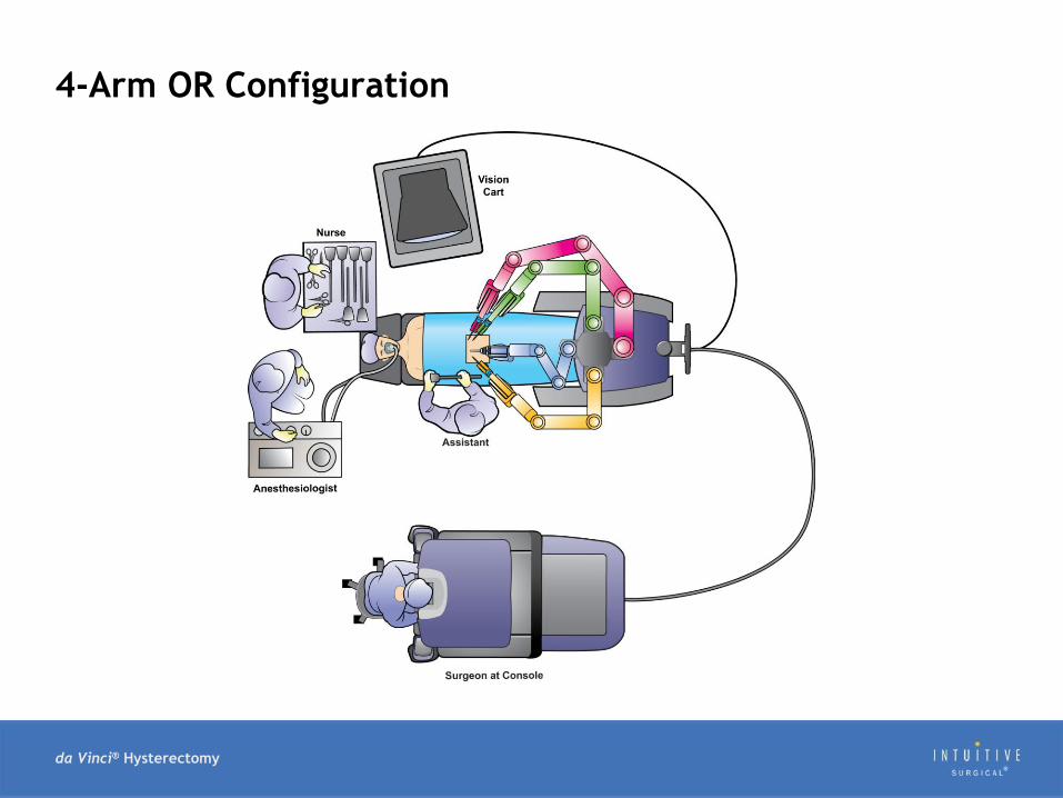
4-Arm OR Configuration
da Vinci® Hysterectomy
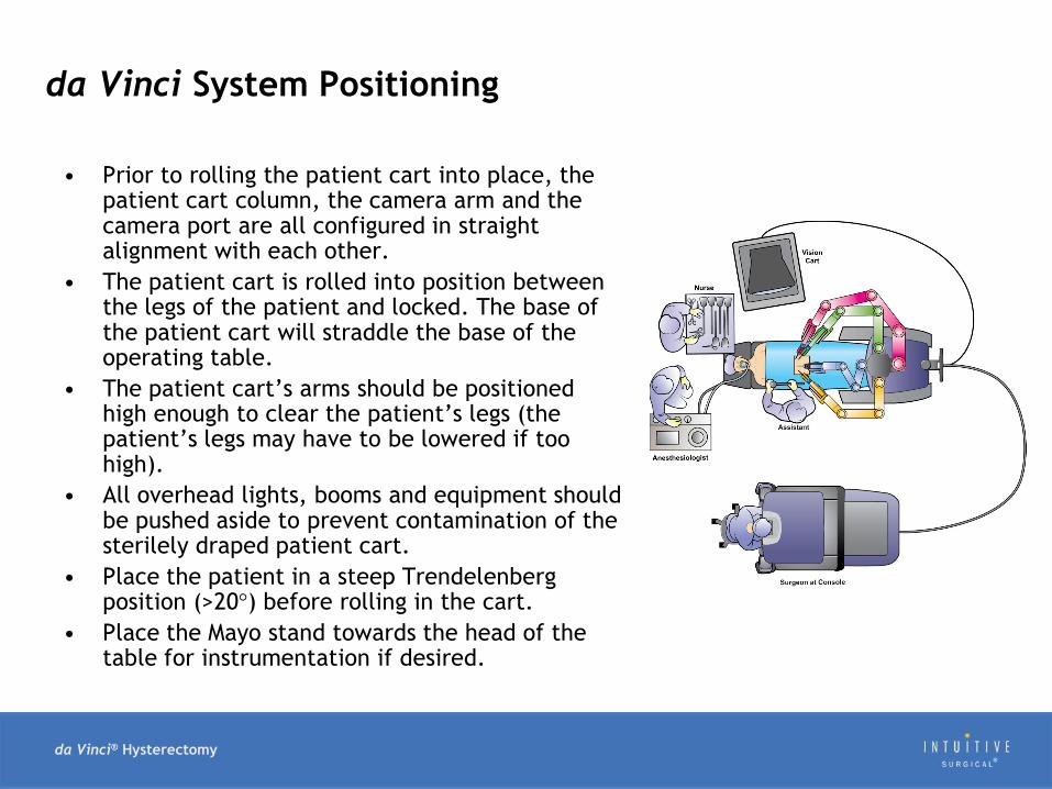
• Prior to rolling the patient cart into place, the patient cart column, the camera arm and the camera port are all configured in straight alignment with each other.
• The patient cart is rolled into position between the legs of the patient and locked. The base of the patient cart will straddle the base of the operating table.
• The patient cart’s arms should be positioned high enough to clear the patient’s legs (the patient’s legs may have to be lowered if too high).
• All overhead lights, booms and equipment should be pushed aside to prevent contamination of the sterilely draped patient cart.
• Place the patient in a steep Trendelenberg position (>20°) before rolling in the cart.
• Place the Mayo stand towards the head of the table for instrumentation if desired.
da Vinci System Positioning

da Vinci® Hysterectomy
Assistant Positioning
• The patient-side surgical assistant
stands on either the left or right side
of the operating table based on the
surgeon’s preferred OR configuration.
• The scrub nurse stands on the opposite
side of the patient-side surgical
assistant.
• Each person is able to access and
exchange their respective da Vinci
instruments.

da Vinci® Hysterectomy
Role of the Surgical Assistant
• Surgical assistants are an important part of the team.
• It’s important to establish good communication.
• Alternating roles provides valuable experience to accelerate the learning curve.
• Tasks include: Facilitating uterine manipulation
Instrument changes
Applying traction and countertraction
Passage/retrieval of suture
Using the suction/irrigator
Cleaning the endoscope
Trocar depth repositioning

da Vinci® Hysterectomy
Patient Preparation &
Positioning

da Vinci® Hysterectomy
Patient Preparation
General Tips:
• Pad the pressure points; use the shoulder supports and anti-skid aids (e.g., gel pad or bean bag).
• DVT prophylaxis
• Decompress the stomach using the orogastric or nasogastric tube.
• Insufflate the abdomen to 15 mmHg.
Anesthesia Tips:
• Run the patient dry to collapse vessels and aid in lymphadenectomy (reduces the
risk of pulmonary and facial edema).
Pelvic Preparation:
• Insert a 16 Fr Foley catheter for bladder drainage.
• Sterilely prepare and drape the abdomen, upper thighs, vaginal and peri-anal
region.
• Drape the legs individually.
• Place the colpotomy ring, uterine manipulator and pneumo-occluder balloon.

da Vinci® Hysterectomy
Patient Positioning
• Place the patient in the dorsal
lithotomy position with her legs in
adjustable boot stirrups.
• Abduct her legs, flex her knees and
place her thighs at the table level.
• Tuck her arms, pad her bony
prominences and place adequate
restraints.
• Place her in a maximum
Trendelenburg position (>20°) before
docking.
NOTE: The use of shoulder blocks with the patient’s arms extended to her side can result in nerve injury; therefore, her
arms should be tucked at her side.

da Vinci® Hysterectomy
Uterine/Vaginal Manipulation

da Vinci® Hysterectomy
Uterine Manipulation
• Uterine Positioning System (CooperSurgical®): Designed to mount, position and secure RUMI® uterine manipulators
– Securely holds the uterus in position for optimal visualization, access and patient safety
– Frees your assistant to perform other tasks during the procedure
– Works with the RUMI® and RUMI Arch™ uterine manipulators
– Connects to any operating table
• Various uterine manipulators and colpotomy rings exist to help identify the interface of the cervix and vagina, thereby delineating the vaginal fornices during colpotomy.
• Planning is required to maintain intra-operative access to the uterine manipulator.
• The surgical assistant will have to reach over the patient’s thigh and under the instrument arm to position the uterine manipulator intra-operatively.
Uterine Artery
Ureter
Pneumo-Occluder Balloon
Uterine Manipulator
Colpotomy Ring

da Vinci® Hysterectomy
Vaginal Manipulation
• Vaginal manipulation is necessary for da Vinci
Sacrocolpopexy.
• Special planning is required to maintain intra-operative access
to the vaginal and rectal manipulators:
– Use rounded EEA™ (End-to-End Anastomosis) sizers to
manipulate the vagina
– An EEA™ sizer in both the vagina (31-33 mm) and rectum (29
mm) allows for clear identification and easy dissection of the
rectovaginal septum
– Other forms of vaginal stents can be used as well (e.g.,
Lucite stents, narrow malleable, Breisky-Navratil retractors)
EEA™ sizers from Autosuture™
da Vinci® Hysterectomy
Port Placement
da Vinci® Hysterectomy
Port Placement – Benign Hysterectomy
• Critical to the success of your GYN case!
• Perform a bimanual exam under anesthesia to
facilitate the planning of port placement.
• The endoscope is placed after primary insufflation.
• The remaining ports are placed after insufflation and under direct
visualization
– The endoscopic trocar is placed 8-10 cm superior to the uterine
fundus.
– The instrument ports are placed in the right and left lower quadrants.
– The accessory port is lateral and cephalad to the umbilical port.
– There are special considerations for large uteri.
– Use long cannulae for high BMI patients.
da Vinci® Hysterectomy
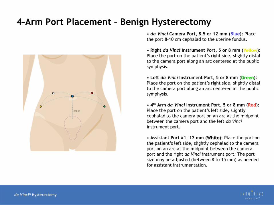
4-Arm Port Placement – Benign Hysterectomy• da Vinci Camera Port, 8.5 or 12 mm (Blue): Place
the port 8-10 cm cephalad to the uterine fundus.
• Right da Vinci Instrument Port, 5 or 8 mm (Yellow):
Place the port on the patient’s right side, slightly distal
to the camera port along an arc centered at the public
symphysis.
• Left da Vinci Instrument Port, 5 or 8 mm (Green):
Place the port on the patient’s right side, slightly distal
to the camera port along an arc centered at the public
symphysis.
• 4th Arm da Vinci Instrument Port, 5 or 8 mm (Red):
Place the port on the patient’s left side, slightly
cephalad to the camera port on an arc at the midpoint
between the camera port and the left da Vinci
instrument port.
• Assistant Port #1, 12 mm (White): Place the port on
the patient’s left side, slightly cephalad to the camera
port on an arc at the midpoint between the camera
port and the right da Vinci instrument port. The port
size may be adjusted (between 8 to 15 mm) as needed
for assistant instrumentation.

da Vinci® Hysterectomy
da Vinci Endoscope Port, 12 mm (Blue): Place superior
to the umbilicus, 24-28 cm from the pubic symphysis.
Right da Vinci Instrument Port, 8 mm (Yellow): Place
on the patient's right side, 8-10 cm lateral and 3-5 cm
inferior to the endoscope port.
Left da Vinci Instrument Port, 8 mm (Green): Place on
the patient's left side, 8-10 cm lateral and 3-5 cm
inferior to the endoscope port.
3rd da Vinci Instrument Port, 8 mm (Red): Place 8-10
cm from the left instrument port on a diagonal line, 1-2
cm superior to the left anterior iliac spine.
Assistant Port, 12 mm (White): Place 1 cm inferior to
the subcostal margin on the left mid-clavicular line.
4 Arm Port Placement – Cancer Hysterectomy
NOTE: Measurements should be made AFTER insufflation to 15 mmHg.
Camera
da Vinci
da Vinci
da Vinci
Assistant
da Vinci® Hysterectomy
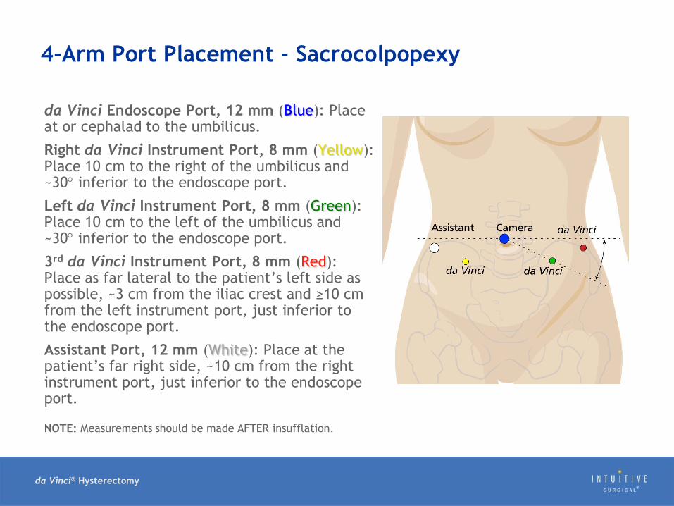
4-Arm Port Placement - Sacrocolpopexy
da Vinci Endoscope Port, 12 mm (Blue): Place at or cephalad to the umbilicus.
Right da Vinci Instrument Port, 8 mm (Yellow): Place 10 cm to the right of the umbilicus and ~30 inferior to the endoscope port.
Left da Vinci Instrument Port, 8 mm (Green): Place 10 cm to the left of the umbilicus and ~30 inferior to the endoscope port.
3rd da Vinci Instrument Port, 8 mm (Red): Place as far lateral to the patient’s left side as possible, ~3 cm from the iliac crest and ≥10 cm from the left instrument port, just inferior to the endoscope port.
Assistant Port, 12 mm (White): Place at the patient’s far right side, ~10 cm from the right instrument port, just inferior to the endoscope port.
NOTE: Measurements should be made AFTER insufflation.

da Vinci® Hysterectomy
4-Arm Port Placement - Myomectomy
da Vinci Endoscope Port, 12 mm (Blue): Place the cannula at the umbilicus; maintain 8-10 cm between the endoscope port and the uterine fundus/myoma.
Right da Vinci Instrument Port, 8 mm (Yellow): Place the cannula on the patient’s right side, 2-3 cm medial to the anterior superior iliac spine along the line diagonal to the endoscope port.
Left da Vinci Instrument Port, 8 mm (Green): Place the cannula on the patient’s left side; offset it ~15°superior to the endoscope port and 8-10 cm from the 3rd da Vinciinstrument port.
3rd da Vinci Instrument Port, 8 mm (Red): Place the cannula on the patient’s left side, 2-3 cm medial to the anterior superior iliac spine along the line diagonal to the endoscope port.
Assistant Port, 12 mm (White): Place the cannula on the patient’s right side; offset it ~15°superior to the endoscope port and 8-10 cm from the right instrument port.
NOTE: Move all ports cephalad with the large pathology to avoid losing optical working space. Measurements should be made AFTER insufflation.
Allow adequate working
distance between the superior-
most border of the enlarged
myoma/uterus and the primary
port site (camera).

da Vinci® Hysterectomy
Docking
da Vinci® Hysterectomy
Docking the Patient Cart
• Dock the camera arm first
– Align the camera port, target anatomy (uterus) and patient
cart center column.
• Dock the remaining instrument arms
– Keep the instrument arms in their center range of motion.
– Point the da Vinci instrument ports toward the center of the
target anatomy using setup joint release or port-clutch
maneuvers.
– Maximize the spacing between all instrument arms.

da Vinci® Hysterectomy
Surgical Videos
da Vinci® Hysterectomy
da Vinci Hysterectomy

da Vinci® Hysterectomy
da Vinci Hysterectomy for Endometrial Cancer with
Staging

da Vinci® Hysterectomy
da Vinci Sacrocolpopexy

da Vinci® Hysterectomy
da Vinci Myomectomy

da Vinci® Hysterectomy
Myomectomy Case Overview Examples

da Vinci® Hysterectomy
Examples of Myoma Removals: Before, During, After
• Pedunculated Myoma
Before
Enucleation
After

da Vinci® Hysterectomy
Examples of Myoma Removals: Before, During, After
• Anterior Subserosal MyomaBefore
Enucleation
After

da Vinci® Hysterectomy
Examples of Myoma Removals: Before, During, After
• Posterior Subserosal MyomaBefore
Enucleation
After

da Vinci® Hysterectomy
Examples of Myoma Removals: Before, During, After
• Cervical MyomaBefore
Enucleation
After

da Vinci® Hysterectomy
Tips & Tricks for Success

da Vinci® Hysterectomy
General Tips
• Establish a dedicated team– Nurses, a surgical technician and an anesthesiologist
• Follow an established clinical pathway:– Onsite training
– Laboratory training
– Case observation
– Proctoring
– Record and review cases together
– Stay committed!

da Vinci® Hysterectomy
Technical Pearls or Potential Pitfalls
• Vaginal cuff closure Ensure adequate tissue bites and incorporate vaginal mucosa into each
bite.
Use interrupted figure-of-eight knots to ensure a strong cuff closure.
Counsel patients to wait 8 weeks before resuming sexual intercourse.
• Open surgical technique!
• “Don’t bite off more than you can chew” Start with simple cases and proceed to more complex cases as you
develop robotic proficiency.
• Know your instruments PK™ Dissecting Forceps (advanced bipolar): Tension-free coagulation
followed by a cold transection.
Skeletonize the vascular pedicles.
“Too much of a good thing (energy) can be bad”
da Vinci® Hysterectomy
da Vinci Sacrocolpopexy Tips & Tricks
• Retract the small bowel and omentum; identify the sacrum BEFORE docking the patient cart.
• The thick black band, indicating the remote center, should be just visible at the level of the peritoneum.
• Begin with the 0 scope to place ports, hysterectomy +/- BSO and dissection.
• Use the 30 down scope for presacral dissection.
• Extend the presacral peritoneal dissection inferiorly to the vagina.
• Use the wide-weave polypropylene mesh (Y-configuration).
• Attach the anterior mesh first.
• Roll the sacral end of the mesh and lift anteriorly.
• Confirm the appropriate tension before suturing the mesh.

da Vinci® Hysterectomy
Managing Presacral Hemorrhage
• Remain calm
• Communicate with the patient-side team
• Apply pressure with an available da Vinci instrument.
• Use the Ray-Tec™ X-Ray Detectable 4” x 4” Sponge to apply pressure.
• Consider attempting to control bleeding with the Maryland Bipolar
Forceps
– May require switching instruments
• Consider using FloSeal™
• “Emergency” undock as a last resort:
– Release da Vinci instruments from tissue.
– Remove instruments and trocars all in one step by depressing
the set-up joint button.
– Keep the assistant port holding pressure.

da Vinci® Surgeryfor both Oncologic and Benign Gynecologic Conditions
Including Simple & Complex Cases
Thank You