D. L. Gilbert et al. (eds.), Squid as Experimental Animals ...
68
235 Chapter 14 The Cytoskeleton of the Squid Giant Axon ANTHONY BROWN and RAYMOND J. LASEK 1. Introduction The squid giant axon is useful to biologists because of its very large size, but it is the continued applicability of studies on this cell to much smaller diameter mammalian axons that have sustained our interest in this mollusc. For example, in electrophysiology the large size of the squid giant axon has enabled manipulations that would have been difficult or impossible with smaller axons. More recently, new methods, such as patch clamping, have made the detailed study of mammalian cell membranes possible, and these have shown that the basic mechanisms established for the squid giant axon also occur in the excitable cells of other metazoa, including mammals and presumably man. In addition to its usefulness for studies on electrically excitable membranes, the squid giant axon has also proven useful for many other kinds of cell biological studies. In particular, its large diameter allows axoplasm to be separated from the axonal plasma membrane and its surrounding glial sheath without chemical treatment or mechanical disruption. This accessibility offers cell biologists an unusual opportunity to investigate the properties of living cytoplasm. Many of these opportunities have yet to be exploited and we hope to point the way to some of them in the course of this article. Since the publication of A guide to the laboratory use of the squid Loligo pealei (Arnold et a/., 1974), there has been much progress in research on squid axoplasm, and some of this has already been summarized in two reviews by Rosenberg (1981) and Gainer et a/. (1984). Particular progress has been made during this period in studies on the cytoskeleton of the squid giant axon. In this article we present a practical guide to the special possibilities that the giant axon offers for the study of the cytoskeleton, and we summarize much of our current knowledge about the structure and organization of the proteins in squid axoplasm. We hope that this article will encourage people to make experimental use of squid axoplasm in their research. However, most cell biologists who might be interested in the information obtained from the giant axon have never had the opportunity to see or use this remarkable preparation. For this large group of scientists, we offer a detailed practical guide to the experimental manipulation of the axon and axoplasm. This partial recreation of the experience of working with the giant axon can not substitute for the actual experience of investigation. Nonetheless, by recreating A. BROWN and R. J. LASEK • Bio-architectonics Center, School of Medicine, Case Western Reserve University, Cleveland, Ohio 44106. D. L. Gilbert et al. (eds.), Squid as Experimental Animals © Springer Science+Business Media New York 1990
Transcript of D. L. Gilbert et al. (eds.), Squid as Experimental Animals ...

235
Chapter 14
The Cytoskeleton of the Squid Giant Axon
ANTHONY BROWN and RAYMOND J. LASEK
1. Introduction
The squid giant axon is useful to biologists because of its very large size, but it is the continued applicability of studies on this cell to much smaller diameter mammalian axons that have sustained our interest in this mollusc. For example, in electrophysiology the large size of the squid giant axon has enabled manipulations that would have been difficult or impossible with smaller axons. More recently, new methods, such as patch clamping, have made the detailed study of mammalian cell membranes possible, and these have shown that the basic mechanisms established for the squid giant axon also occur in the excitable cells of other metazoa, including mammals and presumably man.
In addition to its usefulness for studies on electrically excitable membranes, the squid giant axon has also proven useful for many other kinds of cell biological studies. In particular, its large diameter allows axoplasm to be separated from the axonal plasma membrane and its surrounding glial sheath without chemical treatment or mechanical disruption. This accessibility offers cell biologists an unusual opportunity to investigate the properties of living cytoplasm. Many of these opportunities have yet to be exploited and we hope to point the way to some of them in the course of this article.
Since the publication of A guide to the laboratory use of the squid Loligo pealei (Arnold et a/., 1974), there has been much progress in research on squid axoplasm, and some of this has already been summarized in two reviews by Rosenberg (1981) and Gainer et a/. (1984). Particular progress has been made during this period in studies on the cytoskeleton of the squid giant axon. In this article we present a practical guide to the special possibilities that the giant axon offers for the study of the cytoskeleton, and we summarize much of our current knowledge about the structure and organization of the proteins in squid axoplasm.
We hope that this article will encourage people to make experimental use of squid axoplasm in their research. However, most cell biologists who might be interested in the information obtained from the giant axon have never had the opportunity to see or use this remarkable preparation. For this large group of scientists, we offer a detailed practical guide to the experimental manipulation of the axon and axoplasm. This partial recreation of the experience of working with the giant axon can not substitute for the actual experience of investigation. Nonetheless, by recreating
A. BROWN and R. J. LASEK • Bio-architectonics Center, School of Medicine, Case Western Reserve University, Cleveland, Ohio 44106.
D. L. Gilbert et al. (eds.), Squid as Experimental Animals
© Springer Science+Business Media New York 1990

236 A. Brown and R. J. Lasek
some aspects of the actual physical experience of experimentation with the giant axon, we hope that investigators who have never had this experience may be better able to understand experimental results obtained with this unique preparation.
This chapter is divided into 22 Sections. Sections 1 and 2 give general aspects of the squid. Sections 3 to 8 deal with the anatomy of the giant nerve fiber and with methods for obtaining axoplasm. In these Sections we present a detailed practical guide to the methods and considerations involved in isolating axoplasm from the giant axon. Next, Sections 9 to 11 cover the composition and physical properties of axoplasm, and Sections 12 and 13 deal with methods for the fixation of axoplasm for electron microscopy of the cytoskeleton, and with methods for analysis of the proteins in axoplasm by gel electrophoresis. Then, in Sections 14 to 19, we describe the structure and organization of the cytoskeleton and its elements. In these Sections we summarize much of the current knowledge on the cytoskeleton of the giant axon. Finally, in Sections 20 and 21, we illustrate the potential of the giant axon for studies on the cytoskeleton by describing two paradigms that we have developed for studying the stability and mechanical properties of the axonal cytoskeleton. These latter studies illustrate new ways in which the squid giant axon can aid our understanding of the cell biology of axons. Section 22 is the summary.
2. The squid
Squid do not survive for very long in captivity and it is generally necessary to use them within a day of capture. A healthy squid has a transparent mantle (body wall) and a lively escape response. Unless otherwise mentioned the contents of this chapter apply to the squid, Loligo pealei, which are obtainable at the Marine Biological Laboratory, Woods Hole, Massachusetts. For research on axoplasm, no major differences with other members of the squid family are anticipated.
We can encourage the uninitiated investigator who would like to conduct investigations on the squid giant axon to consider visiting the Marine Biological Laboratory at Woods Hole. The Marine Biological Laboratory has many mechanisms for fostering research on squid. The squid are collected regularly from May through September, and they continue to be reasonably plentiful. Many investigators who work on the giant axon are willing to assist in carrying out a pilot experiment that can be useful in testing the feasibility of a full scale study.
3. The anatomy and development of the giant nerve fiber
To understand how much the squid giant axon can tell us about mammalian axons it is important to understand its anatomy and development. The squid giant axons represent one of the few examples in nature of neurons that are true syncytia. However, unlike other syncytial neurons, such as those found in annelids, all the cell bodies are located at one end of the axon. Thus the squid giant axons share the polarized morphology of other non-syncytial neurons.
There are three orders of giant nerve fibers in squid (Young, 1939). Together, these form part of a motor system that triggers the animal's escape response by causing rapid and synchronous contraction of the body wall. Two first order giant axons

Axonal Cytoskeleton 237
Figure 1. Diagram of the stellate ganglion in the squid Loligo. Each stellate nerve contains a single (third order) giant axon that arises from many small cell bodies located in the giant fiber lobe of the stellate ganglion. The second order pre-ganglionic giant axons form giant synapses directly with the giant stellate axons. Abbreviations: mantle connective (m.c.); second order or pre-ganglionic fiber (p.gf); giant fiber lobe (gf.l.); stellate nerve (st.n.); giant axon of the giant nerve fiber (g f). Drawing reproduced with permission from Young (1936).
emerge from the magnocellular lobes of the brain and synapse with the second order giant axons ih the palliovisceral lobes. The second order giant axons course through the pallial nerves of the mantle connectives and synapse with the third order giant axons in the two bilateral stellate ganglia, from which the stellate nerves emanate.
In Loligo each stellate nerve contains a single third order giant axon that is surrounded by many smaller axons. The giant axons arise from clusters of cell bodies in the giant fiber lobe of the stellate ganglion (see Fig. 1). Apparently each giant axon is formed from a fascicle of ordinary-sized axons that course together in each of the stellate nerves of the developing squid. These axons fuse with each other laterally to form one common axoplasmic mass that is surrounded by a single plasma membrane. The process of fusion is documented in the case of the first order giant fibers (Martin, 1965, 1969). Only the axons fuse; the cell bodies in the stellate ganglia retain their separate identities and each one contributes to the maintenance of the giant axon. Young (1936) estimated that between 300 and 1500 cells fuse to form each giant axon and it is likely that these cells remain transcriptionally active. Thus each giant axon is the product of hundreds of ordinary-sized molluscan neurons that each contribute axoplasmic elements to the single giant axon.
Each giant axon is surrounded by a single layer of adaxonal glial cells (Schwann cells) that are closely apposed to the outer surface of the axolemma (see Fig. 2); a narrow 10 nm periaxonal space separates the axon from the glial cells. The glial cells are extensively folded and interdigitate laterally to form a continuous mosaic sheet that envelops the axon, but a gap of about 10 nm separates adjacent cells forming long tortuous intercellular clefts that provide continuity between the surface of the axon and the extracellular space (Villegas and Villegas, 1984). Diffusion between the periaxonal space and extracellular space is apparently facilitated by a system of tubular trans-glial channels that penetrate the glial cells in a sponge-like fashion (Zwahlen et al., 1988). These channels are about 40 nm in diameter and are continuous with the periaxonal and extracellular spaces. Thus the glial cells do not provide a barrier to the diffusion

238 A. Brown and R. J. Lasek
Comective Tissue
B
Figure 2. The anatomy of the squid giant nerve fiber. A. Schematic drawing of the giant nerve fiber. Like most other axons, the giant axon (A) is surrounded by a cellular sheath, and for the giant axon this sheath is composed of a single layer of Schwann cells (SC), and an outer layer of connective tissue (E) that corresponds to the endoneurium. The giant axon in Loligo pealei can be up to 0.8 mm in diameter. Reproduced from Villegas and Villegas (1984) with permission. B. Schematic drawing of a sector of the giant nerve fiber sheath in cross-section. The outer connective tissue layer contains isolated fibrocytes and is separated from the inner layer of adaxonal glia by a thick basement membrane (the basal lamina). The glial cytoplasm is densely packed with rough endoplasmic reticulum and membranous organelles.
of small molecules from the surroundings. In Loligo pealei the Schwann cell layer is about 1 ~m thick (Adelman et al., 1977).
Surrounding the layer of glial cells is a basal lamina around which is a layer of loose connective tissue containing isolated fibrocytes. Our observations of axons using video-enhanced differential interference contrast microscopy indicate that the collagen fibers crisscross to produce a lattice-like arrangement that confers mechanical strength and elasticity to the nerve fiber. Many hundreds of smaller nerve fibers (less than 50

Axonal Cytoskeleton 239
11m in diameter) surround the giant nerve fiber, and together these constitute a stellate nerve.
4. Dissection of the giant axons
In general usage the hindmost and medialmost giant axon is referred to as the squid giant axon. In each squid there are two of these axons that emerge from the two bilateral stellate ganglia. These giant axons are used because they are the largest and most accessible of the stellate axons. In Loligo pealei, they range from 300 to 800 11m (typically 350 to 500 jlm) in diameter (Rosenberg, 1981). In larger squid, such as Loligo forbesi, which can have mantle lengths of greater than 50 em, it may also be worthwhile to dissect the next largest of the stellate axons. At the Marine Biological Laboratories in Woods Hole, large specimens of Loligo pealei are around 20 to 25 em in mantle length.
The axons can be dissected from the mantle in natural or artificial seawater using a dissecting tray with illumination from beneath. This is a standard procedure which has been refined over the years by numerous investigators. At the Marine Biological Laboratory in Woods Hole, we use a flowing seawater table. The hindmost giant axons run for a few centimeters along the inner surface of the mantle before they course into the mantle musculature. Generally we dissect the axons up to this point or to the first major branch point within the mantle wall. There are smaller branches more proximal to this point, particularly near the point that the fin nerve and giant axon diverge, and care should be taken in the dissection to cut these, preferably a millimeter or so from the axon, rather than tear them when the axon is excised. The dissection of the giant axon is described in detail by Adelman and Gilbert (1990).
It is preferable to dissect the axons from the mantle immediately after the squid has been killed. Once excised from the mantle the axons can be kept in cold seawater (in the refrigerator at 4 °C, for example) for many hours. At room temperature the survival time is shorter. After excision of axons from the mantle, the subsequent dissections are generally performed in Millipore™-filtered natural seawater or in artificial seawater with or without calcium.
4.1. Fine-cleaning the giant axons
Before excision from the mantle, the ends of the axon are tied with cotton thread to prevent seawater leaking into the axoplasm. If the axon is excised with the stellate ganglion attached, then it is not necessary to ligate the proximal end. The axons are then transferred to a dish containing fresh seawater. For the subsequent dissection we use a large Petri dish containing a layer (about 5 mm thick) of Sylgard™, which is a transparent rubbery resin into which dissecting pins can be secured. For this procedure fine dissecting scissors are required. For most investigations it is necessary to dissect away the surrounding smaller nerve fibers and any small pieces of attached muscle, blood vessels etc. before the axon can be used. For extrusion of axoplasm in the conventional manner (see Section 6.1) this need only be done at the proximal end of the fiber, but for some studies it may be necessary to dissect the whole fiber in this way. In both cases, the dissection is done under a low power binocular microscope with illumination from beneath. We typically use a magnification of 25 x. Dark-field

240 A. Brown and R. J. Lasek
illumination improves the visibility of the smaller nerve fibers but bright-field illumination provides better visibility of the fine branches of the giant axon. Either of these sources of illumination can be used with success, but we prefer bright-field illumination.
A careful dissection generates afine-cleaned fiber, which is a fiber that has been freed of surrounding small nerves. The fine-cleaned fiber consists of the axon enveloped by a sheath of Schwann cells and an outer layer of loose connective tissue (see Fig. 2). Usually some tissue is left around the branches of the giant axon to minimize the risk of damaging the axon. It is important to realize that the fine-cleaned giant fiber includes the Schwann cell sheath, which adheres tightly to the axolemma of the giant axon and cannot be removed by conventional dissection. Removal of the sheath requires a special procedure, which was developed by Metuzals et al. (1981b) and is described in detail in Section 13.4. Briefly, they used mild chemical fixation to stabilize the axon and sheath, and osmotic shrinkage to separate the axolemma from the Schwann cell layer. This procedure enables the surface topography of the axon to be viewed by scanning electron microscopy (see Section 14.2).
5. Handling the giant axons
The visualization of the isolated squid giant nerve fiber is a remarkable experience for those used to dealing with smaller axons. The giant axon itself is about the size of a medium size mouse nerve or a small rat nerve, yet it is a single axon that can be seen with the naked eye and can be handled between two lengths of ordinary cotton thread. The connective tissue sheath supports the giant axon sufficiently that it can bear a small amount of stress. Because of this, the giant fiber can be transferred between dissecting dishes or solutions by grasping the attached cotton thread at one end and simply lifting the axon from one solution to the other, but care should be taken not to stretch it too much. When removed from a solution, the fiber conveys a thin covering layer of liquid with it, but it should not be left in air longer than absolutely necessary, in order to avoid drying. When a nerve fiber is transferred into a new solution it often floats at the surface and care should be taken to ensure that it is thoroughly wetted and submerged. If an axon appears more turgid or flaccid after it has been transferred from natural seawater into artificial seawater, this is probably caused by differences in the osmolarity of the two solutions. Axoplasm is in osmotic equilibrium with the external solution (see Section 9) and the axon volume will rapidly shrink, or to some extent swell, in response to changes in the osmolarity of the medium.
6. Methods for obtaining axoplasm from the giant axons
The large diameter of the squid giant axons allows axoplasm to be separated from the axolemma and axonal sheath in milligram quantities. An axon with a diameter of 500 11m contains about 1 J.tl (1 mg wet weight) of axoplasm for a 5 mm length. Three major factors should be considered in choosing the method for isolation of axoplasm: purity, preservation of structural integrity, and time. In this Section

Axonal Cytoskeleton 241
Table I. Comparison of three methods for isolating axoplasm.
Method
Extrusion
Slitting
Cannulation
Advantages
Quick, essentially free of contamination
Preservation of shape and cytoskeletal organization
Preservation of plasma membrane, access to cortical cytoskeleton, no contamination from sheath
Disadvantages
Some distortion of cytoskeletal organization
Some contamination from sheath tissue
Time consuming, special equipment and expertise required, axoplasm disrupted
we describe three methods for isolating axoplasm from the giant axon; extrusion, slitting and cannulation. Some advantages and disadvantages of these methods are summarized in Table I.
In all cases the axon should be undamaged along its length or seawater will leak in and produce white spots in the axoplasm. These are most apparent when axons are viewed under dark-field illumination. The white spots may be due to a precipitation of axoplasmic protein in the presence of seawater that results in an increase in light scattering. The precipitation is apparently caused by the high concentration of calcium in seawater, since calcium forms insoluble sticky precipitates with sulfates, which are present in seawater, and with phosphates, which are present in the axoplasm. Damaged regions are most often caused during the dissection by tearing branches at or near their junction with the axon.
6.1. Extrusion
The quickest way to obtain axoplasm is by extrusion. This method is simple and is widely used by many investigators. For this method the giant axon does not need to be fine-cleaned of associated nerve fibers along its entire length, but it is necessary to fine-clean a region of about 1 em at the proximal end so as to avoid contamination with extracellular fluid or tissue from the sheath. At the proximal end there are fewer branches than more distally and it is possible, with practice, to fme-clean this region in about five minutes without damaging the axon.
It is also possible to extrude axoplasm from the axon without removing the axon from the mantle. This can be done simply by cutting the axon at its proximal end and then applying pressure to the mantle in a distal-to-proximal direction (the axon is widest at its proximal end). Unfortunately this procedure is very messy and it is difficult to avoid contamination of the axoplasm with extracellular fluids during the extrusion process. Thus, we do not recommend this procedure, and in practice it is necessary to dissect out the axon prior to extrusion and to fine-clean its proximal end as described above.

242 A. Brown and R. J. Lasek
giant axon Figure 3. Schematic illustration of axoplasm being extruded from a giant axon. In the method
axoplasm shown here, the giant axon is placed on filter paper on a solid support, such as a glass slide, with the cut end protruding over the edge. For diagrammatic purposes, the axon is shown here as though it has been fme-cleaned along its entire length, though in practice it is only necessary to fine-clean the proximal end. The axoplasm is extruded by gentle pressure in a distal-to-proximal
direction (indicated by arrow) towards the cut end. As the axoplasm is extruded it falls away from the cut end. The axoplasm can be collected on a piece of Parafilm™ or, as shown here, in a watch glass containing a small volume of artificial axoplasm solution. In an alternative extrusion method, the cut end of the axon can be placed directly onto a piece of Parafilm™, as described in the text. After extrusion, the flattened sheath is generally discarded. From Morris and Lasek (1982).
For extrusion, the axon is removed from the seawater and excess fluid removed by gently and briefly blotting on a glass coverslip or filter paper. If desired, the axon can be rinsed in artificial axoplasm solution or calcium-free seawater prior to blotting. The axon is then placed on a fresh region of filter paper and cut at its proximal end, leaving at least 5 mm of fme-cleaned axon distal to the cut. For minimal resistance to extrusion and optimal preservation of the structure of the axoplasm, the cut should be made about 5 mm distal to the stellate ganglion, where the axon is at its widest. Nearer the ganglion the axon diameter is smaller and extrusion from this point distorts the axoplasm as it is forced through the narrower region of axon.
After cutting the axon, it can then be lifted by the cotton thread at its distal end, and the fine-cleaned cut end placed onto a small piece of Parafilm TM with the rest of the axon on filter paper. Extrusion is achieved by applying gentle even pressure in a distal-to-proximal direction along the axon. As pressure is applied along the fiber the axoplasm is extruded from the cut end of the axon onto the Parafilm™, like toothpaste from a tube. The filter paper under the axon serves to absorb excess fluid in the sheath as it is compressed. The axon can also be placed on a solid support with the cut end protruding over the edge so that the axoplasm falls away from the cut end as it is extruded (see Fig. 3). Fig. 4 shows a piece of axoplasm that was extruded in this manner and collected in a small volume of artificial axoplasm solution.
In the original method of Bear et al. (1937b), the axoplasm was extruded with a pair of bent forceps. Others, such as Baker et al. (1962) and Rosenberg (1981), have used a rubber roller. In our hands, a small piece of polyethylene tubing (such as Intramedic PE250 tubing) is preferable, and it is likely to cause less damage to the axon. However, the instrument used is not as important as the protocol employed. For studies where purity is essential, we advise using short lengths of axon and extruding in a single pressure stroke (see Section 7.1). The stroke should not be continued all the way to the end of the axon so that the axoplasm nearest to the site of compression is not extruded. In addition, axoplasm should not be extruded from regions of axon

Axonal Cytoskeleton 243
Figure 4. Photograph of a piece of extruded axoplasm. This piece of axoplasm was extruded and immersed in artificial axoplasm solution as shown in Fig. 3, and photographed with dark-field illumination. Axoplasm is almost transparent and therefore it is almost invisible under bright-field illumination. The axoplasm is about 10 mm long and about 0.5 mm in diameter. The S-shape of the axoplasm is due to distortion of the longitudinal alignment of the cytoskeletal polymers caused by the compressive forces during extrusion (see text).
within a few millimeters of the knot of cotton thread since these regions may be damaged, thereby producing contamination.
We have observed that when extrusion pressure is applied to the axon, the axoplasm initially resists, and the axon becomes turgid before the axoplasm begins to move. This back pressure on the axoplasm apparently compresses the cytoskeletal elements longitudinally and can distort the cytoskeletal organization of the axoplasm during extrusion. For example, measurements of the length and diameter of the axon and the axoplasmic cylinder indicate that axoplasm extruded onto Parafilm™ is shorter and thicker than the axon from which it came. Maximum change in dimensions is seen if the axoplasm is extruded directly into a liquid rather than onto a surface to which the axoplasm can adhere. In measurements of 36 extrusions directly into artificial axoplasm solution (Brown, 1989), the mean length of the axon segments was 10.2 mm and the mean length of the extruded axoplasms was 5.9 mm. Thus the axoplasms were on average 58% shorter than the original axon (minimum = 26%, maximum = 88%, standard deviation = 14%). Since the volume remains constant, the diameters of the

244 A. Brown and R. J. Lasek
extruded axoplasms were correspondingly larger. Electron microscopy of axoplasm extruded in this way revealed that the cytoskeletal polymers in the center of the extruded axoplasm were orientated obliquely to the long axis of the cylinder of axoplasm. Apparently, the cytoskeletal elements tend to buckle in response to the compressive forces of the extrusion. An indication of the distortion of the extruded cytoskeleton is that the extruded axoplasm often assumes a macroscopic curved or twisted shape (see Fig. 4), which is not seen when axoplasm is isolated without compression (see Section 6.2 and Fig. 5).
For studies that require preservation of the structural integrity of the axoplasm, the distortion of the longitudinal organization of the axoplasmic cytoskeleton can be partially offset by extruding the axoplasm under gentle tension in order to straighten the cytoskeletal polymers as they emerge from the axon. One way to achieve this is by using the natural adhesiveness of the axoplasm to help pull the axoplasm out of the axon. To do this, the roller or plastic tubing is held still and the axon is drawn backwards away from the cut end. As the axoplasm is extruded, it adheres very slightly to the Parafilm™ and with a little practice, this method can yield long unbroken lengths of axoplasm (centimeters in length). Continuous unbroken lengths of axoplasm are necessary for investigations in which the organization and mechanical integrity of the axoplasm is important, such as for our stretching studies on axoplasm (see Section 21) and for the preparation of fibers for X-ray diffraction studies on the cytoskeletal polymers (Wais-Steider et al., 1988).
Extrusion of axoplasm using tension is best achieved on a more adhesive surface such as glass (see Section 8), but in this case the adhesion is so good that the axoplasm tends to maximize its contact with the glass and flatten out. If the axoplasm then needs to be lifted off the glass, further mechanical disruption is inevitable, so extrusion onto glass is not suitable for any studies that require further handling or preservation of the cylindrical shape of the axoplasm. One application in which we have used extrusion onto glass is for obtaining improved longitudinal organization of the cytoskeletal polymers for electron microscopy of extruded axoplasm (Miller and Lasek, 1985).
6.2. Slitting
One of us (A. B.) has experimented with an alternative method for isolating axoplasm that avoids extrusion and the complication of compression. This is the slitting method, in which the axon is slit along its length to gain access to the axoplasm. In this procedure, the axon is first fine-cleaned of associated small nerve fibers along its entire length (except around the branches) and rinsed in several changes of 1 - 2 ml artificial axoplasm solution. The axon is then placed in a small Sylgard™ dish containing artificial axoplasm solution and pinned at either end under gentle tension (if required, the dissection can also be done in oil to prevent extraction of diffusible components). Using fine dissecting scissors and a dissecting microscope, it is then possible to carefully slit the sheath open along the length of the axon. Scissors with fine blades and pointed tips have proven to be most effective.
The scissors must be held at a low angle to minimize penetration of the axoplasm. As the sheath is cut it tends to retract so as to expose the axoplasm. The axoplasm can then be gently peeled away from the sheath, while still submerged in solution, and thereby lifted out of the axon (see Fig. 5). The solution can then be removed and replaced with fixative for electron microscopy, or the axoplasm can be used directly for

Axonal Cytoskeleton 245
Figure 5. A cylinder of axoplasm being removed from a slit axon. This axon was slit from right to left and the axoplasm was then grasped at one end with fme forceps and gently peeled away from the axon. Note that the axoplasm is removed as a smooth and undistorted cylinder. The white arrow shows the limit of the slit. The axoplasm (Ax.), sheath (Sh.) and inner surface of the axon (i.s .) are labelled. Photograph taken under dark.field illumination. The axoplasm is about 0.5 mm in diameter.
other studies. Some contamination with sheath material is likely in this procedure but contamination with extracellular fluid, which can be destructive to the axoplasm (see Section 11.2), can be avoided by rinsing the fine-cleaned fiber well with artificial axoplasm solution before slitting. In many studies the possibility of slight contamination with extracellular fluid or sheath material is not important.
Axoplasm is almost transparent and, thus, it is almost invisible under bright-field illumination. However, the edges of axoplasm obtained both by extrusion and by slitting appear sharp under the light microscope in dark-field illumination, and this suggests that the axoplasm is removed along a natural annular cleavage plane in the cortex of the axon (see Section 15). Ultrastructural studies have shown that extrusion removes greater than 95% of the axoplasm and leaves behind a 10 !J.m thick layer of cortical axoplasm. It is likely that the axoplasm peeled from a slit axon also cleaves at this plane, but we ha~e not examined this.

246 A. Brown and R. J. Lasek
6.3. Cannulation
A third method for obtaining axoplasm from the giant axon is by cannulation. This procedure was developed to allow internal perfusion of the giant axon for electrophysiological studies on the membrane and is described in detail by Adelman and Gilbert (1990). Briefly, the axon is fine-cleaned and then cannulated with tapered glass cannulae at one or both ends. In one method, a long and narrow glass capillary is then inserted into one of the cannulae and advanced axially along the inside of the axon. As the capillary is advanced, a central core of axoplasm is drawn into it by applying suction pressure. When the capillary has been advanced to the other end of the axon, the axoplasm is expelled by applying positive pressure and the capillary is then withdrawn. Because axoplasm adheres to glass, complete expulsion from the capillary requires flow of internal perfusion solution. The axoplasm expelled in this way tends to wash out in fragments.
The main advantage of the cannulation procedure is that it does not damage the axon membrane and thus it affords access to the subaxolemmal cortical cytoplasm by internal perfusion of the axon, as is described in Section 15.2. The intactness of the membrane can be verified throughout the procedure by monitoring the excitability of the axon and by the use of a cell-impermeant extracellular marker such as radio-labelled sucrose or mannose (see Section 7.1). However, cannulation is not practical for routine isolation of axoplasm since it requires special effort and equipment and furnishes a low yield of axoplasm. The cannulation procedure is also not suitable for studies in which preservation of the organization of the cytoskeleton is important since the axoplasm obtained is disrupted and is also diluted with the perfusion solution used to expel it from the core capillary.
7. How pure is axoplasm isolated from the giant axon?
The two possible sources of contamination of axoplasm are the extracellular fluid and the tissue of the sheath.
7 .1. Extrusion
Compression of the axon during extrusion may result in damage to the axolemma and sheath cells and this could give rise to contamination of the axoplasm with extracellular fluids or extracellular material such as Schwann cell cytoplasm. One measure of the extent of damage to the axon during extrusion is whether the remaining sheath will propagate a biphasic action potential, though this is not an absolute test since it is known that action potentials can propagate through damaged regions. Baker et at. (1962) reported that extrusion of axoplasm using a rubber roller resulted ·in electrically inexcitable sheaths in 60 out of 207 attempts. Thus, in their hands about one third of the axons were so badly damaged that they became inexcitable. However, in practice the extrusion procedure is satisfactory if these precautions are taken:
1. To avoid contamination with extracellular fluids: fine-clean proximal end of axon, rinse axon in artificial axoplasm solution or calcium-free seawater, and blot well to remove excess fluid before extruding axoplasm.

Axonal Cytoskeleton 247
2. To avoid contamination from sheath: extrude with gentle pressure in a single stroke using shorter lengths where possible, and always end the extrusion stroke before reaching the end of the axon so that the axoplasm in the region of compression is not extruded.
One way to test the amount of contamination from extracellular material is to incubate the giant nerve fiber in an extracellular marker prior to extrusion, and then to measure how much of the marker is present in the extruded axoplasm. For example, Larrabee and Brinley (1968) incubated axons in radioactive inulin and then extruded the axoplasm, taking precautions similar to those listed above. They found that there was a slight contamination of the axoplasm that could be accounted for by the short portion of the axon (near the cut end) that was placed on the plastic film onto which the axoplasm was extruded. Rosenberg and Hoskin (1963) and Hoskin and Rosenberg (1965) tested a variety of other radiolabelled molecules that are also considered to be impermeable to cells, such as sucrose and acetylcholine. Using partially fine-cleaned axons and a slightly different extrusion protocol (see Rosenberg, 1981), they found less than 1% penetration compared to that expected if the plasma membrane of the axon presented no permeability barrier. It is possible that the axon is indeed slightly permeable to these compounds, but nevertheless these determinations place an upper limit on the amount contamination of extruded axoplasm from extracellular fluids.
7 .2. Slitting
Contamination with extracellular fluids should not be a problem in the slitting procedure because the fine-cleaned axon can be rinsed in artificial axoplasm solution prior to cutting the axon open. However, slight contamination of the axoplasm with tissue from the sheath tissue is likely.
7 .3. Cannulation
In the cannulation procedure, contamination by extracellular fluids and by tissue from the sheath can be avoided, but this procedure is not practical for many studies. One example of its use is in an investigation of the glia-neuron transfer hypothesis by Gainer et a/. (1977), in which cannulation and internal perfusion of the giant axon was used to provide evidence that glial cells are the sole site of protein synthesis in the giant nerve fiber. The perfused axon preparation has also been used to investigate the composition and properties of the cortical membrane-associated cytoskeleton of the giant axon (see Section 15.2).
8. Handling axoplasm
Axoplasm is gelatinous in appearance and has sufficient structural integrity to be handled gently with flattened or rounded forceps. Nevertheless, axoplasm is, after all, naked cytoplasm, and any handling will locally disrupt its microscopic structure. Thus, it is preferable to reduce the number of times it is handled to the minimum possible. The precautions taken during handling depend on the nature of the study. For

248 A. Brown and R. J. Lasek
biochemical experiments, full recovery of the axoplasm at each stage of handling is the main requirement, while for structural or mechanical investigations, preservation of the organization and shape of the axoplasm is more important.
One consideration in handling axoplasm is its adhesiveness. Axoplasm adheres strongly to glass and tends to spread out and flatten so as to maximize its area of contact when extruded onto a glass surface. We find that better preservation of the cytoskeletal organization, and easier handling of the axoplasm, is afforded by extrusion onto a hydrophobic plastic surface such as a piece of Parafilm™ or Nescofilm™. If necessary, the plastic film can be stretched over a glass slide to provide rigidity and a solid base for extrusion. Handling axoplasm on plastic film also maximizes recovery of the axoplasm when it is subsequently removed, so we also recommend plastic for general handling of axoplasm, such as for weighing or for axoplasm that is being pooled for subsequent treatment.
Axoplasm rapidly dries if left in air. There are a number of ways to avoid or minimize this. The choice of method again depends on what the axoplasm is to be used for. One way is to immediately place the freshly isolated axoplasm in a moist atmosphere on ice. For example, axoplasm can be placed on Parafilm TM over moistened filter paper in a petri dish on ice. We often place axoplasm in a small envelope made by folding a piece of Parafilm™ and pinching together the open sides.
The shape and organization of the axoplasm can also be disrupted by surface tension forces when axoplasm is transferred through an air-water interface. Typically these forces cause the axoplasm to flatten to some extent. In investigations where the structural integrity of the axoplasm is important, we avoid this by extruding the axoplasm directly into a drop of solution placed over the cut end of the axon, or by submerging a fine-cleaned axon in the solution during extrusion. Two examples where we have extruded axoplasm in this manner are in our stretch experiments on axoplasm (see Section 21), in which mechanical integrity is important, and also in our experiments on the assembly dynamics of the cytoskeletal polymers, in which the cylindrical shape of the axon is important for the kinetic analyses (see Section 20).
9. The composition of axoplasm
The accessibility of axoplasm from the giant axon has enabled a detailed analysis of the chemical composition of the axon. Axoplasm contains about 2 - 2.5% (w/w) protein (Huneeus and Davison, 1970; Morris and Lasek, 1984) and 86.5 ± 5% (w/w) water (Koechlin, 1955). The axoplasm is iso-osmotic with seawater (about 980 mOs/kg) and has a pH of about 7 .2, with published estimates ranging from 7.0 to 7.35 (Caldwell, 1958; Spyropoulos, 1960; Eicher and Ohki, 1972; Boron and De Weer, 1976).
9.1. Ions and small molecules
Table II is a comparison of the composition of axoplasm and blood from Loligo pealei, and of seawater. The values are given as millimoles/kg axoplasm (i.e. millimolal). Note that since the water content of axoplasm is about 865 g/kg, the actual molar (moles/liter) concentrations of the elements are somewhat greater than their molal (moles/kilogram) concentration. The values for the amino acids, sugars and

Axonal Cytoskeleton 249
Table ll. The composition of axoplasm and blood in Loligo pealei, and of seawater.
Substance Axoplasm
Water (g/kg) 865 Protein4"6 (g/kg) 25 pH 7.2
Inorganic ions2.l.S,6
K 340 Na 65 Cl 150 Ca 3.5 Mg 10 Sulfate Phosphate 18
Amino acids2
Gly 12 Ala 8.6 Val 2.4 Leu/lle 2.9 Pro 1.1 Ser 4.0 Thr 2.1 Asp 79 Glu 21 Arg 3.5 Lys 2.6 Ornithine 2.0 Others 2.7
Other organic metabolites2
Isethionate 170 Taurine 110 Homarine 20 Betaine 74 Cysteic acid amide 4.9
Carbohydrates2
Glucose 0.24 Sucrose 0.24 Fructose 0.24 Myo-inositol 7.6 Glycerol 4.4
Others1
ATP 0.7-1.7 Arginine phosphate 1.8-5.7
Values are given as millimoles/kg unless otherwise noted.
Blood
870 0.15 7.5
22 460 580
11 55
8.1
2.5 2.9 2.6 4.4 2.1 1.3 1.2 1.8 2.2 0.6 1.0 0.40 7.2
1.6 4.6 3.3 4.3 0.4
0.3
0.5
Seawater
966
7.9
10 460 540
10 53 28
References: 1 Caldwell (1960), 2 Deffner (1961), 3 Hodgkin (1958), 4 Morris and Lasek (1984), s Koechlin (1955), and 6 Robertson (1949).
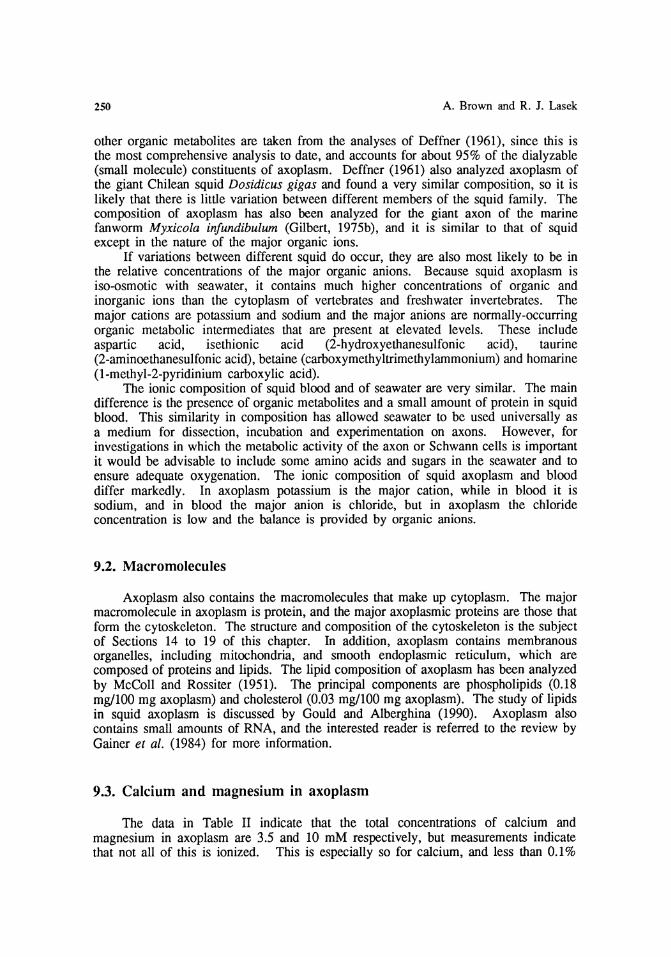
250 A. Brown and R. J. Lasek
other organic metabolites are taken from the analyses of Deffner (1961), since this is the most comprehensive analysis to date, and accounts for about 95% of the dialyzable (small molecule) constituents of axoplasm. Deffner (1961) also analyzed axoplasm of the giant Chilean squid Dosidicus gigas and found a very similar composition, so it is likely that there is little variation between different members of the squid family. The composition of axoplasm has also been analyzed for the giant axon of the marine fanworm Myxicola infundibulum (Gilbert, 1975b), and it is similar to that of squid except in the nature of the major organic ions.
If variations between different squid do occur, they are also most likely to be in the relative concentrations of the major organic anions. Because squid axoplasm is iso-osmotic with seawater, it contains much higher concentrations of organic and inorganic ions than the cytoplasm of vertebrates and freshwater invertebrates. The major cations are potassium and sodium and the major anions are normally-occurring organic metabolic intermediates that are present at elevated levels. These include aspartic acid, isethionic acid (2-hydroxyethanesulfonic acid), taurine (2-aminoethanesulfonic acid), betaine (carboxymethyltrimethylammonium) and homarine (1-methyl-2-pyridinium carboxylic acid).
The ionic composition of squid blood and of seawater are very similar. The main difference is the presence of organic metabolites and a small amount of protein in squid blood. This similarity in composition has allowed seawater to be used universally as a medium for dissection, incubation and experimentation on axons. However, for investigations in which the metabolic activity of the axon or Schwann cells is important it would be advisable to include some amino acids and sugars in the seawater and to ensure adequate oxygenation. The ionic composition of squid axoplasm and blood differ markedly. In axoplasm potassium is the major cation, while in blood it is sodium, and in blood the major anion is chloride, but in axoplasm the chloride concentration is low and the balance is provided by organic anions.
9.2. Macromolecules
Axoplasm also contains the macromolecules that make up cytoplasm. The major macromolecule in axoplasm is protein, and the major axoplasmic proteins are those that form the cytoskeleton. The structure and composition of the cytoskeleton is the subject of Sections 14 to 19 of this chapter. In addition, axoplasm contains membranous organelles, including mitochondria, and smooth endoplasmic reticulum, which are composed of proteins and lipids. The lipid composition of axoplasm has been analyzed by McColl and Rossiter (1951). The principal components are phospholipids (0.18 mg/100 mg axoplasm) and cholesterol (0.03 mg/100 mg axoplasm). The study of lipids in squid axoplasm is discussed by Gould and Alberghina (1990). Axoplasm also contains small amounts of RNA, and the interested reader is referred to the review by Gainer et al. (1984) for more information.
9.3. Calcium and magnesium in axoplasm
The data in Table II indicate that the total concentrations of calcium and magnesium in axoplasm are 3.5 and 10 mM respectively, but measurements indicate that not all of this is ionized. This is especially so for calcium, and less than 0.1%

Axonal Cytoskeleton 251
of the total calcium in axoplasm is ionized. Baker and Schlaepfer (1978) identified two distinct components of calcium binding in squid axoplasm. One is an energy-dependent process with a high capacity but low affinity for calcium. This component probably represents the mitochondria and endoplasmic reticulum in axoplasm (Baker and Schlaepfer, 1978, Henkart et al., 1978), which can sequester calcium in large amounts (probably as a calcium phosphate or calcium oxalate precipitate). The other component is independent of metabolic energy and has a higher affinity for calcium, but is readily saturated. This binding may be accounted for by proteins in the cytoplasm. For example, squid axoplasm contains calmodulin (Alema et al., 1973; Head and Kaminer, 1980), and it may constitute as much as 1% of the total protein in the axon (Alema et al., 1973), which is about 0.25 mg protein/g axoplasm wet weight. Another likely source of calcium-binding are the peripheral domains of the neurofilament proteins because these domains are known to be highly phosphorylated (see Section 17.3). In support of this, Abercrombie et al. (1986) have reported that neurofilament proteins from the giant axon of the marine fanworm Myxicola infundibulum bind calcium, and that this can account for most of the energy-independent calcium binding in these axons.
As an aside in this regard, a little-known study by Krishnan and Singer (1974) is also of interest. They used the reagent potassium pyroantimonate, which forms an electron-dense precipitate with sodium, calcium and magnesium, to localize the sites of cation binding in amphibian myelinated nerve. In addition to precipitation of cations in the Schwann cell cytoplasm, precipitate was also observed in the axoplasm. This precipitate was located predominantly on neurofilaments and in a periodic fashion along their length. The periodicity was often regular and measured 45 - 75 nm, which is similar to the axial spacing of the neurofilament side-arms that contain the highly phosphorylated peripheral domains of the neurofilament proteins.
The regulation of the ionized calcium concentration in cells is important because of the large number of cellular processes that are regulated by this ion. One such process in axons is calcium-dependent proteolysis (see Section 17.4). The calcium-dependent protease in squid axoplasm cleaves the cytoskeletal proteins in the presence of millimolar concentrations of calcium causing liquification of the axoplasm, but the low levels of ionized calcium in the axon ensure that the enzyme is not normally active.
Measurements in Loligo pealei using the calcium indicators aequorin and arsenazo III (DiPolo et al., 1976), and using calcium-selective electrodes (DiPolo et al., 1983) indicate a resting ionized calcium concentration in freshly dissected axons of about 100 nM (a pCa of 7). In contrast, the calcium concentration in squid blood and seawater is about 10 mM, though the presence of calcium-binding compounds such as sulfate ions result in a free ionized calcium concentration of nearer 4 mM (Blaustein et al., 1974). Thus the ratio of ionized calcium in the extracellular space to that in the axoplasm is about 40,000:1. A similar level of ionized calcium is found in other cells, such as resting muscle, and it seems that under normal conditions the bulk of the calcium inside cells is sequestered. The high calcium concentration in seawater and the biological activity of calcium in axoplasm underscore the importance of avoiding contamination of axoplasm with extracellular fluids during isolation.
For magnesium in axoplasm, a much larger fraction is ionized than for calcium. Measurements indicate that between one third and one half of the total axoplasmic magnesium is free, with estimates varying from 2 - 4 mmoles/kg axoplasm (Baker and Crawford, 1972; Brinley and Scarpa, 1975; De Weer, 1976). The remainder is probably bound to charged molecules in the axoplasm such as proteins and nucleotides. The

252 A. Brown and R. J. Lasek
large proportion of magnesium that is ionized means that the ratio of free magnesium to calcium in axoplasm is about 30,000:1.
10. Artificial axoplasm solutions
Because the composition of squid axoplasm is known it is possible to design solutions that closely approximate many of the important physical and chemical parameters of this cytoplasm. We consider inorganic molecules, small organic molecules, macromolecules, concentration of protons (pH), ions such as calcium (pCa) and magnesium, ionic strength, and colligative properties such as osmotic strength. In many studies it is not practical nor necessary to approximate all of these parameters. The choice of an appropriate medium therefore depends on defining those factors that are important for the phenomenon being studied. Since we do not know how critically the chemical environment affects many of the processes in cells, we have attempted to simulate the known chemical composition of squid axoplasm in many of our studies, as far as is practical.
10.1. Buffer P
Originally we developed Buffer P, which adheres closely to the known concentrations of small molecules and ions in axoplasm (Morris and Lasek, 1982). Initially Buffer P contained 15 amino acids (ala, ile, leu, met, phe, pro, val, gly, ser, thr, tyr, asp, glu, arg and lys), four carbohydrates (glucose, fructose, myo-inositol and glycerol), five other organic metabolites (isethionate, taurine, citrulline, hypoxanthine and ornithine), and five inorganic ions (K+, Na+, Mg++, CI- and phosphate). Excess taurine was added to substitute for homarine and cysteic acid amide. Subsequently, in a simplification of this solution, alanine, glycine, aspartic acid and arginine were used in appropriate amounts to substitute for their non-polar, polar, acidic or basic counterparts. Also, citrulline, hypoxanthine, myo-inositol and ornithine were eliminated from the solution. Adenosine-5'-triphosphate (ATP) and guanosine 5'-triphosphate (GTP) were included at their approximate physiologic levels to fulfill the energy requirements of the axoplasm. Ethylene glycol-bis(2-amino ethyl ether)N,N,N',N'-tetraacetic acid (EGTA) and phenyl-methylsulfonyl fluoride (PMSF) were added to chelate calcium and inhibit proteolysis, respectively, but the inclusion of protease inhibitor is not necessary, providing the free calcium concentration is low. The composition of the simplified Buffer P is shown in Table III. Note that in calculating the composition of this artificial axoplasm solution, millimoles/kg axoplasm was taken to be equivalent to millimoles/liter of buffer, though axoplasm is in fact only 86.5% water (w/w) (see Section 9.1).
10.2. Buffer X
A simpler alternative to Buffer P is Buffer X which was developed for our studies on organelle translocation in axoplasm (Brady et al., 1984). In this solution, isethionate is omitted and replaced with aspartate, and alanine and arginine are omitted and
Axonal Cytoskeleton 253
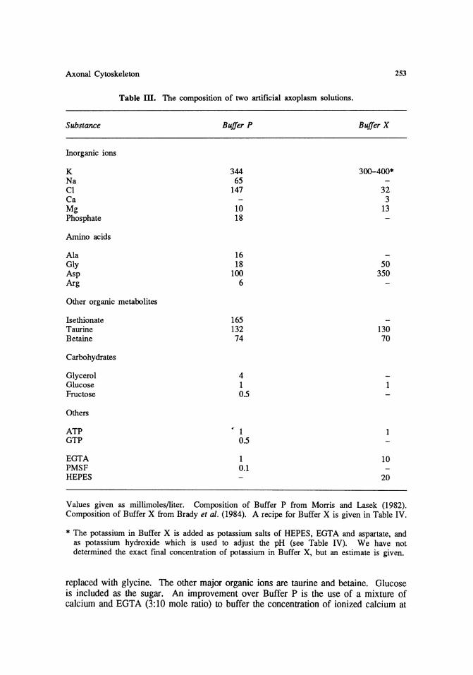
Table ill. The composition of two artificial axoplasm solutions.
Substance Buffer P Buffer X
Inorganic ions
K 344 300-400* Na 65 Cl 147 32 Ca 3 Mg 10 13 Phosphate 18
Amino acids
Ala 16 Gly 18 50 Asp 100 350 Arg 6
Other organic metabolites
lsethionate 165 Taurine 132 130 Betaine 74 70
Carbohydrates
Glycerol 4 Glucose 1 Fructose 0.5
Others
ATP • 1 GTP 0.5
EGTA 1 10 PMSF 0.1 HE PES 20
Values given as millimoles/liter. Composition of Buffer P from Morris and Lasek (1982). Composition of Buffer X from Brady et al. (1984). A recipe for Buffer X is given in Table IV.
* The potassium in Buffer X is added as potassium salts of HEPES, EGTA and aspartate, and as potassium hydroxide which is used to adjust the pH (see Table IV). We have not determined the exact final concentration of potassium in Buffer X, but an estimate is given.
replaced with glycine. The other major organic ions are taurine and betaine. Glucose is included as the sugar. An improvement over Buffer P is the use of a mixture of calcium and EGTA (3:10 mole ratio) to buffer the concentration of ionized calcium at

254 A. Brown and R. J. Lasek
Table IV. Recipe for Buffer X (Developed by Scott T. Brady)
Buffer X stock stock ml of Substance mM M, M g/100 ml stock/} 00 ml
Aspartate 350 171.2 1 17.12 35 Taurine 130 125.1 1.626 g* Betaine· H20 70 135.2 1 13.52 7 Glycine 50 75.1 1 7.51 5 HE PES 20 238.3 1 23.83 2 MgC12·6HzO 13 203.3 1 20.33 1.3 EGTA 10 380.4 0.1 3.804 10 CaC}z·2H20 3 147.03 1 14.703 0.3 Glucose 1 180.16 0.1 1.802 1 ATP 1 553.18 55 mg*
The table has six columns. These represent (from left to right): the substance, its molarity in Buffer X (mM), its relative molecular mass (M.), the molarity of the stock solution (M), the amount of substance required to make 100 ml of stock solution (g/100 ml), and the volume of that stock solution required to make 100 ml of Buffer X (ml of stock/100 ml). Stock solutions are prepared for all components, except for taurine and A TP which are added as the solid. The stock solutions are stored frozen and thawed immediately before use. When all the components have been added, the pH of the mixture is adjusted to pH 7.2 with a 20% solution of potassium hydroxide, and then made up to final volume with distilled/deionized water. The Buffer is stored frozen in aliquots at -20 °C. Aspartate is the mono-potassium salt of the L-isomer. The HEPES and EGTA stocks are prepared with the free acids and adjusted to pH 7.2 with potassium hydroxide. ATP is used as the disodium salt. Abbreviations:
HEPES (N-2-hydroxyethlypiperazine-N' -2-ethanesulfonic acid) EGTA (ethylene glycol-bis(2-arnino ethyl ether)N,N,N',N'-tetraacetic acid) ATP (adenosine-5'-triphosphate)
* Add by weight.
physiological levels. In an artificial axoplasm solution containing 3 mM calcium, 10 mM EGTA, 12.9 mM magnesium, and methane-sulfonate, taurine and glutamate as the major anions, Rubinson and Baker (1979) determined the ionized calcium concentration to be 65 nM, which is within . the physiological range (see Section 9 .3). Of the magnesium, 2.9 mM was bound to the EGTA, leaving 10 mM ionized magnesium. The composition of Buffer X is shown in Table III, and a recipe for Buffer X that we use in our laboratory is given in Table IV.
Both Buffer P and Buffer X should be iso-osmotic with axoplasm in order to preserve the membranous vesicles, mitochondria and smooth endoplasmic reticulum. These solutions are approximately iso-osmotic with axoplasm but the osmolarity should be checked and, if necessary, the difference should be made up with sucrose rather than salt in order to keep the ionic strength the same. It should be noted, however, that the composition of these artificial axoplasm solutions still differs from axoplasm in at least one very important way in that they contain no macromolecules, and in particular no protein, which is the major macromolecule in axoplasm.

Axonal Cytoskeleton 255
11. The solubility and stability of axoplasm
Axoplasm is normally of gel-like consistency and is strong enough to be handled with forceps. These mechanical properties are due largely to the great length of the neurofilaments and microtubules, which makes them particularly important elements in the axoplasmic architecture. If axoplasm is immersed in physiological solutions that approximate the solute composition of axoplasm, the neurofilaments and many of the microtubules remain stably polymerized, and the axoplasm retains its mechanical integrity (see Section 10 for a description of these solution conditions). However, ionic conditions that differ from the physiological state, such as those produced by increasing the ionic strength or pH, can alter the stability of the cytoskeletal polymers and in this way affect the mechanical properties of the axoplasm.
11.1. Salt and pH
High salt concentrations tend to depolymerize axonal cytoskeletal polymers and thereby destabilize axoplasmic architecture, and comparisons of the effects of different salts on both the mechanical properties of axoplasm and the stability of the cytoskeletal polymers indicate that the chemical nature of the salt is more important than the ionic strength. For example, Baumgold et al. (1981) showed, using centrifugation, that the cytoskeletal polymers in squid axoplasm are stable in 360 mM potassium fluoride at pH 7.3 but are completely soluble in 360 mM potassium iodide at the same pH and ionic strength. In potassium fluoride, centrifugation of homogenized axoplasm produced a pellet that was highly enriched in neurofilament proteins, tubulin and actin; these proteins comprised 81% of the sedimentable protein. In potassium iodide, the axoplasm dispersed and none of these proteins sedimented.
In fact, the relative efficacy of different ions for preserving the structure of axoplasm appears to increase in the order of the classical Lyotropic Series of colloid chemistry. In this series, ions are ranked according to their chao tropic effect, which is their ability to destabilize interactions between hydrophilic colloids such as proteins. The chaotropic effect is a consequence of effects on the structure of the water which in turn modify the solvent-macromolecule interactions that are involved in the stabilization of macromolecular assemblies and associations (von Hippe! and Wong, 1964; Friefelder, 1985). Physical studies on the interactions between macromolecules have indicated the following anion series:
fluoride > phosphate > aspartate/glutamate > sulfate > citrate > tartrate > acetate > chloride > nitrate > bromide > iodide > thiocyanate
and the following cation series:
rubidium > potassium > ammonium > sodium > lithium
Those ions that are most chaotropic are most effective at solubilizing proteins. Thus solutions that contain fluoride and phosphate salts of potassium are much more favorable for the preservation of the cytoskeleton than those containing iodide or thiocyanate salts of sodium or lithium.

256 A. Brown and R. J. Lasek
Studies on neurofilaments and microtubules in vitro confirm that the stability of these cytoskeletal polymers is dependent on the chaotropic nature of the ionic environment. Apparently, solvent interactions are critical for the stability of these cytoskeletal polymers. For example, in studies on the assembly/disassembly properties of neurofilaments purified from axoplasm and squid brain, we have found that potassium thiocyanate is more effective than potassium chloride at solubilizing filaments, and a more detailed comparison of different potassium halide salts revealed a direct correspondence to the Lyotropic Series (Brown, 1989). In addition, Sakai and Matsumoto (1978) observed a similar relationship in their studies on the assembly of microtubules from squid axoplasm in the presence of various potassium halide salts.
Through their effect on the stability of the protein filaments, chaotropic salts can affect the mechanical properties of the axoplasm. For example, Rubinson and Baker (1979) investigated the effect of various salts on the mechanical properties of axoplasm by measuring the viscous resistance of extruded axoplasm to flow under pressure through a porous cellulose acetate tube. They used an artificial axoplasm solution containing methanesulfonate (0.3 M), taurine (0.15 M) and glutamate (0.1 M) as the major anions. Methanesulfonate was used as a substitute for isethionate (2-hydroxyethanesulfonate), which is the major anion in squid axoplasm (see Section 9.1). Substitution of methanesulfonate in the artificial axoplasm solution with more chaotropic anions had a marked effect on the mechanical properties of the axoplasm; chloride, bromide, iodide and thiocyanate (in order of increasing efficacy) all caused a reversible decrease in the viscosity.
The protein interactions that stabilize the cytoskeletal polymers in axoplasm are also sensitive to pH. The microtubules and neurofilaments in squid axoplasm are optimally stable between pH 6 and 7. These polymers tend to precipitate or aggregate at around pH 5, which is near their isoelectric point, but are readily solubilized at alkaline pH (Brown, 1989).
11.2. Calcium-dependent proteolysis
In addition to their physical effects, ions may also have effects due to their specific chemical or biological activity. This is so for calcium, which has a specific biological action on axoplasm. In early studies on extruded axoplasm it was noted that axoplasm rapidly liquified in seawater. Hodgkin and Katz (1949) showed that this disintegration was caused specifically by calcium at concentrations of 1 mM or more; axoplasm does not disintegrate in calcium-free seawater. These observations were confirmed by the observations of Chambers and Kao (1952) and by the rheological measurements of Rubinson and Baker (1979). It is now known that this effect is due to the action of an endogenous calcium-dependent protease that preferentially cleaves the neurofilament proteins in axoplasm (see Section 17.4). The protease is not active in the axon in vivo since the ionized calcium concentrations in axoplasm are very low, and do not exceed micromolar levels due to the strong buffering capacity of the intracellular calcium stores (see Section 9.3).

Axonal Cytoskeleton 257
12. Preparation of axoplasm and sheath for SDS PAGE technique
The protein composition of the axoplasm and sheath of the giant axon can be analyzed by sodium dodecyl sulfate polyacrylamide gel electrophoresis (SDS PAGE). In this technique proteins are dissolved in the ionic detergent, sodium dodecyl sulfate (SDS), in the presence of a sulfhydryl reducing agent, ~-mercaptoethanol, which reduces any disulfide bonds. The denatured SDS-polypeptide complexes are then applied to a porous polyacrylamide gel and subjected to an electric field. The proteins move through the gel toward the anode because of the negative charge of the bound SDS. For most proteins, the rate of movement has been found empirically to be related to the size of the denatured polypeptide chain, which in turn is related to its mass. Therefore, this technique has proven useful for the separation of polypeptides of different molecular masses. In addition, SDS PAGE allows an empirical estimation of the molecular mass of a protein by comparison of its mobility with the mobility of proteins of known mass. A comprehensive treatment of the principles and practice of electrophoresis can be found in Andrews (1981).
Axoplasm readily dissolves in a standard sample buffer containing SDS and ~mercaptoethanol, with brief heating in a hot water bath, as originally described by Laemmli (1970). No homogenization is necessary. For standard SDS PAGE with Coomassie Blue (CBBR-250) staining, 1.2 mg axoplasm wet weight (equivalent to about 30 f..l.g protein) gives a good loading on a typical 1 mm thick Studier slab gel (Studier, 1973), and 0.3 mg axoplasm wet weight (equivalent to about 7.5 f..l.g protein) gives a suitable loading for a typical 0.9 mm thick minigel. These loadings will, of course, depend also on the width of the wells on the gel.
In contrast to the axoplasm, the sheath is difficult to solubilize since it contains connective tissues. We recommend homogenization with a small volume glass-glass homogenizer with ground glass surfaces, followed by heating with sample buffer. High speed centrifugation may then be necessary to clarify the sample of insoluble material such as collagen, which can disturb the protein separation in SDS PAGE and cause smearing of the bands. For analysis of the residual cortical axoplasm in the sheath (see Section 15), without extracting glial and extracellular proteins, the sheath can be slit open after axoplasm extrusion and extracted with salt solutions as described by Sakai and Matsumoto (1978). Alternatively, the proteins of the cortical axoplasm can be extracted by the intracellular perfusion technique (see Section 15.2).
13. Preparation of axoplasm for electron microscopy
13.1. A general fixation protocol
For routine preparation of axoplasm for thin sectioning, we use the following protocol (Miller and Lasek, 1985). Axoplasm is fixed for 1 - 2 hours at room temperature in SPME (0.6 M sucrose, 0.1 M potassium phosphate, 10 mM magnesium chloride, 1 mM EGTA, pH 7.1) solution containing 2.5% glutaraldehyde. The SPME solution has an osmolarity of 980 mOs/kg, which is approximately iso-osmotic with axoplasm. The osmolarity of the glutaraldehyde fixative is ignored since it readily crosses membranes during fixation and therefore is not osmotically active (Glauert, 1975). For preservation of membranous organelles, it is better that the fixative solution

258 A. Brown and R. J. Lasek
be slightly hyper-osmotic than hypo-osmotic. The osmolarity should be adjusted with sucrose rather than salt since it is desirable to keep the ionic strength low in order to stabilize the cytoskeletal filaments. Artificial axoplasm solutions containing molecules with amine groups, such as solutions containing amino acids, are not suitable since these molecules react with aldehydes and interfere with the fixation. Phosphate is a good choice for the buffer since it ranks low in the Lyotropic Series and therefore does not destabilize protein interactions (see Section 11.1). After glutaraldehyde fixation the axoplasm is post-fixed with 1% osmium tetroxide in SPME solution, stained en bloc with 0.5% (aq) uranyl acetate, and finally dehydrated through a graded ethanol series into propylene oxide, and embedded in Epon (Poly/Bed 812, Polysciences, Inc.). Thin sections are stained with aqueous uranyl acetate and lead citrate. Since axoplasm poses no permeability barrier, many of the steps in the preparation, including the fixation, could probably be shortened. Some other fixation protocols that have been used for squid axoplasm and for the giant axon are described by Metuzals (1969), Hodge and Adelman (1980) and Metuzals et al. (1983).
13.2. Preservation of microfilaments
The above conventional fixation, dehydration and embedding protocol preserves microtubules and neurofilaments in axoplasm, but actin microfilaments are more susceptible to destruction during fixation and dehydration, and a modified procedure is necessary to ensure good preservation. This is important because our recent studies indicate that microfilaments are a major component of the inner and cortical cytoskeletons of axons (see Section 19).
The two treatments that are critical for microfilament preservation are post-fixation with osmium tetroxide, and dehydration. Conventional osmication and dehydration protocols can fragment and destroy microfilaments, even after aldehyde fixation. McDonald (1984) obtained improved preservation of microfilaments by post-fixation with osmium ferricyanide rather than osmium tetroxide. In addition, the destructive effects of dehydration were minimized by further fixation with tannic acid and uranyl acetate, and by dehydration in acetone rather than ethanol.
Fath and Lasek (1988) used this protocol for the visualization of actin microfilaments in squid axoplasm. Axoplasm was fixed by immersion for 30 minutes in SPME solution (see Section 13.1) containing 1% glutaraldehyde. In some cases, axoplasm was extruded directly into the fixative. In other cases the axoplasm was first extruded into Buffer X (see Section 10.2) containing 10 J.1M phalloidin (to stabilize the microfilaments), and extracted with gentle stirring for 4 hours at ambient temperature prior to immersion in the fixative. Extraction of the soluble proteins in this way improved the visibility of the microfilaments by removing some of the dense matrix that surrounds them in axoplasm (see Section 19). After primary fixation, the axoplasm was post-fixed according to the osmium ferricyanide procedure of McDonald (1984). Briefly, the fixed axoplasm was first washed for 5 minutes in 0.1 M sodium phosphate buffer (pH 7.2), and then reacted for 5 minutes with a solution of 0.8% potassium ferricyanide in 0.1 M sodium phosphate buffer. The axoplasm was then post-fixed for 15 minutes in a mixture of 0.5% osmium tetroxide and 0.8% potassium ferricyanide in 0.1 M sodium phosphate (on ice, in the dark), followed by 15 minutes in 0.5% tannic acid in 0.1 M sodium phosphate, and rinsed in deionized water. Finally, the axoplasm was stained en bloc with aqueous uranyl acetate, dehydrated through graded acetones, and embedded in plastic for sectioning.

Axonal Cytoskeleton
sf s
2
3
13.3. Fixative penetration
259
Figure 6. Removal of the Schwann cell sheath from the giant axon. 1. Small nerve fibers (sf) are removed from a portion of the giant nerve fiber. The sheath (S), axolemma (A) and needle (N) are labelled. 2. The sheath is cut circumferentially with the needle. 3. The sheath (S) is pulled back from the opening on either side to expose the surface of the axon (A). Reproduced with permission from Metuzals et al. (198lb).
Good fixation also depends on good penetration of the fixative into the axoplasm. Best fixation is achieved if the fixative is in direct contact with the axoplasm. The simplest approach is to extrude axoplasm as described in Section 6.1 , and then to fix by immersion. However, the compressive forces during extrusion in the conventional manner tend to cause some disruption of the longitudinal organization of the cytoskeletal polymers. Some improvement is achieved by extruding the axoplasm onto glass, and then immersing the axoplasm on the glass into fixative. The adhesive effect of the glass allows the axoplasm to be extruded under tension which helps to restore the longitudinal alignment of the polymers that is present in the axon (see Section 6.1). Compression of the cytoskeleton can also be avoided by isolating axoplasm by the slitting method described in Section 6.2, but for optimum preservation of the organization of the cytoskeleton, the axoplasm must be fixed in situ. However, fixation by immersion of the whole giant axon in fixative does not produce good preservation of the axoplasm. Apparently, the collagenous sheath becomes a barrier to the diffusion of the fixative during fixation.
13.4. Removing .. the axon sheath
Improved preservation of axoplasm in situ has been obtained by first dcsheathing the axon according to a method developed by lchiji Tasaki in 1975 and described by Metuzals et al. (1981b); see Fig. 6. The close association of the glial cells with the axon means that the sheath cannot be removed from the axon by conventional dissection procedures. To desheath the axon, the giant nerve fiber is first fine-cleaned to remove associated small nerve fibers and then immersed for 1 hour, without loss of excitability, in seawater containing 1 mg/ml trypsin. The trypsinized fiber is then subjected to mild fixation for 15 minutes in a 0.5% solution of glutaraldehyde in seawater. After fixation the fiber is placed in a hypertonic solution of 0.4 M sucrose
260 A. Brown and R. J. Lasek
in seawater, which causes the axon to shrink away from the sheath. The elevated sheath, consisting of the Schwann cell layer, basement membrane and outer collagenous layer (see Section 3), can then be transected with a needle by pinching the sheath between the needle and the base of the dissecting dish. A circumferential incision of the sheath is then made in the same manner by inserting the tip of the needle into this opening. The sheath is then everted, exposing about 1 - 2 em of desheathed axon. Desheathed axons can then be fixed by direct immersion in seawater containing aldehyde fixative and processed for transmission electron microscopy in the normal manner, or the axon can be prepared for scanning electron microscopy of the surface of the axon (see Section 14.2).
Improved fixation of axons may also be facilitated by pretreatment of the fine-cleaned fiber with collagenase to remove the collagenous layer of the sheath (Lasek, 1989), though this procedure renders the fiber extremely fragile.
13.5. Cannulation
Hodge and Adelman (1980) obtained much improved fixation and preservation of organization of the axoplasm by internal irrigation of axons with fixative following cannulation (see Section 6.3). One could also slit a fine-cleaned axon along its length as described in Section 6.2 and then immediately flood with fixative. Both of these techniques allow the fixative direct access to the axoplasm in situ, without the need for separation of the axoplasm from the sheath.
13.6. Negative staining
The cytoskeletal elements in axoplasm can also be visualized by negative staining. We have used two preparations. In one, the axoplasm is gently homogenized at a concentration of about 10 mg/ml wet weight (200 Jlg/ml protein) in a suitable solution, and then a small aliquot is applied to a carbon-coated grid and stained with 1% aqueous uranyl acetate for electron microscopy (see Huxley, 1963, for procedure). If solutions containing high salt concentrations are used, such as Buffer P and Buffer X, then it is necessary to rinse the grids with a low-salt solution in order to obtain good staining. If required, the proteins can be fixed with glutaraldehyde before applying them to the grid. Phosphate buffer is not recommended since it forms a precipitate with uranyl acetate.
In another approach, a carbon-coated grid can be gently pressed directly against the surface of freshly extruded axoplasm, allowing proteins to stick to the carbon support (Wais-Steider et al., 1988). The proteins are then stained with uranyl acetate as described above. This procedure provides excellent negatively-stained images of the cytoskeletal polymers in axoplasm, without the need for dilution or homogenization, and essentially in their native state.

Axonal Cytoskeleton 261
14. The organization of the cytoskeleton
14.1. Longitudinal organization
Axons are cylindrical in shape, and their three-dimensional structure can be usefully described by two aspects, one radial and the other longitudinal. The cylindrical shape of the axon, in its longitudinal dimension, is formed and largely maintained by the long polymers of the axonal cytoskeleton (neurofilaments and microtubules), which are orientated along the long axis of the axon. The high degree of alignment of these cytoskeletal elements is the most striking feature of the organization of the axonal cytoskeleton. This structural property of axoplasm was first inferred over 50 years ago by Bear et al. (1937a), based on their observations on the birefringence of axoplasm in polarized light.
One good way to appreciate the longitudinal alignment of the cytoskeletal elements in the giant axon is to examine an intact axon by differential interference contrast microscopy. Electron microscopy provides far more structural detail, but light microscopy gives a much better impression of the long-range order. Using this technique, Metuzals and Izzard (1969) showed that axoplasm in situ was composed of highly linear elements that were orientated along the long axis of the axon. These linear elements apparently represent individual bundles of microtubules that are now known to be refractile enough to be detected by this type of light microscopy (see Section 18.2). Electron microscopy confirms that the microtubules in axoplasm in situ are very straight and that they are aligned in parallel to each other along the long axis of the axon. The neurofilaments, in contrast, trace a slightly more sinuous path through the axon. In extruded axoplasm, the longitudinal organization of the axoplasm is essentially preserved, but the straightness of the long polymers is lost due to the compressive forces that are employed in the extrusion process (see Section 6.1).
14.2. Helicity
Axons often have a natural internal helicity that is superimposed on the longitudinal alignment of the cytoskeletal polymers in the axoplasm (for discussion, see Lasek et a/., 1983). This helicity is particularly evident in the axoplasm of squid and marine fanworm giant axons. For example, Metuzals et al. (1981b) demonstrated helicity in the cytoskeleton in the squid giant axon by scanning electron microscopy of the surface of the axon after removal of the sheath (see Section 13.4 for description of procedure). The surface of the desheathed axon had a ridge-and-groove topology, which traced a right-handed helical path along the axon. The angle of the helix was about 10° to the long axis, which corresponds to a pitch of about 18 mm for an axon with a diameter of 0.5 mm. The ridges and grooves may have been an artifact due to the osmotic shrinkage employed in the desheathing procedure, but nevertheless, they did reveal an underlying helical twist to the cytoskeletal polymers in the periphery of the axon. This helicity has also been observed directly by differential interference contrast microscopy of axoplasm in situ (Metuzals and Izzard, 1969).
A more striking example of helical organization in axoplasm is the giant axon of the marine fanworm, Myxicola infundibulum, which is also unusual in that it contains a very high proportion of neurofilaments and relatively few microtubules. The helicity

262 A. Brown and R. J. Lasek
is so pronounced in these axons that three orders of helical configuration can be identified (Gilbert, 1972; 1975a); a ripple helix that has a pitch of about 13 Jlm, a segmental helix that corresponds to the helicity described above for the squid giant axon, and a macroscopic gel helix, which forms the axon into large coils during the substantial length changes that occur when the worm contracts. The ripple helix may also be weakly expressed in squid axoplasm (Gilbert, 1972).
Gilbert was able to isolate axoplasm from the fanworm and he showed that the neurofilament-rich axonal cytoskeleton retains the helical properties of the axon. These observations indicate that helicity may be an intrinsic property of the axonal cytoskeleton. This proposal is supported by the fact that, in squid, giant axons from the right and left sides of the animal both have the same helical handedness (Metuzals et at., 1981b). The abundance of neurofilaments in the squid and fanworm giant axons, and in other axons with helical cytoskeletons, suggests that neurofilaments contribute to the intrinsic helicity of the axonal cytoskeleton (Gilbert, 1972; 1975b; Metuzals et at., 1981a; Lasek et at., 1983). For example, Gilbert showed that fanworm axoplasm behaves like a weakly coiled spring. This property is apparently due to the neurofilaments, which trace a helical path that forms the ripple helix of the axon (Gilbert, 1975a). If the axoplasm is stretched, the neurofilaments straighten, and if the stretch is released, they then tend to recoil.
In addition, the molecular structure of neurofilaments also supports the proposal that these fibrous polymers contribute to the helical organization of the cytoskeleton. Like other intermediate filaments, neurofilaments are composed of rod-shaped subunits that are arranged in a rope-like helical pattern along the axis of the neurofilament (Krishnan et at., 1979; Aebi et at., 1983), and this helical arrangement of the fibrous subunits within the neurofilaments may generate a slight torque within these structures so that they tend to twist when force is applied to them by the cytoskeletal translocation mechanisms of the slow axonal transport system. In this way, the helical characteristics of the neurofilaments may largely determine the higher order helical properties of the axonal cytoskeleton, in axons with large numbers of neurofilaments (Gilbert, 1975a).
The cytoskeletal helicity is a very weak property of axoplasm in squid, since observations on axoplasm isolated by the axon slitting procedure (see Section 6.2) show no sign of helical recoil after separation from the sheath, and the axoplasm retains the original proportions of the axon (see Fig. 5). In contrast to the squid giant axon, the helical properties of axoplasm in the giant axon of the fanworm Myxicola infundibulum are more pronounced, and they may have an important role in the mechanics of the axonal cytoskeleton when these axons are compressed and stretched during contraction and elongation of the animal. This difference between squid and the fanworm axoplasm may reflect the large number of microtubules in squid giant axons compared to the fanworm; microtubules are more rigid than neurofilaments and they tend to trace a straighter path and are therefore less extensible than axoplasmic neurofilaments (see Section 18).
In addition, the fanworm axoplasm in situ is actually subjected to repeated stretch and compression by the motility of the worm, and the continual working of the cytoskeleton in this way probably exaggerates the intrinsic potential of neurofilaments to generate the higher order helical twist that is observed in these and other annelid axons. In contrast with the extensible tissues surrounding the nerve cords of annelids, the giant axons of the squid are embedded in relatively inextensible tissues that are supported by a rigid chitinous pen (which serves as the backbone of the animal). Thus,

Axonal Cytoskeleton 263
in the squid giant axon, working of the cytoskeleton by external forces probably contributes little, if at all, to the helicity of the cytoskeleton.
14.3. Radial organization
In the radial dimensions of axons, two regions of the cytoplasm can be defmed; a central or inner region, and a peripheral or cortical region (see Lasek, 1984 and 1988, for further discussion). The cytoskeleton in these regions is distinct in both composition and organization. The large size of the squid giant axon enables separation of these two regions of the axoplasm and direct analysis of their biochemistry and ultrastructure.
15. The cortical cytoskeleton
15.1. Axoplasm is attached to the plasma membrane
The axoplasmic cytoskeletal elements in the cortex of the axon are connected to the plasma membrane. In squid it is possible to demonstrate this by direct mechanical manipulation. For example, Metuzals and Tasaki (1978) have used polarizing microscopy to observe the cannulation of giant axons for internal perfusion. In this process, a glass cannula is inserted into the cut end of an axon and then advanced axially along the inside of the axon, using gentle suction to remove a central core of axoplasm (see Section 6.3). Metuzals and Tasaki noted that in some cases strong suction produced a birefringent distortion of the axoplasm that extended to the inner surface of the plasma membrane, where the axolemma became indented towards the tip of the cannula. Apparently suction produces a strain on fibrous elements in the cortical axoplasm that are attached to the inner surface of the plasma membrane, and this causes an orientation of these elements towards the tip of the cannula, producing the birefringence. Sato et al. (1984) reported similar evidence for attachment based on their observations of the movement of magnetic spheres in axoplasm under a magnetic field. Movement of the spheres from the periphery of the axon towards the center was seen to cause a local inward distortion of the axolemma. If sufficient force was applied the spheres would overcome this resistance and move more freely.
Observations on the extrusion process also indicate an association of the peripheral cytoskeleton with the plasma membrane. When axoplasm is extrUded from the giant axon it shears at an annular plane beneath the axolemma. This plane of shear defines a cortical layer of axoplasm within the axon, the membrane-associated cortical cytoskeleton, that has some distinct properties from the inner cytoplasm. We have used video-enhanced differential interference contrast microscopy to examine the extrusion process (Brady and Lasek, 1989). A fine-cleaned giant axon was placed on a glass slide in the specimen plane of the microscope and pressure applied at one end so as to extrude the axoplasm from the cut end (see Section 6.1). By slowly increasing the pressure it was possible to reach a point at which the extrusion of the axoplasm could be controlled. With just sufficient pressure it was possible to observe the axoplasm moving through the axon. Our observations indicate that the axoplasm shears at a relatively sharp boundary that is located roughly 10 Jlffi beneath the plasma membrane.

264 A. Brown and R. J. Lasek
This is in agreement with the observations of Baker et al. (1962), using electron microscopy and action potential velocity measurements on extruded nerve fibers, and of Kobayashi et al. (1986), using electron microscopy. Thus, for an axon 500 J..IIIl in diameter, extrusion removes about 92% of the total axoplasm.
The plane of shear produced by the extrusion process may not correspond directly to a pre-existing natural partition between cortical and inner axoplasm. For example, electron microscopic studies of the organization of the axoplasm within intact axons indicate that the cortical cytoskeletal elements that are directly adherent to the plasma membrane are arranged within a thin 0.1 - 1.0 J.l.m layer immediately beneath the membrane (see Section 15.4). Perfusion studies, which are described in Section 15.2, indicate that the remainder of the axoplasm within the axon is not tightly attached to the plasma membrane since it can readily be removed by cannulation and the flow of perfusion solution. Although the axoplasmic cytoskeletal polymers that are deeper than 1 - 2 J..IIIl from the plasma membrane are not attached to the plasma membrane, they may experience considerable resistance to movement under extrusion pressure (or suction pressure in the case of cannulation) because of frictional interactions with the adjacent stationary polymers of the membrane-associated cortical cytoskeleton. This viscous resistance to sliding under pressure may contribute to the tendency of the axoplasm to shear in a plane that is farther from the plasma membrane than that which separates it from the membrane-associated cytoskeleton. This subject of the interaction between axonal cytoskeletal elements within the axon and their resistance to longitudinal sliding is discussed further in Section 21.
15.2. Internal perfusion of axons
Internal perfusion of the squid giant axon is a useful technique for studying the membrane-associated cortical cytoskeleton. Methods for perfusing the giant axon are described by Adelman and Gilbert (1990). In brief, the procedure involves removing a central core of axoplasm from the giant axon, thereby allowing the flow of solutions through the inside of the axon (see Section 6.3). The perfused axon can then be preserved by internal perfusion with fixative and processed for transmission electron microscopy (see Section 13), or it can be cut open to expose the inner surface of the axolemma for scanning electron microscopy.
By altering the composition of the perfusion solution, the perfusion technique can be used to chemically dissect the cortical axoplasm (Metuzals and Tasaki, 1978; Baumgold et al., 1981). Perfusion with an isotonic solution containing potassium fluoride (360 mM potassium fluoride, 40 mM potassium phosphate, 4% glycerol, pH 7 .3) flushes away most of the inner axoplasm, but preserves a meshwork of subaxolemmal filaments associated with the inner surface of the membrane. In contrast, perfusion with more chaotropic salts, such as potassium iodide, which solubilize cytoskeletal protein filaments (see Section 11.1), causes extraction of the subaxolemmal meshwork. This membrane-associated filamentous meshwork can also be removed by proteolysis. For example, media containing pronase or millimolar calcium, which activates the endogenous calcium-dependent protease (see Sections 11.2 and 17 .4), also extract the cortical cytoplasm (Metuzals and Tasaki, 1978). Scanning electron microscopy after perfusion with chaotropic salts or proteases shows that much of the inner surface of the axolemma is devoid of filaments, and extensive blebbing of the membrane is observed in these regions. These observations indicate that the cortical cytoskeleton is composed of protein filaments that are associated with the

Axonal Cytoskeleton 265
plasma membrane of the axon through non-covalent interactions that can be overcome by altering the ionic environment. The blebbing of the membrane in regions devoid of these filaments suggests that the cortical cytoskeleton may be important in maintaining membrane stability.
15.3. Protein composition of the cortical cytoskeleton
Since the association of the cortical cytoskeleton with the axolemma is preserved by potassium fluoride but is extracted by potassium iodide, perfusion with these salts can be used to specifically analyze the composition of the cortical cytoplasm. Baumgold et al. (1981) and Kobayashi et al. (1986) used silver staining of polyacrylamide gels to analyze the protein composition of fractions after perfusion with potassium iodide. Baumgold et al. used the squid Loligo pealei and Kobayashi et al. used the squid Doryteuthis bleekeri. Because of the small amounts of protein that elute it is necessary to dialyze the salts from the fractions and then concentrate them by lyophilization before analysis by SDS PAGE. These studies indicated that the cortical cytoskeleton contained tubulin and actin and a variety of other proteins, including a number of high molecular weight.
The separation afforded by mechanical extrusion allows more extensive biochemical analysis of the composition of the cortical and inner axoplasm. In order to analyze the cortical axoplasm remaining in the sheath after extrusion, Kobayashi et al. (1986) slit open the extruded sheaths of Doryteuthis bleekeri axons and extracted them, without homogenization, in a solution containing 0.6 M NaCl, 10 mM CaC12 and 10 mM sodium 2-(N-morpholino)ethane sulfonate (MES) buffer, pH 6.8, as described by Sakai and Matsumoto (1978). In this way the proteins in the cortical axoplasm were extracted but the proteins in the sheath cells (fibroblasts and glia) were not.
Two high molecular weight polypeptides were identified that are present in high concentration in the cortical axoplasm. One protein of M, 260,000, a putative microtubule-binding protein named axolinin, was shown to co-sediment with microtubules but not with microfilaments, and to produce bundling of microtubules in vitro (Murofushi et al., 1983). The other, a putative microfilament-binding protein of M, 255,000, was shown to co-sediment with microfilaments but not with microtubules and to increase the low-shear viscosity of actin filament suspensions in vitro, which suggests that it can act as a cross-linker. Both proteins have been purified from axonal sheaths and from a bundle of small nerve fibers called the fin nerve. The proteins have distinct solubility properties and the M, 255,000 protein showed no reactivity with antisera raised to axolinin. Both proteins are concentrated in the cortex, but are also present in the inner axoplasm; axolinin and the M, 255,000 protein constitute 1.5 and 1.7%, respectively, of the total axoplasmic protein in extruded axoplasm, and 8.9 and 5.2%, respectively, of the axonal sheath extract. Assuming that the cortical axoplasm represents 8% of the total axoplasm (see Section 15.1), we calculate that about 32% of the axolinin and 20% of the M, 255,000 protein in the axon are present in the cortical region.
Another high-molecular weight cytoskeletal protein that is present in the cortex of the giant axon is brain spectrin (fodrin), which migrates as a doublet of M, 235,000 and 240,000 on SDS-polyacrylamide gels. The identity of these two polypeptides has been confirmed on nitrocellulose blots using a polyclonal antiserum to mammalian brain spectrin (Fath and Lasek, 1989). The apparent molecular weights (by SDS PAGE) of

266 A. Brown and R. J. Lasek
Table V. The distribution of some proteins in cortical and inner axoplasm.
Total axoplasm1
Actinz Axolinin3
M. 255,000 protein3
Spectrin4
Source of data:
Inner cytoskeleton
92% 92% 68% 80% 80%
Cortical cytoskeleton
8% 8%
32% 20% 20%
1 Based on calculation for a 500 j.1m diameter axon with a 10 j.lm-thick region of cortical axoplasm.
z Fath and Lasek (1988). 3 Calculated from Kobayashi et al. (1986). 4 Fath and Lasek, (1989)
the two polypeptides are identical to those for spectrin from avian and mammalian brain. Like axolinin and the M. 255,000 protein, spectrin may also cross-link cytoskeletal proteins within the membrane-associated cytoskeleton.
Our studies also indicate that significant amounts of spectrin are present in extruded axoplasm. To quantify the distribution of spectrin, we have compared the amounts of spectrin in the inner and cortical axoplasm by densitometry of SDS-polyacrylamide gels (Path and Lasek, 1989). Spectrin has been considered to be principally localized subjacent to the plasma membrane (Levine and Willard, 1981). Our analyses indicate that 80% of the spectrin in the giant axon is recovered in the extruded axoplasm and 20% in the remaining 8% of the axoplasm in the sheath. Thus spectrin is 2 - 3 fold more concentrated in the cortex, but most of the protein is present in the inner cytoplasm.
Table V compares the distribution of these three high-molecular weight proteins in the cortical·and inner axoplasm of the giant axon. Axolinin, theM, 255,000 protein and spectrin are all present in higher concentrations in the cortex. However, because the cortical axoplasm is only a small fraction of the total axoplasmic volume, most of each of these proteins is present in the inner cytoplasm and is not associated with the plasma membrane. Thus these proteins apparently contribute to the inner cytoskeleton as well as to the cortex. For example, in addition to cross-linking the membrane cytoskeleton, these proteins may also cross-link filaments in the inner cytoskeleton (See Goodman and Zagon, 1986, for review).
15.4. Architecture of the cortical cytoskeleton
Tsukita et al. (1986) have studied the morphology of the subaxolemmal cytoskeleton in giant axons from the squid Doryteuthis bleekeri after internal perfusion with solutions containing potassium fluoride, which stabilizes the polymers and their associations. For thin section electron microscopy, axons were fixed by internal

Axonal Cytoskeleton 267
perfusion with aldehyde fixative containing tannic acid. For freeze-etch replica electron microscopy, perfused axons were fixed with glutaraldehyde and then cut open along their length and rapidly frozen by touching the inner surface against a copper block cooled by liquid helium.
These methods have revealed that the cortical cytoskeleton has a distinct architecture from that of the inner cytoplasm. A dense filamentous meshwork, a few micrometers in thickness, lines the inner surface of the axolemma. The major cytoskeletal polymers in this zone are microfilaments and microtubules; neurofilaments, which are the major cytoskeletal element in the inner axoplasm, are relatively few in number. The high concentration of microtubules in the cortex of the giant axon has also been noted by Sakai et al. (1985). They found that the mean microtubule density within 1 llJll of the axolemma was about 80 per J..Lm2 (cross-sectional area), which is about 3 - 4 fold greater than the density in the inner axoplasm.
Within the cortex, the cytoskeletal polymers are organized into discrete microfilament- and microtubule-associated membrane domains. In the microtubule domains, microtubules run in parallel beneath the axolemma along the long axis of the axon, and sometimes appear to be connected to the plasma membrane by fine filamentous strands. Locally the microtubules are often displaced inwards from the axolemma by punctate clusters of microfilaments that lie directly beneath the membrane. These microfilament domains occupy about 15% of the surface of the axolemma, and also appear to be connected to the plasma membrane by fine filamentous strands. In addition, microfilaments are seen outside these clusters in association with a matrix surrounding the microtubules, and this is also a feature of the microtubules in the inner cytoplasm (see Sections 18 and 19). A schematic illustration of the inner and cortical axoplasm is shown in Fig. 7.
The fine filamentous matrix that surrounds the microtubules and microfilaments may be composed of proteins such as axolinin, the M, 255,000 protein and spectrin, which are all present at higher concentrations in the cortex of the axoplasm. To examine this possibility, Tsukita et at. (1986) used immunogold electron microscopy in thin section to localize the microtubule-associated protein, axolinin, in the cortical axoplasm. Their results indicate that axolinin is concentrated in the microtubuleassociated membrane domains within the cortex. This distribution is consistent with a role for this protein in the organization of the cytoskeleton in regions where microtubules are the principal cytoskeletal polymers.
15.5. The cortical cytoskeleton and membrane excitability
The intimate association of the cortical cytoskeleton with the plasma membrane is probably important in providing mechanical stability to the lipid bilayer. However, in addition there are indications that the membrane-associated cytoskeleton may also be important in the maintenance of the electrical excitability of the axon. The evidence for this rests primarily on the observation that many agents which extract the cortical cytoskeletal elements also alter the ionic permeability of the membrane. For example, proteases and chaotropic salts, which are known to remove the cortical cytoskeleton (see Section 15.2), also abolish the excitability of the axon (Tasaki and Takenaka, 1964; Tasaki et al., 1965; Metuzals and Tasaki, 1978). Much of this evidence is equivocal, however, because many of the treatments that abolish excitability may also have direct effects on channel proteins within the membrane. For example, the effects of intracellular perfusion with proteases on the sodium conductance of the giant axon

268 A. Brown and R. J. Lasek
Figure 7. A schematic illustration of the inner and cortical regions of the axonal cytoskeleton. The number and relative diameter of the neurofilaments, microtubules and microfilaments are shown as they are in axoplasm. The polymers are orientated along the long axis of the axon. Note that the microfilaments are much shorter than the microtubules and neurofilaments, but that they are much more numerous. The lateral projections from the polymers represent side-arms and associated proteins. The small globular particles represent soluble proteins of the cytomatrix, such as metabolic enzymes. A. Inset showing a microfJ.lament-associated domain directly apposed to the inner surface of the axolemma. The microfilaments are attached to integral membrane proteins via radial cross-links. In other regions microtubules can also be directly apposed to the membrane (see text). B. Inset showing organization characteristic of the inner cytoskeleton. Microfilaments cluster in the microtubule domains, which are present throughout the inner axoplasm and are interspersed by neurofilaments.
have widely been attributed to direct proteolytic cleavage of the sodium channel (Armstrong et a/., 1973; Sevcik and Narahashi, 1975; Rojas and Rudy, 1976).
However, studies using more specific treatments also suggest an involvement of the cortical cytoskeleton in the maintenance of membrane excitability. For example, the effect of chaotropic ions on the sodium conductance of the axon can be mimicked by a variety of agents that depolymerase microtubules (Matsumoto and Sakai, 1979a; Matsumoto et a/., 1984). These agents include drugs such as colchicine, podophyllotoxin and vinblastine, and submillimolar concentrations of calcium ions. Furthermore, studies reported by Matsumoto and Sakai (1979b) and Matsumoto et a/. (1979) indicate that excitability can be restored to these inexcitable axons by subsequent perfusion with media containing microtubules and various cofactors. These results have been interpreted to indicate a specific requirement for cortical microtubules in the maintenance of the electrical excitability of the squid axon. For example, it is possible

Axonal Cytoskeleton 269
that microtubules in the cortical cytoskeleton may interact directly or indirectly with channel proteins in the membrane, and that this interaction may be necessary for the proper functioning of these channels. Indeed, there is considerable precedent for the interaction of the membrane cytoskeleton with integral membrane proteins in other cells (for example, see Goodman and Zagon, 1986), and it seems plausible that this interaction might have specific functional consequences for these proteins. The reader who is interested in these studies is referred to a review by Sakai et al. (1985) for more information.
16. The inner cytoskeleton
The inner cytoskeleton forms the bulk of the axoplasm in the axon and it has a different protein composition than the cortex; though all the axonal proteins that have been identified in squid axoplasm appear to be present in both the inner and cortical regions, their relative concentrations can differ greatly. For example, Table V shows that certain cytoskeletal binding proteins are more concentrated in the cortical axoplasm. In contrast, other proteins such as neurofilaments appear to be partially excluded. This difference in composition is reflected by a different cytoskeletal organization.
In the inner axoplasm, neurofilaments are the major cytoskeletal polymer and together with microtubules and microfilaments, these polymers largely determine the architecture of the inner cytoskeleton. The neurofilaments and microtubules are both very long (greater than tens of micrometers), while the microfilaments are much shorter (less than 1 jlm). The long polymers (microtubules and neurofilaments) are both aligned parallel to the long axis of the axon. Estimates based on analysis of the amounts of cytoskeletal protein as monomer and polymer in axoplasm (see Section 20) indicate that the ratio of neurofilaments to microtubules may be as low as 2: 1 (Fath and Lasek, 1988), which represents a much larger proportion of microtubules than in large vertebrate axons. For example, the ratio of neurofilaments to microtubules in the internodal regions of the large somatic motor axons of the chick oculomotor nerve is about 75:1 (Price and Lasek, 1989). However, in other respects the organization of the cytoskeleton in these axons appears to be very similar.
Neurofilaments, microtubules and microfilaments are present throughout the inner cytoplasm, but their distribution is not homogeneous. Microtubules often cluster together and are surrounded by a dense filamentous matrix, and this is most apparent in longitudinal sections (for example, see Miller and Lasek. 1985). Often membranous organelles and endoplasmic reticulum are associated with these domains. This filamentous matrix is composed of actin microfilaments and other cytoplasmic proteins. The microfilaments in the inner axoplasm are concentrated in these domains forming longitudinal columns through the axoplasm (Fath and Lasek, 1988). A schematic illustration of the inner cytoskeleton is shown in Fig. 7.
17. Neurofilaments in axoplasm
Neurofilaments are the major cytoskeletal elements of the inner axoplasm of the squid giant axon. Because of their abundance in the giant axon and the relatively large amount of pure axoplasm that can be obtained from the squid giant axon, studies of

270 A. Brown and R. J. Lasek
squid neuroftlaments have been important in understanding neuroftlament structure and physiology. The neuroftlament proteins in axoplasm are essentially fully polymerized (see Section 20) and studies in vitro indicate that they are very stable to changes in the ionic composition of the medium. To achieve full disassembly of squid neurofilaments requires high concentrations of chaotropic salt and high pH (see Section 11.1). Forexample, complete disassembly can be achieved by dialysis of axoplasm against solutions containing 1 M potassium chloride or 0.5 M potassium thiocyanate, at pH 8.
Electron microscopy of thin sections of intact squid axoplasm reveals that the neurofilaments are aligned in parallel along the long axis of the axon, though they trace a somewhat more wavy path than the microtubules. Neuroftlaments can also be visualized in homogenized axoplasm by electron microscopy of negatively-stained preparations (see Section 13.6). By this procedure the neurofilaments measure about 8.5 nm in diameter and are smooth in appearance (Lasek et al., 1979; Wais-Steider et al., 1983). In homogenates the filaments are many micrometers in length, but they are likely to be much longer in intact axoplasm. In marked contrast to microtubules, the neurofilaments in homogenized axoplasm appear to be very flexible polymers that often entangle.
17 .1. Purification of squid neurofilaments
Neurofilaments have been purified from squid axoplasm by a variety of procedures, including precipitation with cytochrome-c (Gilbert, 1976), centrifugation on discontinuous sucrose gradients (Lasek et al., 1979), Millipore™ filtration (Roslansky et al., 1980) and cycles of disassembly and reassembly of the neurofilament subunits (Steinert et al., 1982; Brown, 1989). Enriched preparations of neurofilaments can also be readily obtained from axoplasm by a simple washing procedure (Wais-Steider et al., 1983). Freshly extruded axoplasm is quickly weighed on a piece of Parafilm™ and then placed in about 30 volumes of Glu-500 washing solution (500 mM glutamate, 10 mM magnesium chloride, 2 mM EGTA, 2 mM dithiothreitol, 0.01% azide, pH 7.2) at 4 °C. The axoplasm in this solution is kept on ice and the solution changed frequently over the course of a day in order to extract the soluble proteins. During this time the axoplasm swells greatly but retains its mechanical integrity. These enriched preparations may be used directly for investigations, or as a starting material for further purification of neuroftlaments.
Neurofilaments can also be purified from squid brain optic lobes by cycles of disassembly and reassembly (Zackroff and Goldman, 1980; Steinert et al., 1982). Two cycles yielded about 0.5 mg of protein from 30 optic lobes. The major polypeptides in this preparation appear to correspond to those in neurofilament preparations from axoplasm (see Section 17 .2), though some of this protein may also originate from intermediate filaments of glial origin.
17.2. Polypeptide composition of squid neurofilaments
Unlike microfilaments and microtubules, which are highly conserved, neuroftlaments from different phyla differ both in the number and size of their component polypeptides. For example, the neurofilaments of warm blooded vertebrates (birds and mammals) are composed of three polypeptides (the neurofilament triplet)

Axonal Cytoskeleton 271
with an apparent M. by SDS PAGE of about 200,000, 150,000 and 70,000. In contrast, neurofilaments from the giant axon of the annelid fanworm Myxicola infundibulum are composed of a single parent polypeptide of apparent M,. by SDS PAGE of 172,000 (Eagles et al., 1981).
Squid neurofilaments have a distinct composition from those of both annelids and vertebrates. Purification of neurofilaments from squid axoplasm indicates that they are composed of two polypeptides of M. 60,000 (P60)and 200,000 (P200), and a high molecular weight component (Band 1). Estimates of the apparent M. of Band 1 have varied from 300,000 (Gallant et al., 1986) to 800,000 (Eagles et al., 1980). This variation is probably due to difficulties in the calibration of M,. in this region of the gel, rather than to an actual variation in molecular mass.
What is the relationship between the three squid neurofilament proteins? Peptide mapping of the neurofilament proteins from Loligo forbesi using proteolytic enzymes indicates that P60 and P200 are distinct proteins, but that P200 and Band 1 show great similarity (Eagles et al., 1980). These similarities and differences have been confirmed by staining Western blots of SDS-polyacrylamide gels (on which the neurofilament proteins have been resolved) with monoclonal antibodies. Western blotting is a procedure for the electrophoretic transfer of proteins from SDS-polyacrylamide gels on to nitrocellulose sheets, thereby affording better accessibility of the proteins to antibodies (Towbin et al., 1979). In this way, it has been shown that aNFP, which is an antibody that was raised against squid axoplasm, binds to P200 and Band 1, but not to P60 (Cohen et al., 1987). Dephosphorylation of these polypeptides (see Section 17.3) abolishes the binding of this antibody, which suggests that it recognizes a common phosphorylated epitope on the two high-M. proteins. In contrast, Anti-IFA, an antibody that recognizes a conserved epitope amongst all intermediate filament proteins (Pruss et al., 1981), binds to P200 and P60, but not to Band 1 (Brown and Eagles, 1986; Gallant et al., 1986). It is likely that Band 1 is a cross-linked oligomer of P200 in which the Anti-IFA epitopes are masked.
As with the neurofilaments of the fanworm Myxicola infundibulum and with those of vertebrates, neurofilaments in squid axoplasm are substrates for two axoplasmic enzymic activities; phosphorylation and calcium-dependent proteolysis. These activities were first identified in squid axoplasm but they have also been identified in other neurons, and they are thought to play an important part in the physiology and metabolism of neurofilaments in axons (for example, see Roots, 1983; Nixon et al., 1987). Though there is still much to be learned about the physiological role of these enzymes, their mode of action has helped to define structural similarities between neurofilaments of different phyla, in spite of phylogenetic differences in the polypeptide composition of these cytoskeletal elements.
17 .3. Phosphorylation of squid neurofilaments
Neurofilaments in axoplasm are actively phosphorylated by a specific endogenous Mg++-dependent, Ca++_ and cyclic-nucleotide-independent protein kinase (Pant et al., 1978, 1979b, 1986; Eagles et al., 1980). 32J>-radiolabelled neurofilaments can be prepared by incubating freshly extruded axoplasm with A TP labeled with p32 in the y position ("fl-P-ATP) or with P2P]Pi. In the latter case the inorganic radioactive phosphate is incorporated into ATP by endogenous mitochondrial oxidative phosphorylation. Phosphorylation can be achieved in intact axoplasm or in homogenized axoplasm. For labelling intact axoplasm, the isotope can be injected
272 A. Brown and R. J. Lasek
directly into the extruded axoplasm or added in a small volume of artificial axoplasm solution. It is also possible to achieve labelling by incubating intact axons in [32P)P; but larger amounts of isotope are necessary to achieve equivalent incorporation (Brown, 1989).
In these 32P-phosphorylated preparations, P200 and Band 1 are the major labelled polypeptides and there is no detectable phosphorylation of P60. Indeed, extensive phosphorylation of the high molecular mass polypeptides is a characteristic feature of neurofilaments from all sources. An indication of the extent of phosphorylation of the squid polypeptides has been provided by dephosphorylation with exogenous alkaline phosphatase (Brown and Eagles, 1986; Cohen et al., 1987). Treatment with this enzyme causes a substantial increase in the mobility of both P200 and Band 1 by SDS PAGE, but no detectable change for P60. For example, the apparent M, by SDS PAGE of the dephosphorylated P200 is about 160,000 (Brown, 1989). Thus the apparent M, values by SDS PAGE are overestimates. In fact, this is also a characteristic property of the high molecular mass polypeptides of neurofilaments from other sources. For mammalian neurofilaments this has been shown to be due in part to the high phosphate content of these proteins (Julien and Mushynski, 1982; Carden et al., 1985).
Studies on the phosphorylation of neurofilaments in other organisms have shown that neurofilaments in the cell bodies of neurons are not heavily phosphorylated, and that phosphorylation of neurofilaments is associated with their entry into the axon (for example, see Oblinger, 1988; Shaw et al., 1986). This has lead to speculation that phosphorylation causes structural changes in the peripheral domains of the neurofilament proteins that are associated with the space-filling properties of neurofilaments in axons. In squid it is possible to excise the stellate ganglion that contains the cell bodies of the giant axon and compare its protein composition to axoplasm. Using the intermediate-filament antibody Anti-IFA, Gallant et al. (1986) have shown that the major reactive proteins in the ganglion are a polypeptide of M, 60,000, that presumably corresponds to P60, and a polypeptide of M, 65,000. In addition there was a minor a polypeptide of M, 180,000, and another of M, 74,000 that probably corresponds to a protein of similar M, that copurifies with neurofilaments from axoplasm (Brown and Eagles, 1986; and see Section 20.2). Neither P200 (M, 220,000 on their gels) nor Band 1 were detectable in the ganglion.
It is likely that the immunoreactive M,180,000 protein in the ganglion corresponds to a non-phosphorylated precursor to the P200 polypeptide in axoplasm. On the other hand, the identity of the M, 65,000 is not clear. One possibility is that it is a precursor of the P60 polypeptide, though alternatively it may be the intermediate filament protein of tl'le glial cells that also populate the ganglion. In addition, it is interesting to speculate that the absence of Band 1 in the ganglion may also reflect a posttranslational modification associated with entry into the axon, but in this case there is a covalent cross-linking of two or more P200 polypeptides or their non-phosphorylated precursors (see Section 17 .2).
17 .4. Calcium-dependent proteolysis
Neurofilaments in axoplasm are also substrates for an endogenous calcium-dependent proteinase (Gilbert et al., 1975; Pant et al., 1979a and 1982; Pant and Gainer, 1980). The protease is only active at calcium concentrations greater than 0.5 mM and it preferentially cleaves the neurofilament proteins and spectrin in

Axonal Cytoskeleton 273
axoplasm, though it may also cleave other cytoskeletal proteins. Based on its calcium-sensitivity, the enzyme is probably equivalent to the protein calpain II that has been identified in a wide variety of vertebrate tissues (Murachi, 1983). Exposure of axoplasm to millimolar concentrations of calcium, such as those present in seawater, results in rapid dissolution of the axoplasm due to fragmentation and destruction of the cytoskeletal polymers (see Section 11.2).
17.5. The structure of the squid neurofilament
Brown and Eagles (1986) investigated the calcium-dependent proteolysis of [ 32J>]phosphorylated neurofilaments in axoplasm of the squid Loligo forbesi in order to localize the major sites of phosphorylation and probe the arrangement of the polypeptides in the filament. In the presence of millimolar concentrations of calcium, the protease preferentially cleaves the P200 and Band 1 neurofilament proteins, liberating the phosphorylated domains from the filament. Intact filaments remain after limited proteolytic cleavage has removed essentially all of the phosphorylated domains. These studies indicate that proteolysis defines two distinct filament domains; a core domain that forms the backbone of the filament and a peripheral domain that is highly phosphorylated and is not essential for filament integrity.
The organization of the neurofilament polypeptides was probed further using the monoclonal antibody Anti-IFA, which recognizes both P200 and P60 on Western blots of SDS-polyacrylamide gels. This antibody is known to bind to a conserved epitope present in the filament-forming core domain of all intermediate filament proteins (Pruss et al., 1981; Geisler et al., 1983). Axoplasm was subjected to limited calciumdependent proteolysis and the neurofilaments were then separated from soluble proteolytic fragments by centrifugation. Anti-IFA was shown to bind to the proteolytic fragments in the filament fraction, but not to the phosphorylated fragments that are liberated from the filament.
These results indicate that, despite their distinct polypeptide composition, neurofilaments from squid conform to the same structural principles as those found for other phyla (for example, see Geisler et al., 1983). Specifically, all neurofilament proteins share a common alpha-helical rod-shaped domain that is analogous to the core domains of other intermediate filament proteins and that forms the filament core. In addition, the neurofilament polypeptides are unique in possessing large highly charged carboxy-terminal extensions of the rod domain that lie peripheral to the filament core and that are not required for filament formation or the maintenance of filament integrity. Variations in the length of this peripheral domain account for the large differences in polypeptide size for neurofilaments from different phyla. Such variations also explain the relationship between the multiple neurofilament polypeptides that comprise neurofilaments in animals such as squid and mammals.
The proposed structure of the squid neurofilament is summarized in the diagram in Fig. 8. The neurofilament is depicted as having a cylindrical core with a laterally projecting peripheral domain. Single P200 and P60 polypeptides are shown that are meant to be representative of the location of these polypeptides throughout the neurofilament. Limited proteolysis defines two major domains in P200, one in the filament core that binds Anti-IFA (P100p), and the other located peripheral to the filament core that is phosphorylated (P110s). P60 resides at least predominantly within
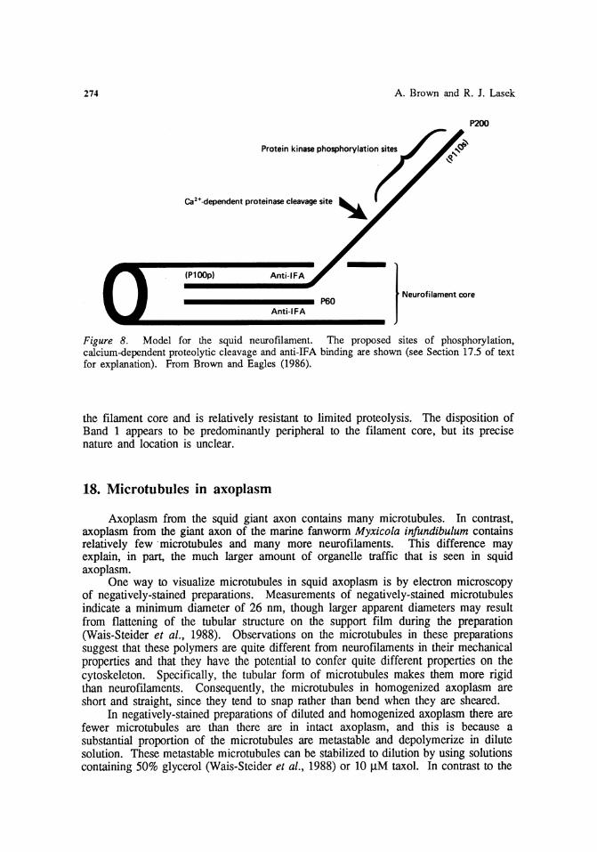
274 A. Brown and R. J. Lasek
P200
Protein kinase phosphorylation sites
ea>+-dependent proteinase cleavage site ~
}-·--(P100p) AntHFA
................. ~0 Anti· IFA
Figure 8. Model for the squid neurofilament. The proposed sites of phosphorylation, calcium-dependent proteolytic cleavage and anti-IF A binding are shown (see Section 17.5 of text for explanation). From Brown and Eagles (1986).
the filament core and is relatively resistant to limited proteolysis. The disposition of Band 1 appears to be predominantly peripheral to the filament core, but its precise nature and location is unclear.
18. Microtubules in axoplasm
Axoplasm from the squid giant axon contains many microtubules. In contrast, axoplasm from the giant axon of the marine fanworm Myxicola infundibulum contains relatively few · microtubules and many more neurofilaments. This difference may explain, in part, the much larger amount of organelle traffic that is seen in squid axoplasm.
One way to visualize microtubules in squid axoplasm is by electron microscopy of negatively-stained preparations. Measurements of negatively-stained microtubules indicate a minimum diameter of 26 nm, though larger apparent diameters may result from flattening of the tubular structure on the support film during the preparation (Wais-Steider et al., 1988). Observations on the microtubules in these preparations suggest that these polymers are quite different from neurofilaments in their mechanical properties and that they have the potential to confer quite different properties on the cytoskeleton. Specifically, the tubular form of microtubules makes them more rigid than neurofilaments. Consequently, the microtubules in homogenized axoplasm are short and straight, since they tend to snap rather than bend when they are sheared.
In negatively-stained preparations of diluted and homogenized axoplasm there are fewer microtubules are than there are in intact axoplasm, and this is because a substantial proportion of the microtubules are metastable and depolymerize in dilute solution. These metastable microtubules can be stabilized to dilution by using solutions containing 50% glycerol (Wais-Steider et al., 1988) or 10 J.1M taxol. In contrast to the
Axonal Cytoskeleton 275
metastable population, some microtubules in axoplasm are very stable and resist depolymerization by a variety of conditions including cold, submillimolar calcium concentrations, and some anti-mitotic drugs (Brady, 1989). The stability and assembly dynamics of microtubules and the other cytoskeletal polymers is discussed in Section 20.
Electron microscopy of thin sections of intact axoplasm shows that the microtubules cluster forming longitudinal bundles that are aligned parallel to the long axis of the axon. Surrounding these microtubules is a fine filamentous matrix that is denser than the surrounding cytoplasm, and is composed of actin microfllaments and other proteins (see Section 19). These microtubules domains are most apparent in longitudinal sections, and they are often associated with vesicles and smooth endoplasmic reticulum.
18.1. Purification of microtubules
Several investigators have purified microtubules from squid tissues for biochemical studies. For example, Sakai and Matsumoto (1978) purified microtubules from squid axoplasm by one cycle of temperature-dependent disassembly and reassembly and studied their properties in vitro. However, it should be noted that some of the microtubules in axoplasm are stable (see Section 20) and may not purify by this protocol. Thus purification by this procedure will select for soluble tubulin and metastable microtubules. For studies that require larger quantities of protein (a single axon contains only about 55 j.lg of tubulin), tubulin can also be purified from the optic lobes of squid brain (see Vale, Schnapp, Reese, and Sheetz, 1985). Squid optic lobes, which form the major mass of the squid brain, provide a good source of tissue for protein purification. The lobes lie immediately behind the squid eyes and can be dissected out in a few minutes. About 0.5 g wet weight of lobe tissue can be obtained from a single squid.
18.2. Video-enhanced contrast light microscopy of axoplasm
The study of microtubules in squid axoplasm has been greatly facilitated by the development of video-enhanced differential interference contrast light microscopy (Allen and Allen, 1983). This technique allows the detection of single microtubules and membranous organelles in living cytoplasm and it has allowed rapid progress in understanding the mechanisms of organelle translocation in axoplasm (Weiss, Meyer, and Langford, 1990). Neurofllaments and microfilaments are below the limits of detection by this technique and can only be visualized by electron microscopy.
Observation of intact axons by differential interference contrast microscopy gives a striking impression of the longitudinal alignment of the microtubules in the axoplasm, but the density of the cytoplasm does not allow the clear resolution of the individual polymers. Single microtubules can only be delineated at the periphery of extruded axoplasm or in dispersed preparations. The lengths of microtubules in squid axoplasm have not been accurately measured, but our observations of many microtubules at the periphery of extruded axoplasm suggest that they are in excess of 50 J.1.ffi in length. In homogenized or gently dispersed preparations, the microtubules are much shorter, which indicates that they are very susceptible to shearing.

276 A. Brown and R. J. Lasek
Weiss et al. (1988) have used video-enhanced differential interference contrast microscopy to examine the length changes of microtubules in extruded axoplasm. Their observations indicate that length changes are a rare event. This is consistent with the existence of stable microtubules in axoplasm (see Section 20) and suggests that most of the microtubules in axoplasm do not display the dynamic instability characteristic of some other cell types, such as actively crawling epithelial cells, in which many microtubules rapidly grow and shrink in length (Mitchison and Kirschner, 1984). This observation underlines one of the many specific differences between the cytoskeletal elements of axons and those of cells, such as fibroblasts, that are capable of mitosis.
18.3. Microtubule-associated proteins in axoplasm
A number of proteins that bind to microtubules have been isolated from squid axoplasm. One of these proteins is kinesin, which is an oligomeric protein with a native relative molecular mass of about 600,000 and is composed of M. 110 - 120,000 and M, 60 - 70,000 polypeptides (Vale, Reese, and Sheetz, 1985). Kinesin has been purified from squid axoplasm on the basis of its affinity for microtubules in the presence of 5'-adenylyl imidodiphosphate (AMP-PNP), which is a non-hydrolyzable analog of ATP (Lasek and Brady, 1985; Vale, Reese, and Sheetz, 1985). The kinesin preparation is sufficient to cause unidirectional A TP-dependent movement of latex beads along microtubules, but additional proteins appear to be required for vesicle translocation in axoplasm. The direction of translocation is towards the plus-ends of the microtubules (Vale, Schnapp, Mitchison, et al., 1985). Studies on microtubules in other axons indicate that microtubules are orientated with their plus-ends away from the cell body (Burton and Paige, 1981; Heidemann et al., 1981). This suggests that kinesin is a component of the cross-bridges that mediate anterograde vesicle transport along microtubules in squid axoplasm (Miller and Lasek, 1985; Vale, 1987).
Subsequent to its identification in squid axoplasm, kinesin has also been identified in many other cells (see Hollenbeck, 1988, for review). Studies on this protein from these sources have indicated that kinesin is an adenosine-5'-triphosphatase (ATPase) and that its ability to hydrolyze ATP is stimulated in the presence of microtubules (i.e., see Kuznetsov and Gelfand, 1986). In the future it is likely that much of the biochemical characterization of kinesin will be done on protein purified from sources other than the squid nervous system because the amount of tissue available from squid is limited. However, squid axoplasm will continue to be important in elucidating the mechanisms and dynamics of axoplasmic transport since there are many features of axoplasm that cannot be reconstituted in cell-free assays, and there are likely to be many more components of the translocating mechanisms that have yet to be identified (Brady et al., 1985).
Just as for the axons of vertebrates, squid axoplasm also contains a number of high-molecular weight microtubule-associated proteins. For example, Sakai and Matsumoto (1978) identified five high molecular weight polypeptides on SDS-polyacrylamide gels that all sedimented with microtubules from extruded axoplasm (Doryteuthis bleekeri) after one cycle of temperature-dependent disassembly and reassembly. One of these proteins is axolinin, which has a M. of 260,000 and is present in large amounts in the cortical axoplasm (see Section 15.3). Another of these proteins is the M, 292,000 protein that has been identified in axoplasm of the squid Loligo pealei by Gilbert and Sloboda (1986). This protein cross-reacts with antibodies to porcine brain MAP-2 and copurifies with microtubules from squid brain optic lobes.

Axonal Cytoskeleton 277
The M. 292,000 protein was identified as an ATP-binding protein using 8-azido-ATP, and Pratt (1986) showed that it associated in an ATP-dependent manner with a sedimentable fraction containing microtubules and vesicles, but it is not clear whether or not it is an ATPase. Pratt (1986) also identified a number of other high-molecular weight proteins that sediment with fractions containing microtubules and vesicles from squid axoplasm, but these proteins were not characterized further.
Because of the current interest in microtubule-based organelle translocation, rapid progress is being made on the identification and characterization of microtubule-associated proteins in squid axoplasm. For further discussion of the role of microtubule-associated proteins in organelle transport, consult Weiss, Meyer, and Langford (1990).
19. Microfilaments in axoplasm
19.1. Microfilarnents are numerous in the inner cytoskeleton
Actin microfilaments are the least studied of the cytoskeletal polymers in axons, though much is known about their structure and function in some other cell types, such as muscle cells, fibroblasts, and red blood cells. Actin comprises about 6% of the protein in the squid giant axon. Analysis of the axoplasm and sheath after extrusion indicates that about 92% of the total actin in the giant nerve fiber is extruded; only 8% remains in the sheath, and a portion of this actin is located in extra-axonal cells. Thus greater than 92% of the actin in the giant axon is present within the inner axoplasm (Path and Lasek, 1988).
Path and Lasek (1988) used quantitative biochemistry together with light and electron microscopy to investigate the distribution and organization of the microfilaments in extruded axoplasm. Electron microscopy of negatively-stained preparations of extruded axoplasm revealed the presence of many 4 - 6 nm filaments that were decorated by myosin subfragment-1 (S-1) with the arrowhead pattern that is characteristic of actin microfilaments. For negative staining, axoplasm from one axon (about 3 - 4 J.l.l) was extruded directly into 200 - 500 J.l.l Buffer X and gently dispersed in a small-volume teflon/glass homogenizer. Negative staining was performed as described in Section 13.6. For S-1 decoration, a drop of dispersed axoplasm was placed on a carbon-coated grid and then replaced with a drop of 0.4 mg/ml S-1 in standard salts solution (50 mM KCl, 5 mM MgC12, 6 mM sodium phosphate, pH 7.2). The S-1 was prepared by chymotryptic digestion of rabbit muscle myosin. In order to measure the microfilament lengths, the neurofilaments and microtubules, which tend to obscure the microfilaments, were solubilized using 0.6 M potassium iodide in which actin filaments are stable. Measurements made in this way showed that axoplasmic microfilaments are short polymers (mean length of 0.5 J.l.m), in marked contrast to the microtubules and neurofilaments which are tens of micrometers in length.
The observation that microfilaments are substantial components of the inner cytoskeleton in the axon is a further illustration of the usefulness of the extrusion paradigm for quantitative determinations of the distribution of axoplasmic elements in the cortical and inner axoplasm. Studies on vertebrate axons using fluorescent staining methods have suggested that axonal microfilaments are principally located in the cortex. This idea became entrenched in the literature (for example, see review by Pachter et

278 A. Brown and R. J. Lasek
al., 1984), and because microfilaments are essential elements in actomyosin-mediated motility, models of axonal motility were constrained by the notion that most and possibly all of the microfilaments and certain associated proteins, such as spectrin (see Section 15.3), were located subjacent to the plasma membrane. Based on this notion, one of the most actively supported models of slow axonal transport explicitly assumed that slow transport operated through force generating mechanisms located subjacent to the plasma membrane (for example, see Levine and Willard, 1981). Although there were some clues that the cortex was not the only the part of the axon where microfilaments were plentiful (Morris and Lasek, 1982; Heriot et al., 1985), it was not until quantitative studies using the extrusion model were developed that the abundance of microfilaments and their associated proteins in the inner axoplasm was established.
19.2. Two classes of microfilaments in the inner cytoskeleton
The state of actin as monomer and polymer has been investigated by Morris and Lasek (1984). Based on studies using pharmacological agents and a kinetic equilibration paradigm that they developed, they proposed that axoplasmic actin exists in three forms: soluble (monomeric), metastable (dilution-labile) microfilaments and stable microfilaments. Their results suggested that 56% of the actin in extruded axoplasm was polymeric; 26% stable and 30% metastable. The soluble actin concentration in axoplasm is about 0.37 mg/ml, which is greater than the critical concentration in vitro, and this suggests that factors in axoplasm, such as actin binding proteins, may sequester monomeric actin in a form that does not readily polymerize. The stability and assembly dynamics of actin microfilaments and the other cytoskeletal polymers in axoplasm are discussed in more detail in Section 20.
Electron microscopy has indicated that the stable and metastable microfilaments differ in their mean length as well as in their stability. Path and Lasek (1988) measured microfilament length in the presence and absence of phalloidin, which is a pharmacological agent that stabilizes actin filaments, and they showed that the stable microfilaments in axoplasm have a mean length of 0.79 J.l.m, and that the metastable microfilaments are shorter, with a mean length of 0.41 J.l.ffi. Thus both morphological and kinetic criteria define two distinct populations of actin filaments in axoplasm. Based on these observations, they proposed that these two populations of microfilaments that differ in their stability may play distinct roles in the dynamic architecture of the inner cytoskeleton in the axon.
19.3. Organization of microfilaments in the inner cytoskeleton
Since phalloidin binds to polymeric actin, but not to monomer, it can be used to localize microfilaments in cells. By staining axoplasm with rhodamine-phalloidin, Path and Lasek (1988) showed that the microfilaments are not distributed uniformly in the inner axoplasm, but are organized in columns that are orientated along the long axis of the axon. Each column is about 1 J.l.m wide and extends for many micrometers through the axoplasm. Microfilaments can also be visualized in intact axoplasm by electron microscopy of thin sections. Axoplasm was stained with S-1 to unambiguously identify the microfilaments, and then fixed using the potassium ferricyanide procedure for optimum preservation (see Section 13.2). Using this procedure it appears that the

Axonal Cytoskeleton 279
microfilaments are concentrated in the vicinity of the microtubules bundles and are often aligned along their long axis. This indicates that the columns of actin may correspond to the microtubule bundles in axoplasm.
A number of experiments on vesicle transport in axoplasm support the localization of actin microfilaments in the vicinity of microtubules. For example, Brady et al. (1984) showed that DNase I and gelsolin both inhibit anterograde and retrograde vesicle transport in extruded axoplasm. Both of these proteins can cause the fragmentation of actin filaments in vitro. Since vesicle transport along microtubules can be reconstituted in vitro, apparently in the absence of actin, it is not likely that microfilaments are involved in force generation for vesicle transport. Rather, these experiments suggest that DNase I and gelsolin inhibit vesicle transport in axoplasm by disrupting the cytoskeletal architecture of the microtubule domains. Another actin-binding protein that interferes with organelle movement in axoplasm is synapsin I, which has been shown to inhibit vesicle transport in extruded axoplasm, and in a phosphorylation-dependent manner (McGuinness et al., 1989). It is thought that the dephosphorylated form of synapsin I can bind vesicles to the microfilament matrix that surrounds microtubules, thereby interfering with their translocation through the axoplasm. These experiments suggest that microfilaments in the inner axoplasm play an important role in the functional architecture of those microtubule domains that are associated with the transport of membranous organelles.
19.4. Myosin from squid brain
With the current interest in microtubule-based organelle transport in axoplasm, little attention has been given to the role of myosin in the squid giant axon. This is particularly surprising given the large number of microfilaments in axoplasm and the fact that neurons were among the first non-muscle cells in which actin and myosin were identified (Berl et al., 1973). See and Metuzals (1976) have purified and characterized myosin from squid brain optic lobes. Analysis of the purified myosin by SDS PAGE revealed a single band that co-migrated with the heavy chain of rabbit muscle myosin, and several minor bands that may have corresponded to myosin light chains. The purified squid brain myosin demonstrated ATPase activity that was stimulated by F-actin from rabbit muscle. In addition, electron microscopy of negatively-stained preparations showed that the squid myosin decorated rabbit muscle F-actin with the characteristic arrowhead pattern, and polymerized to form bipolar myosin filaments at low ionic strength. These observations indicate that if a serious commitment to the study of myosin-mediated mechanisms in the giant axon were made, it could advance our understanding of the biochemistry and organization of intra-axonal dynamics along a path that has been almost completely neglected.
20. Studying the stability of the cytoskeleton
20.1. Assembly dynamics of cytoskeletal polymers in axons
In addition to the opportunities for biochemical and ultrastructural investigations on axoplasm, the squid giant axon offers a special and perhaps unique opportunity for

280 A. Brown and R. J. Lasek
0 MONOMER
(]'-.,. 0 ~-..... ~-.r--"..--.r......,-,-T"""l["--.-,. ~ <l.J /" """"'o METASTABLE POLYMER
Q; 0 v..o ++<r++++OfOfO±»~
o ~" 'tt. ro STABLE POLYMER
Figure 9. Diagram illustrating the three states of cytoskeletal protein in axoplasm. Monomer subunits exchange freely with the ends of the metastable polymer, and the steady-state concentration of metastable polymer is dependent on the monomer concentration. In contrast, monomer can not dissociate from the ends of stable polymers. Reproduced from Lasek (1988).
analyzing the stability and dynamics of the axoplasmic cytoskeletal elements. The axon has three distinct cytoskeletal polymer systems: the neurofilament system, the microtubule system, and the microfilament system. These systems consist of their self-assembling protein subunits (neurofilament proteins, tubulin, and actin) and their respective polymers (neurofilaments, microtubules, and microfilaments). In the axon, these systems operate at a steady state that is apparently fairly constant along the length of the axon between the cell body and axon terminal.
The cytoskeletal polymers themselves can be sub-classified with regard to their assembly dynamics into two distinct populations; metastable polymers and stable polymers (see Fig. 9). Metastable polymers (also termed dynamic or equilibrium polymers) continually exchange protein subunits with the diffusible monomer pool, and their length is a function of the amount of monomer available for polymerization. Metastable polymers are very sensitive to decreases in the concentration of available free monomer, and if the monomer concentration decreases sufficiently, they will disassemble entirely, so that no metastable polymers remain. In contrast with metastable polymers, stable polymers are insensitive to decreases in the concentration of available monomeric subunits.
If extruded axoplasm is immersed in an artificial axoplasm solution, the monomeric proteins are free to diffuse out of the axoplasm, and the concentration of monomer in the axoplasm decreases accordingly. This decreases the availability of monomer for assembly onto the ends of the metastable polymers. Thus the subunits that are continually dissociating from the metastable polymers are not replaced, and the polymers decrease in length. If the volume of the solution is large relative to the volume of the axoplasm then net disassembly will continue until all of the metastable polymers have been extinguished.

Axonal Cytoskeleton
NF1-
NF 220-
-90k
:TUBULIN
-ACTIN
-35k
-14k
t t GHOST SOLUBLE
281
Figure 10. Stable and soluble proteins in axoplasm. Axoplasm was extracted in Buffer P for 2 - 3 hours as described in the text and then analyzed by SDS PAGE and stained with Coomassie Blue. The polypeptide composition of the extracted axoplasm (GHOST) and of the extraction medium (SOLUBLE) are shown. The three neurofilament proteins are labelled on the left, and a number of other proteins in axoplasm are labelled on the right. NF-1 , NF220 and NF60 correspond to Band 1, P200 and P60 in the nomenclature used in Section 17. Reproduced from Morris and Lasek (1982).
Morris and Lasek (1982, 1984) used this experimental approach to quantitatively analyze the elution of proteins from isolated axoplasm. For these studies a solution (Buffer P) that closely simulates the solution conditions inside the axon was developed to preserve the types of protein associations that occur in axoplasm in situ (see Section 10.1). Because of the large diameter of the axoplasm, the egress of diffusible proteins occurs over a time course of minutes and can therefore be measured readily. In this Section we describe these studies and show how analysis of the extent and kinetics of elution can be used to infer the amount of cytoskeletal protein as monomer, metastable polymer, and stable polymer in axons. These methods could also be applied to the study of the association of other cytoplasmic proteins of the cytoskeleton, and this is an opportunity that has yet to be exploited.
20.2. The axoplasmic ghost
When axoplasm is extruded into a solution that simulates the solution conditions of the axon, the loss of soluble proteins (including free cytoskeletal subunits) by diffusion occurs rapidly, and the loss due to the disassembly of dilution-labile cytoskeletal polymers occurs more slowly. Af~~r a few hours the axoplasm approaches equilibrium with the surrounding medium, a11d what remains is comprised of the stable

282 A. Brown and R. J. Lasek
Table VI. The protein composition of axoplasm and the axoplasmic ghost
Axoplasm Ghost
Tubulin 22% 12% Neurofilament 13% 54% Actin 6% 5% 35kD protein 6% n.d. 90kD protein 1% n.d.
Other proteins 52% 29%
The values are percentages of the total protein in axoplasm and in the axoplasmic ghost. Not detected (n.d.). Data from Morris and Lasek (1982, 1984).
cytoskeletal polymers of axoplasm, their associated proteins, and the axoplasmic membranous elements: mitochondria, vesicles, and endoplasmic reticulum. Though many of the axoplasmic proteins have been lost, the extracted axoplasm, or axoplasmic ghost, retains its original cylindrical shape and mechanical integrity.
In order to determine the proportions of stable polymer for the cytoskeletal proteins in axoplasm, Morris and Lasek have quantitatively analyzed the protein composition of the extracted axoplasm and extraction medium (Buffer P). The amount of each protein was determined by densitometry of 50S-polyacrylamide gels using bovine serum albumin as a standard. The volume of .the extraction medium determines the extent of dilution of the axoplasmic proteins. Thus, in order to achieve full disassembly of metastable polymers, the solution volume must be large relative to the axoplasm volume. In a typical experiment, axoplasm from one axon (about 5 Jl}) was extruded into 1 or 2 ml of Buffer P, which represents a 200 to 400-fold dilution.
Fig. 10 shows the polypeptide composition of the extracted and stable proteins after the axoplasm had equilibrated with the Buffer P, and Table VI shows the relative proportions of a number of the major proteins in axoplasm and in the axoplasmic ghost About 80% of the axoplasmic proteins are soluble in Buffer P and essentially all of these soluble proteins are extracted within 2 hours. The remaining 20% of the protein constitutes the axoplasmic ghost. All three neurofilament polypeptides remain associated with this stable axonal cytoskeleton, which indicates that the neurofilaments are stable to dilution. In contrast, only 17% of the tubulin and 26% of the actin are present as stable polymer (see Section 20.3), and many other proteins in axoplasm, such as the M. 35,000 protein, are completely lost from the axoplasm. Because of the stability of the neurofilaments, the neurofilament proteins are the major component of the axoplasmic ghost, and these polypeptides constitute 54% of the total ghost protein. Together, neurofilament protein, tubulin and actin, constitute over 70% of the ghost protein. Electron microscopy of the axoplasmic ghost indicates that neurofilaments are the major cytoskeletal structures; microtubules are much fewer than in unextracted axoplasm and much of the filamentous matrix that surrounds them also appears to have been extracted. These observations support the conclusion that the cytoskeletal polymers are indeed the morphological correlate of the stable cytoskeletal proteins.
A number of other cytomatrix proteins remain in the axoplasmic ghost, and this may indicate that they have a high affinity for the stable cytoskeletal polymers. For

Axonal Cytoskeleton 283
example, very little spectrin (see Section 15.3) is extracted in Buffer P. In fact, we have noted that spectrin often copurifies with neurofilaments (for example, see Brown and Eagles, 1986), and this suggests that it may bind tightly to these cytoskeletal elements. Another protein that remains in the axoplasmic ghqst is a protein with an approximate M, of 70,000, which we have also consistently observed in squid neurofilament preparations. This protein binds the antibody Anti-IFA (Brown and Eagles, 1986) and therefore may be related to internexin, an intermediate filament-associated protein that is present in intermediate filament preparations from a wide variety of sources (Pruss et al., 1981; Napolitano et al., 1985; Pachter and Liem, 1985).
20.3. Monomer-polymer equilibria in axoplasm
We have shown above that a large proportion of the actin and tubulin in axoplasm is extracted in dilute solution. This extracted protein includes both monomer and metastable polymer. Thus the amount of monomer and metastable polymer in axoplasm cannot be determined directly. To determine the concentration of actin and tubulin as monomer and metastable polymer in axoplasm, Morris and Lasek (1984) took two approaches. In both approaches, densitometry of SDS-polyacrylamide gels was used to quantify the amount of protein in the same way as for the studies described in Section 20.2.
One method, the kinetic equilibration paradigm (KEP), employs the basic principles of diffusion to distinguish freely diffusible monomer from proteins that are present in the form of polymer. In this approach axoplasm is extruded into a large volume (0.5 - 1 ml) of artificial axoplasm solution (Buffer P) and the rate at which proteins elute from the cylinder of axoplasm is measured by analyzing the amount of protein in the bathing medium at different times. The KEP analysis can be applied to a single piece of axoplasm (the single axon KEP) by transferring the axoplasm through a series of baths, or alternatively separate pieces of axoplasm can be used for each time-point determination. The latter method (the ratio KEP) is preferable because repeated transfer between solutions makes the axoplasm more fragile and small fragments of axoplasm can break off and remain in the bath. For the ratio KEP analysis, axoplasm is extruded into 1 ml of Buffer P, extracted for a measured length of time,. and then removed from the bath with forceps. The axoplasm and bath are analyzed by SDS PAGE and the amount of protein is quantified by densitometry of the polyacrylamide gel. For each extraction time the proportion of protein extracted is calculated and the results for a range of times are used to plot the elution kinetics.
Morris and Lasek (1984) found that the rate of elution of actin and tubulin from the axoplasm was not monophasic, but that it could be modelled as the sum of two exponential components. One component elutes at the rates predicted by diffusion theory for actin and tubulin monomer. 95% of this component elutes from the axoplasm within the first 10 minutes. The other component elutes more slowly and corresponds to the elution of monomer from the pool of metastable polymers. This rate of elution is limited by the rates of disassembly for these polymers, which is slower than the rate of diffusion from the axoplasm. 95% of the actin and tubulin in this component elutes within two hours. Quantification of the amount of protein in these two rate components yields the total amounts of protein as monomer and metastable polymer in the axoplasm.

284 A. Brown and R. J. Lasek
Table VII. Cytoskeletal proteins as monomer and polymer in axoplasm.
Stable polymer Metastable polymer Monomer
Actin
26% 30% 44%
Tubulin
17% 55% 28%
N euroji.lament
>95%
Values given as percentage of the total amount of that protein present in axoplasm. Data from Morris and Lasek (1984 ).
The other method is pharmacological and employs taxol and phalloidin to stabilize the microtubules and microfilaments, respectively. Taxol and phalloidin are stored at room temperature as a 10 mM stock solution in dimethylsulfoxide (DMSO) and diluted in Buffer P to give a final drug concentration of 10 J.lM in 0.1% DMSO. Axoplasm is extruded into 1 ml Buffer P either with or without the stabilizing drug and then extracted to equilibrium (2 or 3 hours). Analysis of the extracted axoplasm and bathing medium by SDS PAGE and densitometry allows comparison of the amount of actin or tubulin that elutes in the presence and absence of the appropriate drug. The difference between the amount of that protein in extracted axoplasm in the presence and absence of the stabilizing drug represents the amount of metastable polymer in the axon.
Morris and Lasek (1984) found that the results of the kinetic and pharmacological methods showed good agreement, and the mean values are shown in Table VII. These results indicate that 56% of the actin in axoplasm and 72% of the tubulin in axoplasm are present as polymer. Since axoplasm contains 25 mg/ml protein (see Section 9), we can calculate the actual monomer and polymer concentrations from the known proportions of actin and tubulin in axoplasm (shown in Table VI). These calculations indicate that the monomer concentration in axoplasm is about 0.6 mg/ml for actin and about 1.6 mg/ml for tubulin. These concentrations are greater than the critical concentrations for assembly of these proteins in vitro and this suggests that factors are present in the axoplasm that inhibit the polymerization of actin and tubulin, or that elevate the critical concentration for their assembly. No detectable neurofilament protein elutes in these experiments and this supports the proposal that essentially all of the neurofilament protein in axoplasm is stably polymerized.
21. Mechanical studies on axoplasm
21.1. Axoplasm has mechanical integrity
The ability to isolate intact axoplasm from the squid giant axon provides one of the rare opportunities in nature for the biologist to see and handle pure cytoplasm. Biologists who have had this opportunity tend to be struck by the mechanical integrity of the axoplasm when they are reminded that it is composed mostly of water and contains only 2.5% protein by weight (see Section 9). This mechanical integrity is due

Axonal Cytoskeleton 285
to the long polymers of the cytoskeleton, and treatments that disrupt the cytoskeletal polymers, such as proteolytic degradation or chaotropic ions, cause the axoplasm to liquify (see Section 11). The cytoskeletal polymers therefore confer solid-like properties on the axoplasm so that it maintains its cylindrical fonn after isolation, even when it is handled, albeit gingerly, with forceps.
21.2. Axoplasm is anisotropic
The axoplasmic cytoskeleton appears homogeneous to the naked eye, but light and electron microscopy indicate that the cytoskeletal polymers are highly aligned along the long axis of the axon (see Section 14.1). This organization of the cytoskeleton makes axoplasm highly anisotropic. For example, the longitudinal alignment of the polymers confers distinct mechanical properties on the radial and longitudinal dimensions of axoplasm.
A graphic illustration of this structural and mechanical anisotropy of axoplasm was described by Chambers and Kao (1952) based on their observations on the microinjection of substances into axons. They showed that bubbles of air or oil injected into the axoplasm assumed an ovoid shape with their long axes orientated parallel to the long axis of the axon. Injection of millimolar concentrations of calcium into the axons, which is now known to activate the calcium-dependent protease (see Sections 11.2 and 17.4 ), caused the axoplasm to liquify, and the bubbles then assumed a spherical shape. These observations indicate that axoplasm has a mechanical anisotropy. The oil and air tend to form bubbles in an aqueous environment because of the surface tension forces of the water. Apparently the axoplasm offers less resistance to these surface tension forces in its longitudinal dimension than in its radial dimension. With these observations, Chambers and Kao were able to infer a linear ultrastructure in axoplasm at a time before electron microscopy had revealed the existence of cytoskeletal polymers. We now know that this linear ultrastructure is due to the cytoskeletal polymers that are orientated along the long axis of the axon. These observations therefore indicate that axoplasm has different mechanical properties in the dimension parallel to the cytoskeletal polymers than perpendicular to the polymers.
21.3. The macroscopic mechanical properties of axoplasm
Two approaches have been taken for the quantitative analysis of the mechanical properties of axoplasm from the squid giant axon. In one approach, Rubinson and Baker (1979) measured the resistance of axoplasm to flow through small-bore cellulose acetate capillary tubing. They showed that the axoplasm behaved in a viscous manner and was characterized by a yield-strength, which is a force below which the axoplasm did not flow. However, it is likely that the methods used in these measurements, which involved high rates of shear, resulted in considerable disruption of the organization of the cytoskeleton. Thus it is likely that many factors contributed to the resistance to flow, including frictional interactions with the capillary wall and shearing of entangled polymers.
Using a different approach, Sato et al. (1984) implanted single magnetic spheres (100 11m in diameter) into axons and then measured their movement in a magnetic field. Their analyses indicated that axoplasm had viscoelastic properties. In other

286
B
1
2
A. Brown and R. J. Lasek
Figure 11. The apparatus for stretching axoplasm. A. Photograph of the apparatus. B. Schematic diagram illustrating the essential features of the apparatus. In the upper drawing (1) the apparatus is shown with the chamber lowered to reveal the two angled clips. To stretch axoplasm, the axoplasm is immersed in liquid in the chamber and then mounted between the clips. The lower drawing (2) shows a close-up view of a piece of axoplasm being stretched in the chamber. The right clip is attached to a the force transducer (F7). The left clip is attached via a metal rod to the drive carriage that is driven by a stepper motor (D). The movement of the left clip is measured by a linear position transducer (LPT) that is attached to the drive carriage by a retractable sprung cable. The straight arrows indicate the direction of movement during stretch. The resistance of the axoplasm to stretch is measured by the extent of displacement (curved arrow) of the vertical tongue of the force transducer, to which the right clip is attached. The axoplasm is
observed and photographed from above with a binocular microscope (M).
words, the magnetic spheres experienced both viscous and elastic resistances to their movement through the axoplasm. Since these studies used intact axons and very low rates of shear, it is likely that they involved less disruption of the cytoskeletal organization than the studies of Rubinson and Baker (1979). However, the large size of the magnetic spheres caused visible disruption of the axoplasm and entanglement of the cytoskeletal polymers, and it is likely that polymer shearing was also a contributing factor to the viscous resistance that was measured in these studies. Nevertheless, by changing the orientation of the axon relative to the poles of the magnet, it was possible to see differences in the resistance to movement in the radial and longitudinal dimensions. These analyses confirmed observations of Chambers and Kao (1952) that the axoplasm is mechanically anisotropic. Specifically, they showed that there was a greater elastic resistance to movement in the radial dimension of the axon (perpendicular to the long axis of the cytoskeletal polymers) than in the longitudinal dimension of the axon (parallel to the cytoskeletal polymers).

Axonal Cytoskeleton 287
The anisotropy of axoplasm, and specifically its mechanical properties in the longitudinal dimension, are likely to be important in many aspects of the mechanics of axonal motility. For example, studies on squid axoplasm have shown that there is a continuous and bustling traffic of membranous vesicles along the microtubules in the axon, and these constitute the fast component of axoplasmic transport (Weiss, Meyer, and Langford, 1990). In order for the vesicles to move through the axoplasm, they have to overcome the viscous and elastic resistances that they encounter as they move past adjacent cytoplasmic structures along the long axis of the axon (Lasek and Miller, 1986).
In order to more effectively study the mechanical properties of axoplasm in its longitudinal dimension, we have developed a novel approach in which axoplasm is stretched along its long axis at slow rates (George and Lasek, 1989; Brown and Lasek, 1989). By measuring the resistance of the axoplasm to stretch, the mechanical properties of the axoplasm can be investigated. This method does not involve disruption of the anisotropic organization of the axoplasm, and the measurements therefore reflect the mechanical properties along the long axis of the cytoskeletal polymers. In this Section we describe the apparatus and experimental design, and indicate how these studies may aid our understanding of the dynamics of the axonal cytoskeleton.
21.4. The stretch apparatus
Three essential requirements for the stretch measurements are a pulling device, a force transducer, and a means to amplify and record the transducer output. Fig. 11 shows a photograph and a schematic diagram of the stretch apparatus. A segment of axoplasm is suspended between two metal clips (Schwartz temporary clip, Roboz Surgical Instrument Company) in a clear plexiglass (perspex) chamber. The dimensions of the chamber are 60 mm long, 12 mm wide and 10 mm deep. The plexiglass is 2 mm thick. Inside the chamber is a block of plexiglass cut at an angle of 45 degrees onto which a thin mirror is mounted. This provides an additional image of the axoplasm from the side when the axoplasm is viewed from above. The mirror is close enough to the axoplasm that the two images appear in the same field of view, though at slightly different depths of focus. We use an aluminum first-surface mirror (12 x 49 x 0.8 mm) which was purchased from Edmund Scientific and cut to size using a glass knife. The mirror can be glued to the plexiglass mount or, less permanently, it can be secured with an inert silicone lubricant (Dow Coming Ill, Dow Coming Corporation). The chamber is mounted on a micromanipulator to allow adjustment of its position relative to the clips. Axoplasm is transparent but can be seen in dark-field because the long cytoskeletal polymers scatter light. We illuminate the axoplasm from the back of the chamber using fiber optic light guides and view the chamber from above using a low power binocular microscope. The base of the chamber is made of black plexiglass to provide a black background for the dark-field image. The internal volume of the chamber with the mirror block in place is about 4.5 ml, but 2 ml of liquid is sufficient to cover the axoplasm.
The force is measured using a Model FT03 force displacement transducer (Grass Instrument Company) without the isotonic springs. The transducer gives a voltage output that is proportional to the displacement of the transducer tongue. The transducer is mounted vertically to the steel base of the stretch apparatus. The position of the mount and the height of the transducer are adjustable to allow alignment with the

288 A. Brown and R. J. Lasek
pulling device. The clip is mounted to the transducer in such a way that the axis of the axoplasm is in line with the shaft of the transducer so that no torque is applied to the transducer tongue. To prevent damage to the transducer when the axoplasm is being attached to the clips, we designed a small clamp consisting of two set screws that attaches to the housing of the transducer and allows the tongue to be temporarily secured. The maximum sensitivity of the FT03 transducer is about 1 mg force. Transducers of much higher sensitivity can be obtained (for example, from the Cambridge Instrument Company), but we still use the FT03 because the metal clip is easily attached to the transducer tongue, and its construction is rugged enough to withstand the handling when the axoplasm is mounted in the clips. The FT03 is also comparatively low cost.
The pulling device is a converted Sage syringe pump (Model 352, Orion Instrument Company). In the version of the stretch apparatus shown in Fig. 11, the stepper motor and drive carriage have been removed from the pump control and mounted on the raised platform of the stretch apparatus. The stepper motor is connected to the motor control panel via a cable. This allows mechanical isolation of the stretch apparatus from the operating unit. The Sage syringe pump controller that we use allows a broad range of pull rates from 2 f...Lm/sec to greater than 1 mm/sec, and different ranges of rates can be obtained by substituting motors with different gear ratios. A switch on the control panel allows the direction of the motor to be reversed. Stepper motors are preferable to DC motors for this application because they have a long life, are variable over a wider range of rates, and are highly reproducible independent of the load. A gear wheel on the shaft of the motor engages with a toothed bar in the drive carriage, converting the rotational motion of the motor into a linear movement of the drive carriage along the raised platform. The clip that holds the left end of the axoplasm is attached to the drive carriage via a metal rod. The position and movement of the puller rod are monitored with a linear position transducer (LPT, Unimeasure, Inc.) that is attached to the drive carriage via a retractable sprung cable. The LPT gives a voltage output that is proportional to the extension of the cable.
The design and placement of the apparatus must allow operation of the pump controller, microscope and camera without transmitting vibration to the transducer. This is achieved by placing the camera and pump controls and the microscope base on a separate adjacent table or shelf. The microscope is mounted on a stand that swings over the apparatus to allow free access to the chamber. More recently, we have also enclosed the apparatus in a transparent plexiglass box in order to protect the transducer from air currents and to minimize temperature changes over time. Recently, we have modified the stretch apparatus by removing the rack-and-pinion drive carriage and replacing it with a worm-drive (Stoelting Company, 5 mm pitch thread), which allows a tighter coupling of the motor to the puller-rod, with less lateral play. In this version of the stretch apparatus, the motor shaft is attached directly to the worm-drive via a close-fitting chuck. The puller rod that holds the clip at the left end of the axoplasm now attaches to the translating portion of the worm-drive. For our experiments at the Marine Biological Laboratory in Woods Hole, we have recorded the voltage output of the FT03 force transducer and linear position transducer on a two-channel strip chart recorder (Grass Instrument Company Model 79 Polygraph equipped with a Model 7P1 low level DC pre-amplifier and Model 7DA DC pen driver amplifier on each channel).
It is important for these studies to isolate axoplasm as a continuous unbroken length and to minimize any distortion of the organization of the cytoskeletal polymers.

Axonal Cytoskeleton 289
Figure 12. Photograph of a piece of axoplasm suspended in liquid between the clips of the stretch apparatus. The length of the axoplasm between the clips is about 4.5 mm and its diameter is about 0.4 mm. The upper image is of the axoplasm viewed from above and the lower image is a reflection of the same axoplasm from the side. The two images can be seen and photographed in the same field of view of the microscope.
In our stretch experiments so far we have used axoplasm isolated by the extrusion method, but the slitting method may be preferable since it allows axoplasm to be isolated without applying tension or compression to the cytoskeleton (see Section 6.2).
To stretch axoplasm, the isolated axoplasm is immediately transferred into 2 ml of liquid in the chamber. In our experiments we have stretched the axoplasm in paraffin oil so as to maintain a constant volume and prevent the extraction of diffusible proteins that occurs in aqueous solution (see Section 20). A minimum axoplasm length of about 10 mm is required to successfully mount the axoplasm in the clips, but longer lengths are preferable. The axoplasm is first mounted in the right clip, which is attached to the force transducer. To mount the axoplasm, the clip is held open with a pair of hemostatic forceps and the axoplasm is raised in between the clip blades using fine forceps. The clip blades are then allowed to close onto the axoplasm. The inside of the clip blades can be slightly roughened with abrasive paper to improve the grip. Then the left clip, which is auached to the puller rod, is advanced to a distance of 4 mm from the right clip and the left end of the axoplasm is also attached. Angled clips are used so that the tips are not obscured when viewed from above. The axoplasm can be photographed during the experiment through a trinocular attachment to the microscope. A photograph of a piece of axoplasm between the clips is shown in Fig. 12.

290
4.2 mm ,.----.
20 l c >--o
(j) 10
~ 0 0 '>-
pull
0 5
8.7 mm
~----------------~~ I
A. Brown and R. J. Lasek
8.0 mm Figure 13. A typical stretch profile for axoplasm. This segment of axoplasm was extruded onto parafilm and then stretched in paraffin oil at a rate of 17.2 J.Lm/sec. The stretch profile represents the
reverse voltage output from the force transducer that is attached to one end of the axoplasm.
10 15
time (min)
20 The movement of the puller, which is attached to the other end of the axoplasm. is indicated below
the trace. The small arrows on the stretch profile indicate the points at which the puller motor was started and stopped. The units on the ordinate are force in dynes and the units on the abscissa are time in minutes. The axoplasm was stretched for about 4 minutes (pull). When the force reached a plateau, the motor was switched off and the force was allowed to decay at constant extension. After about 12 minutes, the puller was reversed (reverse) at the same rate in order to remove the remaining elastic tension in the axoplasm. The lengths are shown for the axoplasm before the stretch (4.2 mm), at the maximum extent of the stretch (8.7 mm), and at its new resting length after the reversing the puller (8.0 mm).
21.5. Stretch analysis
Fig. 13 shows a typical stretch profile for axoplasm. The output from the force transducer and the movement of the puller are shown. The puller was off at the beginning of this trace, and this resting force is taken to be zero. About 40 seconds into the trace, the puller was switched on, and the axoplasm was stretched at a constant rate of 17.2 J.lm/sec. The force rose slowly at first and then more rapidly. As the stretch continued, the rate of rise of force then began to decrease until it reached a constant value (or plateau) of about 12 dynes (1 dyne = w-s Newtons) of force. When the force reached this plateau value, the length of the axoplasm was 8.7 mm, which was greater than twice the original length. In one batch of thirteen stretch experiments, we observed plateau forces ranging from 5 to 25 dynes.
As the axoplasm is stretched it becomes longer and narrower in diameter. Because the axoplasm is in oil, which is immiscible with water, the overall volume of the axoplasm remains constant. If we continue to stretch the axoplasm past the point at which plateau is reached, the force very gradually decreases as the axoplasm becomes thinner and thinner. Our observations of the axoplasm through the microscope during stretching indicate that, in practice, certain regions thin more than others. This indicates that the axoplasm is not uniform along its length. This could be caused by slight variations in the diameter of the axoplasm along its length and also by local weakening of the axoplasm caused by local compression or tension during its isolation.

Axonal Cytoskeleton 291
Measurements of the axoplasm using periodic carbon-dust markings indicate that all regions of the axoplasm elongate, but that the narrow regions undergo the greatest elongation and the greatest thinning. Since the force produced by the stretch is maintained along the whole length of the axoplasm, the narrower regions receive proportionally more force per unit cross-sectional area than thicker regions, and this probably explains why they undergo the greatest thinning. Eventually the thin regions become so narrow that the axoplasm breaks. This may arise if insufficient polymer overlap remains to maintain the continuity of the axoplasm, or if the force borne by the small number of polymers in the thin region exceeds their breaking strength, causing them to snap.
In the stretch analysis in Fig. 13, the axoplasm was stretched until a constant plateau force had been reached, and then the motor was switched off and the axoplasm was held at its new extended length of 8.7 mm. As soon as we stopped stretching the axoplasm, the force rapidly decayed. The decay is most rapid initially and becomes slower with time. This time-dependent decay in force at constant length has a characteristic and very reproducible shape that can be modelled as a complex exponential function. If the axoplasm is left for long enough (many hours), the force can eventually decay completely, whereupon the axoplasm is seen to sag between the clamps.
In practice we generally allow the axoplasm to relax for long enough to clearly establish the kinetics of the decay in force, and then we advance the motor in reverse at the same rate until the remaining tension in the axoplasm has been removed. Baseline drift during the experiment sometimes makes it difficult to judge the point of zero force, but we have found that a reliable indicator is to look for the point at which the axoplasm begins to sag between the clamps. At this point the axoplasm is no longer under tension and its length represents a resting length. In fact, we have found that the axoplasm routinely increased about two-fold in resting length during such stretch-hold-reverse cycles. We have analyzed many pieces of axoplasm in this way over two consecutive summers at Wood's Hole and we have found that the shape of this profile is highly reproducible. In a few cases, we have been able to repeat this stretch-hold-reverse cycle up to three times before the axoplasm finally thins to breaking point. Each time the plateau force is lower, but the general form of the stretch profile is the same. The decrement in the plateau force is apparently due to the thinning of the axoplasm in successive stretches.
21.6. Interpretation of the stretch profile
The general form of the stretch profile that we have described is immediately recognizable to mechanical engineers and materials scientists as characteristic behavior of a viscoelastic substance. One of the best studied viscoelastic tissues in biology is connective tissue (for example, see Silver, 1987). One example is tendon, which is a highly anisotropic tissue that is composed of bundles of collagen polymers which are aligned longitudinally along the tension-bearing axis (Kastelic and Baer, 1980). The mechanical behavior of tendon under stretch has been extensively studied. The mechanical response to stretch can be analyzed into a time-independent (instantaneous) elastic response and a time-dependent viscous response.
The rising phase of the response of tendon to stretch has a similar general form as that seen for axoplasm (see Fig. 13), and is characterized by four regions (Baer et a/., 1986). Analyses of tendon during stretch have allowed determination of the

292 A. Brown and R. J. Lasek
structural basis for this mechanical behavior. The initial part of the curve is called the toe region and it corresponds to an elastic straightening of the collagen fibrils. If the tendon is stretched further, the force rises markedly and enters a linear region which corresponds to a reversible extension of the collagen fibrils themselves. The slope of this region corresponds to the modulus of elasticity for the collagen fibrils. With further stretch, the force enters a yield region in which the force tends towards a plateau. In this region, the force is high enough to cause slippage of the collagen fibrils relative to each other, and this results in an irreversible extension of the tendon. It the force on the tendon is now released, it is found to have a new and longer resting length. By correlating the organizational changes in the tendon with the mechanical behavior, these studies indicate that the structural correlate of elasticity in the tendon is straightening and extension of the collagen fibrils, and the structural correlate of viscous flow in tendon is the slippage of the collagen fibers relative to each other.
However, this mechanical behavior differs from that of axoplasm in a number of important respects. Firstly, the forces required to deform axoplasm are very low compared to those for tendon collagen. For example, a typical piece of axoplasm plateaus at about 20 mg force, while to deform tendon to the same extent would require kilograms of force for the equivalent cross-sectional area of tissue (Baer et al., 1986; Silver, 1987). A second major difference is that tendon is highly elastic and the rising phase of the force is characterized by a linear elastic region that represents a reversible elongation of the collagen fibrils. In contrast, we have found that no phase of the extension of axoplasm is totally elastic. For example, if axoplasm is held at even small extensions of its original length, a time-dependent decay in force is seen similar to that in Fig. 13. Furthermore, we noted earlier that if axoplasm is stretched to plateau and then held at this length, the force can eventually decay to zero. This indicates that the resistance to flow in axoplasm is low and chiefly limited by its time-dependence. Thus, if axoplasm is stretched and then held at this new length for sufficient time, substantial increases in the resting length of the axoplasm are observed. For example, the two-fold increase in resting length of the axoplasm in Fig. 13 (after about 12 minutes of relaxation) corresponded to greater than 90% of the maximum extent of stretch.
21.7. A mechanical model
These observations indicate that the mechanical behavior of axoplasm in its longitudinal dimension can be considered to be a combination of elasticity and time-dependent viscous flow. To emphasize the dominant property of viscous flow, we refer to axoplasm in its longitudinal dimension as elastoviscous rather than viscoelastic. The behavior of axoplasm can be explained by the following mechanical model in which both elastic and viscous forces contribute to the stretch profile: as the axoplasm is stretched, instantaneous extension of elastic components results in an increase in resistive force and this is accompanied simultaneously by a slower time-dependent relaxation due to viscous flow.
In our experiments, we have found that the mechanical behavior of axoplasm under tension depends greatly on the rate of stretch deformation. The above mechanical model, in which instantaneous elastic extension and time-dependent viscous flow both contribute simultaneously to the stretch profile, can explain these observations. Specifically, since the viscous component of the deformation is time-dependent, rapid stretches are essentially elastic and the axoplasm elongates until

Axonal Cytoskeleton 293
the force is sufficient to cause it to break, but if the axoplasm is stretched slowly it can flow to attain a new resting length. This behavior is characteristic of viscoelastic materials in geneml. A common example is putty or plasticine, which extends to great lengths if stretched slowly but snaps if stretched rapidly. We found that the appropriate rate of stretch for axoplasm must be determined empirically. If axoplasm is stretched very slowly (rates less than I Jlm/sec), substantial increases in resting length can be achieved, but the force is too low to measure. On the other hand, if axoplasm is stretched too rapidly then the force rises rapidly and the axoplasm breaks. In our investigations on axoplasm, we have chosen rates of stretch that allow us to measure both the viscous and elastic components. We have found that stretch rates of about 20 Jlm/sec produce a measurable force without causing the axoplasm to break.
In the plateau region, the force generated by the extension of the elastic components is sufficient to balance the rate of stretch with the rate of viscous flow, and thus the plateau represents a purely viscous deformation with the elastic elements maintained at a constant extension. When stretching is stopped in the plateau phase, and the axoplasm held at its new length, viscous flow continues as the elastic components gradually return to their original resting length. If we wait long enough, the force can eventually decay completely, whereupon the axoplasm sags between the clamps. At this point, the tension generated by the stretch has been completely dissipated by viscous flow, and the axoplasm now has a new resting length. Alternatively, the remaining elastic tension can be dissipated more rapidly by reversing the motor, but the longer the force is allowed to decay, the greater the extent of viscous flow and thus the longer the final resting length.
21.8. The structural basis for elasticity and flow
What is the ultrastructural basis for elasticity and flow in axoplasm in these experiments? Based on the analogy with the mechanics of viscoelastic biological materials such as tendon, and on our knowledge of the architecture of axoplasm, we propose that the structuml correlate of flow in axoplasm under longitudinal tension is the sliding of axonal polymers relative to each other, and that the viscous resistance to flow represents the frictional interactions of the polymers with other axoplasmic elements as they slide through the axoplasm (George and Lasek, 1986). By similar analogy, the weak elasticity in axoplasm may arise from a spring-like straightening or elongation of the cytoskeletal polymers during the stretch. Since the force required to stretch axoplasm is many orders of magnitude less than that required to stretch collagen fibrils (see Section 21.6), the contribution of stretching the polymers themselves to the elasticity is likely to be negligible and the major contribution will come from the configurational elasticity of the polymers. For example, the neurofilaments and microtubules are oriented helically along the axon (see Section 14.2), and stretching axoplasm will tend to straighten the polymers, causing an elastic resistance. This elastic effect is particularly dramatic for axoplasm from the giant axon of the marine fanworm Myxicola infundibulum, which has a pronounced helical organization. In addition, the polymers in extruded axoplasm are not perfectly straight, and elasticity may also arise from the tendency of the polymers to return to their slightly wavy configuration.

294 A. Brown and R. J. Lasek
21.9. Polymer sliding in axons
Our measurements using the stretch apparatus indicate that axoplasm readily flows when stretched along the long axis of the cytoskeletal polymers at low rates. This suggests that the lateral interactions between the cytoskeletal polymers in axoplasm may be relatively weak. Support for this has come from observations on the transport of membranous vesicles in axoplasm (fast axoplasmic transport) which indicate that the vesicles experience a similar resistance to movement in axoplasm as they do to movement in vitro along isolated microtubules (Lasek and Miller, 1985).
These considerations are of interest with respect to the mechanism of slow axoplasmic transport in axoplasm. Studies on axons in other organisms indicate that slow transport represents the movement of cytoskeletal and cytomatrix proteins from their site of synthesis in the neuronal cell body along the axon towards the terminals (Lasek et al., 1984). In one model of this process, Lasek (1986) proposed that the cytoskeletal polymers are the vehicle for slow axoplasmic transport, and that the mechanism of movement is the sliding translocation of the polymers along the long axis of the axon. In this model, transported proteins move as components of, or in association with, these cytoskeletal elements.
For cytoskeletal polymers to slide in axons they must be relatively free to move so that the forces applied to each polymer can translocate them through the axon. If the plateau force in our stretch experiments represents the force required to induce polymer sliding in axoplasm (see Section 21.8), then we can estimate the force required to slide an individual polymer in axoplasm. We will consider only the long cytoskeletal polymers (neurofilaments and microtubules) because it is the overlap of these long elements that is mainly responsible for the longitudinal continuity and structural integrity of axoplasm, and thus for the mechanical properties of axoplasm in the longitudinal dimension. In one batch of thirteen separate stretch experiments, the mean plateau force was about 20 dynes, which corresponded to about 50 dynes/mm2
cross-sectional area per mm length of axoplasm. To calculate the force required to slide a single polymer in axoplasm, we need to know exactly how many sliding interactions there are when axoplasm is stretched. At present we cannot determine this directly, but we can make an approximation. We (Brown and Lasek, 1989) know from electron microscopy that the density of neurofilaments and microtubules in axoplasm is about 50/j.tm2 (cross-sectional area). Thus there are about 5 x 10⁷ neurofilaments and microtubules per mm2 in a cross-section of axoplasm. If we assume that all these polymers in the axoplasm slide relative to each other, then we can calculate that the force required to slide a 1 mm length of polymer in axoplasm would be about 1 x IQ-6
dynes, which is equal to 1 x I0-11 Newtons, or 10 pN (pico Newtons). The force generated by a single dynein cross-bridge during microtubule sliding
in cilia is about 1 pN (Kamimura and Takahashi, 1981), and the force generated by a single myosin head during the sliding of actin and myosin filaments in muscle contraction has been estimated to be about 5 pN (Oplatka, 1972). Thus, based on the above assumptions, the force necessary to slide cytoskeletal polymers in axoplasm is low enough to be generated by a small number of myosin or dynein-like cross-bridges per millimeter length of polymer (George and Lasek, 1986). Such calculations are very approximate but they are important in order to illustrate the magnitude of the plateau force in biological terms. They show that the forces required to cause polymer sliding in axoplasm are within the range that could be generated by known biological force-generating proteins. Further studies may allow us to define the assumptions more precisely and obtain more rigorous estimates. By studying the resistance to polymer

Axonal Cytoskeleton 295
sliding in axoplasm under different conditions, the stretch studies may allow us to identify factors that determine the rate of polymer sliding in the axon and thereby affect the rate of slow axoplasmic transport in vivo.
22. Summary
Ultimately we are most interested in understanding the axons of mammals and especially those of man, but often it is difficult or impossible to do the kinds of experiments that we would like to on these organisms. The squid giant axon is especially useful for studies on the cytoskeleton of axons for three reasons. First and foremost is its large size, especially in the radial dimension. This allows biologists to conduct experiments that would be difficult or impossible with smaller cells. Second is the large amount of structural and biochemical information that is now available for the cytoskeleton of the giant axon, which rivals that of any other kind of axon. Thirdly, information obtained for the squid giant axon can lead directly to understanding other kinds of axons, notably those of man.
Seven hundred million years of evolutionary change separate squid from man, and during this time some of the proteins in axons have apparently diverged considerably. Perhaps the most notable differences are for the neurofilament proteins, which differ greatly in both their size and number. In fact, we expect that both man and squid have some new proteins that were not present in the axons of their common ancestral precursor, though no cytoskeletal proteins have yet been identified that are not shared by both organisms. With this cautionary note about the differences between the neurons of squid and humans clearly in mind, we find that at the level of the structure, composition, and organization of axonal cytoskeletons, squid and humans are more alike than they are different, and we can be fairly confident that information obtained about the organization and dynamics of cytoskeletal elements in the squid giant axon will continue to be very useful for understanding the biology of other axons, including our own, whether they are large or small.
DEDICATION. This article is dedicated to the memory of Peter F. Baker F.R.S. (deceased March 1987) for his outstanding contributions to experimental physiology and to the use of the squid as an experimental animal. A.B. is grateful to have worked in his laboratory at the Marine Biological Laboratory in Plymouth, England, for two squid seasons shortly before his death.
References
Abercrombie, R. F., Ganuneltoft, K., Jackson, J., and Young, L., 1986, An intracellular calcium-binding site on neurofilament proteins of Myxicola giant axon. 1. Gen. Physiol. 88:9a.
Adelman, W. J., Jr. and Gilbert, D. L., 1990, Electrophysiology and biophysics of the squid axon, this volume.
Adelman, W. J. Jr., Moses, J., and Rice, R. V., 1977, An anatomical basis for the resistance in series with the excitable membrane of the squid giant axon. 1. Neurocytol. 6:621-646.
Aebi, U., Fowler, W. E., Rew, P., and Sun, T.-T., 1983, The fibrillar structure of keratin unravelled, 1. Cell Biol. 97:1131-1143.

296 A. Brown and R. J. Lasek
Alema, S., Calissano, P., Rusca, G., and Giuditta, A., 1973, Identification of a calcium-binding brain-specific protein in the axoplasm of squid giant axons, J. Neurochem. 20:681-689.
Allen, R. D. and Allen, N. S., 1983, Video-enhanced microscopy with a computer frame memory, J. Microscopy 129:3-17.
Andrews, A. T., 1981, Electrophoresis, Oxford University Press, New York. Armstrong, C. M., Bezanilla, F., and Rojas, E., 1973, Destruction of sodium conductance
inactivation in squid axons perfused with pronase, J. Gen. Physiol. 62:375-391. Arnold, J. M., Gilbert, D. L., Daw, N. W., Summers, W. C., Manalis, R. S., and Lasek, R. J.,
1974, A Guide to the Laboratory Use of the Squid Loligo pealei, Marine Biological Laboratory, Woods Hole, Massachusetts.
Baer, E., Cassidy, J. J., and Hiltner, A., 1986, Hierarchical structure of collagen and its relationship to the physical properties of tendon, in: Collagen: Biochemistry, Biotechnology and Molecular Biology (M. Nimni, ed.), CRC Press, Inc ..
Baker, P. F. and Crawford, A. C., 1972, Mobility and transport of magnesium in squid axons, J. Physiol. 227:855-874.
Baker, P. F. and Schlaepfer, W. W., 1978, Uptake and binding of calcium by axoplasm isolated from giant axons of Loligo and Myxicola, J. Physiol. 276:103-125.
Baker, P. F., Hodgkin, A. L., and Shaw, T. 1., 1962, Replacement of the axoplasm of giant nerve fibers with artificial solutions, J. Physiol. 180:424-438.
Baumgold, J., Terakawa, S., lwasa, K., and Gainer, H., 1981, Membrane-associated cytoskeletal proteins in squid giant axons, J. Neurochem. 36:759-764.
Bear, R. S., Schmitt, F. 0., and Young, J. Z., 1937a, The ultrastructure of nerve axoplasm, Proc. Roy. Soc. Ser. B 123:505-519.
Bear, R. S., Schmitt, F. 0., and Young, J. Z., 1937b, Investigations on the protein constituents of nerve axoplasm, Proc. Roy. Soc. Ser. B 123:520-529.
Berl, S., Puszkin, S., and Nicklas, W. J., 1973, Actomyosin-like protein in brain, Science 179:441-446.
Bicher, H. and Ohki, S., 1972, Intracellular pH electrode experiments on the squid giant axon, Biochim. Biophys. Acta 255:900-904.
Blaustein, M. P., Russell, J. M., and De Weer, P., 1974, Calcium efflux from internally dialysed squid axons, J. Supramol. Struct. 2:558-581.
Boron, W. F. and DeWeer, P., 1976, Intracellular pH transients in squid giant axons caused by C02, NH3, and metabolic inhibitors, J. Gen. Physiol. 67:91-112.
Brady, S. T., 1989, personal commun. Brady, S. T. and Lasek, R. J., 1989, unpub. observ. Brady, S. T., Lasek, R. J., Allen, R. D., Yin, H. L., and Stossel, T. P., 1984, Gelsolin inhibition
of fast axonal transport indicates a requirement for actin microfilaments, Nature 310:56-58. Brady, S. T., Lasek, R. J., and Allen, R. D., 1985, Video microscopy of fast axonal transport
in extruded axoplasm: a new model for study of molecular mechanisms, Cell Motil. Cytoskel. 5:81-101.
Brinley, F. J. and Scarpa, A., 1975, Ionized magnesium concentration in axoplasm of dialysed squid axons, FEES Letts. 50:82-85.
Brown, A., 1989, unpub. observ. Brown, A. and Eagles, P. A. M., 1986, Squid neurofilaments: phosphorylation and
calcium-dependent proteolysis in situ, Biochem. J. 239:191-197. Brown, A. and Lasek, R. J., 1989, in prep. Burton, R. R., and Paige, J. L., 1981, Polarity of axoplasmic microtubules in the olfactory nerve
of the frog, Proc. Nat. Acad. Sci. USA 78:3269-3273. Caldwell, P. C., 1958, Studies on the internal pH of large muscle and nerve fibers, J. Physiol.
142:22-62. Caldwell, P. C., 1960, The phosphorus metabolism of squid axons and its relation to the active
transport of sodium, J. Physiol. 152:561-590. Carden, M. J., Schlaepfer, W. W., and Lee, V. M.-Y., 1985, The structure, biochemical
properties, and imrnunogenicity of neurofilament peripheral regions are determined by their phosphorylation state, J. Bioi. Chern. 260:9805-9817.

Axonal Cytoskeleton 297
Chambers, R. and Kao, C.-Y., 1952, The effect of electrolytes on the physical state of the nerve axon of the squid and of stentor, a protozoan, Exp. Cell Res. 3:564-573.
Cohen, R. S., Pant, H. C., House, S., and Gainer, H., 1987, Biochemical and immunocytochemical characterization and distribution of phosphorylated and nonphosphorylated subunits of neurofilaments in squid giant axon and stellate ganglion, 1. Neurosci. 7:2056-2074.
Deffner, G. G. J., 1961, The dialyzable free constituents of squid blood; a comparison with nerve axoplasm, Biochim. Biophys. Acta 47:378.
De Weer, P., 1976, Axoplasmic free magnesium levels and magnesium extrusion from squid giant axons, 1. Gen. Physiol. 68:159-178.
DiPolo, R., Requena, J., Brinley, F. J., Mullins, L. J., Scarpa, A., and Tiffert, T., 1976, Ionized calcium concentration in squid giant axons, 1. Gen. Physiol. 67:433-467.
DiPolo, R., Rojas, H., Vergara, J., Lopez, R., and Caputo, C., 1983, Measurement of intracellular ionized calcium in squid axons using calcium-selective electrodes, Biochim. Biophys. Acta 728:311-318.
Eagles, P. A. M., Gilbert, D. S., and Maggs, A., 1980, Neurofllament structure and enzymic modification, Biochem. Soc. Trans. 8:484-487.
Eagles, P. A.M., Gilbert, D. S., and Maggs, A., 1981, The polypeptide composition of axoplasm and of neurofllaments from the marine fanworm Myxicola infundibulum, Biochem. 1. 199:89-100.
Fath, K. R. and Lasek, R. J., 1988, Two classes of actin microfilaments are associated with the inner cytoskeleton of axons, 1. Cell Bioi. 107:613-621.
Fath, K. R. and Lasek, R. J., 1989, unpub. observ. Friefelder, D. M., 1985, Principles of Physical Chemistry with Applications to the Biological
Sciences, 2nd ed., Jones and Bartlett Publishers, Inc., Boston, MA, pp. 1-809. Gainer, H., Tasaki, I., and Lasek, R. J., 1977, Evidence for the glia-neuron protein transfer
hypothesis from intracellular perfusion studies of squid giant axons, 1. Cell Bioi. 74:524-530.
Gainer, H., Gallant, P. E., Gould. R. M., and Pant, H. C., 1984, Biochemistry and metabolism of the squid giant axon, in: Current Topics in Membranes and Transport, vol. 22 (P. F. Baker, ed.), pp. 57-90, Academic Press, Inc. (London) Ltd.
Gallant, P. E., Pant, H. C., Pruss, R. M., and Gainer, H., 1986, Calcium-activated proteolysis of neurofilament proteins in the squid giant neuron, 1. Neurochem. 46:1573-1581.
Geisler, N., Kaufmann, G., Fischer, S., Plessmann, U., and Weber, K., 1983, Neurofllament architecture combines structural principles of intermediate filaments with carboxy-terminal extensions of increasing size between triplet proteins, EMBO 1. 2:1295-1302.
George, E. B. and Lasek, R. J., 1986, Rheology of extruded axoplasm: evidence for polymer sliding, Bioi. Bull. 171:469.
George, E. B. and Lasek, R. J., 1989, in prep. Gilbert, D. S., 1972, Helical structure of Myxicola axoplasm, Nature New Bioi. 237:195-198. Gilbert, D. S., 1975a, Axoplasm architecture and physical properties as seen in the Myxicola
giant axon, 1. Physiol. 253:257-301. Gilbert, D. S., 1975b, Axoplasm chemical composition in Myxicola and solubility properties of
its structural proteins, 1. Physiol. 253:303-319. Gilbert, D. S., 1976, Neurofllament rings from giant axons, 1. Physiol. 266:81-83P. Gilbert, D. S., Newby, B. J., and Anderton, B. H., 1975, Neurofilament disguise, destruction and
discipline, Nature 256:586-589. Gilbert, S. P. and Sloboda, R. D., 1986, Identification of a MAP-2-like ATP-binding protein
associated with axoplasmic vesicles that translocate on isolated microtubules, 1. Cell Bioi. 103:947-956.
Glauert, A. M., 1975, Fixation, Dehydration and Embedding of Biological Specimens, Elsevier North-Holland Biomedical Press, pp. 1-207.
Goodman, S. R. and Zagon, I. S., 1986, The neural cell spectrin skeleton: a review, Am. 1. Physiol. 250:C347-C360.

298 A. Brown and R. J. Lasek
Gould, R. M. and Alberghina, M., 1990, Lipid metabolism in the squid nervous system, this volume.
Head, J. F. and Kaminer, B., 1980, Calmodulin from the axoplasm of the squid, Bioi. Bull. 159:485.
Heidemann, S. R., Landers, J. M., and Hamborg, M. A., 1981, Polarity orientation of axonal microtubules, J. Cell Bioi. 91:661-665.
Henkart, M., Reese, T. S., and Brinley, F. J., 1978, Endoplasmic reticulum sequesters calcium in the squid giant axon, Science 202: 1300-1303.
Heriot, K., Gambetti, P., and Lasek, R. J., 1985, Proteins transported in slow components a and b of axonal transport are distributed differently in the transverse plane of the axon, J. Cell Bioi. 100:1167-1172.
Hodge, A. J. and Adelman, W. J. Jr., 1980, The neuroplasmic network in Loligo and Hermisseruia neurons, J. Ultrastruct. Res. 70:220-241.
Hodgkin, A. L. and Katz, B., 1949, The effect of calcium on the axoplasm of giant nerve fibers, J. Exp. Bioi. 26:292-294.
Hodgkin, A. L., 1958, Ionic movements and electrical activity in giant nerve fibers, Proc. Roy. Soc. Ser. B. 148:1-37.
Hollenbeck, P. J., 1988, Kinesin: its properties and possible functions, Protoplasma 145:145-152. Hoskin, F. C. G. and Rosenberg, P., 1965, Penetration of sugars, steroids, amino acids and other
organic compounds into the interior of the squid giant axon, J. Gen. Physiol. 49:47. Huneeus, F. C. and Davison, P. F., 1970, Fibrillar proteins from squid axons. I. Neurofilament
protein, J. Mol. Bioi. 52:415. Huxley, H., 1963, Electron microscope studies on the structure of native and synthetic protein
filaments from striated muscle, J. Mol. Bioi. 7:281-308. Julien, J.-P. and Mushynski, W. E., 1982, Multiple phosphorylation sites in mammalian
neurofilament polypeptides, J. Bioi. Chem. 257:10467-10470. Kamimura, S. and Takahashi, K., 1981, Direct measurement of the force of microtubule sliding
in flagella, Nature 293:566-568. Kastelic, J. and Baer, E., 1980, Deformation in tendon collagen, in: Soc. Exp. Bioi. Symp. 34,
The mechanical properties of biological materials, (J. F. V. Vincent and J. D. Currey, eds.), Soc. Exp. Bioi., Great Britain.
Kobayashi, T., Tsukita, S., Tsukita, S., Yamamoto, Y., and Matsumoto, G., 1986, Subaxolemmal cytoskeleton in squid giant axon. I. Biochemical analysis of microtubules, microfilaments, and their associated high-molecular weight proteins, J. Cell Bioi. 102:1699-1709.
Koechlin, B. A., 1955, On the chemical composition of squid giant nerve fibers with particular reference to its ion pattern, J. Biophys. Biochem. Cytol. 1:511-529.
Krishnan, N. and Singer, M., 1974, Localization of cations in the peripheral nerve fiber by the pyroantimonate method, Exp. Neurol. 42:191-205.
Krishnan, N., Kaiserman-Abramof, I. R., and Lasek, R. J., 1979, Helical substructure of neurofilaments isolated from Myxicola and squid giant axons, J. Cell Bioi. 82:323-335.
Kuznetsov, S. A. and Gelfand, V. 1., 1986, Bovine brain kinesin is a microtubule-activated ATPase, Proc. Nat. Acad. Sci. USA 83:8530-8534.
Laemmli, U. K., 1970, Cleavage of structural proteins during assembly of the head of bacteriophage T4, Nature 227:680-685.
Larrabee, M. G. and Brinley, F. J., 1968, Incorporation of labelled phosphate into phospholipids in squid giant axons, J. Neurochem. 15:533-546.
Lasek, R. J., 1984, The structure of axoplasm, in: Current Topics in Membranes arui Transport, vol. 22 (P. F. Baker, ed.), pp. 39-53, Academic Press, Inc. (London) Ltd ..
Lasek, R. J., 1986, Polymer sliding in axons, J. Cell Sci. [Suppl.] 5:161-179. Lasek, R. J., 1988, Studying the intrinsic determinants of neuronal form and function, in:
Intrinsic Determinants of Neuronal Form arui Function (Lasek, R.J. and M. M. Black, eds.), pp. 1-58, Alan R. Liss, Inc., New York.
Lasek, R. J., 1989, unpub. observ. Lasek, R. J. and Brady, S. T., 1985, Attachment of transported vesicles to microtubules in
axoplasm is facilitated by AMP-PNP, Nature 316:645-647.

Axonal Cytoskeleton 299
Lasek, R. J. and Miller, R. H., 1986, How can vesicles move freely through the filamentous matrix that surrounds the microtubules in axons? in: Microtubules and Microtubule Inhibitors (M. DeBrabander, and J. DeMay, eds.), pp. 197-204, Elsevier, Amsterdam.
Lasek, R. J., Krishnan, N., and Kaisermann-Abramof, I. R., 1979, Identification of the subunit proteins of 10 nm neurofilaments isolated from axoplasm of squid and Myxicola giant axons, J. Cell Bioi. 82:336-346.
Lasek, R. J., Metuzals, J., and Kaiserman-Abramof, I. R., 1983, Cytoskeletons reconstituted in vitro indicate that neurofilaments contribute to the helical structure of axons. In Developing and Regenerating Nervous Systems, pp. 1-18, Alan R. Liss, Inc., NY.
Lasek, R. J., Gamer, J. A., and Brady, S. T., 1984, Axonal transport of the cytoplasmic matrix, J. Cell Bioi. 99:212s-221s.
Levine, J. and Willard, M., 1981, Fodrin: axonally transported polypeptides associated with the internal periphery of many cells, J. Cell Biol. 90:631-643.
Martin, R., 1965, On the structure and embryonic development of the giant fiber system of the squid Loligo vulgaris, Z. Zellforsch. Mikrosk. Anat. 67:77-85.
Martin, R., 1969, The structural organization of the intracerebral giant fiber system of cephalopods. The chiasma of the first order giant axons, Z. Zellforsch. Mikrosk. Anat. 97:5Q-68.
Matsumoto, G. and Sakai, H., 1979a, Microtubules inside the plasma membrane of squid giant axons and their possible function, J. Membrane Bioi. 50:1-14.
Matsumoto, G. and Sakai, H., 1979b, Restoration of membrane excitability of squid giant axons by reagents activating tyrosine-tubulin ligase, J. Membrane Biol. 50:15-22.
Matsumoto, G., Kobayashi, T. and Sakai, H., 1979, Restoration of excitability of squid giant axon by tubulin-tyrosine ligase and microtubule proteins, J. Biochem. 86:1155-1158.
Matsumoto, G., Ichikawa, M., Tasaki, A., Murofushi, H., and Sakai, H., 1984, Axonal microtubules necessary for generation of sodium current in squid giant axons: I. Pharmacological study on sodium current and restoration of sodium current by microtubule proteins and 260 K protein, J. Membrane Bioi. 77:77-91.
McColl, J. D. and Rossiter, R. J., 1951, Lipids of the nervous system of the squid Loligo pealei, J. Exp. Bioi. 28:116-124.
McDonald, K., 1984, Osmium ferricyanide fixation improves microfilament preservation and membrane visualization in a variety of animal cell types, J. Ultrastruct. Res. 86:107-118.
McGuinness, T., Brady, S. T., Gruner, J., Sugimori, M., Llinas, R., and Greengard, P., 1989, Phosphorylation-dependent inhibition by synapsin I of organelle movement in squid axoplasm, J. Neurosci., in press.
Metuzals, J., 1969, Configuration of a filamentous network in the axoplasm of the squid (Loligo pealei L.) giant nerve fiber, J. Cell Bioi. 43:48Q-505.
Metuzals, J. and Izzard, C. S., 1969, Spatial patterns of threadlike elements in the axoplasm of the giant nerve fiber of the squid (Loligo pealei L.) as disclosed by differential interference microscopy and by electron microscopy, J. Cell Bioi. 43:456-479.
Metuzals, J. and Tasaki, I., 1978, Subaxolemmal filamentous network in the giant nerve fiber of the squid (Loligo pealei) and its possible role in excitability, J. Cell Bioi. 78:597-621.
Metuzals, J., Montpetit, V. and Clapin, D. F., 1981a, Organization of the neurofilamentous network, Cell Tiss. Res. 214:455-482.
Metuzals, J., Tasaki, 1., Terakawa, S., and Clapin, D. F., 1981b, Removal of the Schwann cell sheath from the giant nerve fiber of the squid: an electron microscopic study of the axolemma and associated structures, Cell Tiss. Res. 221:1-15.
Metuzals, J., Hodge, A. J., Lasek, R. J., and Kaiserman-Abramof, I. R., 1983, Neurofilamentous network and filamentous matrix preserved and isolated by different techniques from squid giant axon, Cell Tiss. Res. 228:415-432.
Miller, R. H. and Lasek, R. J., 1985, Cross-bridges mediate anterograde and retrograde vesicle transport along microtubules in squid axoplasm, J. Cell Bioi. 101:2181-2193.
Mitchison, T. and Kirschner, M., 1984, Dynamic instability of microtubule growth, Nature 312:237-242.
300 A. Brown and R. J. Lasek
Morris, J. R. and Lasek, R. J., 1982, Stable polymers of the axonal cytoskeleton: the axoplasmic ghost, J. Cell Biol. 92:192-198.
Morris, J. R. and Lasek, R. J., 1984, Monomer-polymer equilibria in the axon: direct measurement of tubulin and actin as polymer and monomer in axoplasm, J. Cell Biol. 98:2064-2076.
Murachi, T., 1983, Calpain and calpastatin, Trends Biochem. Sci. 8:167-169. Murofushi, H., Minami, Y., Matsumoto, G., and Sakai, H., 1983, Bundling of microtubules in
vitro by a high molecular weight protein prepared from the squid axon, J. Biochem. 93:639-650.
Napolitano, E. W., Pachter, J. S., Chin, S. S. M., and Liem, R. K. H., 1985, Beta-intemexin, a ubiquitous intermediate filament-associated protein, J. Biol. Chem. 101:1323-1331.
Nixon, R. A., Lewis, S. E., and Marotta, C. A., 1987, Post-translational modification of neurofilament proteins by phosphate during axoplasmic transport in retinal ganglion cell neurons, J. Neurosci. 7:1145-1158.
Oblinger, M. M., 1988, Characterization of post-translational processing of mammalian high molecular weight neurofilament protein in vivo, J. Neurosci. 7:2510-2521.
Oplatka, A., 1972, On the mechanochemistry of muscle contraction, J. Theor. Biol. 34:379-403. Pachter, J. and Liem, R. K. H., 1985, Alpha-intemexin, a 66 kD intermediate filament binding
protein from mammalian CNS tissues, J. Biol. Chem. 101:1316-1322. Pachter, J., Liem, R. K. H., and Shelanski, M., 1984, The neuronal cytoskeleton, Adv. Cell.
Neurobiol. 5:113-142. Pant, H. C. and Gainer, H., 1980, Properties of a calcium-activated protease in squid axoplasm
which selectively degrades neurofilament proteins, J. Neurobiol. 11:1-12. Pant, H. C., Shecket, G., Gainer, H., and Lasek, R. J., 1978, Neurofilament protein is
phosphorylated in the squid giant axon, J. Cell Biol. 78:R23-27. Pant, H. C., Terakawa, S., and Gainer, H., 1979a, A calcium-activated protease in squid
axoplasm, J. Neurochem. 32:99-102. Pant, H. C., Yoshioka, T., Tasaki, 1., and Gainer, H., 1979b, Divalent cation dependent
phosphorylation of proteins in the squid giant axon, Brain Res. 162:303-314. Pant, H. C., Gallant, P. E., Gould, R. M., and Gainer, H., 1982, Distribution of calcium-activated
protease activity and endogenous substrates in the squid nervous system, J. Neurosci. 2:1578-1587.
Pant, H. C., Gallant, P. E., and Gainer, H., 1986, Characterization of a cyclic-nucleotide- and· calcium-independent neurofilament protein kinase activity in axoplasm from the squid giant axon, J. Biol. Chem. 261:2968-2977.
Pratt, M. M., 1986, Stable complexes of axoplasmic vesicles and microtubules: protein composition and ATPase activity, J. Cell Biol. 103:957-968.
Price, R. L. and Lasek, R. J., 1989, unpub. observ. Pruss, R. M., Mirsky, R., Raff, M. C., Thorpe, R., Dowding, A. J., and Anderton, B. H., 1981,
All classes of intermediate filaments share a common antigenic determinant defined by a monoclonal antibody, Cell 27:419-428.
Robertson, J. D., 1949, Ionic regulation in some marine invertebrates, J. Exp. Biol. 26:182-200. Rojas, E. and Rudy, B., 1976, Destruction of the sodium conductance inactivation by a specific
protease in perfused nerve fibers from Loligo, J. Physiol. 262:501-531. Roots, B. 1., 1983, Neurofilament accumulation induced in synapses by leupeptin, Science
221:971-972. Rosenberg, P., 1981, The squid giant axon: methods and applications, in: Methods in
Neurobiology, vol. 1 (R.Lahue, ed.), pp. 1-133, Plenum Press, New York. Rosenberg, P. and Hoskin, F. C. G., 1963, Demonstration of increased permeability as a factor
in the effect of acetylcholine on the electrical activity of venom-treated axons, J. Gen. Physiol. 46:1065.
Roslansky, P. F., Cornell-Bell, A., Rice, R. V., and Adelman Jr., W. J., 1980, Polypeptide composition of squid neurofilaments, Proc. Nat. Acad. Sci. USA 77:404-408.
Rubinson, K. A. and Baker, P. F., 1979, The flow properties of axoplasm in a defmed chemical environment: influence of anions and calcium, Proc. Roy. Soc. Lond. Ser. B 205:323-345.

Axonal Cytoskeleton 301
Sakai, H. and Matsumoto, G., 1978, Tubulin and other proteins from squid giant axon, J. Biochem. 83:1413-1422.
Sakai, H., Matsumoto, G., and Murofushi, H., 1985, Role of microtubules and axolinin in membrane excitation of the squid giant axon, Adv. Biophys. 19:43-89.
Sato, M., Wong, T. Z., Brown, D. T., and Allen, R. D., 1984, Rheological properties of living cytoplasm: a preliminary investigation of squid axoplasm (Loligo pealei), Cell Mot. Cytoskel. 4:7-23.
See, Y-P. and Metuzals, J., 1976, Purification and characterization of squid brain myosin, J. Biol. Chem. 251:7682-7689.
Sevcik, C. and Narahashi, T., 1975, Effects of proteolytic enzymes on ionic conductances of squid axon membranes, J. Membrane Biol. 24:329-339.
Shaw, G., Osborn, M., and Weber, K., 1986, Reactivity of a panel of neurofilament antibodies on phosphorylated and dephosphorylated neurofilaments, Eur. J. Cell Biol. 42:1-9.
Silver, F. H., 1987, Biological Materials: Structure, Mechanical Properties, and Modelling of Soft Tissues, New York Univ. Press, New York, pp. 1-228.
Spyropoulos, C. S., 1960, Cytoplasmic pH of nerve fibers, J. Neurochem. 5:185-194. Steinert, P. M., Zackroff, R., Aynardi-Whitman, M., and Goldman, R. D., 1982, Isolation and
characterization of intermediate filaments, Methods in Cell Biology, part A 24:399-419. Studier, F. W., 1973, Analysis of bacteriophage T7 early RNAs and proteins on slab gels, J.
Mol. Biol. 79:237-248. Tasaki, I. and Takenaka, T., 1964, Effects of various potassium salts and proteases upon
excitability of intracellularly perfused squid giant axons, Proc. Nat. Acad. Sci. U.SA. 52:804-810.
Tasaki, 1., Singer, 1., and Takenaka, T., 1965, Effects of external and internal environment on excitability of squid giant axons: a macromolecular approach, J. Gen. Physiol. 48:1095-1123.
Towbin, H., Staehelin, T., and Gordon, T., 1979, Electrophoretic transfer of proteins from polyacrylamide gels to nitrocellulose sheets; procedure and some applications, Proc. Nat. Acad. Sci. 76:4350-4353.
Tsukita, S., Tsukita, T., Kobayashi, T., and Matsumoto, G., 1986, Subaxolemmal cytoskeleton in squid giant axon. IT. Morphological identification of microtubule- and microfilament-associated domains of axolemma, J. Cell Biol. 102:1710-1725.
Vale, R. D., 1987, Intracellular transport using microtubule-based motors, Ann. Rev. Cell Biol. 3:347-378.
Vale, R. D., Reese, T. S., and Sheetz, M. P., 1985, Identification of a novel force-generating protein, kinesin, involved in microtubule-based motility, Cell 42:39-50.
Vale, R. D., Schnapp, B. J., Mitchison, T., Steuer, E., Reese, T. S., and Sheetz, M. P., 1985, Different axoplasmic proteins generate movement in opposite directions along microtubules in vitro, Cell 43:623-632.
Vale, R. D., Schnapp, B. J., Reese, T. S., and Sheetz, M. P., 1985, Organelle, bead, and microtubule translocations promoted by soluble factors from the squid giant axon, Cell 40:559-569.
Villegas, G. M. and Villegas, R., 1984, Squid axon ultrastructure. in: Current Topics in Membranes and Transport, vol. 22 (P. F. Baker, ed.), pp. 3-37, Academic Press, Inc. (London) Ltd ..
von Rippel, P. H. and Wong, K-Y., 1964, Neutral salts: the generality of their effects on the stability of macromolecular conformations, Science 145:577-510.
Wais-Steider, C., Eagles, P. A. M., Gilbert, D. S., and Hopkins, J., 1983, Structural similarities and differences amongst neurofilaments, J. Mol. Biol. 165:393-400.
Wais-Steider, C., White, N. S., Gilbert, D. S., and Eagles, P. A. M., 1988, X-ray diffraction patterns from microtubules and neurofilaments in axoplasm, J. Mol. Bioi. 197:205-218.
Weiss, D. G., Langford, G., Seitz-Tutter, D., and Keller, F., 1988, Dynamic instability and motile events of native micro tubules from squid axoplasm, Cell Motil. Cytoskel., 10:1-11.
Weiss, D. G., Meyer, M. A., and Langford, G. M., 1990, Studying axoplasmic transport by video microscopy and using the squid giant axon as a model system, this volume.

302 A. Brown and R. J. Lasek
Young, J. Z., 1936, The giant nerve fibers and epistellar body of cephalopods, Quarterly J. Microsc. Sci. 78:367-386.
Young, J. Z., 1939, Fused neurons and synaptic contacts in the giant nerve fibers of cephalopods, Phil. Trans. Roy. Soc. Ser. B 229:465-503.
Zackroff, R. V. and Goldman, R. D., 1980, In vitro reassembly of squid brain intermediate filaments (neurofilaments): purification by assembly-disassembly, Science 208:1152-1155.
Zwahlen, M. J., Sandri, C., and Greef, N. G., 1988, Transglial pathway of diffusion in the Schwann sheath of the squid giant axon, J. Neurocytol. 17:145-159.