Cytoplasmic lipid droplet accumulation in developing mammary epithelial … · ·...
13
Cytoplasmic lipid droplet accumulation in developing mammary epithelial cells: roles of adipophilin and lipid metabolism Tanya D. Russell, 1, * ,† Carol A. Palmer, 1,§ David J. Orlicky, ** Andreas Fischer, †† Michael C. Rudolph, § Margaret C. Neville, †,§,§§ and James L. McManaman 2, * ,†,§,§§ Graduate Programs in Molecular Biology* and Cell and Developmental Biology, §§ Division of Basic Reproductive Science, Department of Obstetrics and Gynecology, † and Departments of Physiology and Biophysics § and Pathology,** University of Colorado Health Sciences Center, Aurora, CO; and Department of Pharmacology and Toxicology, †† Charite ´-University Medicine, Berlin, Germany Abstract PAT proteins (perilipin, adipophilin, and TIP47) are hypothesized to be critical regulators of lipid accumu- lation in eukaryotic cells. We investigated the developmental relationships between the expression of these proteins and cytoplasmic lipid droplet (CLD) accumulation in differen- tiating secretory epithelial cells in mouse mammary glands. Adipophilin (ADPH) specifically localized to CLD in differ- entiating and lactating mammary glands and was found exclusively in the secreted lipid droplet fraction of mouse milk. ADPH transcripts were selectively detected in secre- tory epithelial cells, and steady-state levels of both ADPH mRNA and protein increased during secretory differentia- tion in patterns consistent with functional linkage to CLD accumulation. TIP47 also was detected in secretory epithe- lial cells; however, it had a diffuse punctate appearance, and its mRNA and protein expression patterns did not correlate with CLD accumulation. Perilipin-positive adipose cells and steady-state levels of perilipin mRNA and protein decreased during mammary gland differentiation, suggesting a pro- gressive loss of adipose lipid storage during this process. Collectively, these data demonstrate that increased ADPH expression is a specialized property of differentiated secre- tory epithelial cells and provide developmental evidence spe- cifically linking increased ADPH expression to increased CLD accumulation. In addition, evidence is presented that the epithelial and adipose compartments of the mammary gland undergo concerted, developmentally regulated shifts in lipid metabolism that increase the availability of fatty acids necessary for lipid synthesis by milk-secreting cells.— Russell, T. D., C. A. Palmer, D. J. Orlicky, A. Fischer, M. C. Rudolph, M. C. Neville, and J. L. McManaman. Cytoplasmic lipid droplet accumulation in developing mammary epithe- lial cells: roles of adipophilin and lipid metabolism. J. Lipid Res. 2007. 48: 1463–1475. Supplementary key words perilipin & mammary gland & adipose & fatty acid & cytoplasmic lipid droplet & differentiation & triglyceride Lipids are a key constituent of milk, providing a large proportion of neonatal calories and essential fatty acids required for membrane synthesis. In many species, the lac- tating mammary gland is among the most active lipogenic organs in the body (1, 2). Functional differentiation of the mammary gland occurs during pregnancy with the forma- tion and growth of alveoli, which are the milk-secreting units of the mammary gland (3, 4). Alveoli are composed of a single layer of secretory epithelial cells that actively synthesize and secrete proteins, sugars, and lipids into milk (5). Unlike milk proteins and sugars, which are se- creted by classical secretory vesicle pathways (5), milk lipids are secreted by a unique membrane envelopment process (5, 6). Milk lipids originate from triglyceride and cholesteryl esters, which are synthesized by enzymes lo- cated on the endoplasmic reticulum and are released as protein-coated cytoplasmic lipid droplets (CLDs) (5). For secretion into milk, CLDs contact and become progres- sively enveloped by the apical plasma membrane (6, 7) before being released into the lumina of mammary alveoli as membrane-coated structures known as milk fat glob- ules (MFGs). Despite the nutritional importance of milk lipids and the prominence of lipid biogenesis and secre- tion to the function of alveolar epithelial cells, the factors regulating the formation and secretion of CLDs remain poorly defined. The PAT (for perilipin, adipophilin, and TIP47) family of lipid droplet-associated proteins is thought to play a key role in regulating the formation and secretion of CLDs (5, 8–10). Although perilipin has limited tissue expression (9, 11–15), adipophilin (ADPH) and TIP47 are thought to be expressed ubiquitously (16–18). TIP47 was coinciden- tally discovered as a placenta-specific gene known as the Manuscript received 26 October 2006 and in revised form 15 March 2007. Published, JLR Papers in Press, April 23, 2007. DOI 10.1194/jlr.M600474-JLR200 1 T. D. Russell and C. A. Palmer contributed equally to these studies. 2 To whom correspondence should be addressed. e-mail: [email protected] Copyright D 2007 by the American Society for Biochemistry and Molecular Biology, Inc. This article is available online at http://www.jlr.org Journal of Lipid Research Volume 48, 2007 1463 by guest, on June 3, 2018 www.jlr.org Downloaded from
Transcript of Cytoplasmic lipid droplet accumulation in developing mammary epithelial … · ·...

Cytoplasmic lipid droplet accumulation in developing
mammary epithelial cells: roles of adipophilin and
lipid metabolism
Tanya D. Russell,1,*,† Carol A. Palmer,1,§ David J. Orlicky,** Andreas Fischer,††
Michael C. Rudolph,§ Margaret C. Neville,†,§,§§ and James L. McManaman2,*,†,§,§§
Graduate Programs in Molecular Biology* and Cell and Developmental Biology,§§ Division of BasicReproductive Science, Department of Obstetrics and Gynecology,† and Departments of Physiology andBiophysics§ and Pathology,** University of Colorado Health Sciences Center, Aurora, CO; andDepartment of Pharmacology and Toxicology,†† Charite-University Medicine, Berlin, Germany
Abstract PAT proteins (perilipin, adipophilin, and TIP47)are hypothesized to be critical regulators of lipid accumu-lation in eukaryotic cells. We investigated the developmentalrelationships between the expression of these proteins andcytoplasmic lipid droplet (CLD) accumulation in differen-tiating secretory epithelial cells in mouse mammary glands.Adipophilin (ADPH) specifically localized to CLD in differ-entiating and lactating mammary glands and was foundexclusively in the secreted lipid droplet fraction of mousemilk. ADPH transcripts were selectively detected in secre-tory epithelial cells, and steady-state levels of both ADPHmRNA and protein increased during secretory differentia-tion in patterns consistent with functional linkage to CLDaccumulation. TIP47 also was detected in secretory epithe-lial cells; however, it had a diffuse punctate appearance, andits mRNA and protein expression patterns did not correlatewith CLD accumulation. Perilipin-positive adipose cells andsteady-state levels of perilipin mRNA and protein decreasedduring mammary gland differentiation, suggesting a pro-gressive loss of adipose lipid storage during this process.Collectively, these data demonstrate that increased ADPHexpression is a specialized property of differentiated secre-tory epithelial cells and provide developmental evidence spe-cifically linking increased ADPH expression to increasedCLD accumulation. In addition, evidence is presented thatthe epithelial and adipose compartments of the mammarygland undergo concerted, developmentally regulated shiftsin lipid metabolism that increase the availability of fattyacids necessary for lipid synthesis by milk-secreting cells.—Russell, T. D., C. A. Palmer, D. J. Orlicky, A. Fischer, M. C.Rudolph, M. C. Neville, and J. L. McManaman. Cytoplasmiclipid droplet accumulation in developing mammary epithe-lial cells: roles of adipophilin and lipid metabolism. J. LipidRes. 2007. 48: 1463–1475.
Supplementary key words perilipin & mammary gland & adipose & fattyacid & cytoplasmic lipid droplet & differentiation & triglyceride
Lipids are a key constituent of milk, providing a largeproportion of neonatal calories and essential fatty acidsrequired for membrane synthesis. In many species, the lac-tating mammary gland is among the most active lipogenicorgans in the body (1, 2). Functional differentiation of themammary gland occurs during pregnancy with the forma-tion and growth of alveoli, which are the milk-secretingunits of the mammary gland (3, 4). Alveoli are composedof a single layer of secretory epithelial cells that activelysynthesize and secrete proteins, sugars, and lipids intomilk (5). Unlike milk proteins and sugars, which are se-creted by classical secretory vesicle pathways (5), milklipids are secreted by a unique membrane envelopmentprocess (5, 6). Milk lipids originate from triglyceride andcholesteryl esters, which are synthesized by enzymes lo-cated on the endoplasmic reticulum and are released asprotein-coated cytoplasmic lipid droplets (CLDs) (5). Forsecretion into milk, CLDs contact and become progres-sively enveloped by the apical plasma membrane (6, 7)before being released into the lumina of mammary alveolias membrane-coated structures known as milk fat glob-ules (MFGs). Despite the nutritional importance of milklipids and the prominence of lipid biogenesis and secre-tion to the function of alveolar epithelial cells, the factorsregulating the formation and secretion of CLDs remainpoorly defined.
The PAT (for perilipin, adipophilin, and TIP47) familyof lipid droplet-associated proteins is thought to play a keyrole in regulating the formation and secretion of CLDs(5, 8–10). Although perilipin has limited tissue expression(9, 11–15), adipophilin (ADPH) and TIP47 are thought tobe expressed ubiquitously (16–18). TIP47 was coinciden-tally discovered as a placenta-specific gene known as the
Manuscript received 26 October 2006 and in revised form 15 March 2007.
Published, JLR Papers in Press, April 23, 2007.DOI 10.1194/jlr.M600474-JLR200
1 T. D. Russell and C. A. Palmer contributed equally to these studies.2 To whom correspondence should be addressed.
e-mail: [email protected]
Copyright D 2007 by the American Society for Biochemistry and Molecular Biology, Inc.
This article is available online at http://www.jlr.org Journal of Lipid Research Volume 48, 2007 1463
by guest, on June 3, 2018w
ww
.jlr.orgD
ownloaded from

48 kDa placental protein 17b, or PP17b (19, 20), as wellas an endocytic vesicle-trafficking protein (21). Subse-quently, others speculated that TIP47 may also localize tolipid droplets (10, 22). Cellular levels of ADPH have beenshown to correlate with lipid accumulation in a varietyof cells and tissues (23–29). This protein localizes to CLDin many mammalian cell types, including mammary epi-thelial cells, and is hypothesized to play a key role in CLDformation (16, 17, 30, 31). Furthermore, recent studiesshowing that exogenous expression of ADPH can increaseCLD and triacylglycerol accumulation in cell culture mod-els provide direct evidence that ADPH can modulate CLDlevels (32–34). Previous studies in our laboratory demon-strating that ADPH forms a stable complex with xanthineoxidoreductase and butyrophilin (35), two proteins criti-cally required for milk lipid secretion (36, 37), implicateADPH in both the formation and secretion of CLDs. How-ever, evidence linking ADPH expression to CLD formationin the mammary gland is lacking, and possible contribu-tions of other PAT proteins to these processes have notbeen investigated.
Histological studies (7) demonstrated that CLDs beginto accumulate in the secretory epithelium of the mousemammary gland during the last half of pregnancy, in con-junction with the differentiation of alveoli; however, themechanisms regulating this accumulation have not beendefined. Cell culture studies have demonstrated thatADPH mRNA levels increase in differentiating adipocytes(16), and previous studies have shown that ADPH localizesto CLDs in differentiating mouse mammary glands (2).However, ADPH expression in the developing mammarygland has not been examined, and it is unclear to whatextent ADPH expression is related to CLD accumulationduring mammary gland differentiation or whether otherPAT proteins are involved. In this study, we define the lo-calization and developmental relationships between PATprotein expression and CLD accumulation in the differ-entiating mouse mammary gland. Our results indicate thatCLD accumulation in differentiating secretory epithelialcells is a sequentially ordered process initiated by in-creased ADPH expression and is developmentally linkedto increased fatty acid availability attributable to the acti-vation of lipid synthesis gene expression in the secretoryepithelium and reduced lipid storage in mammary adi-pose tissue.
MATERIALS AND METHODS
Antibodies
Guinea pig polyclonal antibodies to perilipin were obtainedcommercially (Research Diagnostics Incorporated/FitzgeraldIndustries International, Flanders, NJ). Antibodies to theN-terminal 25 amino acid sequence of murine ADPH were gen-erated in rabbits and affinity-purified (Affinity Bioreagents,Golden, CO) or obtained commercially (Research Diagnostics/Fitzgerald Industries). Antibodies to the N-terminal 16 aminoacids of murine TIP47 were generated in rabbits (Affinity Bio-reagents). Rabbit antibodies to perilipin were obtained commer-cially (Research Diagnostics/Fitzgerald Industries). The specificities
of ADPH and TIP47 antibodies were established by competi-tion assays using specific immunizing peptides in both West-ern blot and immunofluorescence assays and by the absence ofreaction of anti-ADPH and anti-TIP47 with purified recombi-nant mouse TIP47 and mouse ADPH proteins, respectively (datanot shown).
Animals and tissue preparation
CD-1 mice (Charles River, Inc., Wilmington, MA) were main-tained as breeding colonies at the Animal Resource Center of theUniversity of Colorado Health Sciences Center (UCHSC) andhoused individually. Pregnancy was timed by the observation ofvaginal plugs after mating. The first day of pregnancy is taken asthe day of vaginal plug detection. Parturition occurs on approxi-mately day 19 of pregnancy and is also designated day 1 of lacta-tion. Mammary tissue was removed from animals euthanized bycarbon dioxide at the times indicated below and frozen in liquidnitrogen or processed for immunofluorescence microscopy (35).All animal procedures were approved by the Institutional AnimalCare and Use Committee of the UCHSC.
Protein extraction and MFG isolation
Mammary tissue was ground under liquid nitrogen using amortar and pestle. Ground tissue was suspended in lysis buffer(150 mM NaCl, 50 mM Tris, pH 7.0, 0.1% Triton X-100, 1 mg/mlprotease inhibitor, 5 mM sodium orthovanadate, and 50 mMsodium fluoride). Tissue was spun at 3,000 g for 10 min at 4jC.Supernatant was subjected to methanol/chloroform extraction(38). The pellet was rinsed with methanol, dried, and suspendedin 10% SDS solution. Protein concentrations were determined bythe Micro BCA method (Pierce, Rockford, IL).
Whole and skim milk were collected as described previously(39) from continuously bred primiparous and multiparous micemilked on day 10 of lactation. MFGs were isolated from fresh milkby flotation through sucrose (40). Briefly, milk was adjusted to10% sucrose, overlaid with 2 volumes of PBS, and centrifugedat 5,000 rpm for 20 min in a clinical centrifuge at 4jC. The topMFG layer was carefully removed and the process was repeated.The tube was frozen on dry ice, and the MFG fraction was excisedand stored at 280jC before assay.
Immunoblot analysis
Proteins (50 mg) from mammary gland extracts or MFGswere separated on 10% polyacrylamide gels, transferred to nitro-cellulose membranes, and probed with primary antibodies atthe following concentrations: rabbit or guinea pig anti-ADPH,1:1,000; rabbit anti-TIP47, 1:2,000; rabbit anti-perilipin, 1:2,000.Corresponding horseradish peroxidase-conjugated secondaryantibodies (Sigma Chemical Co., St. Louis, MO) were used ac-cording to the manufacturer’s specifications. Western Lighten-ing Chemiluminescence Reagent (Perkin-Elmer, Boston, MA)was used to detect bands, and band intensities were quantified bydensitometry (ChemiDoc system; Bio-Rad Laboratories, Hercules,CA). Preliminary studies were performed to verify that the densi-tometry responses of each protein were in the linear range. Eachtissue was analyzed at least twice with similar results.
Isolation and quantitation of mRNA
Total RNA was extracted from frozen tissue or cultured cellsusing Trizol (Life Technologies, Gaithersburg, MD) according tothe manufacturer’s instructions. The purity, concentration, andintegrity of total RNA were verified using a NanoDrop spectro-photometer (NanoDrop Technologies, Wilmington, DE) and
1464 Journal of Lipid Research Volume 48, 2007
by guest, on June 3, 2018w
ww
.jlr.orgD
ownloaded from

the RNA 6000 Nano Assay (Agilent Technologies, Palo Alto, CA)at the UCHSC Cancer Core. Random primed reverse tran-scription was carried out on 30 ng of total RNA as describedpreviously (35). Perilipin A mRNA was quantified by semiquan-titative RT-PCR on an Applied Biosystems 310 Genetic Analyzerusing the primer pairs indicated in Table 1. mRNA valueswere expressed relative to the levels of mouse b-actin mRNA.Sizes and amounts of the PCR products were calculated usingGeneScan software (Perkin-Elmer Applied Biosystems, FosterCity, CA). Control experiments were performed to optimizesignal linearity. Oligonucleotide primers for perilipin and b-actin were synthesized by the Department of Biochemistry andMolecular Biology at the University of New Mexico, and the fluo-rescent nucleotide tags were obtained from Perkin-Elmer Ap-plied Biosystems.
ADPH and TIP47 transcript copy numbers were determined byquantitative real-time PCR (QRT-PCR) analysis using a multi-plexing strategy to provide an internal standard for normaliza-tion (b-2-microglobulin). Similar results were obtained if copynumber values were normalized to total input RNA (data notshown). Primers, probes, and amplicons used in these experi-ments are listed in Table 1. PCR was carried out on 96-well platesusing the following combination of reagents: a master mixincluding 12.5 ml of PCR mix (Perkin-Elmer Applied Biosystems),2.5 ml of ADPH or TIP47 primer/probe solution, 2.5 ml of b-2-microglobulin primer/probe solution, 0.25 ml of 100 U/mlAmpErase-UNG, 0.125 ml of 50 U/ml AmpliTaqGold polymerase,and 3.85 ml of nuclease-free water (Perkin-Elmer Applied Bio-systems). QRT-PCR data were captured using an Opticon Moni-tor II real-time PCR detection system using the accompanyingsoftware version 2.02.24 (Bio-Rad Laboratories). Copy numbersof the investigated genes and the internal b-2-microglobulin werecalculated from the respective amplicon standard curves usingthe formula 10[(cycle time 2 Y intercept)/(slope)]. Tissues at each timepoint were analyzed at least twice with similar results.
In situ hybridization
In situ hybridization was performed on 5 mm paraffin sectionsof paraformaldehyde-fixed mammary gland, prepared by over-night incubation of freshly dissected mammary tissue in neutralbuffered formalin before embedding in paraffin. Sense and
antisense probes to mouse ADPH were labeled with digoxigeninusing the DIG RNA Labeling Kit (Roche Applied Science, India-napolis, IN) according to the manufacturer’s instructions. Anti-sense probes were generated from SP6 polymerase transcriptsof the Hind III-digested pcDNA vector containing a full-lengthmouse ADPH insert (pADPH[FL]) (33). Sense probes were gen-erated from T7 transcripts of the XhoI-digested pADPH. Toobtain 200–300 bp fragments, the labeled transcripts were in-cubated in hydrolysis buffer (10 mM DTT, 80 mM NaHCO3, and120 mM Na2CO3) for 25 min at 65jC, and the reaction wasstopped on ice by the addition of an equal volume of stop solu-tion (10 mM DTT, 1% acetic acid, and 100 mM sodium acetate).Labeled probes were then stored at 280jC until used.
Sections were deparaffinized in xylene and rehydrated ingraded alcohols, postfixed in 4% paraformaldehyde, and di-gested with 10 mg/ml proteinase K in a buffer containing 20 mMTris and 1 mM EDTA for 10 min. After a second postfixation in4% paraformaldehyde, the sections were washed twice in PBS and23 SSC and incubated at room temperature in Tris-Glycinebuffer (100 mM Tris and 45 mM glycine, pH 7.0) for 30 min andthen with hybridization buffer [50% formamide, 53 Denhardt’ssolution, 200 mg/ml yeast tRNA (Sigma Chemical Co.), and100 mg/ml sonicated salmon sperm DNA] for 4 h at room tem-perature. After addition of the labeled probes, hybridization wasperformed overnight at 72jC. Posthybridization washes werecarried out in 50% formamide and 1% Tween 20 in 23 SSCat 65jC followed by two washes in MABT (100 mM malic acid,150 mM NaCl, and 1% Tween 20) for 30 min each at roomtemperature. Slides were then blocked in MABT containing 1%blocking reagent (Boehringer Mannheim, Indianapolis, IN) and10% normal sheep serum (Jackson ImmunoResearch Laborato-ries, Inc., West Grove, PA) for 1 h at room temperature and in-cubated in a 1:100 dilution of alkaline phosphatase-coupledsheep anti-digoxigenin antibody (Roche Applied Science) over-night at 4jC. After three 30 min washes in MABT at room tem-perature, the sections were equilibrated in 100 mM Tris, pH 9.5,100 mM NaCl, and 5 mM MgCl for 5 min and then incubatedwith BM Purple alkaline phosphatase substrate (Roche AppliedScience) for 1–6 h at room temperature as indicated. The reac-tion was stopped by briefly rinsing in PBS, and the sections weremounted in Permount and analyzed by light microscopy usinga Leitz microscope equipped with a Kodak 290 digital camera.
TABLE 1. Primer list for RT-PCR and QRT-PCR
Assay Gene Primers and Probes
RT-PCR Perilipin 5¶-TETTM– CAA GGG CCC AAC CCT G3¶–CGG AGG CGG GTA GAG ATG G
b-actin 5¶-6-FAMTM– AGC AGC CGT GGC CAT CTC TTG CTC GAA GTC3¶- AAC CGC GAG AAG ATG ACC CAG ATC ATG TTT
QRT-PCR ADPH Forward: 5¶-CAG CCA ACG TCC GAG ATT G-3¶Reverse: 5¶-CAC ATC CTT CGC CCC AGT T-3¶Probe: 5¶-FAM/TGC CAG TGC CAG AGG TGC CG-3¶Amplicon: 5¶-CAG CCA ACG TCC GAG ATT GTT GCC AGT GCC AGA GGTGCC GTA ACT GGG GCG AAG GAT GTG-3¶
TIP47 Forward: 5¶-CTC AAG CTG CTA TGG AGG ACC C-3¶Reverse: 5¶-CAT ACG TGG AAC TGA TAA GAG GCA-3¶Probe: 5¶56/FAM/CGT GGT GGA TCG TGT TGC CGG/36-TAMSp-3¶Amplicon: 5¶-AAC TCA AGC TGC TAT GGA GGA ACC TGT TGT GCA GCCCAG CGT GGT GGA TCG TGT TGC CGG CCT GCC TCT TATCAG TTC CAC GTA TGA A-3¶
B2- Microglobulin Forward: 5¶-GAT CAC ATG TCT CGA TCC CAG TAG-3¶Reverse: 5¶-CAT ACG CCT GCA GAG TTA AGC A-3¶Probe: 5¶-Hex/AGT ATG GCC GAG CCC GAG ACC G-3¶Amplicon: 5¶-AAC ATA CGC CTG CAG AGT TAA GCA TGC CAG TAT GGC CGAGCC CAA GAC CGT CTA CTG GGA TCG AGA CAT GTG ATC AA-3¶
Mammary gland lipid formation and secretion 1465
by guest, on June 3, 2018w
ww
.jlr.orgD
ownloaded from
In situ hybridization analyses of mammary tissue were repeatedat least twice with similar results.
Microarray analysis
Total RNA was acquired, verified as described above, ampli-fied, biotin-labeled (Enzo, Farmingdale, NY), and fragmented ac-cording to the 2002 protocol for eukaryotic target preparation(Affymetrix, Santa Clara, CA). Acceptable samples were hybrid-ized to Affymetrix Mu74Av2 microarray chips. Raw data weregathered from scanned array chips using Affymetrix MicroarraySuite version 5.0. Quality control chip statistics were evaluatedfrom each array, including background intensity, noise, scalingfactor, 3¶–5¶ ratios of the “housekeeping” genes, percentagedistribution of calls, and the presence of “spiked” control genes.Compiled data in the form of 28 individual CEL files, the primaryoutput of scanned Mu74Av2 microarray chips using the contem-porary GeneChip Operating Software version 1.1 (Affymetrix),were imported to GeneSpring (Silicon Genetics, Redwood City,CA) for analysis using the native probe level GC-Robust Multi-Array Average algorithm. The complete results of this analysis areavailable at http://www.ncbi.nlm.nih.gov/geo under the seriesrecord GSE4222.
Data analysis
Genes were removed that scored ,15.0 RAW intensity dataunits in four or more of the conditions analyzed. Genes thatwere expressed within 15% of their mean across all time pointswere also removed, giving a filtered list of 6,560 genes. To definegenes that vary significantly over either the time course or thediet experiment, the Kruskal-Wallis nonparametric statisticaltest was run on the 6,560 gene list using a false discovery rateof 0.05.
Immunofluorescence microscopy
Freshly dissected mouse mammary tissue was fixed by over-night incubation in 4% paraformaldehyde in neutral bufferedsaline and embedded in paraffin at the UCHSC Pathology Corefacility. Microtome sections (8 mm) were mounted on glass slides,deparaffinized and hydrated, and subjected to antigen retrieval(Vector Laboratories, Burlingame, CA) as described (35). Thesections were then permeabilized in 0.2% glycine in PBS andblocked with 5% normal goat serum and 0.1% (w/v) saponin inPBS overnight at 4jC. Perilipin, ADPH, and TIP47 were immu-nolabeled with the appropriate antibodies (1:100 for perilipinand ADPH, 1:200 for TIP47) for 1 h at room temperature. Thesections were then rinsed in PBS and incubated with a solution ofCy3-labeled donkey anti-rabbit IgG (Jackson ImmunoResearchLaboratories) or Alexa Fluor: 594-labeled donkey anti-rabbitIgG (Invitrogen/Molecular Probes, Carlsbad, CA) containing4 ml/ml Alexa Fluor: 488 conjugate of wheat germ agglutinin(Invitrogen/Molecular Probes) and 0.03 mg/ml 4¶,6-diamidino-2-phenylindole (Sigma Chemical Co.) in PBS for 45 min, rinsedwith PBS, and mounted. Cultured cells were fixed in 3.7% forma-lin, washed in PBS, extracted with methyl alcohol at 220jC, andstained for ADPH and nuclei as described above.
Image analysis and CLD quantitation
Immunofluorescence images were captured at room tempera-ture on a Nikon Diaphot fluorescence microscope equipped witha Cooke SensiCam charge-coupled device camera (Tonawand,NY) using Slidebook software (Intelligent Imaging Innovations,Inc., Denver, CO) as described previously (35). All images weredigitally deconvolved using the No Neighbors algorithm (Slide-
book), converted to TIFF files, and processed by Photoshop(Adobe Systems, Inc., Mountain View, CA). Average CLD diame-ters were determined by analysis of individual CLDs in 30–60 ran-domly chosen alveoli from three sections per animal. Threeanimals were analyzed per time point. Individual CLDs wereenumerated using the masking functions of Slidebook. A lowerlimit of 200 nm was used in quantifying CLDs, because structuresbelow this size were not adequately resolved.
Statistical analysis
Statistical significance was determined using Student’s t -test.
RESULTS
ADPH is the PAT protein on CLDs in mouse mammaryepithelial cells and MFGs
ADPH is a prominent surface-associated protein ofCLDs in epithelial cells of bovine and mouse mammaryglands (16, 17, 30) and a major protein constituent of themembrane surrounding MFGs in milk of mice and cattle(30, 35, 41). Recent studies have also suggested that TIP47can associate with CLDs and MFGs (22, 42). To determinewhich PAT proteins are associated with CLDs in milk-producing mammary epithelial cells of the mouse, we com-pared the localizations of ADPH, TIP47, and perilipin inmammary glands of fully lactating mice. During lactation,the mammary gland is composed mainly of a system ofducts connected to terminal alveoli, which are responsiblefor synthesizing and secreting milk. Alveoli are composedof a single layer of secretory epithelial cells surroundinga central lumen. Figure 1A shows immunofluorescencestaining of ADPH, TIP47, and perilipin in cross-sectionsof CD1 mouse mammary glands taken at lactation day 10(panels a–c). Corresponding differential interference con-trast images of each section (Fig. 1A, panels d–f) are shownbelow the fluorescence images to illustrate the relationshipsbetween fluorescence staining and structural features ofthe mammary tissue. The immunofluorescence images inFig. 1A show that ADPH, TIP47, and perilipin differ in theircellular and subcellular localization patterns. ADPH local-ized exclusively to CLD within secretory epithelial cells ofmammary alveoli and to secreted MFGs in alveolar lumens(Fig. 1A, panel a). TIP47 was also found in secretory epi-thelial cells, but in contrast to ADPH, it exhibited a diffuselydistributed punctate staining pattern and did not appear tobe associated with CLDs or MFGs (Fig. 1A, panel b). Fur-thermore, we found TIP47 but not ADPH staining in mam-mary adipose cells (Fig. 1A, arrow). In agreement with theknown localization of perilipin to lipid droplets in adiposetissue (9, 11, 13, 43, 44), perilipin staining was restrictedto a few small islands of lipid-depleted adipose tissue thatremain in the mammary gland during lactation. Immu-noblot analysis of mouse milk fractions showed that ADPHstaining was restricted to MFGs, whereas TIP47 immuno-reactivity was found only in the skim (fat-depleted) milkfraction (Fig. 1B). Consistent with its adipose localization,perilipin was not detected in milk (data not shown). Theseresults indicate that ADPH is the only PAT protein asso-
1466 Journal of Lipid Research Volume 48, 2007
by guest, on June 3, 2018w
ww
.jlr.orgD
ownloaded from
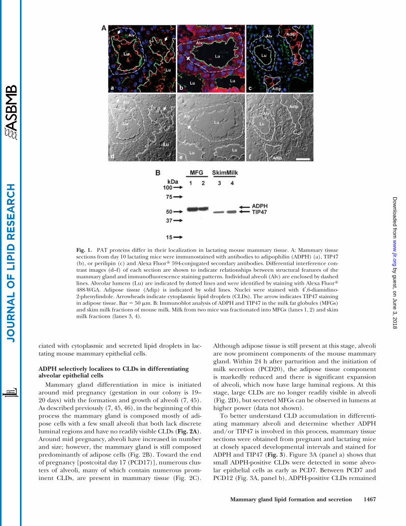
ciated with cytoplasmic and secreted lipid droplets in lac-tating mouse mammary epithelial cells.
ADPH selectively localizes to CLDs in differentiatingalveolar epithelial cells
Mammary gland differentiation in mice is initiatedaround mid pregnancy (gestation in our colony is 19–20 days) with the formation and growth of alveoli (7, 45).As described previously (7, 45, 46), in the beginning of thisprocess the mammary gland is composed mostly of adi-pose cells with a few small alveoli that both lack discreteluminal regions and have no readily visible CLDs (Fig. 2A).Around mid pregnancy, alveoli have increased in numberand size; however, the mammary gland is still composedpredominantly of adipose cells (Fig. 2B). Toward the endof pregnancy [postcoital day 17 (PCD17)], numerous clus-ters of alveoli, many of which contain numerous prom-inent CLDs, are present in mammary tissue (Fig. 2C).
Although adipose tissue is still present at this stage, alveoliare now prominent components of the mouse mammarygland. Within 24 h after parturition and the initiation ofmilk secretion (PCD20), the adipose tissue componentis markedly reduced and there is significant expansionof alveoli, which now have large luminal regions. At thisstage, large CLDs are no longer readily visible in alveoli(Fig. 2D), but secreted MFGs can be observed in lumens athigher power (data not shown).
To better understand CLD accumulation in differenti-ating mammary alveoli and determine whether ADPHand/or TIP47 is involved in this process, mammary tissuesections were obtained from pregnant and lactating miceat closely spaced developmental intervals and stained forADPH and TIP47 (Fig. 3). Figure 3A (panel a) shows thatsmall ADPH-positive CLDs were detected in some alveo-lar epithelial cells as early as PCD7. Between PCD7 andPCD12 (Fig. 3A, panel b), ADPH-positive CLDs remained
Fig. 1. PAT proteins differ in their localization in lactating mouse mammary tissue. A: Mammary tissuesections from day 10 lactating mice were immunostained with antibodies to adipophilin (ADPH) (a), TIP47(b), or perilipin (c) and Alexa Fluor: 594-conjugated secondary antibodies. Differential interference con-trast images (d–f) of each section are shown to indicate relationships between structural features of themammary gland and immunofluorescence staining patterns. Individual alveoli (Alv) are enclosed by dashedlines. Alveolar lumens (Lu) are indicated by dotted lines and were identified by staining with Alexa Fluor:488-WGA. Adipose tissue (Adip) is indicated by solid lines. Nuclei were stained with 4¶,6-diamidino-2-phenylindole. Arrowheads indicate cytoplasmic lipid droplets (CLDs). The arrow indicates TIP47 stainingin adipose tissue. Bar 5 50 mm. B: Immunoblot analysis of ADPH and TIP47 in the milk fat globules (MFGs)and skim milk fractions of mouse milk. Milk from two mice was fractionated into MFGs (lanes 1, 2) and skimmilk fractions (lanes 3, 4).
Mammary gland lipid formation and secretion 1467
by guest, on June 3, 2018w
ww
.jlr.orgD
ownloaded from

relatively small and many alveolar epithelial cells lackedthese structures. In addition, there was considerable cell-cell variability in the number and size of ADPH-positiveCLDs within individual cells. Beginning around PCD14,larger ADPH-positive CLDs began to accumulate in somecells (Fig. 3A, panel c). At this stage, however, there wasstill significant variability in CLD size and number andmany cells still lacked these structures. By PCD16, nearlyall cells possessed ADPH-positive CLDs, and in some cellsthese CLDs had diameters in the 10 mm range (Fig. 3A,panel d). By PCD18, the cytoplasm of alveolar epithelialcells appeared to be completely filled with large ADPH-positive CLDs, many of which had diameters in excess of10 mm (Fig. 3A, panel e). Within 24 h after parturition andthe onset of lactation (PCD20), numerous small ADPH-positive CLDs were observed to be enriched along the api-cal border of alveolar epithelial cells (Fig. 3A, panel f).These changes appeared to be related specifically to alveo-lar differentiation, as ADPH-positive CLDs were not de-tected in ductal epithelial cells during pregnancy and onlysmall numbers of positive CLDs were detected in someductal cells of lactating animals (Fig. 3B). In addition, theabsence of ADPH immunofluorescence in nonepithelialcompartments of the mammary gland (Fig. 3A, B) indi-cates that it is not found in mammary adipocytes frompregnant or lactating animals. Together, these results sug-gest that ADPH is primarily a secretory epithelium-specificprotein in the mouse mammary gland.
TIP47 was also detected in differentiating alveolar epi-thelial cells by immunofluorescence (Fig. 3C). However,rather than being associated with CLDs, like ADPH, it hada diffuse cytoplasmic staining pattern. Moreover, TIP47appeared to be present in all alveolar epithelial cells, evenduring the early stages of differentiation (Fig. 3C, panel a).In addition, there did not appear to be any enrichment ofTIP47 along the apical border of these cells once lactationwas initiated (Fig. 3C, panel d). These data suggest that
TIP47 is not normally associated with CLDs in alveolarepithelial cells either during differentiation or in theirmature milk-secreting state.
ADPH expression and CLD accumulation aredevelopmentally linked during mammarygland differentiation
The results in Fig. 3 suggest that CLD accumulation inalveolar epithelial cells follows a specific developmentalpattern during secretory differentiation. To better definethis developmental pattern, we quantified CLD size in dif-ferentiating and lactating alveoli. Figure 4A shows thatCLD size increased modestly between PCD7 and PCD16;however, the sharpest increase in CLD size occurred nearthe end of pregnancy, when between PCD17 and PCD18the average CLD size increased to 6 mm. However, oncemilk secretion was initiated, CLD size decreased dramati-cally. Figure 4A shows that within 24 h after parturition, theaverage CLD size was only one-sixth that of PCD18 values.These results suggest that CLD size is influenced by thebalance between their rates of formation and secretion.
The immunofluorescence data in Fig. 3 predict thatADPH levels in differentiating mammary tissue will cor-relate with changes in CLD accumulation. In agreementwith this prediction, immunoblot analysis demonstratedthat mammary gland levels of ADPH protein increase withchanges in CLD size and number during differentiationand decrease somewhat after the initiation of lactation andlipid secretion (Fig. 4B). In contrast, we found that TIP47levels were not temporally correlated with CLD accumu-lation during these periods (Fig. 4B). These data suggestthat CLD accumulation in alveolar epithelial cells is devel-opmentally linked to increased ADPH expression. How-ever, ADPH is known to be stabilized by association withtriglycerides in CLDs and to undergo rapid proteasome-mediated degradation in the absence of triglyceride syn-thesis (47). This being the case, changes in ADPH levels
Fig. 2. Histological changes in the differentiating mam-mary gland. Low-power images of hematoxylin and eosin-stained sections of mammary tissue from pregnant andlactating mice show the growth of alveolar structures andthe accumulation of CLDs in secretory epithelial cells inlate pregnancy. A: Postcoital day 3 (PCD3). B: PCD12. C:PCD17. D: PCD20 (lactation day 2). Alveolar regions areoutlined by dotted lines. Alveolar (Alv) and adipose (Adip)tissue and ducts are indicated. Arrowheads indicate CLDs,and the arrow indicates the alveolar lumen. Bar 5 100 mm.
1468 Journal of Lipid Research Volume 48, 2007
by guest, on June 3, 2018w
ww
.jlr.orgD
ownloaded from

could simply reflect posttranslational stabilization ofADPH by CLDs or diminished proteasomal activity. Al-though analysis of specific proteasomal activity is difficultin complex tissues such as the mammary gland, we pre-viously observed that genes encoding components of the26S proteasome complex did not change significantly dur-ing pregnancy (48). In addition, we found little change in
the pattern of ubiquitinated proteins in mammary tissueextracts during this period (data not shown). Together,these results suggest that generalized decreases in pro-teasomal activity are not responsible for increases in ADPHprotein levels during mammary gland differentiation. Todetermine whether ADPH transcript levels increase duringmammary gland differentiation, we quantified steady-state
Fig. 3. ADPH and TIP47 differ in localization in the differentiating mammary gland. A: Immunolocaliza-tion of ADPH (red) in differentiating and lactating mouse mammary tissue. Higher power images at PCD7(a), PCD12 (b), PCD14 (c), PCD16 (d), PCD18 (e), and PCD20 (day 2 of lactation) (f) show that ADPH(red) specifically localizes to CLD (arrowheads, panels a–e) in epithelial cells in differentiating and maturealveoli. Note that ADPH-coated CLDs increase markedly in size during the last half of pregnancy. After theonset of lactation, ADPH-coated CLDs concentrate at the apical membrane (arrowhead, panel f). B: Lowerpower images show the absence of ADPH-stained CLDs in ductal epithelial cells at days 14 (a) and 18 (b) ofpregnancy. Limited numbers of small CLDs were observed in ductal epithelial cells on lactation day 2(arrowhead, panel c). C: Immunolocalization of TIP47 (red) in differentiating mouse mammary tissue.Higher power images of alveolar epithelial cells at PCD12 (a), PCD16 (b), PCD18 (c), and PCD20 (d) showthat, in contrast to ADPH, TIP47 localizes diffusely within the cytoplasm of epithelial cells of developingand mature alveoli but is not enriched around CLDs (arrowheads). Alveoli are indicated by dotted lines in Aand C. Apical borders of alveoli were identified by Alexa Fluor: 488-WGA staining (green). Nuclei (blue)were stained with DAPI. Alveoli (Alv) and alveolar lumens (Lu) are indicated.
Mammary gland lipid formation and secretion 1469
by guest, on June 3, 2018w
ww
.jlr.orgD
ownloaded from

levels of ADPH mRNA between PCD7 and PCD29 (lacta-tion day 10) using QRT-PCR. Figure 5A shows that, likeADPH protein, steady-state levels of ADPH mRNA in-creased dramatically during the last half of pregnancy andreached a peak at PCD18. However, the largest increase inADPH mRNA occurred between PCD12 and PCD14, be-fore the detection of increases in ADPH protein or CLDnumber. The expression of TIP47 was also increased be-tween PCD12 and PCD14; however, steady-state levels ofTIP47 mRNA peaked at PCD17 and were .1 order of mag-
nitude lower than those of ADPH throughout pregnancyand lactation.
Although these results are consistent with the conceptthat ADPH expression is increased during the differen-tiation of alveolar epithelial cells, this interpretation is
Fig. 4. ADPH protein levels correlate with CLD size and accu-mulation. A: Change in CLD diameter in secretory epithelial cellsat the indicated days of pregnancy (PCD7–PCD18) and lactation(PCD20, L2). The values are average diameters (mm) 6 SEM de-termined from an analysis of individual CLDs in 30–60 randomlychosen alveoli from three sections per animal. Three animals wereanalyzed per time point. † The value was significantly different fromthe value of the preceding time point (P , 0.01); - the value wassignificantly different from the value of the preceding time point(P , 0001). B: Relative changes in ADPH (diamonds) and TIP47(squares) protein levels in mammary tissue at the indicated daysafter coitus were determined by immunoblot analysis and quan-tified by densitometry. The values are average (6SEM) densito-metric measurements normalized to b-actin from three to fouranimals per time point. Representative immunoblot analyses foreach protein are shown in the insets. Immunoblot analyses of tissueextracts were performed twice with similar results. - the value wassignificantly different from the value of the preceding time pointfor ADPH (P, 0.00001); † the value was significantly different fromthe value of the preceding time point for ADPH (P, 0.001); 1 thePCD18 value was significantly different from the PCD10 andPCD28 values for TIP47 (P , 0.001). Pregnancy and lactation pe-riods are indicated by double-headed horizontal arrows. Parturi-tion is indicated by the up arrow.
Fig. 5. Induction of ADPH expression during pregnancy. A:Developmental changes of ADPH (solid line) and TIP47 (dashedline) mRNA copy numbers as assessed by quantitative real-timePCR (QRT-PCR). Values are average copy numbers (6SEM) nor-malized to b-2-microglobulin for three to four animals per timepoint. Pregnancy and lactation periods are indicated by horizontaldouble-headed arrows. The up arrow indicates the time of par-turition.- The PCD18 value was significantly different from thePCD14 value (P, 0.005). B: In situ hybridization of ADPH in mam-mary tissue sections from mice at pregnancy days 12, 16, and 18 andlactation day 10. Tissues in panels a, c, e, and g were labeled withdigoxigenin-conjugated sense oligonucleotides to mouse ADPH.Tissues in panels b, d, f, and h were labeled with digoxigenin-conjugated antisense oligonucleotides to mouse ADPH. The insetin panel h shows the lack of reaction in tissues treated with RNasebefore in situ hybridization with antisense oligonucleotides. Tissuesections were incubated for 5 h with substrate (asterisks) or for 1 hwith substrate (double daggers). Note the uniform staining of al-veoli and the lack of adipose staining under these conditions.
1470 Journal of Lipid Research Volume 48, 2007
by guest, on June 3, 2018w
ww
.jlr.orgD
ownloaded from

complicated by changes in the relative proportions of epi-thelium and adipose tissue in the differentiating mam-mary gland and by observations that ADPH transcripts areexpressed in mature adipocytes in culture (16, 23). Todetermine which cell types express ADPH transcripts, andto verify that ADPH transcript expression increases indifferentiating secretory epithelial cells, sections of mam-mary tissue from pregnant and lactating mice were ana-lyzed by in situ hybridization using ADPH-specific probes.Figure 5B shows that ADPH transcripts are detected se-lectively in alveolar epithelial cells in both differentiatingand mature mammary glands. In contrast to the significantcell-cell variability observed in ADPH-positive CLDs inthese cells during the initial phases of mammary glanddifferentiation (Fig. 3A), there appeared to be little or nocell-cell variability in ADPH transcript expression duringthe differentiation process. These observations confirmthat ADPH expression is increased selectively in alveolarepithelial cells during mammary gland differentiation andare consistent with the concept that increased ADPHexpression promotes CLD accumulation in these cells.However, differences in the temporal patterns of ADPH
transcript expression and CLD accumulation suggest thatother factors contribute to this process.
Lipid metabolism is altered in the differentiatingmammary gland
Changes in fatty acid availability and/or lipid metabo-lism are also potential regulators of CLD accumulation.The lipid content of the mammary gland adipose storesbegins to be depleted in late pregnancy and is significantlydepleted in response to lactation (49, 50). It has been pos-tulated that this depletion of adipose lipid is attributableto shunting of fatty acids into secretory epithelial cells formilk lipid synthesis (51). To determine whether functionalrelationships exist between adipose lipid storage capacityand CLD accumulation in differentiating secretory epi-thelial cells, we characterized perilipin protein and mRNAlevels in differentiating mammary tissue. Immunofluores-cence staining (Fig. 6A) shows that there is a decrease inperilipin-positive adipose cells during mammary gland dif-ferentiation and that adipose cells adjacent to differen-tiating alveoli have a shrunken appearance. Immunoblot(Fig. 6B) and QRT-PCR (Fig. 6C) analyses of differentiat-
Fig. 6. Perilipin levels decrease during mammarygland differentiation. A: Immunofluorescence analysis ofperilipin (red) in mammary gland sections showing de-pletion of perilipin-positive adipose cells (Adip) in dif-ferentiating and lactating mammary glands at PCD7 (a),PCD17 (b), and PCD20 (lactation day 2) (c). Alveoli(Alv) were identified by staining their luminal borders(arrow) with Alexa Fluor: 488-WGA (green). Nuclei(blue) were stained with DAPI. Note that the numberof perilipin-positive adipose cells in mammary tissuedeclines dramatically during pregnancy and lactation. B:Change in relative steady-state perilipin protein levels inmammary tissue at the indicated times after coitus.Perilipin protein was determined by immunoblot anal-ysis (inset) and quantified by densitometry. The valuesare average (6SEM) densitometric measurements nor-malized to b-actin from three to four animals per timepoint. † The value was significantly different from thevalue of the preceding time point (P , 0.05); - the valuewas significantly different from the value of the preced-ing time point (P , 0.01). Error bars for PCD21 andPCD29 values are within the size of the symbol. C:Change in steady-state levels of perilipin mRNA in mousemammary tissue during pregnancy and lactation. Rela-tive perilipin mRNA was determined by QRT-PCR.Values are normalized to PCD7 values and are averages6 SEM for three to four animals per time point. † Thevalue was significantly different from the previous value(P , 0.01); - the value was significantly different fromthe previous value (P , 0.005). Pregnancy and lacta-tion periods are indicated in B and C by double-headed horizontal arrows. Up arrows indicate the timeof parturition.
Mammary gland lipid formation and secretion 1471
by guest, on June 3, 2018w
ww
.jlr.orgD
ownloaded from

ing mammary tissue show that perilipin protein relative totissue actin and mRNA relative to total RNA begin to de-crease about mid pregnancy, and by the end of pregnancy(PCD18), relative perilipin protein and mRNA levels areonly 25% and 14%, respectively, of their mid pregnancyvalues (PCD10). The temporal correlation between thedecline in perilipin expression and the increase in ADPHexpression in secretory epithelial cells is consistent withthe possibility that the processes are functionally linkedand suggest that reduced lipid storage capacity by mam-mary adipose may contribute to enhanced lipid accumu-lation in differentiating secretory epithelial cells.
In previously published studies, we reported changes inthe expression of lipid metabolism genes in mouse mam-mary glands during pregnancy and in lactation from mi-croarray analysis of FVB mice, finding that these changeswere consistent with those from another laboratory inC57Bl6 mice (48, 52, 53). In limited microarray analyseson the CD1 mouse strain used for these studies, we againfound comparable changes in lipid metabolism gene ex-pression between late pregnancy and early lactation (datanot shown). For this reason, we are comfortable that theresults of the analysis of three genes involved in lipid syn-thesis, whose expression is likely limited in adipose tissue,are representative of changes in the epithelial compart-ment only of most if not all mouse strains. These genes areGlut1 (gene accession number M22998), the major glucosetransporter in the mammary epithelium (54), and a D5 fattyacid desaturase (Fads1; gene accession number AK083959)and a fatty acid elongase (Elovl1; gene accession numberAI842813) that likely contribute to the desaturation andelongation of fatty acids in the mammary gland to producethe long-chain polyunsaturated fatty acids characteristic ofmilk (53). The expression patterns of these genes are con-sistent (Fig. 7). Starting after PCD7, there is a slow 2-foldincrease in expression, followed by a 2- to 3-fold increasebetween PCD17 and PCD20 (lactation day 2). These obser-vations suggest that there is a slow upregulation of the genesof lipid synthesis in the mammary epithelium during latepregnancy that contributes to CLD accumulation.
DISCUSSION
The stabilization of nascent CLDs by PAT proteins isthought to be an important element in their accumulation(55). Our study provides evidence that ADPH mediatesCLD accumulation in differentiating secretory epithelialcells in the mouse mammary gland. The observations thatboth CLD accumulation and ADPH expression selectivelyincrease in these cells during their differentiation suggestthat they are specific, developmentally induced propertiesof the secretory epithelium. Although TIP47 can localizeto CLDs (56–58) and there have been reports that it isfound on MFGs in human milk (22, 42), our data indicatethat it is not associated with CLDs in milk-secreting cellsof the mouse mammary gland and is not a componentof secreted MFGs. Interestingly, our immunofluorescenceresults indicate that, like ADPH, TIP47 is enriched in
secretory epithelial cells, which suggests that it may beimportant for their differentiation or function. However,observations that TIP47 levels are inversely correlated withCLD levels in these cells argue against a role for it in CLDaccumulation or secretion in the normal mouse mammarygland. At present, it is unclear whether the presence ofTIP47 in MFGs from human milk (22) reflects species dif-ferences in the types of PAT proteins associated with CLDsor whether other mechanisms are involved. For instance, itis known that MFGs in some species, including humans,possess significant amounts of cytoplasmic inclusions, whichcan contain endoplasmic reticulum and Golgi fragments(7). Because TIP47 normally functions as an adaptor pro-tein in endocytic vesicle trafficking (21), it is possible thatvesicular material containing TIP47 becomes trapped dur-ing MFG formation in humans but not in mice. Interest-ingly, we found TIP47 in skim milk fractions, which alsosuggests that mechanisms exist for its transfer into milkindependent of milk lipid secretion. Although it is unclearhow this transfer occurs, skim milk fractions are knownto contain small membrane vesicles (7), and it is possiblethat TIP47 is transferred into milk in association withthis material.
CLD accumulation in secretory epithelial cells was longago observed to be initiated during pregnancy and asso-ciated with the differentiation of the mammary gland intoa secretory organ (7, 45); however, the factors regulatingthis process were poorly defined. Our data show that theaccumulation and growth of these structures correspondto increased ADPH expression and follow a distinct bi-phasic developmental pattern. In agreement with earlierhistological and electron microscopy studies (7, 45), wefound that only limited numbers of secretory epithelialcells possessed ADPH-positive CLDs during the initialphase of differentiation (PCD7–PCD14) and that among
Fig. 7. Expression of genes contributing to lipid synthesis inmouse mammary glands during pregnancy and lactation. Relativeexpression levels of Glut-1 (solid line), D5 fatty acid desaturase(mixed dashed line), and fatty acid elongase (dashed line) in dif-ferentiating mouse mammary tissue are shown. Values are meansfrom analysis of four mice per time point. Standard errors, notshown, were ,10% of the mean. † The value was significantly dif-ferent from the preceding value (P , 0.01); - the value was sig-nificantly different from the preceding value (P , 0.005).
1472 Journal of Lipid Research Volume 48, 2007
by guest, on June 3, 2018w
ww
.jlr.orgD
ownloaded from

these cells there was considerable cell-cell variability inboth the number and size of CLDs. The observation thatADPH transcripts appear to be expressed uniformly in allsecretory epithelial cells, even those that lack visible CLDs,suggests that differences in ADPH expression are not thecause of cell-cell variability in CLD accumulation. Impor-tantly, the presence of ADPH mRNA in cells before theappearance of CLDs suggests that ADPH expression may beone initiating event in CLD accumulation. However, thesignificant lag between the increase in ADPH mRNA and theincrease in CLD accumulation indicates that increasedADPH expression is not the only factor driving this process.
The possibility that developmental increases in the lipidsynthetic capacity of secretory epithelial cells contribute toCLD accumulation was suggested by studies showing thatlipid synthetic activity increases in differentiating mousemammary tissue in late pregnancy (59). More recently, arraystudies have demonstrated widespread changes in the ex-pression of genes involved in lipid metabolism at the end ofpregnancy (48, 53). Furthermore, the array data presentedhere show an increase in epithelium-specific genes thatcontribute to the synthesis of milk lipid that is temporallysimilar to changes in CLD accumulation. Together, thesedata are consistent with developmentally driven increasesin lipid production in differentiating secretory epithelialcells, which may contribute to CLD accumulation.
In addition to alterations in secretory epithelial cell lipidmetabolism, our data also indicate that diminished lipidstorage in mammary adipocytes may contribute to CLDaccumulation by shunting of fatty acids to secretory epi-thelial cells. Loss of perilipin is known to decrease tri-glyceride storage in adipose cells and to increase serumfatty acid levels (60, 61). Our observations that relativeperilipin protein and mRNA levels decrease with a timecourse similar to that of CLD accumulation in differenti-ating secretory cells suggest that the two processes may befunctionally related. Indeed, earlier studies documentedthat depletion of lipid in mammary adipose cells duringlate pregnancy and lactation is dependent on the presenceof an epithelial component (49). In this context, it is in-teresting that steady-state levels of perilipin protein andmRNA begin to decrease at mid pregnancy, well beforealveoli are significant cellular components of the mam-mary gland. Thus, it is unlikely that declines in perilipinexpression are attributable simply to displacement of theadipose compartment by alveoli (49). At present, the fac-tors regulating adipose lipid depletion and the reduc-tion of perilipin expression are not well defined, but bothsystemic hormones and/or epithelium-derived paracrinefactors are possible contributors. Collectively, our data sug-gest that CLD accumulation in the differentiating mousemammary gland is a sequentially ordered process that isinitiated around mid pregnancy by increased expressionof ADPH, which functions to stabilize the triglyceride corein nascent CLDs. Subsequent CLD accumulation is pro-posed to depend on increases in intracellular fatty acidlevels, which are likely to be driven by both increased denovo fatty synthesis in secretory epithelial cells and re-duced lipid storage by adipose cells.
In summary, previous studies of mammary gland dif-ferentiation have focused primarily on changes in geneticor biochemical markers, and little was known about thecell biological aspects of this process. Our data provideevidence that CLD accumulation and trafficking are prom-inent features of the differentiating mammary gland andappear to be early and specific markers of the differenti-ation process. We previously demonstrated that ADPHspecifically colocalizes with xanthine oxidoreductase andbutyrophilin at sites of CLD secretion on apical mem-branes of secretory epithelial cells in lactating mousemammary glands and exists as a stable complex with theseproteins on isolated MFGs (35). Together, these observa-tions raise the possibility that ADPH functions in both theformation and secretion of CLD by mammary epithelialcells. Although recent freeze-fracture localization studieshave raised questions about the exact role of ADPH inCLD secretion (42), they nevertheless demonstrate an en-richment of ADPH on the apical membrane at sites thatcontact CLDs, which is consistent with the possibility thatADPH contributes to interactions between CLDs and theapical plasma membrane.
Whether or not this interaction is caused by a specificassociation with a xanthine oxidoreductase-butyrophilincomplex, as hypothesized previously (6, 35), the ability ofADPH to associate with the CLD surface as well as withthe plasma membrane implies that it possesses distinctbinding functions (5). Previous studies have identifiedCLD targeting regions in the N-terminal portion of ADPH(33, 62), and it will be important to define the in vivo rolesof these, as well as other, regions of the ADPH molecule inthe formation and secretion of CLDs. Of interest, thecrystal structure of the C-terminal region of TIP-47, whichis structurally similar to that of ADPH, has been shown tobe composed of a four helix bundle similar to the putativemembrane binding domain of apolipoprotein E (63).Thus, it will be important to determine whether this struc-ture contributes to interactions between CLD and mem-brane structures or other physiological functions of ADPH(5). As yet, there is relatively limited information on thephysiological roles of ADPH, although recent data haveshown that loss of ADPH is protective against the develop-ment of fatty liver (64). Our data suggest that the differen-tiating mammary gland is a useful model in which to studydevelopmental mechanisms regulating CLD accumulationand secretion, and it will be interesting in future studies todetermine how the loss of ADPH affects these processes.
This research was supported by grants from the National Insti-tutes of Health, RO1 HD-045962 (J.L.M.) and PO1 HD-38129(J.L.M., M.C.N.). C.A.P. was supported by National ResearchService Award Postdoctoral Fellowship HD-044359. The au-thors thank W. Zabaronick for excellent technical assistance.
REFERENCES
1. Allen, J. C., R. P. Keller, P. C. Archer, and M. C. Neville. 1991.Studies in human lactation. VI. Milk composition and daily secre-
Mammary gland lipid formation and secretion 1473
by guest, on June 3, 2018w
ww
.jlr.orgD
ownloaded from
tion rates of macronutrients in the first year of lactation. Am. J. Clin.Nutr. 54: 69–80.
2. Schwertfeger, K. L., J. L. McManaman, C. A. Palmer, M. C. Neville,and S. M. Anderson. 2003. Expression of constitutively activatedAkt in the mammary gland leads to excess lipid synthesis duringpregnancy and lactation. J. Lipid Res. 44: 1100–1112.
3. Neville, M. C., J. Morton, and S. Umemora. 2001. Lactogenesis: thetransition from pregnancy to lactation. Pediatr. Clin. North Am. 48:35–52.
4. Neville, M. C., and C. W. Daniel. 1987. The Mammary Gland:Development, Regulation and Function. Plenum Press, New York.
5. McManaman, J. L., M. E. Reyland, and E. C. Thrower. 2006. Se-cretion and fluid transport mechanisms in the mammary gland:comparisons with the exocrine pancreas and the salivary gland.J. Mammary Gland Biol. Neoplasia. 11: 249–268.
6. Mather, I. H., and T. W. Keenan. 1998. Origin and secretion of milklipids. J. Mammary Gland Biol. Neoplasia. 3: 259–273.
7. Hollmann, K. H. 1974. Cytology and fine structure of the mammarygland. In Lactation. B. L. Larson and V. R. Smith, editors. AcademicPress, New York. 3–95.
8. Londos, C., D. L. Brasaemle, C. J. Schultz, J. P. Segrest, and A. R.Kimmel. 1999. Perilipins, ADRP, and other proteins that associatewith intracellular neutral lipid droplets in animal cells. Semin. CellDev. Biol. 10: 51–58.
9. Lu, X., J. Gruia-Gray, N. G. Copeland, D. J. Gilbert, N. A. Jenkins,C. Londos, and A. R. Kimmel. 2001. The murine perilipin gene:the lipid droplet-associated perilipins derive from tissue-specific,mRNA splice variants and define a gene family of ancient origin.Mamm. Genome. 12: 741–749.
10. Miura, S., J. W. Gan, J. Brzostowski, M. J. Parisi, C. J. Schultz,C. Londos, B. Oliver, and A. R. Kimmel. 2002. Functional conser-vation for lipid storage droplet association among Perilipin, ADRP,and TIP47 (PAT)-related proteins in mammals, Drosophila, andDictyostelium. J. Biol. Chem. 277: 32253–32257.
11. Arimura, N., T. Horiba, M. Imagawa, M. Shimizu, and R. Sato. 2004.The peroxisome proliferator-activated receptor gamma regulatesexpression of the perilipin gene in adipocytes. J. Biol. Chem. 279:10070–10076.
12. Bertile, F., F. Criscuolo, H. Oudart, Y. Le Maho, and T. Raclot.2003. Differences in the expression of lipolytic-related genes in ratwhite adipose tissues. Biochem. Biophys. Res. Commun. 307: 540–546.
13. Brasaemle, D. L., G. Dolios, L. Shapiro, and R. Wang. 2004. Pro-teomic analysis of proteins associated with lipid droplets of basaland lipolytically stimulated 3T3-L1 adipocytes. J. Biol. Chem. 279:46835–46842.
14. Fong, T. H., C. C. Yang, A. S. Greenberg, and S. M. Wang. 2002.Immunocytochemical studies on lipid droplet-surface proteins inadrenal cells. J. Cell. Biochem. 86: 432–439.
15. Greenberg, A. S., J. J. Egan, S. A. Wek, M. C. Moos, Jr., C. Londos,and A. R. Kimmel. 1993. Isolation of cDNAs for perilipins A and B:sequence and expression of lipid droplet-associated proteins ofadipocytes. Proc. Natl. Acad. Sci. USA. 90: 12035–12039.
16. Brasaemle, D. L., T. Barber, N. E. Wolins, G. Serrero, E. J.Blanchette-Mackie, and C. Londos. 1997. Adipose differentiation-related protein is an ubiquitously expressed lipid storage droplet-associated protein. J. Lipid Res. 38: 2249–2263.
17. Heid, H. W., R. Moll, I. Schwetlick, H. R. Rackwitz, and T. W.Keenan. 1998. Adipophilin is a specific marker of lipid accumula-tion in diverse cell types and diseases. Cell Tissue Res. 294: 309–321.
18. Carroll, K. S., J. Hanna, I. Simon, J. Krise, P. Barbero, and S. R.Pfeffer. 2001. Role of Rab9 GTPase in facilitating receptor recruit-ment by TIP47. Science. 292: 1373–1376.
19. Bohn, H., W. Kraus, and W. Winckler. 1983. Purification and char-acterization of two new soluble placental tissue proteins (PP13 andPP17). Oncodev. Biol. Med. 4: 343–350.
20. Than, N. G., B. Sumegi, G. N. Than, G. Kispal, and H. Bohn. 1998.Cloning and sequence analysis of cDNAs encoding human placen-tal tissue protein 17 (PP17) variants. Eur. J. Biochem. 258: 752–757.
21. Diaz, E., and S. R. Pfeffer. 1998. TIP47: a cargo selection device formannose 6-phosphate receptor trafficking. Cell. 93: 433–443.
22. Than, N. G., B. Sumegi, S. Bellyei, T. Berki, G. Szekeres, T. Janaky,A. Szigeti, H. Bohn, and G. N. Than. 2003. Lipid droplet andmilk lipid globule membrane associated placental protein 17b(PP17b) is involved in apoptotic and differentiation processes ofhuman epithelial cervical carcinoma cells. Eur. J. Biochem. 270:1176–1188.
23. Jiang, H. P., and G. Serrero. 1992. Isolation and characterization of
a full-length cDNA coding for an adipose differentiation-relatedprotein. Proc. Natl. Acad. Sci. USA. 89: 7856–7860.
24. Steiner, S., D. Wahl, B. L. Mangold, R. Robison, J. Raymackers,L. Meheus, N. L. Anderson, and A. Cordier. 1996. Induction of theadipose differentiation-related protein in liver of etomoxir-treatedrats. Biochem. Biophys. Res. Commun. 218: 777–782.
25. Gao, J., H. Ye, and G. Serrero. 2000. Stimulation of adipose dif-ferentiation related protein (ADRP) expression in adipocyte pre-cursors by long-chain fatty acids. J. Cell. Physiol. 182: 297–302.
26. Vosper, H., L. Patel, T. L. Graham, G. A. Khoudoli, A. Hill, C. H.Macphee, I. Pinto, S. A. Smith, K. E. Suckling, C. R. Wolf, et al.2001. The peroxisome proliferator-activated receptor delta pro-motes lipid accumulation in human macrophages. J. Biol. Chem.276: 44258–44265.
27. Buechler, C., M. Ritter, C. Q. Duong, E. Orso, M. Kapinsky, and G.Schmitz. 2001. Adipophilin is a sensitive marker for lipid loadingin human blood monocytes. Biochim. Biophys. Acta. 1532: 97–104.
28. Gupta, R. A., J. A. Brockman, P. Sarraf, T. M. Willson, and R. N.DuBois. 2001. Target genes of peroxisome proliferator-activatedreceptor gamma in colorectal cancer cells. J. Biol. Chem. 276:29681–29687.
29. Exil, V. J., R. L. Roberts, H. Sims, J. E. McLaughlin, R. A. Malkin,C. D. Gardner, G. Ni, J. N. Rottman, and A. W. Strauss. 2003. Very-long-chain acyl-coenzyme A dehydrogenase deficiency in mice.Circ. Res. 93: 448–455.
30. Wu, C. C., K. E. Howell, M. C. Neville, J. R. Yates, 3rd, and J. L.McManaman. 2000. Proteomics reveal a link between the endo-plasmic reticulum and lipid secretory mechanisms in mammaryepithelial cells. Electrophoresis. 21: 3470–3482.
31. Liu, P., Y. Ying, Y. Zhao, D. I. Mundy, M. Zhu, and R. G. Anderson.2004. Chinese hamster ovary K2 cell lipid droplets appear to bemetabolic organelles involved in membrane traffic. J. Biol. Chem.279: 3787–3792.
32. Imamura, M., T. Inoguchi, S. Ikuyama, S. Taniguchi, K. Kobayashi,N. Nakashima, and H. Nawata. 2002. ADRP stimulates lipid accu-mulation and lipid droplet formation in murine fibroblasts. Am. J.Physiol. Endocrinol. Metab. 283: E775–E783.
33. McManaman, J. L., W. Zabaronick, J. Schaack, and D. J. Orlicky.2003. Lipid droplet targeting domains of adipophilin. J. Lipid Res.44: 668–673.
34. Larigauderie, G., C. Cuaz-Perolin, A. B. Younes, C. Furman,C. Lasselin, C. Copin, M. Jaye, J. C. Fruchart, and M. Rouis. 2006.Adipophilin increases triglyceride storage in human macrophagesby stimulation of biosynthesis and inhibition of beta-oxidation.FEBS J. 273: 3498–3510.
35. McManaman, J. L., C. A. Palmer, R. M. Wright, and M. C. Neville.2002. Functional regulation of xanthine oxidoreductase expressionand localization in the mouse mammary gland: evidence of a role inlipid secretion. J. Physiol. 545: 567–579.
36. Ogg, S. L., A. K. Weldon, L. Dobbie, A. J. Smith, and I. H. Mather.2004. Expression of butyrophilin (Btn1a1) in lactating mammarygland is essential for the regulated secretion of milk-lipid droplets.Proc. Natl. Acad. Sci. USA. 101: 10084–10089.
37. Vorbach, C., A. Scriven, and M. R. Capecchi. 2002. The house-keeping gene xanthine oxidoreductase is necessary for milk fatdroplet enveloping and secretion: gene sharing in the lactatingmammary gland. Genes Dev. 16: 3223–3235.
38. Wessel, D., and U. I. Flugge. 1984. A method for the quantitativerecovery of protein in dilute solution in the presence of detergentsand lipids. Anal. Biochem. 138: 141–143.
39. Drews, R., R. K. Paleyanda, T. K. Lee, R. R. Chang, A. Rehemtulla,R. J. Kaufman, W. N. Drohan, and H. Lubon. 1995. Proteolyticmaturation of protein C upon engineering the mouse mammarygland to express furin. Proc. Natl. Acad. Sci. USA. 92: 10462–10466.
40. Patton, S., and G. E. Huston. 1986. A method for isolation of milkfat globules. Lipids. 21: 170–174.
41. Heid, H. W., M. Schnolzer, and T. W. Keenan. 1996. Adipocytedifferentiation-related protein is secreted into milk as a constituentof milk lipid globule membrane. Biochem. J. 320: 1025–1030.
42. Robenek, H., O. Hofnagel, I. Buers, S. Lorkowski, M. Schnoor, M. J.Robenek, H. Heid, D. Troyer, and N. J. Severs. 2006. Butyrophilincontrols milk fat globule secretion. Proc. Natl. Acad. Sci. USA. 103:10385–10390.
43. Blanchette-Mackie, E. J., N. K. Dwyer, T. Barber, R. A. Coxey,T. Takeda, C. M. Rondinone, J. L. Theodorakis, A. S. Greenberg,and C. Londos. 1995. Perilipin is located on the surface layer of in-tracellular lipid droplets in adipocytes. J. Lipid Res. 36: 1211–1226.
1474 Journal of Lipid Research Volume 48, 2007
by guest, on June 3, 2018w
ww
.jlr.orgD
ownloaded from

44. Greenberg, A. S., J. J. Egan, S. A. Wek, N. B. Garty, E. J. Blanchette-Mackie, and C. Londos. 1991. Perilipin, a major hormonally reg-ulated adipocyte-specific phosphoprotein associated with theperiphery of lipid storage droplets. J. Biol. Chem. 266: 11341–11346.
45. Wooding, F. B. P. 1977. Comparative mammary fine structure. InComparative Aspects of Lactation. M. Peaker, editor. AcademicPress, London. 1–41.
46. Richert, M. M., K. L. Schwertfeger, J. W. Ryder, and S. M. Anderson.2000. An atlas of mouse mammary gland development. J. MammaryGland Biol. Neoplasia. 5: 227–241.
47. Xu, G., C. Sztalryd, X. Lu, J. T. Tansey, J. Gan, H. Dorward, A. R.Kimmel, and C. Londos. 2005. Post-translational regulation of adi-pose differentiation-related protein by the ubiquitin/proteasomepathway. J. Biol. Chem. 280: 42841–42847.
48. Rudolph, M. C., J. L. McManaman, L. Hunter, T. Phang, and M. C.Neville. 2003. Functional development of the mammary gland: useof expression profiling and trajectory clustering to reveal changesin gene expression during pregnancy, lactation, and involution.J. Mammary Gland Biol. Neoplasia. 8: 287–307.
49. Elias, J. J., D. R. Pitelka, and R. C. Armstrong. 1973. Changes infat cell morphology during lactation in the mouse. Anat. Rec. 177:533–547.
50. Neville, M. C., D. Medina, J. Monks, and R. C. Hovey. 1998. Themammary fat pad. J. Mammary Gland Biol. Neoplasia. 3: 109–116.
51. Jensen, D. R., S. Gavigan, V. Sawicki, D. Witsell, R. H. Eckel,and M. C. Neville. 1994. Regulation of lipoprotein lipase activityin the mammary gland of the lactating mouse. Biochem. J. 298:321–327.
52. Stein, T., J. S. Morris, C. R. Davies, S. J. Weber-Hall, M. A. Duffy,V. J. Heath, A. K. Bell, R. K. Ferrier, G. P. Sandilands, and B. A.Gusterson. 2004. Involution of the mouse mammary gland is asso-ciated with an immune cascade and an acute-phase response, in-volving LBP, CD14 and STAT3. Breast Cancer Res. 6: R75–R91.
53. Rudolph, M. C., J. L. McManaman, T. Phang, T. Russell, D. J.Kominsky, N. J. Serkova, T. Stein, S. M. Anderson, and M. C.Neville. 2007. Metabolic regulation in the lactating mammarygland: a lipid synthesizing machine. Physiol. Genomics. 28: 323–336.
54. Burnol, A-F., A. Leturque, M. Loizeau, C. Postic, and J. Girard.
1990. Glucose transporter expression in rat mammary gland. Bio-chem. J. 270: 277–279.
55. Murphy, D. J. 2001. The biogenesis and functions of lipid bodiesin animals, plants and microorganisms. Prog. Lipid Res. 40: 325–438.
56. Sztalryd, C., M. Bell, X. Lu, P. Mertz, S. Hickenbottom, B. H.Chang, L. Chan, A. R. Kimmel, and C. Londos. 2006. Functionalcompensation for adipose differentiation-related protein (ADFP)by TIP47 in an ADFP null embryonic cell line. J. Biol. Chem. 281:34341–34348.
57. Ohsaki, Y., T. Maeda, M. Maeda, K. Tauchi-Sato, and T. Fujimoto.2006. Recruitment of TIP47 to lipid droplets is controlled by theputative hydrophobic cleft. Biochem. Biophys. Res. Commun. 347:279–287.
58. Wolins, N. E., B. Rubin, and D. L. Brasaemle. 2001. TIP47 associateswith lipid droplets. J. Biol. Chem. 276: 5101–5108.
59. Baldwin, R. L., and Y. T. Yang. 1974. Enzymatic and metabolicchanges in the development of lactation. In Lactation. B. L. Larsonand V. R. Smith, editors. Academic Press, New York. 349–407.
60. Martinez-Botas, J., J. B. Anderson, D. Tessier, A. Lapillonne, B. H.Chang, M. J. Quast, D. Gorenstein, K. H. Chen, and L. Chan. 2000.Absence of perilipin results in leanness and reverses obesity inLepr(db/db) mice. Nat. Genet. 26: 474–479.
61. Tansey, J. T., C. Sztalryd, J. Gruia-Gray, D. L. Roush, J. V. Zee,O. Gavrilova, M. L. Reitman, C. X. Deng, C. Li, A. R. Kimmel, et al.2001. Perilipin ablation results in a lean mouse with aberrant adi-pocyte lipolysis, enhanced leptin production, and resistance to diet-induced obesity. Proc. Natl. Acad. Sci. USA. 98: 6494–6499.
62. Targett-Adams, P., D. Chambers, S. Gledhill, R. G. Hope, J. F. Coy,A. Girod, and J. McLauchlan. 2003. Live cell analysis and targetingof the lipid droplet-binding adipocyte differentiation-related pro-tein. J. Biol. Chem. 278: 15998–16007.
63. Hickenbottom, S. J., A. R. Kimmel, C. Londos, and J. H. Hurley.2004. Structure of a lipid droplet protein: the PAT family memberTIP47. Structure. 12: 1199–1207.
64. Chang, B. H., L. Li, A. Paul, S. Taniguchi, V. Nannegari, W. C.Heird, and L. Chan. 2006. Protection against fatty liver but normaladipogenesis in mice lacking adipose differentiation-relatedprotein. Mol. Cell. Biol. 26: 1063–1076.
Mammary gland lipid formation and secretion 1475
by guest, on June 3, 2018w
ww
.jlr.orgD
ownloaded from