Cytoplasmic DNA-activity during vitellogenesis in spiderAraneus nauticus koch (Aranae: Argiopidae)
2
976 Specialia EXPERIENTIA 31/8 Cytoplasmic DNA-Activity During Vitellogenesis Koch (Aranae: Argiopidae) Cytoplasmic DNA has been well reported in the oocytes of different animals by ELSON and CHARGAFF 1, HOFF- JORGENSEN a n d ZEUTHEN 2, SZE 3, BRACHET and QUER- TIER4, SAREEN 5-7, BALTUS a n d BRACHET8, HANOCQ- QUARTIER et al. 9 and BALTUS et al. 1~ However, to our knowledge, there is no record of whether or not the cytplasmic DNA is involved in the process of vitello- genesis. In the present communication, the possible involvement of cytoplasmic DNA during vitellogenesis in spider Araneus nauticus has been deduced on the basis of histochemical analysis. Specimens of A. nauticus were collected from the corners of an old abandoned house. The ovaries from the females were dissected out, fixed and processed by stan- dard methods n-13 for the detection of DNA with its usual trichloroacetic acid control, protein and RNA. The precursors of yolk develope at the periphery of the oocyte, which normally measures 30 ~tm in diameter. These precursors are granular and give strong reaction for DNA (Figure 1) and weak reaction for protein. These granules now disperse in the ooplasm (Figure 2). In later stages, when an individual oocyte measures on average 130 b~m in diameter, the precursors at the periphery enlarge into small globules (Figure 3). The latter now fragment into very small granules, which disperse randomly in the ooplasm, and in between which, the yolk bodies develope (Figure 4). These bodies measure on average 15 [zm in diameter and give a strong reaction for 'protein and RNA. In mature oocytes, the DNA content in Spider Araneus nauticus of the precursors appears to be greatly depleted, being barely visible along the margin of the yolk bodies (Fig- ure 5). Considering these observations, the possibility of involvement of the cytoplasmic DNA in the formation of yolk in .d. nauticus cannot be ignored. The cortical DNA +re granules increase in size with a concurrent increase in the amount of DNA. The DNA increase, as is discernable by the increase of size and intensity of the DNA+ve granules, is assumed to be by a process of rapid replication, to feed t D. ELSON and E. CHARGAFF, Experientia 8, 143 (1952). 2 E. HOFF-JORGENSEN and E. ZEUTHEN, Nature, Lond. 169, 245 (1952). 3 L. C. SzE, J. exp. Zool. /22, 577 (1953). 4 j. BRACHETand J. QUERTIER~Expl Cell Res. 32, 410 (1963). 5 M. L. SAREEN, Res. Bull. Panjab Univ. ld, 273 (1963). M. L. SAREEN, Res. Bull. Panjab Univ. 15, 169 (1964). 7 M. L. SAREEN, Res. Bull. Panjab Univ. 16, 1 (1965). 8 E. BALTUS and J. BRACHET, Biochim. biophys. Acta 61, 157 (1962). 9 j. HANOCQ*QUERTIER,E. BALTUS, A. FICQ and J. BRACHET, J. Embryol. exp. Morph. 19, 273 (1968). 10 E. BALTUS, J. HANOCQ-QUERTIERand J. BRACHET, Proc. natn. Acad. Sci., USA 61,469 (1968). 11 R. FEULGENand H. ROSSENBECK, Z. phys. Chem. 135, 203 (1924). 12 D. MAZIA, P. A. BREWER and M. ALFERT, Biol. Bull. lOd, 57 (1953). 13 N. B. KURNICK,Stain Tech. 30, 213 (1955). Fig. 1-5. Photomicrographs of section of oocytes of Araneus nauticus. Feulgen. • 800. 1. DNA+re yolk precursors along peripheral ooplasm. 2. Cytoplasmic DNA granules. 3. DNA+ve globules. 4. DNA+re granules between developing yolk bodies. 5. Traces of DNA along the margin of yolk bodies.
Transcript of Cytoplasmic DNA-activity during vitellogenesis in spiderAraneus nauticus koch (Aranae: Argiopidae)
976 Specialia EXPERIENTIA 31/8
Cytoplasmic DNA-Activity During Vitellogenesis Koch (Aranae: Argiopidae)
Cytoplasmic D N A has been well r epor ted in the oocytes of d i f fe rent animals by ELSON and CHARGAFF 1, HOFF- JORGENSEN and ZEUTHEN 2, SZE 3, BRACHET and QUER- TIER4, SAREEN 5-7, BALTUS and BRACHET8, HANOCQ- QUARTIER et al. 9 and BALTUS e t al. 1~ However , to our knowledge, there is no record of w h e t h e r or no t t he cy tp lasmic D N A is involved in the process of vitel lo- genesis. In the p resen t communica t ion , t he possible i n v o l v e m e n t of cy top lasmic D N A dur ing vi tel logenesis in spider Araneus nauticus has been deduced on the basis of h i s tochemica l analysis.
Specimens of A . naut icus were collected f rom the corners of an old abandoned house. The ovaries f rom the females were dissected out, f ixed and processed b y s tan- da rd m e t h o d s n-13 for t he de tec t ion of D N A wi th its usual t r ichloroacet ic acid control , p ro te in and RNA.
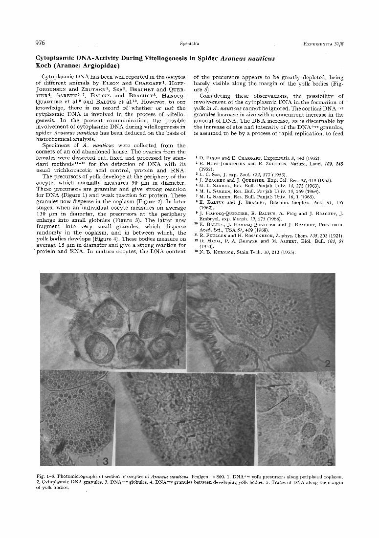
The precursors of yolk develope at the pe r iphe ry of the oocyte, which normal ly measures 30 ~tm in d iameter . These precursors are granular and give s t rong reac t ion for D N A (Figure 1) and weak react ion for protein . These granules now disperse in the ooplasm (Figure 2). In la ter stages, when an indiv idual oocyte measures on average 130 b~m in d iameter , t he precursors a t t he pe r iphe ry enlarge in to small globules (Figure 3). The la t t e r now f r a g m e n t in to ve ry small granules, which disperse r a n d o m l y in the ooplasm, and in be tween which, t he yolk bodies develope (Figure 4). These bodies measure on average 15 [zm in d iamete r and give a s t rong react ion for
' p r o t e i n and RNA. In ma tu r e oocytes, t he D N A co n t en t
in Spider Araneus nauticus
of t he precursors appears to be grea t ly deple ted, be ing bare ly visible along the marg in of the yolk bodies (Fig- ure 5).
Considering these observa t ions , the possibi l i ty of i nvo lvemen t of the cy top lasmic D N A in the fo rmat ion of yolk in .d. naut icus canno t be ignored. The cortical D N A +re granules increase in size w i th a concur ren t increase in t he a m o u n t of DNA. The D N A increase, as is discernable by the increase of size and in t ens i ty of the DNA+ve granules, is a ssumed to be by a process of rap id repl icat ion, to feed
t D. ELSON and E. CHARGAFF, Experientia 8, 143 (1952). 2 E. HOFF-JORGENSEN and E. ZEUTHEN, Nature, Lond. 169, 245
(1952). 3 L. C. SzE, J. exp. Zool. /22, 577 (1953). 4 j . BRACHET and J. QUERTIER~ Expl Cell Res. 32, 410 (1963). 5 M. L. SAREEN, Res. Bull. Panjab Univ. ld, 273 (1963).
M. L. SAREEN, Res. Bull. Panjab Univ. 15, 169 (1964). 7 M. L. SAREEN, Res. Bull. Panjab Univ. 16, 1 (1965). 8 E. BALTUS and J. BRACHET, Biochim. biophys. Acta 61, 157
(1962). 9 j . HANOCQ*QUERTIER, E. BALTUS, A. FICQ and J. BRACHET, J.
Embryol. exp. Morph. 19, 273 (1968). 10 E. BALTUS, J. HANOCQ-QUERTIER and J. BRACHET, Proc. natn.
Acad. Sci., USA 61,469 (1968). 11 R. FEULGEN and H. ROSSENBECK, Z. phys. Chem. 135, 203 (1924). 12 D. MAZIA, P. A. BREWER and M. ALFERT, Biol. Bull. lOd, 57
(1953). 13 N. B. KURNICK, Stain Tech. 30, 213 (1955).
Fig. 1-5. Photomicrographs of section of oocytes of Araneus nauticus. Feulgen. • 800. 1. DNA +re yolk precursors along peripheral ooplasm. 2. Cytoplasmic DNA granules. 3. DNA+ve globules. 4. DNA +re granules between developing yolk bodies. 5. Traces of DNA along the margin of yolk bodies.

15.8. 1975 Specialia 977
the cellular mach ine ry wi th the required a m o u n t of raw mater ia l s dur ing the syn the t i c act ivi t ies of the oocyte in the e labora t ion of yolk. The D N A granules b reak up and the yolk appa ren t l y fo rmed under the i r inf luence reac t r a the r s t rong ly for pro te in and RNA. I t is no t unl ikely t h a t these two cons t i tuen ts of yolk, enough of which m u s t be p roduced to cope wi th the d e m a n d of vitel logenesis, are assembled under ins t ruc t ions f rom DNA. This in- ference is fu r the r suppor t ed by the fact tha t , in t he yolk- c r a m m e d ma tu re oocytes, t he DNA is visible j u s t in t races ind ica t ing its d i s in tegra ta t ion soon af ter t he com- ple t ion of vitellogenesis.
Summary. Cytoplasmic D N A is possibly involved in the synthes is of yolk in spider Araneus nauticus.
G. P. VERMA, K. C. PATRAI4 and C. C. DAS 14
P. G. Department o/Zoology, Bihar University, Langal Singh College, Muzaffarpur (India), and P. G. Department of Zoology, Berhamp~r University, Orissa (India), 3 February 1975.
14 p. G. Department of Zoology, Berhampur University, Orissa, India.
Local H a e m o s t a s i s in Bra in T u m o u r s
Li t t l e in te res t has been dedica ted up to now to the local haemos tas i s in t u m o u r s l , 2. Only recen t ly have repor t s appeared on the occurrence in se rum and o ther f luids of f ibr in / f ibr inogen degrada t ion p roduc t s (FDP) as the resul ts of coagulat ion and f ibr inolyt ic processes a t the site of t he neoplasm e, 3. De te rmina t ion of F D P in neoplas t ic diseases has p roved useful in the diagnosis and t r e a t m e n t of m a l i g n a n t diseases 2, 4, 5.
The p resen t inves t iga t ion was unde r t aken to elucidate the local haemos tas i s of two di f ferent cerebral tumours , viz mening iomas and gliomas. We examined 13 supra ten - torial tu rnouts (7 men ing iomas and 6 gliomas) ope ra ted upon at the Neurosurgical Clinic of Ume&.
Th rombop la s t i c ac t iv i ty was d e t e r m i n e d essent ia l ly according to ASTRUP et al 6. The piece of t issue was homog- enized wi th 0.15 M NaC1, 0.9 ml per 100 mg of t issue. Larger specimens were d iv ided into f r agmen t s weighing 2-3 g, which were t e s t ed separately . Af ter homogen iza t ion the suspens ion was frozen overnight , t hawed and re- homogenized . The larger par t ic les of the resul t ing suspen- sion were a f te rwards r emoved by f i l t ra t ion t h ro u g h co t ton wool. Serial di lut ions of the f i l t ra te wi th saline were prepared . The assay sys tem consis ted of: 0.1 ml saline ; 0.2 ml h u m a n p la te le t -poor c i t ra ted h u m a n p l a sma ; 0,2 ml 0.03 ~r CaC12; 0.1 ml t issue suspension. The m i x t u r e of saline, p l a sma and t issue ex t rac t was first hea t ed for 3 min a t 37~ in a t he rmos t a t i c b a t h af ter which CaCI 2 was added and double de te rmina t ions were made of the
c lo t t ing t ime. The c lo t t ing t ime is given in seconds as the mean of two de te rmina t ions . Resul t s a r e expressed as percentages of the ac t iv i ty of a s t a n d a r d suspens ion of h u m a n bra in t h r o m b o p l a s t i n as de t e rmined by in te rpo la - t ion on the s t r a igh t line ob ta ined by p lo t t ing di lu t ions against c lo t t ing t imes in a double logar i thmic graph.
F ibr inoly t ic ac t iv i ty . The specimens were examined by a modif ica t ion 7 of TODD'S f ibrin slide t echn ique s which pe rmi t s local izat ion and assay of f ibr inolyt ic ac t iv i ty in tissues. A series of 4 slides was p repa red for each specimen. The slides of each series were incuba ted for increas ing per iods of t ime, i.e. 15, 30, 45, 60 rain respect ively , and a f te rwards f ixed in formal in and s ta ined wi th Har r i s ' haema toxy l in . 3 fair ly d i s t inc t degrees of f ibrin d iges t ion were recognized, n a m e l y grade I: microscopical p u n c t a t e
1 H. I. PETERSON, Acta chir. scand, suppl. 1969, 394. 2 L, SVANBERG, F. LINELL, M. PANDOLFI a n d B. ~STEDT, A e t a p a t h .
mierobiol, scand., in press. a [. M. NILSSON, Scand. J. Haemat. 13, 317 (197l). 4 L. SVANBERG, B. aSTI*2DT, l. GYNNING a n d 1. M. NILSSON, A c t a
obst. gynee, stand. 52, 141 (1973). 5 B. ~STEDT 7 g. SVANBERG and I. M. NILSSON, Br. reed. J. 4, 458
(1971). Ii T. ASTRUP, O. ](.. ALBRECHTSEN a n d .J. RASMUSSEN, C i rcu la t ion
Res. 7, 969 (1969). 7 M. I)ANDOLI,'I~ B. ROBERTSON, S. ISACSON a n d 1. M. NII.SSON,
Thromb. I)iath. haemorrh. 20, 247 (]969). s A. S. TODD, J. path. Bact. 78, 281 (1959).
Fig. 1. Meningioma. Numerous areas of fibrinolysis confined to connectival septa and to blood vessel. Incubation time 30 rain. •