CYTOPATHOLOGY-1 DR. MAHA AL-SEDIK. Objectives: 1- Introduction to cytology. 2- Differences between...
33
CYTOPATH OLOGY-1 DR. MAHA AL-SEDIK
-
Upload
clarence-haynes -
Category
Documents
-
view
246 -
download
4
Transcript of CYTOPATHOLOGY-1 DR. MAHA AL-SEDIK. Objectives: 1- Introduction to cytology. 2- Differences between...

CYTOPA
THOLO
GY-
1
DR
. M
AH
A A
L -SED
IK

Objectives:
1- Introduction to cytology.
2- Differences between cytology and histopathology.
3- Advantage and disadvantage of cytology.
4- Sampling techniques.
5- Indications of cytology.

I examine the structure of few separated cells not an intact tissue.

Introduction to cytology
Cytology :
Diagnostic cytology is the microscopic study of cells
for the purpose of diagnosing disease.
It differs importantly from histopathology in that
cytology examines only discrete cells or small
groups of cells, but histopathology examines intact
tissues.

Histopathology
Cytopathology
I. Deals with the form and the structure of the tissue.
I. Deals with the changes within the nucleus and cytoplasm of cells.
II. Evaluation usually begins with a tissue biopsy so it is more invasive and traumatic.
II. Fine needles with small gauge are usually preferred so it is less invasive and traumatic.

III. Basic stain is H&E
IV. Diagnosis obtained after days.
IV. Rapid diagnosis that could be obtained within minutes.
V. Inexpensive simple means of diagnosis which allows frequent repetition of cellular sampling
V. Expensive means of diagnosis do not allow for repetition
III. Basic stain is Pap stain (however H&E could be used as well)

Advantages of diagnostic cytology:
Superior morphologic detail.
Ability to characterize the cellular components of
various fluids.
Speed of sample collection and diagnosis .
Excellent cost effectiveness.
Minimally invasive diagnostic procedure not requiring
surgery or specialized anesthesia.

The disadvantage of diagnostic cytology:
Tumors cannot be graded.
Margins cannot be evaluated.
Limited information about structural arrangement
(architecture) of the cells within the lesion.
Less definitive diagnosis as to the specific tumor
type or the distribution of an inflammatory infiltrate.

Very bad negative test: A problem related to the
small sample size and "blind" nature of many
common collection techniques, is the possibility
that the specimen may not represent the primary
lesion. I am sad because you are taking the sample away from the primary lesion

The following points should be kept in mind when
starting a cytological evaluation:
1) Cytology is an aid to clinical diagnosis, and results
must be interpreted in the context of the overall
case.
2) Negative results should be viewed as inconclusive
( bad negative).
3) Cytology can be but is not always a substitute for
histopathology.
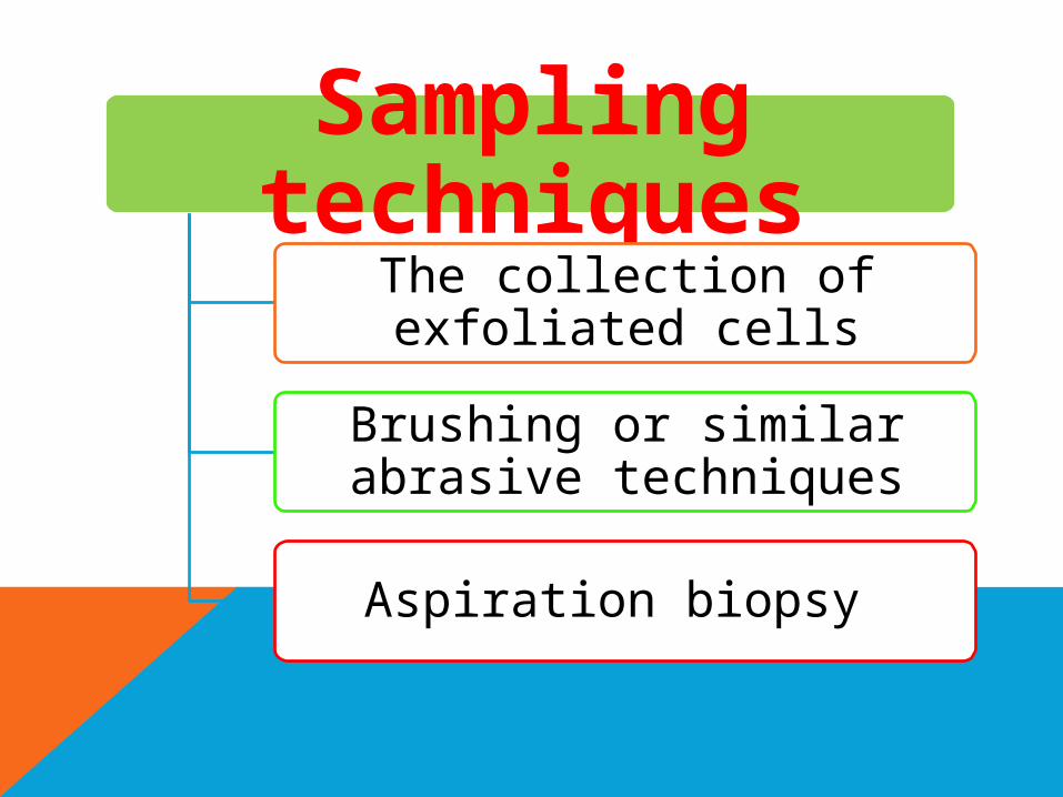
Sampling techniques
The collection of exfoliated cells
Brushing or similar abrasive techniques
Aspiration biopsy

Sampling Techniques
(1) the collection of exfoliated cells e.g. vaginal
discharge, sputum and urine examination.
(2) the collection of cells removed by brushing or
similar abrasive techniques e.g. buccal smear or pap
smear.
(3) the aspiration biopsy or removal of cells from non
surface –bearing tissue (or deep organs) by means of a
needle, with or without a syringe.

1- Exfoliative Cytology:
Exfoliative cytology is the simplest of the three
sampling techniques. To prevent the deterioration of
cells by air-drying or by enzymatic or bacterial
actions, the material should be processed
immediately without delay.

Typical examples are:
I- vaginal smear which is prepared from cells
removed from the posterior fornix of the vagina. The
cells that accumulate in the vaginal fornix are derived
from several sources: from the squamous epithelium
that lines the vagina, the vaginal portion of the uterine
cervix, from the epithelial lining of the endocervical
canal, and from other more distant sources such as
the endometrium.

II- Sputum examination: The sputum is a
collection of mucoid material that contains cells
derived from the buccal cavity, the pharynx, larynx,
trachea, the bronchial tree and the pulmonary
alveoli.
The same principle applies to:
Voided urine
Body fluids
Nipple discharge

2- Abrasive Cytology:
The purpose of this procedure is to enrich the sample
with cells obtained directly from the surface of the
target of interest. Hence, cell specimens are usually
obtained through superficial scraping of the lesion.
Examples:
Pap smear
Buccal mucosal smear
Skin scraping of various lesions
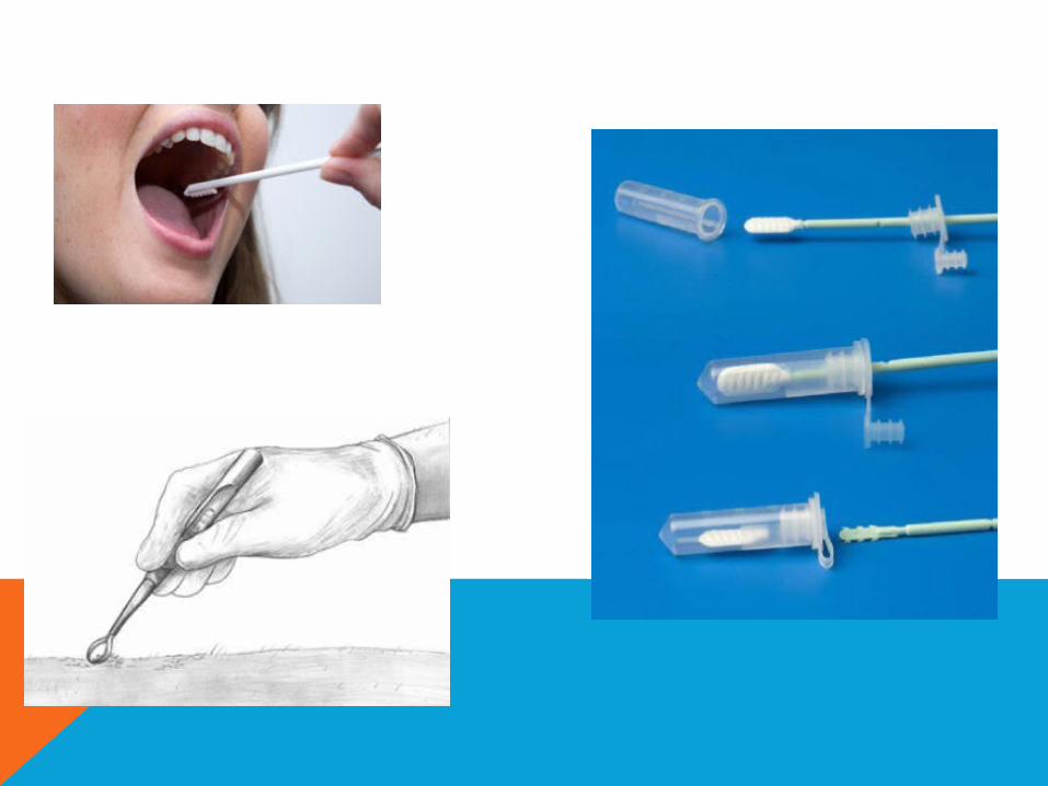


3- Fine Needle Aspiration Cytology (FNAC):
FNAC provides many advantages to the surgeons
being an easy, reliable, cost effective diagnostic
technique which could give rapid results.
The procedure could be performed in an office
setting without anesthesia.
It is usually not more painful than a venipuncture
and can be repeated immediately if the acquired
material is inadequate.


FNAC consists of:
Using a needle and syringe to remove material from
a mass.
Smearing it on a glass slide.
Applying a routine stain.
Examining it under the microscope.


inflammationPremalignant lesion
malignancyhormonal patterns
Follow-up Barr body
Differentiation between benign and malignant
Indications for
Cytopathology:

Indications for Cytopathology:
1- Differentiation between benign and malignant
lesions.
2- Diagnosis of malignancy and the identification of
the neoplastic cells in primary, metastatic (secondary).
3- Diagnosis of premalignant diseases.
4- Detection of inflammation and certain types of
pathogenic agents.

5- Study of the hormonal patterns through the
examination of the squamous cells in vaginal smears;
which are under the influence of ovarian hormones.
6- Follow-up and monitoring of response to
chemotherapy and irradiation, the latter producing
certain cellular features which could be diagnostic on
cytological examination.

7- The identification of sex chromosome: if a newborn
presents with ambiguous genitalia, one can not tell
whether the sex is male or female. The presence of a
dark dot attached to the nuclear membrane from
inside (Barr body +ve) indicates that a sex
chromosome is present, i.e., the genotype of the baby
is
XX (♀).
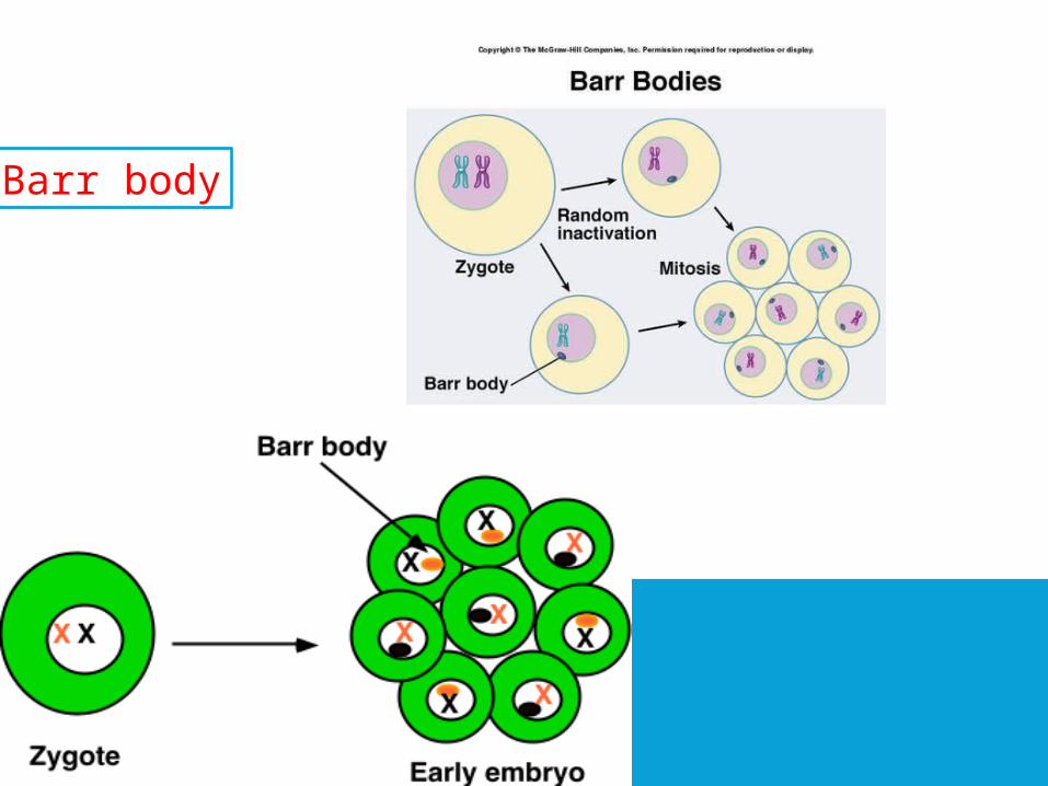
Barr body