Cytogenetic and Molecular Evaluation in Myelodysplastic ... Library/Main Nav/Research and...
7
Cytogenetic and Molecular Evaluation in Myelodysplastic Syndrome and in Acute and Chronic Leukemia Peter R. Papenhausen, PhD, Lynn C. Moscinski, MD, and Cameron G. Binnie, PhD The advent of molecular cytogenetic techniques has provided a new tool for which quantitation of residual disease or transplant monitoring may be the major advantage. Background: The majority of the presently known nonrandom chromosome changes in hematologic malignancy were described during the 1970s and 1980s. The last 10 years have been devoted to the location of oncogenes and tumor suppressor genes altered as a consequence of those changes. New molecular methodology has helped speed this process, which has resulted in DNA sequencing of many of the genes involved, permitting molecular detection of abnormal clones. Methods: This review examines the most common alteration-based subgroups of cytogenetics and molecular genetics in hematologic disorders with the exclusion of lymphoma. Prognosis has been updated to reflect improving treatment protocols. Results: The versatility of cytogenetics for delineating genetic changes is difficult to match by molecular testing. Once a clonal anomaly is identified, molecular methodology can detect residual disease with far greater sensitivity than cytogenetics, but relies on translocation junction targets that exclude clones characterized by deletion or trisomy. Conclusions:Cytogenetic and molecular testing offers independent diagnostic and prognostic evaluation for most patients with hematologic malignancy. Introduction Nonrandom cytogenetic alterations in leukemia provide definitive diagnosis and prognosis in many cases and have been crucially important in localizing oncogenes and tumor suppressor genes. DNA sequencing of many of the breakpoint junctions of the commonly observed translocations in leukemia has provided not only insights into the related oncogenic mechanisms, but also a target for molecular detection. Most translocations reported in this review appear to be capable of causing acute disease without any further genetic changes, although additional changes in chronic myelogenous leukemia (CML) are generally necessary for blast crisis. Conversely, trisomies or deletions of tumor suppressor genes in myelodysplastic syndromes (MDS) appear to progressively accumulate in leukemic clones, with each change increasing the likelihood of acute transformation. It is also clear, however, that genetic changes occur that cannot be identified by any presently known methodology. Evidence of this comes from the large percentage of MDS cases and some acute myeloid leukemia (AML) cases without cytogenetic alterations (approximately 50% and 30%, respectively). Additional evidence stems from the existence of many cases with two apparently unrelated cytogenetically distinct clones. It is likely that the "common denominator" in such cases is a submicroscopic alteration since the cells in these cases can be molecularly confirmed to be monoclonal. These unknown underlying genetic changes are the probable reason for variable prognosis in single trisomy- or single deletion-based MDS/AML cases. Molecular techniques offer specific and highly sensitive diagnostic testing of marrow aspirates or peripheral blood and are not dependent on dividing cells. Polymerase chain reaction (PCR)-based testing, available for many translocations, can also be performed from paraffin-embedded archival samples or frozen sections. The sensitivity of PCR for detection of residual disease can be as great as one malignant cell in a background of 100,000 normal cells. This article is intended to be a concise review of cytogenetic and molecular testing in common hematologic malignancy. There are many nonrandom changes that have not been included due to low incidence rates. Myeloid Disorders Myelodysplasia Immunophenotyping, cytogenetics, and molecular genetics can all provide valuable and often complementary diagnostic and prognostic information in hematologic disorders characterized by overproliferation of various cell lineages. Cytogenetics may be most valuable in MDS. The multilineage dyspoiesis characteristic of these dyscrasias makes flow cytometry impractical, and the relative absence of fusion alterations (translocations) severely limits the use of molecular techniques that target the DNA sequences within the fusion sites. The five accepted subclasses of MDS, as well as the percentage of cases that are cytogenetically abnormal, are listed in Table 1. The cytogenetic changes that are most commonly found in each subgroup are noted in Table 2. The percentage of cases with abnormal chromosomes tends to increase with the blast percentages.
-
Upload
hoangnguyet -
Category
Documents
-
view
214 -
download
0
Transcript of Cytogenetic and Molecular Evaluation in Myelodysplastic ... Library/Main Nav/Research and...
Cytogenetic and Molecular Evaluation in Myelodysplastic Syndrome and in Acute and Chronic Leukemia
Peter R. Papenhausen, PhD, Lynn C. Moscinski, MD, and Cameron G. Binnie, PhD
The advent of molecular cytogenetic techniques has provided a new tool for which quantitation of residual disease or transplant monitoring may be the major advantage.
Background: The majority of the presently known nonrandom chromosome changes in hematologic malignancy were described during the 1970s and 1980s. The last 10 years have been devoted to the location of oncogenes and tumor suppressor genes altered as a consequence of those changes. New molecular methodology has helped speed this process, which has resulted in DNA sequencing of many of the genes involved, permitting molecular detection of abnormal clones. Methods: This review examines the most common alteration-based subgroups of cytogenetics and molecular genetics in hematologic disorders with the exclusion of lymphoma. Prognosis has been updated to reflect improving treatment protocols. Results: The versatility of cytogenetics for delineating genetic changes is difficult to match by molecular testing. Once a clonal anomaly is identified, molecular methodology can detect residual disease with far greater sensitivity than cytogenetics, but relies on translocation junction targets that exclude clones characterized by deletion or trisomy. Conclusions:Cytogenetic and molecular testing offers independent diagnostic and prognostic evaluation for most patients with hematologic malignancy.
Introduction
Nonrandom cytogenetic alterations in leukemia provide definitive diagnosis and prognosis in many cases and have been crucially important in localizing oncogenes and tumor suppressor genes. DNA sequencing of many of the breakpoint junctions of the commonly observed translocations in leukemia has provided not only insights into the related oncogenic mechanisms, but also a target for molecular detection. Most translocations reported in this review appear to be capable of causing acute disease without any further genetic changes, although additional changes in chronic myelogenous leukemia (CML) are generally necessary for blast crisis. Conversely, trisomies or deletions of tumor suppressor genes in myelodysplastic syndromes (MDS) appear to progressively accumulate in leukemic clones, with each change increasing the likelihood of acute transformation. It is also clear, however, that genetic changes occur that cannot be identified by any presently known methodology. Evidence of this comes from the large percentage of MDS cases and some acute myeloid leukemia (AML) cases without cytogenetic alterations (approximately 50% and 30%, respectively). Additional evidence stems from the existence of many cases with two apparently unrelated cytogenetically distinct clones. It is likely that the "common denominator" in such cases is a submicroscopic alteration since the cells in these cases can be molecularly confirmed to be monoclonal. These unknown underlying genetic changes are the probable reason for variable prognosis in single trisomy- or single deletion-based MDS/AML cases.
Molecular techniques offer specific and highly sensitive diagnostic testing of marrow aspirates or peripheral blood and are not dependent on dividing cells. Polymerase chain reaction (PCR)-based testing, available for many translocations, can also be performed from paraffin-embedded archival samples or frozen sections. The sensitivity of PCR for detection of residual disease can be as great as one malignant cell in a background of 100,000 normal cells.
This article is intended to be a concise review of cytogenetic and molecular testing in common hematologic malignancy. There are many nonrandom changes that have not been included due to low incidence rates.
Myeloid Disorders
Myelodysplasia
Immunophenotyping, cytogenetics, and molecular genetics can all provide valuable and often complementary diagnostic and prognostic information in hematologic disorders characterized by overproliferation of various cell lineages. Cytogenetics may be most valuable in MDS. The multilineage dyspoiesis characteristic of these dyscrasias makes flow cytometry impractical, and the relative absence of fusion alterations (translocations) severely limits the use of molecular techniques that target the DNA sequences within the fusion sites. The five accepted subclasses of MDS, as well as the percentage of cases that are cytogenetically abnormal, are listed in Table 1. The cytogenetic changes that are most commonly found in each subgroup are noted in Table 2. The percentage of cases with abnormal chromosomes tends to increase with the blast percentages.

The deletion of the long arm of chromosome 5 and monosomy 7 deserves special mention since these alterations have a consistent clinical presentation.1 Characteristics of these are as follows:
Deletion of the Long-Arm Chromosome 5 (5q– Syndrome)
l Macrocytic anemia resistant to therapy l Normal or increased platelets l Morphologically characteristic megakaryocytic abnormalities (micromegakaryocytes; hypolobulated nuclei) l Generally mild clinical course l Only rarely transform to AML, provided 5q- is the only cytogenetic anomaly present l 5q31 is the minimal common deleted segment suggesting the locus of a tumor suppressor gene)
Monosomy 7 Syndrome
l Male:female ratio: 2:1 l Age at onset:6 months to 19 yrs (median = 7 yrs) l Clinical complications:Infection and bleeding l Background:Usually no exposure to genotoxins, but multiple sibling cases indicate possible predisposition l Presenting hematology:Anemia, decreased platelets, normal or elevated white blood cell count (especially monocytes), marrow hyperplasia l Myelodysplastic stage:Duration up to 12 yrs (median = 2.2 yrs); may be categorized as idiopathic myelofibrosis l Cytogenetics:Percentage of monosomic cells in marrow increases with progression; additional alterations often arise with transformation l Subtype of acute leukemia at progression: frequently transforms to AML M1, M2, and M4
Most MDS-associated aberrations are deletions or trisomies. These types of alterations exist in almost all types of neoplasia in relation to clonal progression. Specific
deleted segments largely correlate with the loss of tumor suppressor genes.2,3 With most tumor suppressor genes, a loss of both copies is necessary to produce an effect; with others, however, a loss of function from a single copy may be sufficient to produce an effect. A second "hit" in the same suppressor gene may occur as a submicroscopic mutation undetectable by cytogenetic analysis. At present, it is not known what causes transformation in the few patients who present with a deletion and no other cytogenetic change but progress to AML (Fig 1). Most patients who do show clonal karyotypic evolution transform to AML.
Translocations are rare in MDS and are generally characterized by a brief MDS phase before blast levels increase to the point where a diagnosis of AML is made. Alterations involving chromosome 3 correlate with dysmegakaryopoesis and a poor prognosis (Table 3). Other translocations occur secondary to previous therapy. Prognosis-correlated cytogenetic subgroups are apparent, although transformation to AML occurs in all groups.
Therapy-Related MDS/AML (t-MDS/t-AML)
The development of MDS or AML secondary to treatment of malignant disease with cytotoxic drugs or radiation has been recognized for some time.4 Distinct
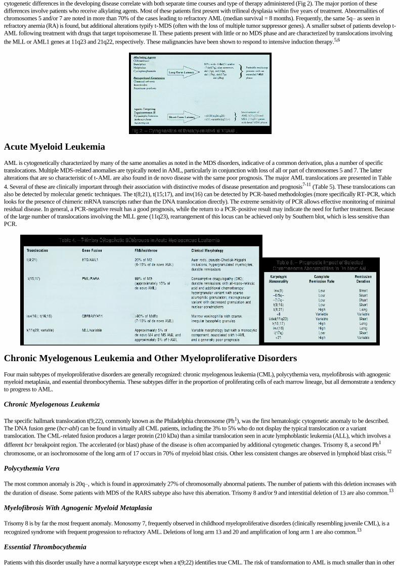
cytogenetic differences in the developing disease correlate with both separate time courses and type of therapy administered (Fig 2). The major portion of these differences involve patients who receive alkylating agents. Most of these patients first present with trilineal dysplasia within five years of treatment. Abnormalities of chromosomes 5 and/or 7 are noted in more than 70% of the cases leading to refractory AML (median survival = 8 months). Frequently, the same 5q– as seen in refractory anemia (RA) is found, but additional alterations typify t-MDS (often with the loss of multiple tumor suppressor genes). A smaller subset of patients develop t-AML following treatment with drugs that target topoisomerase II. These patients present with little or no MDS phase and are characterized by translocations involving
the MLL or AML1 genes at 11q23 and 21q22, respectively. These malignancies have been shown to respond to intensive induction therapy.5,6
Acute Myeloid Leukemia
AML is cytogenetically characterized by many of the same anomalies as noted in the MDS disorders, indicative of a common derivation, plus a number of specific translocations. Multiple MDS-related anomalies are typically noted in AML, particularly in conjunction with loss of all or part of chromosomes 5 and 7. The latter alterations that are so characteristic of t-AML are also found in de novo disease with the same poor prognosis. The major AML translocations are presented in Table
4. Several of these are clinically important through their association with distinctive modes of disease presentation and prognosis7-11 (Table 5). These translocations can also be detected by molecular genetic techniques. The t(8;21), t(15;17), and inv(16) can be detected by PCR-based methodologies (more specifically RT-PCR, which looks for the presence of chimeric mRNA transcripts rather than the DNA translocation directly). The extreme sensitivity of PCR allows effective monitoring of minimal residual disease. In general, a PCR-negative result has a good prognosis, while the return to a PCR-positive result may indicate the need for further treatment. Because of the large number of translocations involving the MLL gene (11q23), rearrangement of this locus can be achieved only by Southern blot, which is less sensitive than PCR.
Chronic Myelogenous Leukemia and Other Myeloproliferative Disorders
Four main subtypes of myeloproliferative disorders are generally recognized: chronic myelogenous leukemia (CML), polycythemia vera, myelofibrosis with agnogenic myeloid metaplasia, and essential thrombocythemia. These subtypes differ in the proportion of proliferating cells of each marrow lineage, but all demonstrate a tendency to progress to AML.
Chronic Myelogenous Leukemia
The specific hallmark translocation t(9;22), commonly known as the Philadelphia chromosome (Ph1), was the first hematologic cytogenetic anomaly to be described. The DNA fusion gene (bcr-abl) can be found in virtually all CML patients, including the 3% to 5% who do not display the typical translocation or a variant translocation. The CML-related fusion produces a larger protein (210 kDa) than a similar translocation seen in acute lymphoblastic leukemia (ALL), which involves a
different bcr breakpoint region. The accelerated (or blast) phase of the disease is often accompanied by additional cytogenetic changes. Trisomy 8, a second Ph1
chromosome, or an isochromosome of the long arm of 17 occurs in 70% of myeloid blast crisis. Other less consistent changes are observed in lymphoid blast crisis.12
Polycythemia Vera
The most common anomaly is 20q–, which is found in approximately 27% of chromosomally abnormal patients. The number of patients with this deletion increases with
the duration of disease. Some patients with MDS of the RARS subtype also have this aberration. Trisomy 8 and/or 9 and interstitial deletion of 13 are also common.13
Myelofibrosis With Agnogenic Myeloid Metaplasia
Trisomy 8 is by far the most frequent anomaly. Monosomy 7, frequently observed in childhood myeloproliferative disorders (clinically resembling juvenile CML), is a
recognized syndrome with frequent progression to refractory AML. Deletions of long arm 13 and 20 and amplification of long arm 1 are also common.13
Essential Thrombocythemia
Patients with this disorder usually have a normal karyotype except when a t(9;22) identifies true CML. The risk of transformation to AML is much smaller than in other
subgroups.14
In general, finding cytogenetic anomalies at diagnosis does not imply that conversion of myeloproliferative disorders to AML is imminent, but further karyotypic evolution has been found to correlate with disease progression. Complex aberrations, regardless of presentation time, clearly correlate with adverse prognoses.
Lymphoid Disorders
In general, lymphoid neoplasms are clonally expanded proliferations of cells arrested at distinct stages of B- or T-cell development. Disorders of the B-cell maturational cycle, depicted in Fig 3, are far more common than T-cell dyscrasias.
The principal change during maturation of immature lymphoblasts to B lymphocytes is the rearrangement of the immunoglobulin genes. The heavy chain locus is located
at 14q32, and the light chain loci are at 2p12(kappa) and 22q11(lambda). All of these loci are involved in chromosomal alterations that characterize B-cell neoplasia.15 In many cases, however, the genetic abnormality is too small to be detected by cytogenetics or the genetic change does not produce a cell surface marker detectable by flow cytometry. The presence of monoclonal malignant cells can then be established by detection of a molecular rearrangement of the immunoglobulin genes. Similarly, malignant T-cell disorders, which are characterized by rearrangement of the T-cell receptor genes (proportional and partial derivative at 14q11, beta at 7q34,
and gamma at 7p13), can be detected by rearrangement of the loci by cytogenetic or molecular methodology.16 Molecular detection of immunoglobulin and T-cell receptor gene rearrangement has to this point been performed by Southern blot. The recent development of PCR-based tests for gene rearrangement promises to not only increase the sensitivity of detection, but also offer the opportunity of testing paraffin-embedded specimens.
Acute Lymphoblastic Leukemia
Specific diagnosis/prognosis-associated chromosomal translocations are found in approximately 40% of B-ALL cases (Tables 6 and 7 -- Please see hard copy of journal for Table 7). These alterations are clonal and directly involved in leukemogenesis. Similar to AML, these recurrent alterations result in fusion genes or transcriptional derepression. Patients with the poor prognosis translocations -- 9;22, 4;11, and 1;19 -- are good candidates for allogeneic bone marrow transplantation following the first remission.
Although most of the ALL translocations bear a poor prognosis,17 this is not true of the newly described t(12;21).18 The subtle nature of the terminal band exchange in this rearrangement and the difficulty of obtaining optimal banded preparations in ALL have likely led to extensive underdiagnosis. New molecular cytogenetic (fluorescence in situ hybridization [FISH]) or molecular testing will be important in detecting this favorable prognostic subgroup.
The hyperdiploid (>50 chromosomes) subgroup is also notable due to the high incidence of this very favorable prognostic subgroup.19 These cases have a CALLA (common acute lymphoblastic leukemia antigen)-positive immunophenotype and a DNA index of 1.14 to 1.20. The invariant nature of specific trisomic chromosomes and tetrasomy 21 facilitates targeted FISH detection methodology, although the pathogenetic mechanism behind such one-step amplification remains difficult to explain.
Chronic Lymphoproliferative Disorders
As already indicated, neoplastic clones can be detected by monoclonal rearrangements of immunoglobulin genes (B-cell) or T-cell receptor genes. Cytogenetic alterations at the site of those genes are the most consistent changes noted in all chronic lymphoid dyscrasias. An inversion of chromosome 14 involving the T-cell receptor gene located at q11 and a putative oncogene (often called TCL1) at q32.1 has been reported in adult T-cell leukemia, T-CLL, and T-PLL (T-prolymphocytic leukemia). TCL1 rearrangement can be observed in random cells of normal individuals and at increased levels in ataxia telangiectasia, so rearrangement alone appears insufficient in producing a malignancy.
Many cytogenetic studies have targeted the most common subgroup. B-cell CLL. (T-cell CLL accounts for less than 2% of all cases.20) The following consensus points are noteworthy:
l B-mitogen culturing has increased the detection of abnormal clones to 40% to 60% of cases. l The most common alteration is trisomy 12, which can be detected in approximately one third of cytogenetically abnormal cases (greater frequency with FISH). l Trisomy 12 is associated with a mixed-cell phenotype often evolving to prolymphocytic leukemia and poorer prognosis.
l Patients characterized by a deletion of long arm 13 (13q–) have typical CLL morphology and a similar survival as those without clonal changes.21,22
l Those patients with the 14q+ (IgH locus) anomaly or complex karyotypes have the poorest prognosis.
Numerous cytogenetic studies in multiple myeloma have also generated considerable data. The percentage of abnormal cases (20% to 60%) has varied considerably, presumably due to the slow proliferation of plasma cells that often require extensive searching to identify the abnormal clone. The following observations are generally
accepted23,24: (1) Survival is longer for patients without a detectable abnormal clone, (2) the most consistent anomaly is a 14q+ alteration with t(11;14)(q13;q32) predominating, (3) the often complex abnormal clones are typified by loss of tumor suppressor genes and nonrandom trisomy but not trisomy 12, and (4) prognosis worsens with increasing proportions of clonally abnormal cells detected.
Molecular Cytogenetics
In situ hybridization using sequence-specific DNA probes with fluorescence (FISH) or enzyme-labeling systems is primarily applied for the segmental chromosome analysis at interphase. The main advantages are (1) no requirement for cell division, (2) many cells can be evaluated and quantified, as opposed to nonquantitative molecular technology, and (3) good results can also be obtained from formalin-fixed paraffin embedded or frozen tissue. The disadvantages are: (1) targeted probes give no information regarding clonal evolution, and (2) low levels of residual disease (<4%) merge with normal background levels.
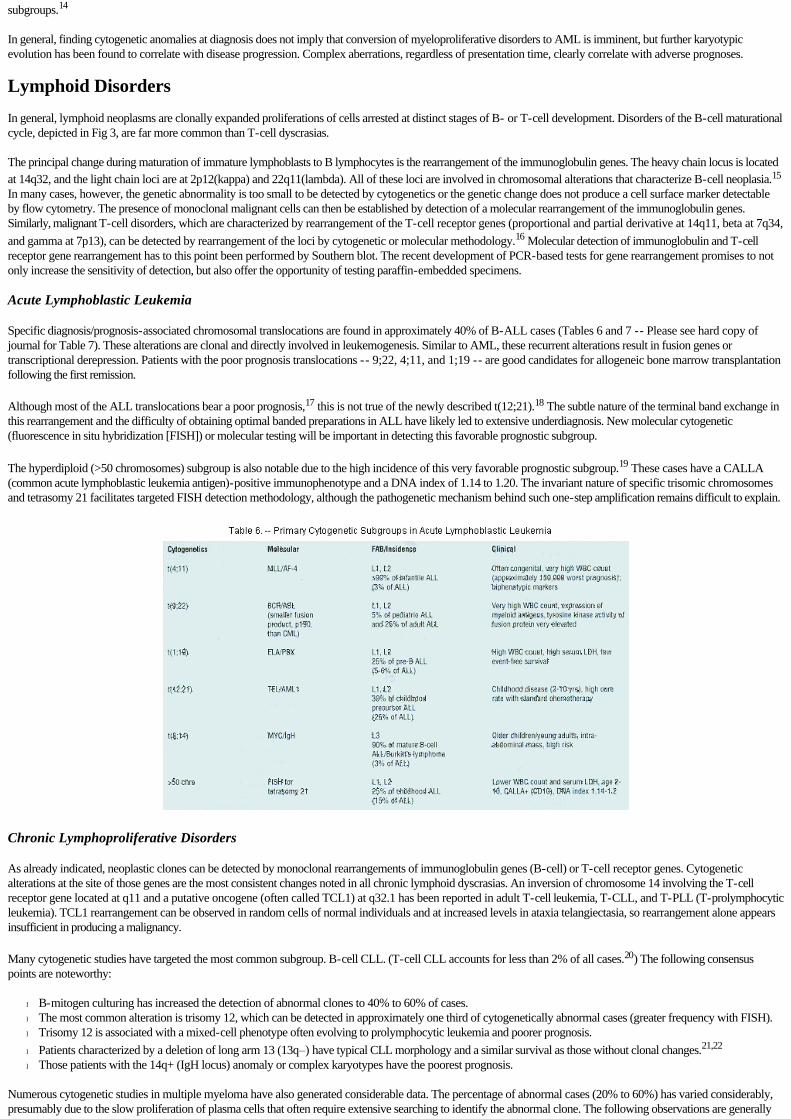
Uses (Interphase)
The primary use involves the detection of residual disease or transplant monitoring (Fig 4). Cloneidentifying probes may be selected by using the information obtained from a pretreatment diagnostic karyotype. These may be translocation specific with dual color merging (Fig 5) with probes specific for DNA sequences adjacent to the breaksites or may be shown by a split of a probe that spans a specific gene leaving an extra signal (Fig 6). Alternately, probes may be used that are specific to appropriate alphoid (centromere) repeats of preidentified clones with monosomy or trisomy.
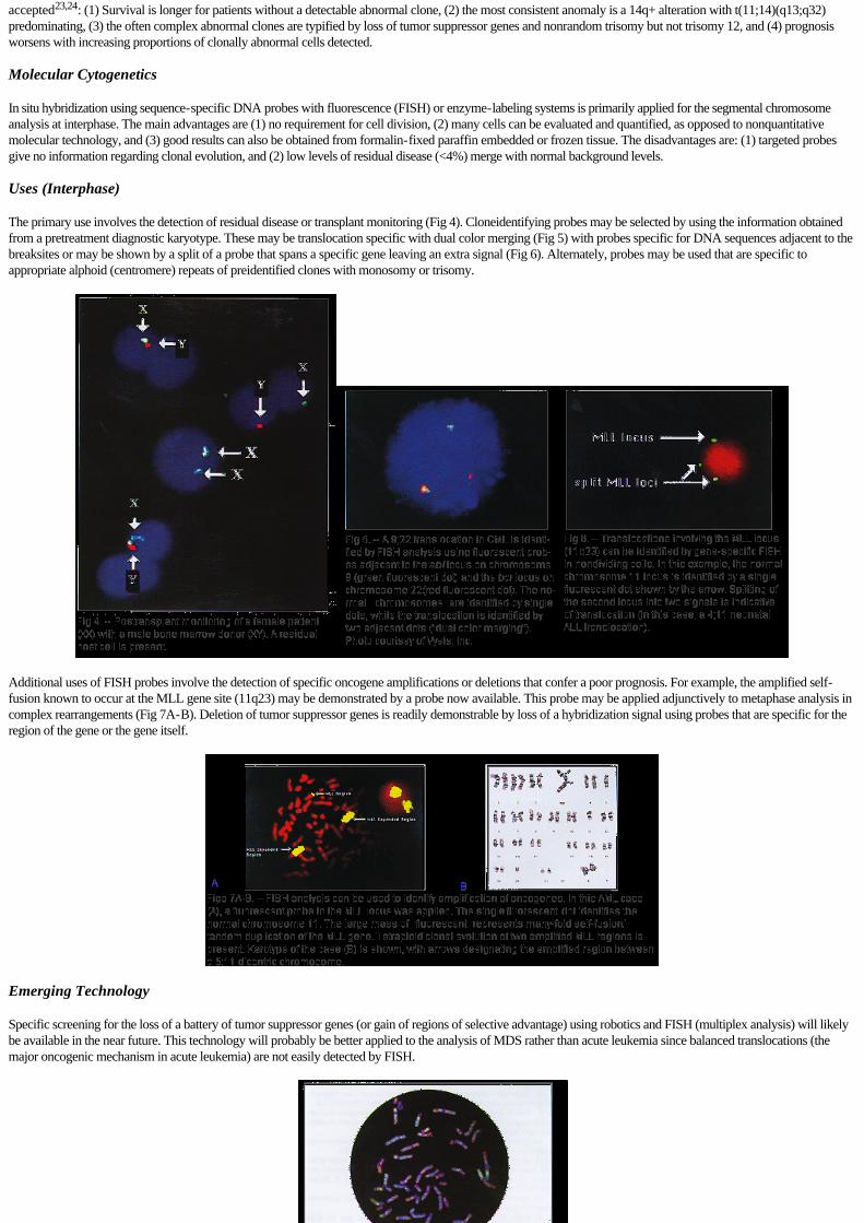
Additional uses of FISH probes involve the detection of specific oncogene amplifications or deletions that confer a poor prognosis. For example, the amplified self-fusion known to occur at the MLL gene site (11q23) may be demonstrated by a probe now available. This probe may be applied adjunctively to metaphase analysis in complex rearrangements (Fig 7A-B). Deletion of tumor suppressor genes is readily demonstrable by loss of a hybridization signal using probes that are specific for the region of the gene or the gene itself.
Emerging Technology
Specific screening for the loss of a battery of tumor suppressor genes (or gain of regions of selective advantage) using robotics and FISH (multiplex analysis) will likely be available in the near future. This technology will probably be better applied to the analysis of MDS rather than acute leukemia since balanced translocations (the major oncogenic mechanism in acute leukemia) are not easily detected by FISH.
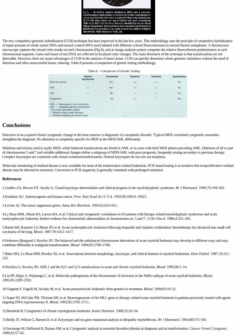
The new competitive genomic hybridization (CGH) technique has been improved in the last few years. This methodology uses the principle of competitive hybridization of equal amounts of whole-tumor DNA and normal-control DNA (each labeled with different-colored fluorochromes) to normal human metaphases. A fluorescence microscope captures the mixed color results on each chromosome (Fig 8), and an image analysis system computes the relative fluorochrome predominance at each chromosomal segment. Gains and losses of test DNA are reflected in localized color changes. The main drawback of the technique is that translocations are not detectable. However, there are major advantages of CGH in the analysis of tumor tissue. CGH can quickly determine whole genomic imbalance without the need of laborious and often unsuccessful tumor culturing. Table 8 presents a comparison of genetic testing methodology.
Conclusions
Detection of an acquired clonal cytogenetic change in the bone marrow is diagnostic of a neoplastic disorder. Typical MDS-correlated cytogenetic anomalies strengthen the diagnosis. No alteration is completely specific for MDS in the MDS/AML differential.
Deletions and trisomy tend to typify MDS, while balanced translocations are found in AML or in cases with brief MDS phases preceding AML. Deletions of all or part of chromosomes 5 and 7 and variable additional changes define a subgroup of MDS/AML with poor prognosis, frequently arising secondary to previous therapy. Complex karyotypes are consistent with clonal evolution/transformation. Normal karyotypes do not rule out neoplasia.
Molecular monitoring of residual disease is now available for most of the translocation-related leukemias. PCR-based testing is so sensitive that nonproliferative residual disease may be detected in remission. Conversion to PCR negativity is generally consistent with prolonged remission.
References
1.Geddes AA, Bowen DT, Jacobs A. Clonal karyotype abnormalities and clinical progress in the myelodysplastic syndrome. Br J Haematol. 1990;76:194-202.
2.Knudson AG. Antioncogenes and human cancer. Proc Natl Acad Sci U S A. 1993;90:10914-10921.
3.Levine AJ. The tumor suppressor genes. Annu Rev Biochem. 1993;62:623-651.
4.Le Beau MM, Albain KS, Larson RA, et al. Clinical and cytogenetic correlations in 63 patients with therapy-related myelodyplastic syndromes and acute nonlymphocytic leukemia: further evidence for characteristic abnormalities of chromosomes no. 5 and 7. J Clin Oncol. 1986;4:325-345.
5.Ratain MJ, Kaminer LS, Bitran JD, et al. Acute nonlymphocytic leukemia following etoposide and cisplatin combination chemotherapy for advanced non-small-cell carcinoma of the lung. Blood. 1987;70:1412-1417.
6.Pedersen-Bjergaard J, Rowley JD. The balanced and the unbalanced chromosome aberrations of acute myeloid leukemia may develop in different ways and may contribute differently to malignant transformation. Blood. 1994;83:2780-2786.
7.Bitter MA, Le Beau MM, Rowley JD, et al. Associations between morphology, karyotype, and clinical features in myeloid leukemias. Hum Pathol. 1987;18:211-225.
8.Nucifora G, Rowley JD. AML1 and the 8;21 and 3;21 translocations in acute and chronic myeloid leukemia. Blood. 1995;86:1-14.
9.Liu PP, Hajra A, Wijmenga C, et al. Molecular pathogenesis of the chromosome 16 inversion in the M4Eo subtype of acute myeloid leukemia. Blood. 1995;85:2289-2302.
10.Grignani F, Fagioli M, Alcalay M, et al. Acute promyelocytic leukemia: from genetics to treatment. Blood. 1994;83:10-25.
11.Super HJ, McCabe NR, Thirman MJ, et al. Rearrangements of the MLL gene in therapy-related acute myeloid leukemia in patients previously treated with agents targeting DNA-topoisomerase II. Blood. 1993;82:3705-3711.
12.Bernstein R. Cytogenetics of chronic myelogenous leukemia. Semin Hematol. 1988;25:20-34.
13.Reilly JT, Wilson G, Barnett D, et al. Karyotypic and ras gene mutational analysis in idiopathic myelofibrosis. Br J Haematol. 1994;88:575-581.
14.Sessarego M, Defferrari R, Dejana AM, et al. Cytogenetic analysis in essential thrombocythemia at diagnosis and at transformation. Cancer Genet Cytogenet. 1989;43:57-65.

15.Lieber MR. The role of site directed recombinases in physiologic and pathologic chromosomal rearrangements. In: Kirsch IR, ed. The Causes and Consequences of Chromosomal Aberrations. Boca Raton, Fla: CRC Press; 1993:239-275.
16.Chan A, Mak TW. Genomic organization of the T-cell receptor. Cancer Detect Prev. 1989;14:261-267.
17.Raimondi SC. Current status of cytogenetic research in childhood acute lymphoblastic leukemia. Blood. 1993;81:2237-2251.
18.Romana SP, Poirel H, Leconiat M, et al. High frequency of t(12;21) in childhood B-lineage acute lymphoblastic leukemia. Blood. 1995;86:4263-4269.
19.Hanson CA, Hoyer JD, Li CY, et al. Similarities between T-cell chronic lymphocytic leukemia and the small-cell variant of T-prolymphocytic leukemia. Blood. 1996;87:3520-3521.
20.Secker-Walker LM. Prognostic and biological importance of chromosome findings in acute lymphoblastic leukemia. Cancer Genet Cytogenet. 1990;49:1-13.
21.Juliusson G, Oscier DG, Fitchett M, et al. Prognostic subgroups in B-cell chronic lymphocytic leukemia defined by specific chromosomal abnormalities. N Engl J Med. 1990;33:720-724.
22.Peterson LC, Lindquist LL, Church S, et al. Frequent clonal abnormalities of chromosome band 13q14 in B-cell chronic lymphocytic leukemia: multiple clones, subclones, and nonclonal alterations in 82 Midwestern patients. Genes Chromosomes Cancer. 1992;4:273-280.
23.Kowalczyk JR, Dmoszynska A, Chobotow M, et al. Cytogenetic studies in patients with multiple myeloma. Cancer Genet Cytogenet. 1991;55:173-179.
24.Weh HJ, Gutensohn K, Selbach J, et al. Karyotype in multiple myeloma and plasma cell leukaemia. Eur J Cancer. 1993;29A:1269-1273.