
Cytocompibility
8
Cytocompatibility of pure metals and experimental binary titanium alloys for implant materials Yeong-Joon Park a, *, Yo-Han Song a , Ji-Hae An a , Ho-Jun Song a , Kenneth J. Anusavice b a Department of Dental Materials and MRC for Biomineralization Disorders, School of Dentistry, Chonnam National University, Gwangju 500-757, South Korea b Department of Restorative Dental Sciences, College of Dentistry, University of Florida, Gainesville, FL 32610-0415, USA 1. Introduction Recently, commercially pure titanium (CP-Ti) has become widely used as a biomaterial for dental implants, orthopaedic implants, cardiovascular appliances, and implant-supported dental crowns because of outstanding characteristics that include high specific strength, high resistance to corrosion, greater biocompatibility, low modulus of elasticity, and high capacity to be osseointegrated with bone. 1–3 However, the use of unalloyed CP-Ti requires further improvement to overcome its limitations including strength, hardness, wear resistance, fatigue strength, and poor grindability. It is desirable also to decrease its elastic modulus as close as possible to that of bone j o u r n a l o f d e n t i s t r y 4 1 ( 2 0 1 3 ) 1 2 5 1 – 1 2 5 8 a r t i c l e i n f o Article history: Received 25 July 2013 Received in revised form 11 September 2013 Accepted 13 September 2013 Keywords: Cytocompatibility Titanium alloy Implant WST-1 test Agar overlay test a b s t r a c t Objective: This study was performed to evaluate the biocompatibility of nine types of pure metal ingots (Ag, Al, Cr, Cu, Mn, Mo, Nb, V, Zr) and 36 experimental titanium (Ti) alloys containing 5, 10, 15, and 20 wt% of each alloying element. Methods: The cell viabilities for each test group were compared with that of CP-Ti using the WST-1 test and agar overlay test. Results: The ranking of pure metal cytotoxicity from most potent to least potent was as follows: Cu > Al > Ag > V > Mn > Cr > Zr > Nb > Mo > CP-Ti. The mean cell viabilities for pure Cu, Al, Ag, V, and Mn were 21.6%, 25.3%, 31.7%, 31.7%, and 32.7%, respectively, which were significantly lower than that for the control group ( p < 0.05). The mean cell viabilities for pure Zr and Cr were 74.1% and 60.6%, respectively ( p < 0.05). Pure Mo and Nb demon- strated good biocompatibility with mean cell viabilities of 93.3% and 93.0%, respectively. The mean cell viabilities for all the Ti-based alloy groups were higher than 80% except for Ti– 20Nb (79.6%) and Ti–10V (66.9%). The Ti–10Nb alloy exhibited the highest cell viability (124.8%), which was higher than that of CP-Ti. Based on agar overlay test, pure Ag, Cr, Cu, Mn, and V were ranked as ‘moderately cytotoxic’, whereas the rest of the tested pure metals and all Ti alloys, except Ti–10V (mild cytotoxicity), were ranked as ‘noncytotoxic’. Significance: The results obtained in this study can serve as a guide for the development of new Ti-based alloy implant systems. # 2013 Elsevier LtdElsevier B.V. All rights reserved. * Corresponding author at: Department of Dental Materials, School of Dentistry, Chonnam National University, 300 Yongbong dong, Buk gu, Gwangju 500 757, South Korea. Tel.: +82 62 530 4871; fax: +82 62 530 4875. E-mail addresses: [email protected], [email protected] (Y.-J. Park). Available online at www.sciencedirect.com ScienceDirect journal homepage: www.intl.elsevierhealth.com/journals/jden 0300-5712/$ – see front matter # 2013 Elsevier LtdElsevier B.V. All rights reserved. http://dx.doi.org/10.1016/j.jdent.2013.09.003
-
Upload
nur-hidayatul-nadhirah -
Category
Documents
-
view
7 -
download
3
description
Implant material
Transcript of Cytocompibility

Cytocompatibility of pure metals and experimentalbinary titanium alloys for implant materials
Yeong-Joon Park a,*, Yo-Han Song a, Ji-Hae Ana, Ho-Jun Song a,Kenneth J. Anusavice b
aDepartment of Dental Materials and MRC for Biomineralization Disorders, School of Dentistry,
Chonnam National University, Gwangju 500-757, South KoreabDepartment of Restorative Dental Sciences, College of Dentistry, University of Florida, Gainesville,
FL 32610-0415, USA
j o u r n a l o f d e n t i s t r y 4 1 ( 2 0 1 3 ) 1 2 5 1 – 1 2 5 8
a r t i c l e i n f o
Article history:
Received 25 July 2013
Received in revised form
11 September 2013
Accepted 13 September 2013
Keywords:
Cytocompatibility
Titanium alloy
Implant
WST-1 test
Agar overlay test
a b s t r a c t
Objective: This study was performed to evaluate the biocompatibility of nine types of pure
metal ingots (Ag, Al, Cr, Cu, Mn, Mo, Nb, V, Zr) and 36 experimental titanium (Ti) alloys
containing 5, 10, 15, and 20 wt% of each alloying element.
Methods: The cell viabilities for each test group were compared with that of CP-Ti using the
WST-1 test and agar overlay test.
Results: The ranking of pure metal cytotoxicity from most potent to least potent was as
follows: Cu > Al > Ag > V > Mn > Cr > Zr > Nb > Mo > CP-Ti. The mean cell viabilities for
pure Cu, Al, Ag, V, and Mn were 21.6%, 25.3%, 31.7%, 31.7%, and 32.7%, respectively, which
were significantly lower than that for the control group ( p < 0.05). The mean cell viabilities
for pure Zr and Cr were 74.1% and 60.6%, respectively ( p < 0.05). Pure Mo and Nb demon-
strated good biocompatibility with mean cell viabilities of 93.3% and 93.0%, respectively. The
mean cell viabilities for all the Ti-based alloy groups were higher than 80% except for Ti–
20Nb (79.6%) and Ti–10V (66.9%). The Ti–10Nb alloy exhibited the highest cell viability
(124.8%), which was higher than that of CP-Ti. Based on agar overlay test, pure Ag, Cr,
Cu, Mn, and V were ranked as ‘moderately cytotoxic’, whereas the rest of the tested pure
metals and all Ti alloys, except Ti–10V (mild cytotoxicity), were ranked as ‘noncytotoxic’.
Significance: The results obtained in this study can serve as a guide for the development of
new Ti-based alloy implant systems.
# 2013 Elsevier LtdElsevier B.V. All rights reserved.
Available online at www.sciencedirect.com
ScienceDirect
journal homepage: www.intl.elsevierhealth.com/journals/jden
1. Introduction
Recently, commercially pure titanium (CP-Ti) has become
widely used as a biomaterial for dental implants, orthopaedic
implants, cardiovascular appliances, and implant-supported
dental crowns because of outstanding characteristics that
* Corresponding author at: Department of Dental Materials, School of Dgu, Gwangju 500 757, South Korea. Tel.: +82 62 530 4871; fax: +82 62 5
E-mail addresses: [email protected], [email protected] (Y.-J. Pa
0300-5712/$ – see front matter # 2013 Elsevier LtdElsevier B.V. All righttp://dx.doi.org/10.1016/j.jdent.2013.09.003
include high specific strength, high resistance to corrosion,
greater biocompatibility, low modulus of elasticity, and high
capacity to be osseointegrated with bone.1–3 However, the use
of unalloyed CP-Ti requires further improvement to overcome
its limitations including strength, hardness, wear resistance,
fatigue strength, and poor grindability. It is desirable also to
decrease its elastic modulus as close as possible to that of bone
entistry, Chonnam National University, 300 Yongbong dong, Buk30 4875.rk).
hts reserved.

j o u r n a l o f d e n t i s t r y 4 1 ( 2 0 1 3 ) 1 2 5 1 – 1 2 5 81252
tissue.4–11 Reports have focused on improved properties of CP-
Ti for use as implant materials. In a study of Ti–Ag and Ti–Cu
alloys,12 Ti–20% Ag and Ti–5% Cu alloys exhibited better
grindability than pure Ti, which is a desirable property for
dental CAD/CAM alloys. The results from a previous study
utilizing experimental Ti–Au (5–20 wt% Au), Ti–Ag (5–20 wt%
Ag), and Ti–Cu (2–10 wt% Cu) alloys suggested that their
hardness and tensile strength were higher than these
properties for pure Ti.13 In another study, Ti–Cr alloys
exhibited greater flexure strength than CP-Ti, which was
associated with the strengthening effect of the v phase.5 The
flexure strength of the Ti–20Cr alloy was about 80% greater
than that for CP-Ti, and the elastic recovery capability was
460% greater than that of CP-Ti. The lower elastic modulus of
CP-Ti and Ti alloys, compared with 316L stainless steel and Cr–
Co alloys, is potentially advantageous for preserving the
surrounding bone by minimizing the reduction in physiologi-
cal stress within bone and a reduction in bone density. This
occurs because of a more favourable match of elastic modulus
between the implants and bone.14–18 The elastic moduli of
recently developed b-Ti alloys range from 55 to 85 GPa, which
are much lower than those of 316L stainless steel, Cr–Co alloys,
and CP-Ti.2 Manganese (Mn), molybdenum (Mo), and niobium
(Nb) have been investigated as b stabilizers for Ti alloys and
studies have suggested that they can decrease the elastic
modulus and increase other important mechanical properties
of Ti-based alloys.19–27 Biocompatibility tests for dental alloys
including Ti alloys have been performed in various stud-
ies.19,28–31 When metallic materials are implanted inside a
body, they may corrode and/or wear. The released metal ions
and/or debris can be toxic or irritating to surrounding tissues.
The release of metallic ions during the destruction of the
passive film can cause side effects in the body. Even though
the Ti–6Al–4V alloy is an established implant material in
orthopaedics, it is reported that ions associated with Ti–6Al–
4V alloy inhibit the normal differentiation of bone marrow
stromal cells to mature osteoblasts in vitro.32 If biologically
relevant molecules are released from the metallic biomaterial
and interact with biologically relevant molecules, biologically
active organo-metallic and metallic salts can be formed.30,33–40
Thus, the biocompatibility of the metallic materials used for
implant treatment should be evaluated during the develop-
ment of Ti alloys.
Cytotoxicity of a biomaterial can be examined using either
a monolith of the material,41 a particulate form,42–44, or
Table 1 – Materials used in the study.
Raw material Specification
CP-Ti (Grade 2) Rod, 10 mm dia. ASTM B265
Titanium Sponge 3 mm and down 99.9%
Aluminium Foil, 1.0 mm thick, annealed, 99.99%
Chromium Pieces, 2–3 mm thick, 99.995%
Copper Shot, 13 mm dia., 99.99%, oxygen free
Manganese Granules, 0.8–10 mm, 99.98%
Molybdenum Foil, 1.0 mm thick, 99.95%
Niobium Sheet, 99.9%
Silver Silver shot, 1–5 mm, Premion1, 99.99%
Vanadium Pieces, 99.7%
Zirconium Foil, 0.02 mm thick Annealed, 99.8%
extracted solutions, which may contain several types of
released metallic compounds.45–48 Most of the biocompatibili-
ty reports for metallic elements have been based on the use of
metal salts or on extracted solutions containing the metal
cations.45,49,50 Because the cytotoxicity evaluations of metallic
compounds are performed at different places by various
methods using several kinds of cell lines, the results cannot be
compared directly to each other. Moreover, the mass release of
a particular element is not expected to be proportional to its
atomic percentage in the alloy, and the lability of an element
can be altered by other elements in the alloy.51,52 Thus, when
developing new Ti alloys, cytotoxicity assays for the fabricated
alloys and the constituent pure metals are useful to study the
effect of each component on the behaviour of cells. The in vitro
cytotoxicity tests are relatively fast and they can be standard-
ized relatively easily. Thus, the in vitro tests can provide highly
reliable data and reproducible measurements even though
their relevance to clinical practice is not always consistent.53
The evaluation of the bulk alloy cytotoxicity in a biological
environment should be performed initially. However, to date,
there is scant information about the cytotoxicity of Ti-alloy
ingots.
The aim of this study was to evaluate the biocompatibility
of candidate Ti-alloys using well-characterized fibroblast-like
cell lines. Candidate alloying elements with Ti (Ag, Al, Cr, Cu,
Mn, Mo, Nb, V, and Zr) were evaluated. Titanium alloys with
varying elemental contents of alloying elements were evalu-
ated for their cytotoxicity, consistent with the aim of
developing new Ti-alloy systems as dental implant materials.
2. Materials and methods
2.1. Sample preparation
Binary Ti–A alloys that varied in the concentrations of element
A (where A was Ag, Al, Cr, Cu, Mn, Mo, Nb, V, and Zr), in
concentrations of 5, 10, 15 or 20 wt% in the Ti alloys, were
fabricated using vacuum arc melting under a high purity argon
atmosphere on a water-cooled hearth (Table 1). To homoge-
nize the alloys, the prepared ingots were melted seven times,
and the alloy specimens were treated for 4 h at temperatures
150 8C below the respective solidus temperatures and furnace
cooled at an approximate rate of 10 8C/min to 600 8C in a high
purity argon atmosphere followed by air cooling to room
Lot no. Manufacturer
04RB-10 Daido Steel Co. Ltd., Nagoya, Japan
129Q001 Alfa Aesar, Ward Hill, USA
40762 Alfa Aesar, Ward Hill, USA
38494 Alfa Aesar, Ward Hill, USA
36686 Alfa Aesar, Ward Hill, USA
K21T034 Alfa Aesar, Ward Hill, USA
36215 Alfa Aesar, Ward Hill, USA
SH-NB04 GMH Stachow-Metall GmbH, Goslar, Germany
12186 Alfa Aesar, Ward Hill, USA
42775 Alfa Aesar, Ward Hill, USA
44752 Alfa Aesar, Ward Hill, USA

j o u r n a l o f d e n t i s t r y 4 1 ( 2 0 1 3 ) 1 2 5 1 – 1 2 5 8 1253
temperature. Prepared alloy buttons were cut into disks with a
diameter of 10 mm and a thickness of 1.2 mm. The disks were
polished by a sequence of coarser to finer abrasives succes-
sively through 2000 grit SiC abrasive. They were ultrasonically
cleaned in acetone, ethanol, and distilled water. CP-Ti disks
were used as a control group.
2.2. Cell viability test
The viability of cells was analysed using a colorimetric assay to
quantify cleavage of the tetrazolium salt WST-1 (Roche,
Germany) by mitochondrial dehydrogenases. The resulting
dye was quantified spectrophotometrically and directly
correlated to the number of metabolically active cells in the
culture.54 For the test, L-929 mouse fibroblast cells were grown
on a 100-mm-diameter Petri dish containing 10 mL of
RPMI1640 supplemented with 10% foetal bovine serum
(FBS). When sufficient number of cells had proliferated,
100 mL of the cell suspension at a cell density of
5 � 104 cells/mL were seeded on the test samples and
incubated at 37 8C under 5% CO2. After 24 h of incubation,
the cells were detached with 10 mL of 0.05% Trypsin–EDTA
solution (Gibco, Canada) for 3 min, and 100 mL of RPMI1640
were added to each well. Then, 100 mL of the mixture were
transferred into a 96-well culture plate, and 10 mL of WST-1
solution were added to each well. The cells were incubated at
37 8C in 5% CO2 for 4 h, shaken for 1 min, and the absorbance
was measured at 450 nm by a microplate reader (Bio-Rad,
USA). The cell viability on each pure metal and their alloy
samples was compared with that of CP-Ti, and this result was
expressed as the cell viability ratio (%) vs. CP-Ti. Analyses were
performed for seven samples per test group.
2.3. Agar overlay test
Ten microlitre aliquots of L-929 cell suspensions at a cell
density of 3 � 105 cells/mL were seeded in 100-mm-diameter
cell culture dishes (Corning, USA) and incubated to confluence
at 37 8C in 5% CO2. After 24 h of incubation, the medium was
replaced with 10 mL of freshly prepared agar/nutrient medium
containing RPMI1640, 5% FBS, and a 3% agarose mixture
(Sigma–Aldrich, USA). Ten millilitres of neutral red solution
(0.01% in phosphate-buffered saline; Sigma–Aldrich, USA)
were added and the cells were incubated for 15 min at room
temperature. Excess dye was removed and the test specimens
were placed on the agar surface. A 0.25% zinc dibutyldithio-
carbamate polyurethane film was used as a positive control
and a polyethylene sheet was used as negative control. The
dishes were incubated for 24 h at 37 8C in 5% CO2. Thereafter,
the cultures were examined under a microscope. Neutral red is
a weak cationic dye that readily diffuses across plasma and
organelle membranes, and accumulated in the lysosomes.
Loss of membrane integrity induced by toxic substances
results in decreased retention of neutral red. Thus, damaged
or dead cells appear decolorized compared with healthy
control cells.50,55
The decolorized zones and cell lysis around and/or under
the specimens were evaluated according to ISO 7405.56 Each
test was repeated five times. The decolorized zones were
scored as follows: 0 for no decolorization detectable; 1 for
decolorization only under the specimen; 2 for a decolorization
zone not greater than 5 mm from the specimen; 3 for a
decolorization zone not greater than 10 mm from the speci-
men; 4 for a decolorization zone greater than 10 mm from the
specimen; and 5 when the total culture was decolorized. Cell
lysis was defined as a loss of cell membrane integrity that was
visible by light microscopy. Cell lysis was scored as follows: 0
when no cell lysis was detectable; 1 for less than 20% cell lysis;
2 for 20–40% cell lysis; 3 for >40% to <60% cell lysis; 4 for 60% to
80% cell lysis; and 5 for more than 80% cell lysis. Analyses were
performed on five samples per test group. For each specimen,
one score was given, and the median score from each
specimen was calculated for both the decolorization zone
index and the lysis index. The cell response was classified as
follows: 0/0 as noncytotoxic; 1/1 as mildly cytotoxic; 2/2 to 3/3
as moderately cytotoxic; and 4/4 to 5/5 as markedly cytotoxic.
The median values were calculated to describe the central
tendency of the scores since the results were expressed as an
index in a ranking scale.56,57
2.4. Statistical analyses
The software, Statistical Package for the Social Sciences (SPSS,
version 19.0, SPSS, Inc., an IBM Company, Chicago, Illinois,
USA), was used to analyse the data from the WST-1 test based
on a Kruskal–Wallis one-way analysis of variance and
Duncan’s multiple range test. The results were expressed as
the mean � standard deviation (SD) for seven separate
experiments. A p-value < 0.05 was considered statistically
significant.58
3. Results
3.1. Cell viability
Compared with the cell viability of cells seeded into culture
wells containing only media, the cell viability for the control
group (cells seeded on CP-Ti) was 89.8 � 19.0%. Pure metals
may be considered biocompatible if the cell viability on the
metals is equivalent to or greater than that of the CP-Ti
control. After a cell culture period of 24 h, the pure metals
exhibited the following decreasing order of cell viability: Mo,
Nb, Zr, Cr, Mn, V, Ag, Al, and Cu (Fig. 1). The cell viabilities for
pure Mn, V, Ag, Al, and Cu, were 32.7 � 12.8%, 31.7 � 8.9%,
31.7 � 16.3%, 25.3 � 7.3%, and 21.6 � 10.5%, respectively,
which were significantly lower than that for the corresponding
control group ( p � 0.001, Table 2). These results demonstrate
the cytotoxicity of these five pure metals.
The cell viability for pure Zr and Cr were 74.1 � 23.3% and
60.6 � 20.9%, respectively, compared with that of CP-Ti. The
pure metals, Mo and Nb, demonstrated good biocompatibility
as evidenced by cell viabilities of 93.3 � 14.4% and 93.0 � 7.6%,
respectively, compared with that of the control group.
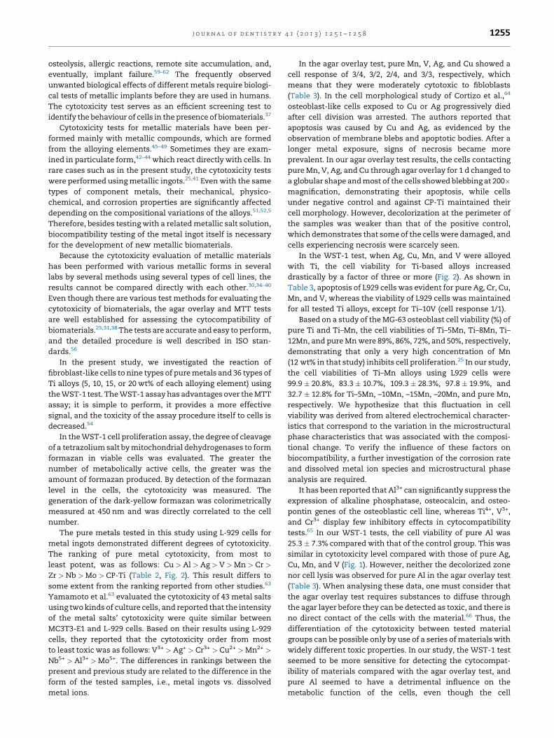
The cell viability for CP-Ti was 100%, which is the
comparison scale limit (Fig. 2). The cell viability for all Ti-
based alloy groups exceeded 80%, except for Ti–20Nb
(79.6 � 7.8%) and Ti–10V (66.9 � 22.0%). When Ag, Al, Cu,
Mn, and V were alloyed with Ti, the cell viability for Ti-based
alloys increased markedly, i.e., by a factor of more than three.
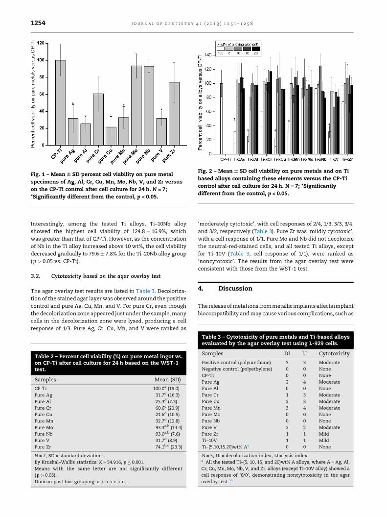
Fig. 1 – Mean W SD percent cell viability on pure metal
specimens of Ag, Al, Cr, Cu, Mn, Mo, Nb, V, and Zr versus
on the CP-Ti control after cell culture for 24 h. N = 7;
*Significantly different from the control, p < 0.05.
Fig. 2 – Mean W SD cell viability on pure metals and on Ti
based alloys containing these elements versus the CP-Ti
control after cell culture for 24 h. N = 7; *Significantly
different from the control, p < 0.05.
j o u r n a l o f d e n t i s t r y 4 1 ( 2 0 1 3 ) 1 2 5 1 – 1 2 5 81254
Interestingly, among the tested Ti alloys, Ti–10Nb alloy
showed the highest cell viability of 124.8 � 16.9%, which
was greater than that of CP-Ti. However, as the concentration
of Nb in the Ti alloy increased above 10 wt%, the cell viability
decreased gradually to 79.6 � 7.8% for the Ti–20Nb alloy group
( p > 0.05 vs. CP-Ti).
3.2. Cytotoxicity based on the agar overlay test
The agar overlay test results are listed in Table 3. Decoloriza-
tion of the stained agar layer was observed around the positive
control and pure Ag, Cu, Mn, and V. For pure Cr, even though
the decolorization zone appeared just under the sample, many
cells in the decolorization zone were lysed, producing a cell
response of 1/3. Pure Ag, Cr, Cu, Mn, and V were ranked as
Table 2 – Percent cell viability (%) on pure metal ingot vs.on CP-Ti after cell culture for 24 h based on the WST-1test.
Samples Mean (SD)
CP-Ti 100.0a (19.0)
Pure Ag 31.7d (16.3)
Pure Al 25.3d (7.3)
Pure Cr 60.6c (20.9)
Pure Cu 21.6d (10.5)
Pure Mn 32.7d (12.8)
Pure Mo 93.3a,b (14.4)
Pure Nb 93.0a,b (7.6)
Pure V 31.7d (8.9)
Pure Zr 74.1b,c (23.3)
N = 7; SD = standard deviation.
By Kruskal–Wallis statistics: K = 54.916, p � 0.001.
Means with the same letter are not significantly different
( p > 0.05).
Duncan post hoc grouping: a > b > c > d.
‘moderately cytotoxic’, with cell responses of 2/4, 1/3, 3/3, 3/4,
and 3/2, respectively (Table 3). Pure Zr was ‘mildly cytotoxic’,
with a cell response of 1/1. Pure Mo and Nb did not decolorize
the neutral-red-stained cells, and all tested Ti alloys, except
for Ti–10V (Table 3, cell response of 1/1), were ranked as
‘noncytotoxic’. The results from the agar overlay test were
consistent with those from the WST-1 test.
4. Discussion
The release of metal ions from metallic implants affects implant
biocompatibility and may cause various complications, such as
Table 3 – Cytotoxicity of pure metals and Ti-based alloysevaluated by the agar overlay test using L-929 cells.
Samples DI LI Cytotoxicity
Positive control (polyurethane) 3 3 Moderate
Negative control (polyethylene) 0 0 None
CP-Ti 0 0 None
Pure Ag 2 4 Moderate
Pure Al 0 0 None
Pure Cr 1 3 Moderate
Pure Cu 3 3 Moderate
Pure Mn 3 4 Moderate
Pure Mo 0 0 None
Pure Nb 0 0 None
Pure V 3 2 Moderate
Pure Zr 1 1 Mild
Ti–10V 1 1 Mild
Ti–(5,10,15,20)wt% Aa 0 0 None
N = 5; DI = decolorization index; LI = lysis index.a All the tested Ti–(5, 10, 15, and 20)wt% A alloys, where A = Ag, Al,
Cr, Cu, Mn, Mo, Nb, V, and Zr, alloys (except Ti–10V alloy) showed a
cell response of ‘0/00, demonstrating noncytotoxicity in the agar
overlay test.56
j o u r n a l o f d e n t i s t r y 4 1 ( 2 0 1 3 ) 1 2 5 1 – 1 2 5 8 1255
osteolysis, allergic reactions, remote site accumulation, and,
eventually, implant failure.59–62 The frequently observed
unwanted biological effects of different metals require biologi-
cal tests of metallic implants before they are used in humans.
The cytotoxicity test serves as an efficient screening test to
identify the behaviour of cells in the presence of biomaterials.37
Cytotoxicity tests for metallic materials have been per-
formed mainly with metallic compounds, which are formed
from the alloying elements.45–49 Sometimes they are exam-
ined in particulate form,42–44 which react directly with cells. In
rare cases such as in the present study, the cytotoxicity tests
were performed using metallic ingots.25,41 Even with the same
types of component metals, their mechanical, physico-
chemical, and corrosion properties are significantly affected
depending on the compositional variations of the alloys.51,52,5
Therefore, besides testing with a related metallic salt solution,
biocompatibility testing of the metal ingot itself is necessary
for the development of new metallic biomaterials.
Because the cytotoxicity evaluation of metallic materials
has been performed with various metallic forms in several
labs by several methods using several types of cell lines, the
results cannot be compared directly with each other.30,34–40
Even though there are various test methods for evaluating the
cytotoxicity of biomaterials, the agar overlay and MTT tests
are well established for assessing the cytocompatibility of
biomaterials.25,31,38 The tests are accurate and easy to perform,
and the detailed procedure is well described in ISO stan-
dards.56
In the present study, we investigated the reaction of
fibroblast-like cells to nine types of pure metals and 36 types of
Ti alloys (5, 10, 15, or 20 wt% of each alloying element) using
the WST-1 test. The WST-1 assay has advantages over the MTT
assay; it is simple to perform, it provides a more effective
signal, and the toxicity of the assay procedure itself to cells is
decreased.54
In the WST-1 cell proliferation assay, the degree of cleavage
of a tetrazolium salt by mitochondrial dehydrogenases to form
formazan in viable cells was evaluated. The greater the
number of metabolically active cells, the greater was the
amount of formazan produced. By detection of the formazan
level in the cells, the cytotoxicity was measured. The
generation of the dark-yellow formazan was colorimetrically
measured at 450 nm and was directly correlated to the cell
number.
The pure metals tested in this study using L-929 cells for
metal ingots demonstrated different degrees of cytotoxicity.
The ranking of pure metal cytotoxicity, from most to
least potent, was as follows: Cu > Al > Ag > V > Mn > Cr >
Zr > Nb > Mo > CP-Ti (Table 2, Fig. 2). This result differs to
some extent from the ranking reported from other studies.63
Yamamoto et al.63 evaluated the cytotoxicity of 43 metal salts
using two kinds of culture cells, and reported that the intensity
of the metal salts’ cytotoxicity were quite similar between
MC3T3-E1 and L-929 cells. Based on their results using L-929
cells, they reported that the cytotoxicity order from most
to least toxic was as follows: V3+ > Ag+ > Cr3+ > Cu2+ > Mn2+ >
Nb5+ > Al3+ > Mo5+. The differences in rankings between the
present and previous study are related to the difference in the
form of the tested samples, i.e., metal ingots vs. dissolved
metal ions.
In the agar overlay test, pure Mn, V, Ag, and Cu showed a
cell response of 3/4, 3/2, 2/4, and 3/3, respectively, which
means that they were moderately cytotoxic to fibloblasts
(Table 3). In the cell morphological study of Cortizo et al.,64
osteoblast-like cells exposed to Cu or Ag progressively died
after cell division was arrested. The authors reported that
apoptosis was caused by Cu and Ag, as evidenced by the
observation of membrane blebs and apoptotic bodies. After a
longer metal exposure, signs of necrosis became more
prevalent. In our agar overlay test results, the cells contacting
pure Mn, V, Ag, and Cu through agar overlay for 1 d changed to
a globular shape and most of the cells showed blebbing at 200�magnification, demonstrating their apoptosis, while cells
under negative control and against CP-Ti maintained their
cell morphology. However, decolorization at the perimeter of
the samples was weaker than that of the positive control,
which demonstrates that some of the cells were damaged, and
cells experiencing necrosis were scarcely seen.
In the WST-1 test, when Ag, Cu, Mn, and V were alloyed
with Ti, the cell viability for Ti-based alloys increased
drastically by a factor of three or more (Fig. 2). As shown in
Table 3, apoptosis of L929 cells was evident for pure Ag, Cr, Cu,
Mn, and V, whereas the viability of L929 cells was maintained
for all tested Ti alloys, except for Ti–10V (cell response 1/1).
Based on a study of the MG-63 osteoblast cell viability (%) of
pure Ti and Ti–Mn, the cell viabilities of Ti–5Mn, Ti–8Mn, Ti–
12Mn, and pure Mn were 89%, 86%, 72%, and 50%, respectively,
demonstrating that only a very high concentration of Mn
(12 wt% in that study) inhibits cell proliferation.25 In our study,
the cell viabilities of Ti–Mn alloys using L929 cells were
99.9 � 20.8%, 83.3 � 10.7%, 109.3 � 28.3%, 97.8 � 19.9%, and
32.7 � 12.8% for Ti–5Mn, –10Mn, –15Mn, –20Mn, and pure Mn,
respectively. We hypothesize that this fluctuation in cell
viability was derived from altered electrochemical character-
istics that correspond to the variation in the microstructural
phase characteristics that was associated with the composi-
tional change. To verify the influence of these factors on
biocompatibility, a further investigation of the corrosion rate
and dissolved metal ion species and microstructural phase
analysis are required.
It has been reported that Al3+ can significantly suppress the
expression of alkaline phosphatase, osteocalcin, and osteo-
pontin genes of the osteoblastic cell line, whereas Ti4+, V3+,
and Cr3+ display few inhibitory effects in cytocompatibility
tests.65 In our WST-1 tests, the cell viability of pure Al was
25.3 � 7.3% compared with that of the control group. This was
similar in cytotoxicity level compared with those of pure Ag,
Cu, Mn, and V (Fig. 1). However, neither the decolorized zone
nor cell lysis was observed for pure Al in the agar overlay test
(Table 3). When analysing these data, one must consider that
the agar overlay test requires substances to diffuse through
the agar layer before they can be detected as toxic, and there is
no direct contact of the cells with the material.66 Thus, the
differentiation of the cytotoxicity between tested material
groups can be possible only by use of a series of materials with
widely different toxic properties. In our study, the WST-1 test
seemed to be more sensitive for detecting the cytocompat-
ibility of materials compared with the agar overlay test, and
pure Al seemed to have a detrimental influence on the
metabolic function of the cells, even though the cell

j o u r n a l o f d e n t i s t r y 4 1 ( 2 0 1 3 ) 1 2 5 1 – 1 2 5 81256
membranes remained intact when they contacted the pure Al
ingot.
Li et al.30 reported that a 72-h extracted solution of Zr metal
(99.8% pure) in the form of powder or bulk was noncytotoxic.
Several reports have demonstrated the favourable cell
viability property of Zr.67,68 However, in our study, the cell
viability for pure Zr and Cr (purity of 99.8% and 99.995%,
respectively) displayed moderate cell viabilities of
74.1 � 23.3% and 60.6 � 20.9%, respectively, which demon-
strates a significant difference when compared with that of
CP-Ti ( p < 0.05, Table 2). Based on the results of our agar
overlay test, pure Zr was mildly cytotoxic (cell response 1/1).
However, pure Cr showed a moderate cytotoxicity (cell
response 1/3), as demonstrated by the appearance of the
decolorization zone just under the sample and the presence of
numerous lysed cells in the decolorization zone (Table 3). The
differences between the present and previous results may be
associated with the experimental form of the metal, i.e., metal
ingots vs. metallic salts or extracts. Based on our results in
which a Cr ingot was used as well as on previous results using
metallic salts,69 the cytotoxicity of pure Cr prompted consid-
eration of its use. However, when pure Cr or Zr were alloyed
with Ti, cell viability increased up to a level similar to that of
CP-Ti ( p > 0.05), and those Ti-alloys were ranked as ‘non-
cytotoxic’ based on the agar overlay test (Table 3, Fig. 2).
A previous investigation of the effects of Al, Cr, Cu, Mo, Nb,
V, and Zr ions at concentrations ranging from 0.05 to 5.0 mM
on human T lymphocytes demonstrated that Nb was the most
toxic metal, inducing <50% viability at a concentration of
approximately 0.5 mM.70 On the other hand, pure Mo and Nb
ingots in our study demonstrated good biocompatibility, as
evidenced by cell viabilities for pure Mo and Nb of 93.3 � 14.4%
and 93.0 � 7.6%, respectively, compared with that of the
control group (Fig. 1). In the agar overlay test, pure Mo and Nb
were noncytotoxic (Table 3). These contradictory results may
have occurred because the cytotoxicity of metallic implant
materials was influenced by the difference in the released
metal ionic forms and their concentrations.34,48,49,63,68–74 Even
though the extent of dissolution for a toxic element deter-
mines the severity of cytotoxicity, when the cytotoxic metal is
alloyed with another metal, the mass release of a particular
element is generally not independent of its atomic concentra-
tion in the alloy.64,75 The cytotoxicity of a metal ingot is
dependent on the concentration of released ions, on the
exposure time of the metal to the cells, and on the sensitivity
of the specific cells or tissues to the released metal.75
Among the tested Ti alloys, the Ti–10Nb alloy exhibited the
highest cell viability (124.8 � 17%), which was even greater
than that of CP-Ti (Fig. 2). However, as the concentration of Nb
in the Ti alloy increased above 10 wt%, the cell viability
decreased gradually to 79.6 � 7.8% for the Ti–20Nb alloy group
( p > 0.05). Also, there was no linear relationship between
alloying element concentration and its cytocompatibility
(Fig. 2). As the concentration of the alloying elements in the
fabricated Ti alloys increased, the crystallographic phase
change and, for some compositions, an intermetallic com-
pound could be formed, and it can influence the physical and
corrosion properties of the alloys along with their resultant
cytocompatibility behaviours.12,25,51,5 This phenomenon indi-
cates that cytotoxicity of the metal alloys is related not only to
the type of component metallic elements but also to their
stability in the biological environment that may vary
considerably because of differences in their microstructures
and electrochemical properties.
5. Conclusions
This study employed WST-1 and agar overlay tests to analyse
the reaction of fibroblast-like cells to nine types of pure metal
ingots and 36 experimental titanium alloys that contained 5,
10, 15, and 20 wt% of alloying elements. The ranking of pure
metal cytotoxicity from most potent to least potent was as
follows: Cu > Al > Ag > V > Mn > Cr > Zr > Nb > Mo > CP-Ti.
In the agar overlay test, pure Mn, V, Ag, and Cu were
moderately cytotoxic. However, the other pure metals and all
tested Ti alloys, except Ti–10V, were noncytotoxic. The Ti–10V
alloy exhibited mild cytotoxicity. The cell viabilities for all of
the Ti-based alloy groups were higher than 80%, except for Ti–
20Nb (79.6 � 7.8%) and Ti–10V (66.9 � 22.0%). Among the tested
Ti alloys, the Ti–10Nb alloy demonstrated the highest cell
viability (124.8 � 16.9%), which was even greater than that of
CP-Ti. Even though Ti and the Ti alloys tested herein, except
for the Ti–10V group, were biocompatible, our cytotoxicity test
results indicate that Ti alloys containing Ag, Al, Cr, Cu, Mn, V,
and Zr need to be investigated further for their long-term
safety in a biological corrosive environment. The results
obtained in this study can serve as a guide for the development
of new Ti-based alloy systems.
Acknowledgements
This study was financially supported by Special Research
Program of Chonnam National University and by the National
Research Foundation of Korea (NRF) grant funded by the Korea
government (MEST) (No. 2012-0009424).
r e f e r e n c e s
1. Geetha M, Singh AK, Asokamani R, Gogia AK. Ti basedbiomaterials, the ultimate choice for orthopaedic implants –a review. Progress in Materials Science 2009;54:397–425.
2. Niinomi M. Recent metallic materials for biomedicalapplications. Metallurgical and Materials Transactions A2002;33:477–86.
3. Brunette DM, Tengvall P, Textor M, Thomsen P. Titanium inmedicine. Berlin: Springer; 2001.
4. Ohkubo C, Shimura I, Aoki T, Hanatani S, Hosoi T, Hattori M,et al. Wear resistance of experimental Ti–Cu alloys.Biomaterials 2003;24:3377–81.
5. Ho WF, Chiang TY, Wu SC, Hsu HC. Mechanical propertiesand deformation behavior of cast binary Ti–Cr alloys. Journalof Alloys and Compounds 2009;468:533–8.
6. Guo Q, Zhan Y, Mo H, Zhang G. Aging response of the Ti–Nbsystem biomaterials with b-stabilizing elements. Materials &Design 2010;31:4842–6.
7. Kim SE, Jeong HW, Hyun YT, Lee YT, Jung CH, Kim SK, et al.Elastic modulus and in vitro biocompatibility of Ti–xNband Ti–xTa alloys. Metals and Materials International2007;13:145–9.

j o u r n a l o f d e n t i s t r y 4 1 ( 2 0 1 3 ) 1 2 5 1 – 1 2 5 8 1257
8. Afonso CRM, Aleixo GT, Ramirez AJ, Caram R. Influence ofcooling rate on microstructure of Ti–Nb alloy for orthopedicimplants. Materials Science and Engineering C 2007;27:908–13.
9. Yao Q, Sun J, Xing H, Guo W-y. Influence of Nb and Mocontents on phase stability and elastic property of b-type Ti–X alloys. Transactions of Nonferrous Metals Society of China2007;17:1417–21.
10. Gabriela SB, Dillec J, Nunesd CA, Soaresa GdA. The effect ofniobium content on the hardness and elastic modulus ofheat-treated Ti–10Mo–xNb alloys. Materials Research2010;13:333–7.
11. He G, Hagiwara M. Bimodal structured Ti-base alloy withlarge elasticity and low Young’s modulus. Materials Scienceand Engineering C 2005;25:290–5.
12. Kikuchi M, Takahashi M, Okabe T, Okuno O. Grindability ofdental cast Ti–Ag and Ti–Cu alloys. Dental Materials Journal2003;22:191–205.
13. Takahashi M, Kikuchi M, Takada Y, Okuno O. Grindabilityand mechanical properties of experimental Ti–Au, Ti–Agand Ti–Cu alloys. International Congress Series 2005;1284:326–7.
14. Teoh SH. Fatigue of biomaterials: a review. InternationalJournal of Fatigue 2000;22:825–37.
15. Sumner DR, Galante JO. Determinants of stress shielding:design versus materials versus interface. ClinicalOrthopaedics and Related Research 1992;274:202–12.
16. Huiskes R. Stress shielding and bone resorption in THA:clinical versus computer-simulation studies. ActaOrthopaedica Belgica 1993;59:118–29.
17. Oh IH, Nomura N, Masahashi N, Hanada S. Mechanicalproperties of porous titanium compacts prepared by powdersintering. Scripta Materialia 2003;49:1197–202.
18. Park CH, Lee CS, Kim YJ, Jang JH, Suh JY, Park JW. Improvedpre-osteoblast response and mechanical compatibility ofultrafine-grained Ti–13Nb–13Zr alloy. Clinical Oral ImplantsResearch 2011;22:735–42.
19. Okazaki Y, Rao S, Asao S, Tateishi T, Katsuda S, Furuki Y.Effects of Ti, Al and V concentrations on cell viability.Materials Transactions JIM 1998;39:1053–62.
20. Khan MA, Williams RL, Williams DF. In-vitro corrosion andwear of titanium alloys in the biological environment.Biomaterials 1996;17:2117–26.
21. Okazaki Y, Kyo K, Ito Y, Tateishi T. Mechanical propertiesand corrosion fatigue of new titanium alloys for medicalimplants in physiological saline solution. Journal of the JapanInstitute of Metals 1995;59:1078–83.
22. Ahmed T, Long M, Silvestri J, Ruiz C, Rack H. A new lowmodulus, biocompatible titanium alloy. Titanium’951995;2:1760–7.
23. Kuroda D, Niinomi M, Fukui H, Morinaga M, Suzuki A,Hasegawa J. Tensile properties and cyto-toxicity of newbiomedical beta-type titanium alloys. Tetsu-To-Hagane2000;86:602–9.
24. Brown SA, Lemons JE. Medical applications of titanium andits alloys: the material and biological issues. WestConshohocken: ASTM; 1996.
25. Zhang F, Weidmann A, Nebe JB, Beck U, Burkel E.Preparation, microstructures, mechanical properties, andcytocompatibility of TiMn alloys for biomedicalapplications. Journal of Biomedical Materials Research Part BApplied Biomaterials 2010;94:406–13.
26. Ho WF, Ju CP, Chern Lin JH. Structure and properties of castbinary Ti–Mo alloys. Biomaterials 1999;20:2115–22.
27. Li S, Yang R, Li S, Hao Y, Cui Y, Niinomi M, et al. Wearcharacteristics of Ti–Nb–Ta–Zr and Ti–6Al–4V alloys forbiomedical applications. Wear 2004;257:869–76.
28. Craig RG, Hanks CT. Reaction of fibroblasts to various dentalcasting alloys. Journal of Oral Pathology 1988;17:341–7.
29. Okazaki Y, Rao S, Tateishi T, Ito Y. Cytocompatibility ofvarious metal and development of new titanium alloys formedical implants. Materials Science and Engineering A1998;243:250–6.
30. Li Y, Wong C, Xiong J, Hodgson P, Wen C. Cytotoxicity oftitanium and titanium alloying elements. Journal of DentalResearch 2010;89:493–7.
31. Assad M, Lemieux N, Rivard CH, Yahia LH. Comparativein vitro biocompatibility of nickel–titanium, pure nickel,pure titanium, and stainless steel: genotoxicity and atomicabsorption evaluation. Bio-Medical Materials and Engineering1999;9:1–12.
32. Thompson GJ, Puleo DA. Ti–6Al–4V ion solution inhibition ofosteogenic cell phenotype as a function of differentiationtime course in vitro. Biomaterials 1996;17:1949–54.
33. Williams DF. On the mechanisms of biocompatibility.Biomaterials 2008;29:2941–53.
34. Elshahawy WM, Watanabe I, Kramer P. In vitro cytotoxicityevaluation of elemental ions released from differentprosthodontic materials. Dental Materials 2009;25:1551–5.
35. Kim DH, Lee SH, Kim KN, Kim KM, Shim IB, Lee YK.Cytotoxicity of ferrite particles by MTT and agar diffusionmethods for hyperthermic application. Journal of Magnetismand Magnetic Materials 2005;293:287–92.
36. Ichinose S, Muneta T, Sekiya I, Itoh S, Aoki H, Tagami M. Thestudy of metal ion release and cytotoxicity in Co–Cr–Mo andTi–Al–V alloy in total knee prosthesis – scanning electronmicroscopic observation. Journal of Materials Science Materialsin Medicine 2003;14:79–86.
37. Hornez JC, Lefevre A, Joly D, Hildebrand HF. Multipleparameter cytotoxicity index on dental alloys and puremetals. Biomolecular Engineering 2002;19:103–17.
38. Sjogren G, Sletten G, Dahl JE. Cytotoxicity of dental alloys,metals, and ceramics assessed by millipore filter, agaroverlay, and MTT tests. Journal of Prosthetic Dentistry2000;84:229–36.
39. Pypen CMJM, Dessein K, Helsen JA, Gomes M, Leenders H,De Bruijn JD. Comparison of the cytotoxicity ofmolybdenum as powder and as alloying element in aniobium–molybdenum alloy. Journal of Materials ScienceMaterials in Medicine 1998;9:761–5.
40. Schmalz G, Arenholt-Bindslev D, Pfuller S, Schweikl H.Cytotoxicity of metal cations used in dental cast alloys.ATLA – Alternatives to Laboratory Animals 1997;25:323–30.
41. Kawahara H, Yamagami A, Nakamura Jr M. Biologicaltesting of dental materials by means of tissue culture.International Dental Journal 1968;18:443–67.
42. Rae T. The haemolytic action of particulate metals (Cd, Cr,Co, Fe, Mo, Ni, Ta, Ti, Zn, Co–Cr alloy). Journal of Pathology1978;125:81–9.
43. Dalal A, Pawar V, McAllister K, Weaver C, Hallab NJ.Orthopedic implant cobalt-alloy particles produce greatertoxicity and inflammatory cytokines than titanium alloyand zirconium alloy-based particles in vitro, in humanosteoblasts, fibroblasts, and macrophages. Journal ofBiomedical Materials Research Part A 2012;100:2147–58.
44. Rae T. A study on the effects of particulate metals oforthopaedic interest on murine macrophages in vitro.Journal of Bone and Joint Surgery British Volume 1975;57:444–50.
45. Wataha JC, Hanks CT, Craig RG. The in vitro effects of metalcations on eukaryotic cell metabolism. Journal of BiomedicalMaterials Research 1991;25:1133–49.
46. Schedle A, Samorapoompichit P, Fureder W, Rausch-FanXH, Franz A, Sperr WR, et al. Metal ion-induced toxichistamine release from human basophils and mast cells.Journal of Biomedical Materials Research 1998;39:560–7.
47. Wataha JC, Hanks CT, Sun Z. Effect of cell line on in vitrometal ion cytotoxicity. Dental Materials 1994;10:156–61.
j o u r n a l o f d e n t i s t r y 4 1 ( 2 0 1 3 ) 1 2 5 1 – 1 2 5 81258
48. Sauvant MP, Pepin D, Bohatier J, Groliere CA, Guillot J.Toxicity assessment of 16 inorganic environmentalpollutants by six bioassays. Ecotoxicology and EnvironmentalSafety 1997;37:131–40.
49. Schedle A, Samorapoompichit P, Rausch-Fan XH, Franz A,Fureder W, Sperr WR, et al. Response of L-929 fibroblasts,human gingival fibroblasts, and human tissue mast cells tovarious metal cations. Journal of Dental Research1995;74:1513–20.
50. Babich H, Borenfreund E. Applications of the neutral redcytotoxicity assay to in vitro toxicology. ATLA – Alternativesto Laboratory Animals 1990;18:129–44.
51. Takada Y, Nakajima H, Okuno O, Okabe T. Microstructureand corrosion behavior of binary titanium alloys with beta-stabilizing elements. Dental Materials Journal 2001;20:34–52.
52. Yamazoe J, Nakagawa M, Matono Y, Takeuchi A, Ishikawa K.The development of Ti alloys for dental implant with highcorrosion resistance and mechanical strength. DentalMaterials Journal 2007;26:260–7.
53. Anusavice KJ. Phillips’ science of dental materials. 11 th ed.New York: Saunders; 2006.
54. Ngamwongsatit P, Banada PP, Panbangred W, Bhunia AK.WST-1-based cell cytotoxicity assay as a substitute for MTT-based assay for rapid detection of toxigenic Bacillus speciesusing CHO cell line. Journal of Microbiological Methods2008;73:211–5.
55. Borenfreund E, Puerner JA. Toxicity determined in vitro bymorphological alterations and neutral red absorption.Toxicology Letters 1985;24:119–24.
56. ISO 7405. Dentistry – preclinical evaluation ofbiocompatibility of medical devices used in dentistry – testmethod for dental materials. 2008.
57. Autian J. Toxicological evaluation of biomaterials: primaryacute toxicity screening program. Artificial Organs 1977;1:53–60.
58. Ailish H, Christopher DL. Statistical methodology in oral anddental research: pitfalls and recommendations. Journal ofDentistry 2013;41:385–92.
59. Wapner KL. Implications of metallic corrosion in total kneearthroplasty. Clinical Orthopaedics and Related Research1991;1:2–20.
60. Okazaki Y, Nishimura E. Effect of metal released from Tialloy wear powder on cell viability. Materials Transactions JIM2000;41:1247–55.
61. Geurtsen W. Biocompatibility of dental casting alloys.Critical Reviews in Oral Biology and Medicine 2002;13:71–84.
62. Manaranche C, Hornberger H. Corrosion andbiocompatibility of dental alloys. European Cells and Materials2005;9:35–6.
63. Yamamoto A, Honma R, Sumita M. Cytotoxicity evaluationof 43 metal salts using murine fibroblasts and osteoblasticcells. Journal of Biomedical Materials Research 1998;39:331–40.
64. Cortizo M, de Mele M, Cortizo A. Metallic dental materialbiocompatibility in osteoblastlike cells. Biological TraceElement Research 2004;100:151–68.
65. Sun ZL, Wataha JC, Hanks CT. Effects of metal ions onosteoblast-like cell metabolism and differentiation. Journalof Biomedical Materials Research 1997;34:29–37.
66. Schmalz G, Arenholt-Bindslev D. Biocompatibility of dentalmaterials. Berlin: Springer; 2009.
67. Fujita M. In vitro study on biocompatibility of zirconium andtitanium. Kokubyo Gakkai Zasshi [Journal of the StomatologicalSociety Japan] 1993;60:54–65.
68. Okazaki Y, Rao S, Asao S, Tateishi T. Effects of metallicconcentrations other than Ti, Al and V on cell viability.Materials Transactions JIM 1998;39:1070–9.
69. Puleo DA, Huh WW. Acute toxicity of metal ions in culturesof osteogenic cells derived from bone marrow stromal cells.Journal of Applied Biomaterials 1995;6:109–16.
70. Caicedo M, Jacobs JJ, Reddy A, Hallab NJ. Analysis of metalion-induced DNA damage, apoptosis, and necrosis inhuman (Jurkat) T-cells demonstrates Ni2+ and V3+ are moretoxic than other metals: Al3+, Be2+, Co2+, Cr3+, Cu2+, Fe3+,Mo5+, Nb5+, Zr2+. Journal of Biomedical Materials Research Part A2008;86:905–13.
71. Hallab NJ, Vermes C, Messina C, Roebuck KA, Glant TT,Jacobs JJ. Concentration- and composition-dependenteffects of metal ions on human MG-63 osteoblasts. Journal ofBiomedical Materials Research 2002;60:420–33.
72. Jennette KW. The role of metals in carcinogenesis:biochemistry and metabolism. Environmental HealthPerspectives 1981;40:233–52.
73. Schmalz G, Langer H, Schweikl H. Cytotoxicity of dentalalloy extracts and corresponding metal salt solutions.Journal of Dental Research 1998;77:1772–8.
74. Schmalz G, Arenholt-Bindslev D, Hiller K, Schweikl H.Epithelium-fibroblast co-culture for assessing mucosalirritancy of metals used in dentistry. European Journal of OralSciences 1997;105:86–91.
75. Wataha JC, Malcolm CT, Hanks CT. Correlation betweencytotoxicity and the elements released by dental castingalloys. International Journal of Prosthodontics 1995;8:9–14.


