Cyclic glucans produced by Agrobacterium tumefaciens are substituted with sn-1-phosphoglycerol...
7
112 Biochimica et Biophysica Acta 901 (1987) 112-118 Elsevier BBA 73590 Cyclic glucans produced by Agrobacterium tumefaciens are substituted with sn-l-phosphoglycerol residues Karen J. Miller a, Vernon N. Reinhold b, Audrey C. Weissborn ~ and Eugene P. Kennedy ~ a Department of Biological Chemistry, Harvard Medical School, Boston, MA and b Division of Biological Sciences, Harvard School of Public Health, Boston, MA (U.S.A.) (Received 22 December 1986) Key words: cyclic Glucan; Polysaccharide; Mass spectrometry,fast atom bombardment; (A. tumefaciens) In a previous study (Miller, ILJ., Kennedy, E.P. and Reinhold, V.N. (1986) Science 231, 48-51) it was reported that the biosynthesis of periplasmic cyclic ~-l,2-gincans by Agrobacterimn tume/aciens is strictly osmoregulated in a pattern closely similar to that found for the membrane-derived oligosaccharides of Escherichia coli (Kennedy, E.P. (1982) Proc. Nail. Acad. Sci. USA 79, 1092-1095). In addition to the well-characterized neutral cyclic glncan, the periplasmic glucans were found to contain an anionic component not previously reported. Biosynthesis of the anionic component is osmotically regulated in a manner indistinguishable from that of the neutral cyclic ~-l,2-glucan. We now find that the anionic component consists of cyclic/~-l,2-glucans substituted with one or more sn-l-phosphoglyceroi residues. The presence of sn-l-phosphoglycerol residues represents an additional, striking similarity to the membrane-derived oligosac- charides of E. coli. Introduction The surface polysaccharides of the two closely- related Gram-negative genera of soil bacteria, Agrobacterium and Rhizobium, have received much attention because of their possible involvement in cell-signaling essential to the infection of specific plant hosts [1,2]. These surface polysaccharides include extracellular and capsular anionic polysac- charides, lipopolysaccharides, and cyclic glucans. The synthesis of such cyclic glucans appears to be unique to species of Agrobacterium and Rhizobium. In a previous paper [3], we reported that the biosynthesis of cyclic ]3-1,2-glucans by Agrobac- terium tumefaciens is subject to strict osmotic reg- Correspondence: E.P. Kennedy, Department of Biological Chemistry,Harvard MedicalSchool,Boston,MA 02115, U.S.A. ulation in a fashion closely similar to that ob- served for the biosynthesis of the membrane-de- rived oligosaccharides of Escherichia coli [4]. In addition, several other similarities between the cyclic fl-l,2-glucans of Agrobacterium and the membrane-derived oligosaccharides of E. coli were noted [3]. These include the following: (1) inter- mediate size, (2) glucose as the sole sugar, (3) a fl-l,2-1inked backbone, and (4) periplasmic locali- zation. These results suggest that the function of periplasmic oligosaccharides in the adaptation to growth in a medium of low osmolarity, although still obscure, is a fundamental one because it is found in bacteria as widely different ecologically as the enteric and soil bacteria. During the course of our analysis of peri- plasmic oligosaccharides of A. tumefaciens, we found that anionic cell-associated glucans are also present at levels comparable to that of the neutral 0005-2736/87/$03.50 © 1987 ElsevierScience Publishers B.V. (BiomedicalDivision)
-
Upload
karen-j-miller -
Category
Documents
-
view
212 -
download
0
Transcript of Cyclic glucans produced by Agrobacterium tumefaciens are substituted with sn-1-phosphoglycerol...
112 Biochimica et Biophysica Acta 901 (1987) 112-118 Elsevier
BBA 73590
Cyclic glucans produced by Agrobacterium tumefaciens are substituted
with sn- l -phosphoglycero l residues
K a r e n J. Mi l l e r a, V e r n o n N . R e i n h o l d b, A u d r e y C. W e i s s b o r n ~
a n d E u g e n e P. K e n n e d y ~
a Department of Biological Chemistry, Harvard Medical School, Boston, MA and b Division of Biological Sciences, Harvard School of Public Health, Boston, MA (U.S.A.)
(Received 22 December 1986)
Key words: cyclic Glucan; Polysaccharide; Mass spectrometry, fast atom bombardment; (A. tumefaciens)
In a previous study (Miller, ILJ., Kennedy, E.P. and Reinhold, V.N. (1986) Science 231, 48-51) it was reported that the biosynthesis of periplasmic cyclic ~-l,2-gincans by Agrobacterimn tume/aciens is strictly osmoregulated in a pattern closely similar to that found for the membrane-derived oligosaccharides of Escherichia coli (Kennedy, E.P. (1982) Proc. Nail. Acad. Sci. USA 79, 1092-1095). In addition to the well-characterized neutral cyclic glncan, the periplasmic glucans were found to contain an anionic component not previously reported. Biosynthesis of the anionic component is osmotically regulated in a manner indistinguishable from that of the neutral cyclic ~-l,2-glucan. We now find that the anionic component consists of cyclic/~-l,2-glucans substituted with one or more sn-l-phosphoglyceroi residues. The presence of sn-l-phosphoglycerol residues represents an additional, striking similarity to the membrane-derived oligosac- charides of E. coli.
Introduction
The surface polysaccharides of the two closely- related Gram-negative genera of soil bacteria, Agrobacterium and Rhizobium, have received much attention because of their possible involvement in cell-signaling essential to the infection of specific plant hosts [1,2]. These surface polysaccharides include extracellular and capsular anionic polysac- charides, lipopolysaccharides, and cyclic glucans. The synthesis of such cyclic glucans appears to be unique to species of Agrobacterium and Rhizobium.
In a previous paper [3], we reported that the biosynthesis of cyclic ]3-1,2-glucans by Agrobac- terium tumefaciens is subject to strict osmotic reg-
Correspondence: E.P. Kennedy, Department of Biological Chemistry, Harvard Medical School, Boston, MA 02115, U.S.A.
ulation in a fashion closely similar to that ob- served for the biosynthesis of the membrane-de- rived oligosaccharides of Escherichia coli [4]. In addition, several other similarities between the cyclic fl-l,2-glucans of Agrobacterium and the membrane-derived oligosaccharides of E. coli were noted [3]. These include the following: (1) inter- mediate size, (2) glucose as the sole sugar, (3) a fl-l,2-1inked backbone, and (4) periplasmic locali- zation. These results suggest that the function of periplasmic oligosaccharides in the adaptation to growth in a medium of low osmolarity, although still obscure, is a fundamental one because it is found in bacteria as widely different ecologically as the enteric and soil bacteria.
During the course of our analysis of peri- plasmic oligosaccharides of A. tumefaciens, we found that anionic cell-associated glucans are also present at levels comparable to that of the neutral
0005-2736/87/$03.50 © 1987 Elsevier Science Publishers B.V. (Biomedical Division)
cyclic fl-l,2-glucans [3]. The synthesis of these anionic glucans was also osmoregulated in a fash- ion exactly parallel to that of the well-characterized neutral cyclic glucans. Anionic derivatives of the neutral cyclic fl-l,2-glucans, however, appear not to have been described previously. We now report that these anionic oligosaccharides are indeed forms of the cyclic fl-l,2-glucans, substituted with variable amounts of sn-l-phosphoglycerol in phos- phodiester linkage. The substitution with sn-1- phosphoglycerol is a further striking similarity to the membrane-derived oligosaccharides of E. coli, which are also substituted with sn-l-phospho- glycerol moieties [5]. In E. coli, the sn-l-phospho- glycerol residues are derived from the hydrophilic head group of phosphatidylglycerol [6,7].
Materials and Methods
Preparation of anionic glucans F2, F3, and F4. Cells of Agrobacterium tumefaciens C58 were grown in 25 liters of YM medium, and cell-associ- ated oligosaccharides were extracted with 1% (w/v) trichloroacetic acid as previously described [3]. The extracts were neutralized, concentrated, and fractionated by gel filtration chromatography on Sephadex G50 followed by ion exchange chro- matography on DEAE-cellulose as previously de- scribed [3]. Fractions F2 and F3 were eluted from DEAE-cellulose at KCI concentrations of 10 mM and 60 mM, respectively, as shown previously [3]. In addition, a third anionic fraction F4, eluting at 90 mM KC1, was also detected in these prepara- tions.
Analysis of anionic glucans F2, F3, and F4. Total carbohydrate was determined by the phenol method [8]. Glucose content was determined by the glucose oxidase method (Sigma Chemical Co. St. Louis, MO) after samples were first hydrolyzed for 4 h in 1.0 M HC1 at 100 ° C. Galactose content was determined with galactose oxidase as de- scribed by Hjelm and De Verdier [9] after hydrol- ysis for 4 h in 1.0 M HCI at 100°C. Pyruvate content was determined by the lactate dehydro- genase method as described by Czok and Lamprecht [10] after mild acid hydrolysis at pH 2.0 (in dilute HC1) for 90 min at 100°C. Total phosphorus was determined as orthophosphate by the method of Chen et al. [11] after digestion with
113
magnesium nitrate by the method of Ames et al. [12]. Phosphomonoester content was determined as orthophosphate after treatment with pure E. coli alkaline phosphatase, specific for monoesters, as described by Kennedy et al. [5]. Succinate was determined by the succinate thiokinase method (Boehringer Mannheim Biochemicals, Indianap- olis, IN) after samples were first treated with 0.1 M NaOH for 30 min at 37 ° C.
Characterization of products of alkaline hydroly- sis of F2, F3, and F4. Samples of anionic glucans F2, F3, and F4 were hydrolyzed in 0.5 M NaOH at 100°C for 80 min. Under these conditions, phosphoglycerol, if present in phosphodiester link- age, should be released, as shown previously for the membrane-derived oligosaccharides of E. coli [5]. After hydrolysis, samples were cooled, diluted with water, and neutralized with CG50 cation exchange resin [5]. The neutralized hydrolysate was then analyzed for total phosphorus content and for phosphomonoester content by treatment with alkaline phosphatase [5]. After the treatment with alkaline phosphatase, the glycerol liberated was determined by the glycerokinase method with [y-32p]ATP as previously described [5].
Fast atom bombardment mass spectrometry. Mass spectra were recorded on a VG-ZAB-SE double focusing instrument (VG Analytical Ltd., Manchester, U.K.) which was operated at 8 kV accelerating voltage. Approx. 100/~g of oligosac- charide sample were dissolved in 50/tl of distilled water and aliquots of these solutions, containing 3-10 #g, were loaded by syringe into a liquid matrix of monothioglycerol (3-mercapto-l,2-pro- panediol). The matrix coated a stainless steel target which was attached to a direct insertion probe. Samples were desorbed by bombardment with a neutral xenon beam (operating parameters: source pressure = 9.10 -6 Tort Xe, FAB tube voltage = 6-8 kV, tube current = 2.0 mA).
Linkage analysis by gas chromatography. A sam- ple of anionic glucan F2 was hydrolyzed in alkali as described above in order to remove phospho- glycerol substituents. The sample was then de- salted on a column of Sephadex G25 (5 ml bed volume) eluted with 7% (v/v) 1-propanol and further analyzed by gas chromatography after per- methylation, reductive cleavage, and acetylation as previously described [3].
114
°I : 0.4
°0.2
O1
0 I0
f2
t 200
F3 ?, , ' " ~150
J e "
20 50 40 50 60 FRACTION NUMBER
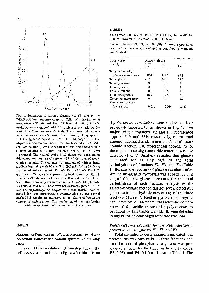
Fig. 1. Separation of anionic glucans F2, F3, and F4 by DEAE-cellulose chromatography. Cells of Agrobacterium tumefaciens C58, derived from 25 liters of culture in YM medium, were extracted with 1% trichloroacetic acid as de- scribed in Materials and Methods. The neutralized extracts were fractionated on a Sephadex G50 column yielding approx. 330 mg (glucose equivalent) of total oligosac.charide. The oligosaccharide material was further fractionated on a DEAE- cellulose column (2 cm × 16.5 cm) that was first eluted with 2 column volumes of 10 mM Tris-HCl (pH 7.4) in 7% (v/v) 1-propanol. The neutral cyclic fl-l,2-glucan was collected in this eluate and comprised approx, 45% of the total oligosac- charide material. The column was next eluted with a linear gradient beginning with 10 mM Tris-HC1 (pH 7.4) in 7% (v/v) 1-propanol and ending with 250 mM KCI in 10 mM Tris-HC1 (pH 7.4) in 7% (v/v) 1-propanol in a total volume of 200 ml. Fractions (3 ml) were collected at a flow rate of 25 ml per hour. Three anionic peaks were eluted at 10 mM KC1, 60 mM KC1 and 90 mM KC1. These three peaks are designated F2, F3, and F4, respectively. An aliquot from each fraction was re- moved for total carbohydrate determination by the phenol method [8]. Results are expressed as the relative carbohydrate content of each fraction. The numbering of fractions begins
with the application of the gradient to the column.
Anionic cell-associated oligosaccharides of Agro- bacterium tumefaciens contain glucose as the only sugar
U p o n DEAE-cel lulose chromatography, the cell-associated, an ionic oligosaccharides from
TABLE I
ANALYSIS OF ANIONIC GLUCANS F2, F3, AND F4 FROM A GROBA CTERIUM TUMEFA CIENS
Anionic glucans F2, F3, and F4 (Fig. 1) were prepared as described in the text and analyzed as described in Materials and Methods.
Constituent Anionic glucan ( ~t mol) F2 F 3 F4
Total carbohydrate (glucose equivalent) 518.4 259.7 65.8
Total glucose 467.1 246.4 63.7 Total galactose 0 0 0 Total pyruvate 0 0 0 Total succinate 0.6 0.6 0.2 Total phosphorus 16.7 19.8 8.9 Phosphate monoester 0 0 0.2 Phosphate : glucose
(mole ratio) 0,036 0.080 0,140
Agrobacterium tumefaciens were similar to those
previously reported [3] as shown in Fig. 1. Two major anionic fractions, F2 and F3, represented
approx. 61% and 32%, respectively, of the total an ionic oligosaccharide material. A third more
anionic fraction, F4, representing approx. 7% of the total anionic oligosaccharide material, was also detected (Fig. 1). Analysis revealed that glucose a c c oun t e d for at least 90% of the total carbohydra te of fractions F2, F3, and F4 (Table
I). Because the recovery of glucose s tandards after similar strong acid hydrolysis was approx. 87%, it
is probable that glucose accounts for the total carbohydrate of each fraction. Analysis by the galactose oxidase method did no t reveal detectable
galactose in acid hydrolysates of any of the three fractions (Table I). Nei ther pyruvate nor signifi-
cant amounts of succinate, characteristic compo- nents of the acidic extracellular polysaccharides produced by this bac te r ium [13,14], were detected in any of the anionic oligosaccharide fractions.
Phosphoglycerol accounts for the total phosphorus present in anionic glucans F2, F3, and F4
Total phosphorus determinat ions indicated that phosphorus was present in all three fractions and that the ratio of phosphorus to glucose was pro- gressively higher for the three fractions F2 (0.036), F3 (0.08), and F4 (0.14) as shown in Table I. The
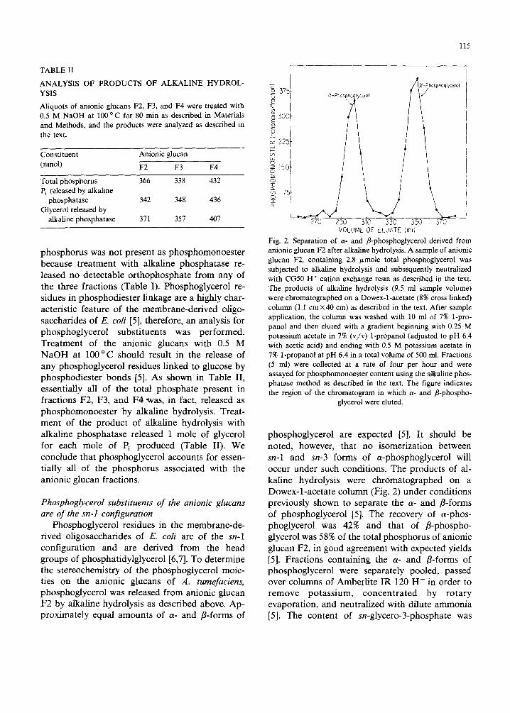
TABLE II
ANALYSIS OF PRODUCTS OF ALKALINE HYDROL- YSIS
Aliquots of anionic glucans F2, F3, and F4 were treated with 0.5 M NaOH at 100 °C for 80 min as described in Materials and Methods, and the products were analyzed as described in the text.
Constituent Anionic glucan
(nmol) F2 F3 F4
Total phosphorus 366 338 432 Pi released by alkaline
phosphatase 342 348 436 Glycerol released by
alkaline phosphatase 371 357 407
phosphorus was not present as phosphomonoester because treatment with alkaline phosphatase re- leased no detectable orthophosphate from any of the three fractions (Table I). Phosphoglycerol re- sidues in phosphodiester linkage are a highly char- acteristic feature of the membrane-derived oligo- saccharides of E. coli [5], therefore, an analysis for phosphoglycerol substituents was performed. Treatment of the anionic glucans with 0.5 M NaOH at 100°C should result in the release of any phosphoglycerol residues linked to glucose by phosphodiester bonds [5]. As shown in Table II, essentially all of the total phosphate present in fractions F2, F3, and F4 was, in fact, released as phosphomonoester by alkaline hydrolysis. Treat- ment of the product of alkaline hydrolysis with alkaline phosphatase released 1 mole of glycerol for each mole of Pi produced (Table II). We conclude that phosphoglycerol accounts for essen- tially all of the phosphorus associated with the anionic glucan fractions.
Phosphoglycerol substituents of the anionic glucans are of the sn-1 configuration
Phosphoglycerol residues in the membrane-de- rived oligosacchafides of E. coli are of the sn-1 configuration and are derived from the head groups of phosphatidylglycerol [6,7]. To determine the stereochemistry of the phosphoglycerol moie- ties on the anionic glucans of A. tumefaciens, phosphoglycerol was released from anionic glucan F2 by alkaline hydrolysis as described above. Ap- proximately equal amounts of a- and r-forms of
115
225
7sf
i _
270 290 310 330 350 370 VOLUME OF ELUATE (mll
Fig. 2. Separation of a- and fl-phosphoglycerol derived from anionic glucan F2 after alkaline hydrolysis. A sample of anionic glucan F2, containing 2.8 ~mole total phosphoglycerol was subjected to alkaline hydrolysis and subsequently neutralized with CG50 H + cation exchange resin as described in the text. The products of alkaline hydrolysis (9.5 ml sample volume) were chromatographed on a Dowex-l-acetate (8% cross linked) column (1.1 cm x 40 cm) as described in the text. After sample application, the column was washed with 10 ml of 7% 1-pro- panol and then eluted with a gradient beginning with 0.25 M potassium acetate in 7% (v/v) 1-propanol (adjusted to pH 6.4 with acetic acid) and ending with 0.5 M potassium acetate in 7% 1-propanol at pH 6.4 in a total volume of 500 ml. Fractions (5 ml) were collected at a rate of four per hour and were assayed for phosphomonoester content using the alkaline phos- phatase method as described in the text. The figure indicates the region of the chromatogram in which a- and /3-phospho-
glycerol were eluted.
phosphoglycerol are expected [5]. It should be noted, however, that no isomerization between sn-1 and sn-3 forms of a-phosphoglycerol will occur under such conditions. The products of al- kaline hydrolysis were chromatographed on a Dowex-l-acetate column (Fig. 2) under conditions previously shown to separate the a- and r-forms of phosphoglycerol [5]. The recovery of a-phos- phoglycerol was 42% and that of fl-phospho- glycerol was 58% of the total phosphorus of anionic glucan F2, in good agreement with expected yields [5]. Fractions containing the a- and B-forms of phosphoglycerol were separately pooled, passed over columns of Amberlite IR 120 H + in order to remove potassium, concentrated by rotary evaporation, and neutralized with dilute ammonia [5]. The content of sn-glycero-3-phosphate was
116
TABLE III
ANALYSIS OF ~- AND /3-PHOSPHOGLYCEROL OB- TAINED BY ALKALINE HYDROLYSIS OF ANIONIC GLUCAN F2.
A sample of anionic glucan F2, containing 2.8 #mole total phosphoglycerol, was subjected to alkaline hydrolysis as de- scribed in the text. The a- and fl-phosphoglycerol products released by alkaline hydrolysis were separated by chromatogra- phy on Dowex-l-acetate as shown in Fig. 2. Fractions con- taining a-phosphoglycerol and fl-phosphoglycerol were sep- arately pooled and assayed for total phosphomonoester by the alkaline phosphatase method as described in the text. Samples were then assayed for sn-3-phosphoglycerol by the dehydro- genase method both before and after acid racemization as described in Materials and Methods.
Constituent a-Phospho- fl-Phospho- (nmol) glycerol glycerol
Total phosphomonoester 1072 1458 sn-3-Phosphoglycerol
before acid racemization 0 0 sn-3-Phosphoglycerol
after acid racemization 463 681
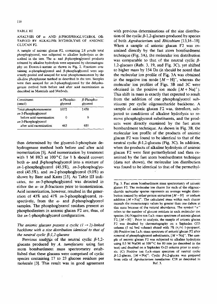
then determined by the glycerol-3-phosphate de- hydrogenase method both before and after acid isomerization [5]. Acid isomerization by treatment with I M HC1 at 100 °C for 1 h should convert both a- and fl-phosphoglycerol into a mixture of sn-l-phosphoglycerol (45.5%), sn-3-phosphoglyc- erol (45.5%), and sn-2-phosphoglycerol (9.0%) as shown by Baer and Kates [15]. As Table III indi- cates, no sn-3-phosphoglycerol was detected in either the ~t- or t-fractions prior to isomerization. Acid isomerization, however, resulted in the gener- ation of 43% and 47% sn-3-phosphoglycerol, re- spectively, from the ct- and fl-phosph0glycerol samples. The phosphoglycerol residues present as phosphodiesters in anionic glucan F2 are, thus, of the sn-l-phosphoglycerol configuration.
The anionic glucans possess a cyclic (1 ~ 2)-linked backbone with a size distribution identical to that of the neutral cyclic fl-t,2-glucans
Previous analysis of the neutral cyclic fl-l,2- glucans produce¢(by A. tumefaciens using fast atom bombardment mass spectrometry estab- lished that these glucans were comprised of cyclic species containing 17 to 23 glucose residues per molecule [3}. This result was in good agreement
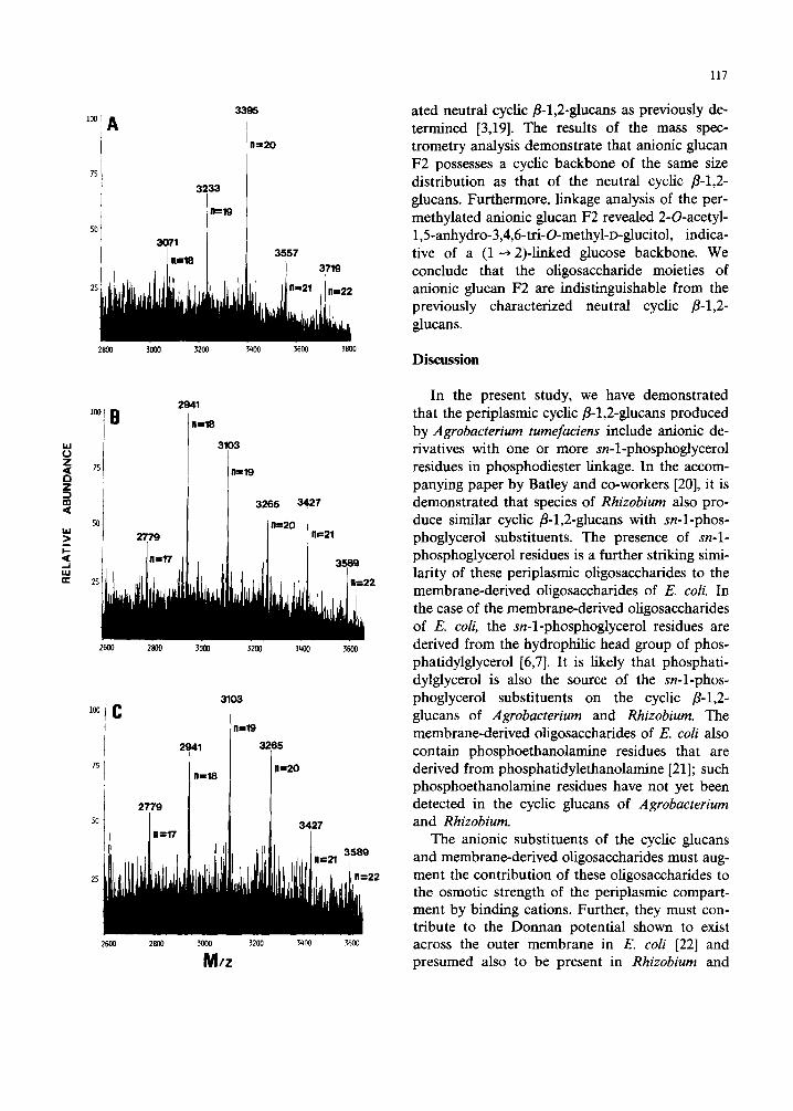
with previous determinations of the size distribu- tion of the cyclic fl-l,2-glucans produced by species of both Agrobacterium and Rhizobium [13,16-18]. When a sample of anionic glucan F2 was ex- amined directly by the fast atom boml~ardment technique (Fig. 3A), the molecular ion distribution was comparable to that of the neutral cyclic fl- 1,2-glucans (Refs. 3, 19, and Fig. 3C), yet shifted to higher mass by 154 Da (it should be noted that the molecular ion profile of Fig. 3A was obtained in the negative ion mode [ M - H I - , whereas the molecular ion profiles of Figs. 3B and 3C were obtained in the positive ion mode [ M + Na]+). This shift in mass is exactly that expected to result from the addition of one phosphoglycerol sub- stituent per cyclic oligosaccharide backbone. A sample of anionic glucan F2 was, therefore, sub- jected to conditions of alkaline hydrolysis to re- move phosphoglycerol substituents, and the prod- ucts were directly examined by the fast atom bombardment technique. As shown in Fig. 3B, the molecular ion profile of the products of anionic glucan F2 was found to be identical to that of the neutral cyclic fl-l,2-glucans (Fig. 3C). In addition, when the products of alkaline hydrolysis of anionic glucan F2 were first permethylated and then ex- amined by the fast atom bombardment technique (data not shown), the molecular ion distribution was found to be identical to that of the permethyl-
Fig. 3. Fast atom bombardment mass spectrometry of anionic glucan F2. The molecular ion cluster for each of the oligosac- charide molecular species represents an average weight distri- bution ionized by either proton extraction [ M - H ] - or sodium addition [M + Na] +. The calculated mass within each cluster exceeds the monoisotopic values by greater than one dalton at this mass because of the natural abundance. The symbol " n " refers to the number of glucose residues in each molecular ion species. (A) Negative ion f.a.b.-mass spectrum of anionic glucan F2, [ M - H]-. Prior to analysis, the sample of anionic glucan F2 was desalted by chromatography on a Sephadex G25 column (5 ml bed volume) ehited with 7% (v/v) 1-propanol. (B) Positive ion f.a.b.-mass spectrum of aniohic'glucan F2 after removal of phosphoglycerol substituents, [M +Na] +. The sam- ple of anionic glucan F2 was subjected to alkaline hydrolysis using 0.5 M NaOH at 100 ° C for 80 rain (as described in the text) and desalted on a Sephadex G-25 column prior to analy- sis. (C) Positive ion f.a.b.-mass spectrum of neutral cyclic fl-l,2-ghican, [ M + N a ] +. Cyclic fl-l,2-glucan was prepared from cells of Agrobacterium tumefaciens C58 as described in
Ref. 3.
UJ (3 Z
Z 3 m ,<
kU > I-- < IJJ r r
100 1
75 I
A
3233
3395
11==20
2800 ~ 3200 3400 3600 3800
2941
11==18
3103
l~r I]
r
2779
11=19
t 3265
11=20
3427
n==21
2600 2800 3000 3200 3400 3600
75
2800
3103
n=19
3000 3200
M/z 2600
2941 3265
3400
g
:22
3600
117
ated neutral cyclic/3-1,2-glucans as previously de- termined [3,19]. The results of the mass spec- trometry analysis demonstrate that anionic glucan F2 possesses a cyclic backbone of the same size distribution as that of the neutral cyclic fl-l,2- glucans. Furthermore, linkage analysis of the per- methylated anionic glucan F2 revealed 2-O-acetyl- 1,5-anhydro-3,4,6-tri-O-methyl-D-glucitol, indica- tive of a (1---, 2)-linked glucose backbone. We conclude that the oligosaccharide moieties of anionic glucan F2 are indistinguishable from the previously characterized neutral cyclic fl-l,2- glUCanS.
Discussion
In the present study, we have demonstrated that the periplasmic cyclic fl-l,2-glucans produced by Agrobacterium tumefa¢iens include anionic de- rivatives with one or more sn-l-phosphoglycerol residues in phosphodiester linkage. In the accom- panying paper by Barley and co-workers [20], it is demonstrated that species of Rhizobium also pro- duce similar cyclic fl-l,2-glucans with sn-l-phos- phoglycerol substituents. The presence of sn-1- phosphoglycerol residues is a further striking simi- larity of these periplasmic oligosaccharides to the membrane-derived oligosaccharides of E. coli. In the case of the membrane-derived oligosaccharides of E. coli, the sn-l-phosphoglycerol residues are derived from the hydrophilic head group of phos- phatidylglycerol [6,7]. It is likely that phosphati- dylglycerol is also the source of the sn-l-phos- phoglycerol substituents on the cyclic /3-1,2- glucans of Agrobacterium and Rhizobium. The membrane-derived oligosaccharides of E. coil also contain phosphoethanolamine residues that are derived from phosphatidylethanolamine [21]; such phosphoethanolamine residues have not yet been detected in the cyclic glucans of Agrobacterium and Rhizobium.
The anionic substituents of the cyclic glucans and membrane-derived oligosaccharides must aug- ment the contribution of these oligosaccharides to the osmotic strength of the periplasmic compart- ment by binding cations. Further, they must con- tribute to the Donnan potential shown to exist across the outer membrane in E. coli [22] and presumed also to be present in Rhizobium and
118
Agrobacterium. The fundamen ta l reason why such osmot ic regula t ion of the pe r ip lasmic compar t - men t is advan tageous to the o rgan ism remains obscure. N o r emarkab l e pheno typ ic consequences have as ye t been de tec ted in strains of E. coli b locked in the p roduc t i on of m e m b r a n e - d e r i v e d o l igosacchar ides [23].
In add i t i on to a poss ib le role for the cyclic glucans in the osmot ic adap t a t i on of Agro- bacterium and Rhizobium, it should be no ted that o ther invest igators have p r o p o s e d that the cyclic
f i - l ,2 -g lucans m a y con t r ibu te to the eff iciency of the p lan t infect ion process [24-26]. A b e and co-
workers [24,25] r epor ted that cyclic f l - l ,2 -g lucans may enhance fo rma t ion of infect ion threads and increase numbers of nodules in the Rhizobium trifolii-white clover symbiosis . Avi ru len t mu tan t s of Agrobacterium tumefaciens, which m a p to the c h r o m o s o m a l locus chvB, were found by Puvane- sa ra jah et al. [26], to be defect ive in the synthesis of cyclic f l - l ,2-glucans. W h e n these mu tan t s were res tored to virulence by complemen ta t i on with c loned D N A , the abi l i ty to p roduce the cyclic f l - l ,2 -g lucans was also res tored. I t was conc luded tha t the cyclic glucans may be impor t an t for the pa thogen ic p roper t i e s of A. tumefaciens. N o w that it has been d e m o n s t r a t e d that the cyclic glucans m a y become modi f i ed with subs t i tuents such as sn - l -phosphog lyce ro l , it will be of in teres t to ex- amine the poss ib le con t r ibu t ion of such sub- s t i tuents to the p l an t infect ion process.
Acknowledgements
This r e sea rch was s u p p o r t e d b y g ran t s GM19822, GM22057, and RR01494 f rom the N a - t ional Ins t i tu tes of Hea l th and PCM-88300342 f rom the N a t i o n a l Science F o u n d a t i o n . K.J .M. is a Medica l F o u n d a t i o n Research Fe l low of the Med ica l F o u n d a t i o n , Inc., Boston, MA. The au thors would l ike to express their apprec ia t ion to Dr . S. San t ika rn for the ope ra t ion of the mass spec t rometer .
References
1 Bauer, W.D. (1981) Annu. Rev. Plant Physiol. 32, 407-449 2 Halverson, L.J. and Stacey, G. (1986) Microbiol. Rev. 50,
193-225
3 Miller, K.J., Kennedy, E.P. and Reinhold, V.N. (1986) Science 231, 48-51
4 Kennedy, E.P. (1982) Proc. Natl. Acad. Sci. USA 79, 1092-1095
5 Kennedy, E.P., Rumley, M.K., Schulman, H. and Van Golde, L.M.G. (1976) J. Biol. Chem. 251, 4208-4213
6 Jackson, B.J. and Kennedy, E.P. (1983) J. Biol. Chem. 258, 2394-2398
7 Jackson, B.J., Bohin, J.-P. and Kennedy, E.P. (1984) J. Bacteriol. 160, 976-981
8 Hanson, R.S. and Phillips, J.A. (1981) in Manual of Meth- ods for General Bacteriology (Gerhardt, P., Murray, R.G.E., Costilow, R.N., Nester, E.W., Wood, W.A., Krieg, N.R. and Phillips, G.B., eds.), pp. 328-364, American Society for Microbiology, Washington, DC
9 Hjelm, M. and De Verdier, C.H. (1974) in Methods of Enzymatic Analysis (Bergmeyer, H.U., ed.), Vol. 3, pp. 1282-1287, Academic Press Inc., New York
10 Czok, R. and Lamprecht, W. (1974) in Methods of En- zymatic Analysis (Bergmeyer, H.U., ed.), Vol. 3, pp. 1446-1451, Academic Press Inc., New York
11 Chen, P.S., Toribara, T.Y. and Warner, H. (1956) Anal. Chem. 28, 1756-1758
12 Ames, B.N. and Dubin, D.T. (1960) J. Biol. Chem. 235, 769-775
13 Hisamatsu, M., Amemura, A., Matsuo, T., Matsuda, H. and Harada, T. (1982) J. Gen. Microbiol. 128, 1873-1879
14 Staneloni, R.J., Tomalsky, M.E. and Leloir, L.F. (1984) J. Gen. Microbiol. 130, 869-879
15 Baer, E. and Kates, M. (1948) J. Biol. Chem. 175, 79-88 16 Dell, A., York, W.S., McNeil, M., Darvill, A.G. and AI-
bersheim, P. (1983) Carbohydr. Res. 117, 185-200 17 Hisamatsu, M., Amemura, A., Koizumi, K., Utamura, T.
and Okada, Y. (1983) Carbohydr. Res. 121, 31-40 18 Koizumi, K., Okada, Y., Horiyama, S. and Utamura, T.
(1983) J. Chromatogr. 265, 89-96 19 Reinhold, V.N. (1986) in Mass Spectrometry in Biomedical
Research (Gaskell, S.J., ed.), pp. 181-213, John Wiley and Sons Ltd., New York
20 Batley, M., Redmond, J.W., Djorjevic, S.P. and Rolfe, B.G. (1987) Biochim. Biophys. Acta 901, 119-126
21 Miller, K.J. and Kennedy, E.P. (1987) J. Bacteriol. 169. 682-686
22 Stock, J.B., Rauch, B. and Roseman, S. (1977) J. Biol. Chem. 252, 7850-7861
23 Bohin, J.-P. and Kennedy, E.P. (1984) J. Bacteriol. 157, 956-957
24 Higashi, S. and Abe, M. (1980) Appl. Environ. Microbiol. 39, 297-301
25 Abe, M., Amemura, A. and Higashi, S. (1982) Plant Soil 64. 315-324
26 Puvanesarajah, V., ScheU, F.M., Stacey, G., Douglas, C.J. and Nester, E.W. (1985) J. Bacteriol. 164, 102-106