
Cyclic beta-1,6-1,3 glucans ofBradyrhizobium: Functional analogs of the cyclic beta-1,2-glucans...
4
CURRENT MICROBIOLOGY Vol. 24 (1992), pp. 101-104 Current Microbiology Springer-Verlag New York Inc. 1992 Cyclic Beta-l,6-1,3 Glucans of Bradyrhizobium: Functional Analogs of the Cyclic Beta- 1,2-Glucans of Rhizobium? Karen J. Miller 1'2'3 and Richard S. Gore 2 IDepartment of Food Science and Graduate Programs in 2Genetics and 3plant Physiology, The Pennsylvania State University, University Park, Pennsylvania, USA Abstract. We have previously shown that species of Bradyrhizobium synthesize a novel class of cyclic beta glucans which contains both beta-1,6 and beta-1,3 glycosidic linkages [Miller K J, Gore RS, Johnson R, Benesi AJ, Reinhold VN (1990) J Bacteriol 172:136-142]. In the present study we show that these cell-associated glucans are localized within the periplasmic compart- ment and that the biosynthesis of these glucans is osmotically regulated. Bacteria of the Rhizobiaceae family are distin- guished by their ability to infect higher plants. The cell-surface carbohydrates of these bacteria are be- lieved to provide important roles in the plant infec- tion process. These cell-surface carbohydrates in- clude extracellular polysaccharides, capsular polysaccharides, lipopolysaccharides, and oligosac- charides. Studies in our laboratory have focused upon one class of cell surface carbohydrates, the cyclic beta glucans. The cyclic beta glucans produced by species of Rhizobium and Agrobacterium are composed of 17-24 glucose residues linked solely by beta-l,2- glycosidic bonds. These molecules are localized within the periplasmic compartment and may be- come substituted with anionic moieties [1, 3, 11, 14-16]. In addition, the biosynthesis of these mole- cules is osmotically regulated, with maximal glucan synthesis occuring when cells are cultured at low osmolarity [14]. Because cyclic beta-l,2-glucan biosynthesis ap- pears to be a distinguishing feature of the Rhizo- biaceae family, it is believed that these molecules may provide functions during plant infection. In- deed, evidence for such functions derives from stud- ies of mutants of Rhizobium and Agrobacterium de- fective for cyclic beta-1,2-glucan biosynthesis. Such mutants of Agrobacterium have been shown to be avirulent and impaired for plant attachment [5, 19]. Furthermore, cyclic beta-l,2-glucan-deficient mu- tants of Rhizobium are also impaired for plant attach- ment and are unable to form effective nodules [6, 8]. Recently, Dylan and coworkers [8] have presented Address reprint requests to: Dr. Karen J. Miller, 105 Borland University, University Park, PA 16802, USA. evidence that the cyclic beta-l,2-glucans may act as signal molecules during legume nodulation. How- ever, these researchers also provide evidence that the cyclic beta-l,2-glucans may not be essential for nodulation. Although the functions of the cyclic beta-l,2- glucans during plant infection remain unclear, recent studies have indicated that these molecules provide osmoregulatory functions for the free-living bacte- ria. Specifically, cyclic beta-l,2-glucan-deficient mutants of both Agrobacterium and Rhizobium have been shown to be impaired in their ability to grow within low osmolarity environments [4, 7]. Such an osmoregulatory function is consistent with the fact that the biosynthesis of these molecules is maximal when cells are cultured at low osmolarity. On the basis of evidence that the cyclic beta- 1,2-glucans provide osmoregulatory functions, ap- pear to act as signal molecules during plant infection, and are apparently synthesized by all species of Rhi- zobium and Agrobacterium, it is surprising that spe- cies of the closely related, legume-nodulating genus Bradyrhizobium lack the ability to synthesize these molecules. Instead, recent studies from our labora- tory have revealed that these bacteria synthesize a novel class of glucan which is cyclic in structure and composed of beta-l,6 and beta-l,3 glycosidic bonds [17]. Interestingly, the levels of the cell-associated cyclic beta-l,6 -1,3 glucans of Bradyrhizobium spe- cies are similar to the levels of the cyclic beta-l,2- glucans present within the periplasmic compartment of species of Rhizobium and Agrobacterium [17]. Thus, it is possible that the cyclic beta-l,6 -1,3 glu- Laboratory, Department of Food Science, The Pennsylvania State
-
Upload
karen-j-miller -
Category
Documents
-
view
212 -
download
0
Transcript of Cyclic beta-1,6-1,3 glucans ofBradyrhizobium: Functional analogs of the cyclic beta-1,2-glucans...

CURRENT MICROBIOLOGY Vol. 24 (1992), pp. 101-104 Current Microbiology �9 Springer-Verlag New York Inc. 1992
Cyclic Beta-l,6-1,3 Glucans of Bradyrhizobium: Functional Analogs of the Cyclic Beta- 1,2-Glucans of Rhizobium? Karen J. Miller 1'2'3 and Richard S. Gore 2
IDepartment of Food Science and Graduate Programs in 2Genetics and 3plant Physiology, The Pennsylvania State University, University Park, Pennsylvania, USA
Abstract. We have previously shown that species of Bradyrhizobium synthesize a novel class of cyclic beta glucans which contains both beta-1,6 and beta-1,3 glycosidic linkages [Miller K J, Gore RS, Johnson R, Benesi AJ, Reinhold VN (1990) J Bacteriol 172:136-142]. In the present study we show that these cell-associated glucans are localized within the periplasmic compart- ment and that the biosynthesis of these glucans is osmotically regulated.
Bacteria of the Rhizobiaceae family are distin- guished by their ability to infect higher plants. The cell-surface carbohydrates of these bacteria are be- lieved to provide important roles in the plant infec- tion process. These cell-surface carbohydrates in- clude extracellular polysaccharides, capsular polysaccharides, lipopolysaccharides, and oligosac- charides. Studies in our laboratory have focused upon one class of cell surface carbohydrates, the cyclic beta glucans.
The cyclic beta glucans produced by species of Rhizobium and Agrobacterium are composed of 17-24 glucose residues linked solely by beta-l,2- glycosidic bonds. These molecules are localized within the periplasmic compartment and may be- come substituted with anionic moieties [1, 3, 11, 14-16]. In addition, the biosynthesis of these mole- cules is osmotically regulated, with maximal glucan synthesis occuring when cells are cultured at low osmolarity [14].
Because cyclic beta-l,2-glucan biosynthesis ap- pears to be a distinguishing feature of the Rhizo- biaceae family, it is believed that these molecules may provide functions during plant infection. In- deed, evidence for such functions derives from stud- ies of mutants of Rhizobium and Agrobacterium de- fective for cyclic beta-1,2-glucan biosynthesis. Such mutants of Agrobacterium have been shown to be avirulent and impaired for plant attachment [5, 19]. Furthermore, cyclic beta-l,2-glucan-deficient mu- tants of Rhizobium are also impaired for plant attach- ment and are unable to form effective nodules [6, 8]. Recently, Dylan and coworkers [8] have presented
Address reprint requests to: Dr. Karen J. Miller, 105 Borland University, University Park, PA 16802, USA.
evidence that the cyclic beta-l,2-glucans may act as signal molecules during legume nodulation. How- ever, these researchers also provide evidence that the cyclic beta-l,2-glucans may not be essential for nodulation.
Although the functions of the cyclic beta-l,2- glucans during plant infection remain unclear, recent studies have indicated that these molecules provide osmoregulatory functions for the free-living bacte- ria. Specifically, cyclic beta-l,2-glucan-deficient mutants of both Agrobacterium and Rhizobium have been shown to be impaired in their ability to grow within low osmolarity environments [4, 7]. Such an osmoregulatory function is consistent with the fact that the biosynthesis of these molecules is maximal when cells are cultured at low osmolarity.
On the basis of evidence that the cyclic beta- 1,2-glucans provide osmoregulatory functions, ap- pear to act as signal molecules during plant infection, and are apparently synthesized by all species of Rhi- zobium and Agrobacterium, it is surprising that spe- cies of the closely related, legume-nodulating genus Bradyrhizobium lack the ability to synthesize these molecules. Instead, recent studies from our labora- tory have revealed that these bacteria synthesize a novel class of glucan which is cyclic in structure and composed of beta-l,6 and beta-l,3 glycosidic bonds [17]. Interestingly, the levels of the cell-associated cyclic beta-l,6 -1,3 glucans of Bradyrhizobium spe- cies are similar to the levels of the cyclic beta-l,2- glucans present within the periplasmic compartment of species of Rhizobium and Agrobacterium [17]. Thus, it is possible that the cyclic beta-l,6 -1,3 glu-
Laboratory, Department of Food Science, The Pennsylvania State

102 CURRENT MICROBIOLOGY Vol. 24 (1992)
cans of bradyrhizobial species may represent func- tional analogs of the cyclic beta-l,2-glucans of Rhi- zobium and Agrobacterium species. In the present study we provide evidence that supports this possi- bility by demonstrating that the biosynthesis of the cyclic beta-l ,6 -1,3 glucans of Bradyrhizobium ja- ponicum is osmotically regulated and that these mol- ecules are localized within the periplasmic com- partment.
Materials and Methods
Bacterial strain and growth conditions. Bradyrhizobium japoni- cure USDA 110 was cultured in YM medium [17] or YM medium of high osmolarity containing one of a variety of nonionic solutes (0.5 M sucrose, 0.5 M mannitol, or 0.5 M glycerol). Ionic solutes such as NaC1 were not used because the growth of B.japonicum USDA 110 was found to be inhibited by low concentrations of these solutes (e.g., growth was strongly inhibited at concentra- tions of NaC1 greater than 50 raM). Cells were grown at 30~ on a rotary shaker.
Analysis of cell-associated and extraceUular beta glucans. Cell- associated cyclic beta-1,6 -1,3 glucans were extracted from cells at the mid-logarithmic phase of growth (cell density of approxi- mately 50/~g of total cell protein per ml) by a methanol extraction procedure [171. Extracts were assayed for glucan content by gel filtration chromatography on Sephadex G50 and Sephadex G25 columns with methods previously described [17].
Extracellular glucan analyses were performed by gel filtra- tion chromatography on Sephadex G50 and Sephadex G25 col- umns as above; however, prior to chromatographic analysis, cell- free supernatants were concentrated approximately 30-fold at 4~ by ultrafiltration with a 400-ml stirred cell apparatus (Amicon Inc., Danvers, Massachusetts) and a Diaflow ultrafilter YCO5 or YM1 membrane with nominal molecular weight cut-offs of 500 or 1000, respectively (Amicon, Inc.). For analyses of cell-free supernatants obtained from cultures grown in YM medium con- taining no added solutes, high-molecular-weight extracellular polysaccharides were removed prior to ultrafiltration by precipi- tation with ethanol (90% final concentration).
Results and Discussion
Biosynthesis of cell-associated cyclic beta- l ,6 -1,3 glucans by Bradyrhizobiumjaponicum is osmotically regulated. The results presented in Table 1 reveal that the amount of cell-associated cyclic beta-l ,6 -1,3 glucans synthesized by B.japonicum USDA 110 decreased when cells were grown in high osmolarity media containing a nonpermeating solute (e.g., 0.5 M sucrose or 0.5 M mannitol). This decrease was particularly dramatic when cells were grown in the presence of 0.5 M sucrose (where no cell- associated cyclic beta- 1,6 - 1,3 glucan was detected). In contrast, when cells were grown in media con- taining glycerol, a readily permeable solute that is
Table 1. Cyclic beta-l,6 -1,3 glucan biosynthesis by Bradyrhizobium japonicum USDA 110 as a function of growth medium osmolarity
Growth medium Cell-associated
cyclic beta glucan a
YM 43 YM + 0.5 M mannitol 30 YM + 0.5 M sucrose b YM + 0.5 M glycerol 49
a Cell-associated cyclic beta-l,6 -1,3 glucan content is expressed as milligrams of glucose per gram of total cell protein. b--, none detected.
osmotically inactive [12], the amount of cell-associ- ated cyclic beta-l ,6 -1,3 glucans did not decrease.
It is noted that Tully and coworkers [22] have also recently reported that the biosynthesis of beta glucans by Bradyrhizobium japonicum is osmoti- cally regulated. Although the glucans described by these researchers share structural features with the glucans in the present study (e.g., both glucans are composed predominantly of beta-l ,6 and beta-l,3 glycosidic linkages), the size of the two glucan prep- arations is substantially different. Specifically, Tully and coworkers [22] have reported the presence of three glucan subfractions, all of which are appar- ently similar and/or greater in size than the cyclic beta-l,2-glucans produced by Agrobacterium and Rhizobium species. In contrast, the cyclic beta-l ,6 -1,3 glucans identified in our studies [17] are substan- tially smaller (10-13 glucose residues). The relation- ship between the glucans described in the present study and those identified by Tully and coworkers [22] remains to be further clarified.
Biosynthesis of extracellular beta glucans is also os- motically regulated. Because it was possible that reduced levels of cell-associated cyclic beta-1,6 -1,3 glucans detected within cultures grown at elevated osmolarity may have resulted from a loss of glucans to the extracellular medium, culture supernatants were examined for the presence of extracellular beta glucans. Upon examination of the extracellular me- dia from cultures grown in YM medium, relatively high levels of extracellular glucans were detected (e.g., 300 mg of glucose equivalent per gram of total cell protein). Indeed, the amount of extracellular glucan produced by mid-logarithmic cultures was approximately sevenfold greater than the amount of cell-associated cyclic beta-1,6 -1,3 glucans (Table 1). Further analysis revealed that the biosynthesis of
K.J. Miller and R.S. Gore: Cyclic Beta-l,6-1,3 Glucans of Bradyrhizobium 103
the extracellular glucan fraction was dramatically repressed when cells were grown at high osmolarity. Under these conditions, we were unable to detect the presence of extracellular beta glucans within cul- tures.
The relationship between the cell-associated cy- clic beta- 1,6 -1,3 glucans and the extracellular glu- cans produced by B. japonicum USDA 110 was fur- ther examined by gel filtration chromatography, ion exchange chromatography, and NMR spectroscopy. The combined results of these analyses indicate that the extracellular glucan fraction is similar in struc- ture to the cell-associated cyclic beta- l ,6 -1,3 glu- cans. First, chromatography of the extracellular glu- can fraction on both Sephadex G50 and Sephadex G25 columns revealed this glucan fraction to be the same size as that of the cell-associated cyclic beta- l ,6 -1,3 glucans. Second, like the cell-associ- ated cyclic beta-1,6 -1,3 glucans [17], the extracellu- lar glucan fraction did not adsorb to DEAE-cellulose (thus, indicating an uncharged character). Third, 1H and 13C NMR spectroscopy of the extracellular glu- can fraction revealed the presence of beta- l ,6 and beta-l ,3 glycosidic linkages, an absence of beta-l ,2 glycosidic linkages, and an absence of reducing glu- cose residues (data not shown). Thus, the extracellu- lar glucan fraction shares structural features with the cell-associated cyclic beta-1,6 -1,3 glucans. Ongoing studies in our laboratory are aimed at establishing the arrangement of the beta-l ,6 and beta-l ,3 glyco- sidic linkages in both the cell-associated and extra- cellular beta glucans produced by this bacterium.
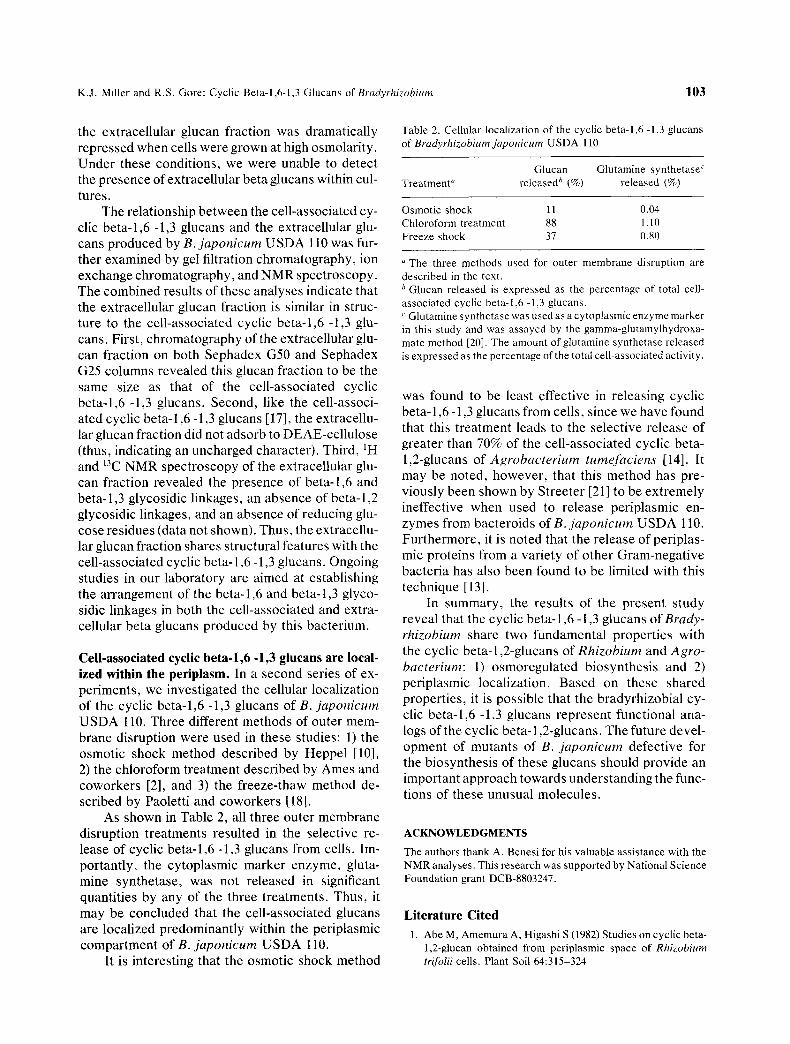
Cell-associated cyclic beta-l,6 -1,3 glucans are local- ized within the periplasm. In a second series of ex- periments, we investigated the cellular localization of the cyclic beta-l ,6 -1,3 glucans of B. japonicum USDA 110. Three different methods of outer mem- brane disruption were used in these studies: 1) the osmotic shock method described by Heppel [10], 2) the chloroform treatment described by Ames and coworkers [2], and 3) the freeze-thaw method de- scribed by Paoletti and coworkers [18].
As shown in Table 2, all three outer membrane disruption treatments resulted in the selective re- lease of cyclic beta- l ,6 -1,3 glucans from cells. Im- portantly, the cytoplasmic marker enzyme, gluta- mine synthetase, was not released in significant quantities by any of the three treatments. Thus, it may be concluded that the cell-associated glucans are localized predominantly within the periplasmic compartment of B. japonicum USDA 110.
It is interesting that the osmotic shock method
Table 2. Cellular localization of the cyclic beta-l,6 -1,3 glucans of Bradyrhizobium japonicum USDA 110
Glucan Glutamine synthetase ~ Treatment '~ reteased b (%) released (%)
Osmotic shock 11 0.04 Chloroform treatment 88 1. l0 Freeze shock 37 0.80
The three methods used for outer membrane disruption are described in the text. ~ Glucan released is expressed as the percentage of total cell- associated cyclic beta-l,6 -1,3 glucans. < Glutamine synthetase was used as a cytoplasmic enzyme marker in this study and was assayed by the gamma-glutamylhydroxa- mate method [20]. The amount of glutamine synthetase released is expressed as the percentage of the total cell-associated activity.
was found to be least effective in releasing cyclic beta-1,6 -1,3 glucans from cells, since we have found that this treatment leads to the selective release of greater than 70% of the cell-associated cyclic beta- 1,2-glucans of Agrobacterium tumefaciens [14]. It may be noted, however, that this method has pre- viously been shown by Streeter [21] to be extremely ineffective when used to release periplasmic en- zymes from bacteroids o fB . japonicum USDA 110. Furthermore, it is noted that the release of periplas- mic proteins from a variety of other Gram-negative bacteria has also been found to be limited with this technique [13].
In summary, the results of the present study reveal that the cyclic beta-1,6 -1,3 glucans of Brady- rhizobium share two fundamental properties with the cyclic beta-1,2-glucans of Rhizobium and Agro- bacterium: 1) osmoregulated biosynthesis and 2) periplasmic localization. Based on these shared properties, it is possible that the bradyrhizobial cy- clic beta-l ,6 -1,3 glucans represent functional ana- logs of the cyclic beta-1,2-glucans. The future devel- opment of mutants of B. japonicum defective for the biosynthesis of these glucans should provide an important approach towards understanding the func- tions of these unusual molecules.
ACKNOWLEDGMENTS The authors thank A. Benesi for his valuable assistance with the NMR analyses. This research was supported by National Science Foundation grant DCB-8803247.
Literature Cited 1. Abe M, Amemura A, Higashi S (1982) Studies on cyclic beta-
1,2-glucan obtained from periplasmic space of Rhizobium trifolii cells. Plant Soil 64:315-324

104 CURRENT MICROBIOLOGY Vol. 24 (1992)
2. Ames GF-L, Prody C, Kustu S (1984) Simple, rapid, and quantitative release of periplasmic proteins by chloroform. J Bacteriol 160:1181-1183
3. Batley M, Redmond JW, Djordjevic SP, Rolfe BG (1987) Characterization of glycerophosphorylated cyclic beta-l,2- glucans from a fast-growing Rhizobium species. Biochim Bio- phys Acta 901:119-126
4. Cangelosi GA, Martinetti G, Nester EW (1990) Osmosen- sitivity phenotypes of Agrobacterium tumefaciens mutants that lack periplasmic beta-l,2-glucan. J Bacteriol 172:2172- 2174
5. Douglas CJ, Staneloni RJ, Rubin RA, Nester EW (1985) Identification and genetic analysis of an Agrobacterium tu- mefaciens chromosomal virulence region. J Bacteriol 161:850-860
6. Dylan T, lelpi L, Stanfield S, Kashyap L, Douglas C, Yanof- sky M, Nester E, Helinski DR, Ditta G (1986) Rhizobium meliloti genes required for nodule development are related to chromosomal virulence genes in Agrobacterium tumefa- ciens. Proc Natl Acad Sci USA 83:4403-4407
7. Dylan T, Helinski DP, Ditta GS (1990) Hypoosmotic adapta- tion in Rhizobium meliloti requires beta-(1-2)-glucan. J B acte- riol 172:1400-1408
8. Dylan T, Nagpal P, Helinski DR, Ditta GS (1990) Symbiotic pseudorevertants ofRhizobium meliloti ndv mutants. J Bacte- riol 172:1409-1417
9. Hanson RS, Phillips JA (1981) Chemical composition, In: Gerhardt P, Murray RGE, Costilow RN, Nester EW, Wood WA, Krieg NR, Phillips GB (eds) Manual of methods for general bacteriology. Washington, DC: American Society for Microbiology, pp 328-364
10. Heppel LA (1968) Preparation of cells of Escherichia coli with altered permeability. Methods Enzymol 12B:841-846
11. Hisamatsu M, Yamada T, Higashiura T, Ikeda M (1987) The production of acidic, O-acylated cyclosophorans [cyclic(1- 2)-beta-D-glucans] by Agrobacterium and Rhizobium species. Carbohydr. Res 163:115-122
12. Laimins LA, Rhoads DB, Epstein W (1981) Osmotic control of kdp operon expression in Escherichia coli. Proc Natl Acad Sci USA 78:464-468
13. Lall SD, Eribo BE, Jay JM (1989) Comparison of four meth- ods for extracting periplasmic proteins. J Microbiol Methods 9:195-199
14. Miller KJ, Kennedy EP, Reinhold VN (1986) Osmotic adapta- tion by gram-negative bacteria: possible role for periplasmic oligosaccharides. Science 231:48-51
15. Miller KJ, Reinhold VN, Weissborn AC, Kennedy EP (1987) Cyclic glucans produced by Agrobacterium tumefaciens are substituted with sn-l-phosphoglycerol residues. Biochim Bi- ophys Acta 901:112-118
16. Miller KJ, Gore RS, Benesi AJ (1988) Phosphoglycerol sub- stituents present on the cyclic beta-l,2-glucans of Rhizobium meliloti 1021 are derived from phosphatidylglycerol. J Bacte- riol 170:4569-4575
17. Miller KJ, Gore RS, Johnson R, Benesi AJ, Reinhold VN (1990) Cell-associated oligosaccharides of Bradyrhizobium spp. J Bacteriol 172:136-142
18. Paoletti LC, Short KA, Blakemore N, Blakemore RP (1987) Freeze-thawing of Aquaspirillum magnetotacticum cells se- lectively releases periplasmic proteins. Appl Environ Micro- biol 53:2590-2592
19. Puvanesarajah V, Schell FM, Stacey G, Douglas CJ, Nester EW (1985) Role for 2-1inked-beta-o-glucans in the virulence of Agrobacterium tumefaciens. J Bacteriol 164:102-106
20. ShapiroB, StadtmanER(1970)Glutaminesynthetase(Esche- richia coli), Methods Enzymol 17A:910-922
21. Streeter JG (1989) Analysis of periplasmic enzymes in intact cultured bacteria and bacteroids of Bradyrhizobium japoni- cure and Rhizobium legurninosarum biovar phaseoli. J. Gen. Microbiol. 135:3477-3484
22. Tully RE, Keister DL, Gross KC (1990) Fractionation of the beta-linked glucans of Bradyrhizobium japonicum and their response to osmotic potential. Appl Environ Microbiol 56:1518-1522