Cyanosis in newborn
46
CYANOSIS By P.Padma Priyanka
-
Upload
padmapriyanka999 -
Category
Health & Medicine
-
view
331 -
download
0
Transcript of Cyanosis in newborn

CYANOSISBy
P.Padma Priyanka

Contents Introduction
Factors affecting detection of cyanosis
Etiology
Types
Cardiac vs pulmonary
Approach
Conclusion

Introduction Cyanosis is the bluish discoloration of the skin and
mucous membranes due to increased concentration of
reduced hemoglobin to about >5g/100 mL in the
cutaneous veins
Desaturation of arterial blood
Increased extraction of oxygen by peripheral tissue in
the presence of normal arterial saturation
Detected –lips,fingernails,oral mucous
membranes,conjuctiva and tip of tongue

Factors affecting detection of
cyanosis in newborn
Hemoglobin concentration
Fetal hemoglobin
Skin pigmentation

Hemoglobin
concentration
The arterial oxygen saturation level at which cyanosis is detectable at different total hemoglobin concentrations is illustrated above. The
solid red portion of each bar represents 3 gm/dL reduced hemoglobin.

Fetal hemoglobin
The oxygen-dissociation curve of human blood and the effects of changes in the H+ ion concentration, Pco2 temperature and level of
2, 3-diposphoglycerate (2,3-DPG) are depicted above. For fetal hemoglobin, the normal curve (a) is shifted to the left (b).

Skin pigmentation Less apparent in the skin of babies with darker
pigmentation.
Examination should include the nail beds, tongue,
and mucous membranes, which are less affected by
pigmentation.

Cyanosis
Pulmonary
Central CNS depression
Local
Ventilation-perfusion mismatch
Alveolar hypoventilation
Diffuse impairment
Cardiac
Increased pulmonary vascularity
Decreased pulmonary vascularity
Hemoglobi-nopathies

Ventilation/perfusion mismatch
Airway disease: transient tachypnea of the newborn
(TTN), respiratory distress syndrome (RDS),
pneumonia, aspiration (meconium, blood, amniotic
fluid), atelectasis, diaphragmatic hernia, pulmonary
hypoplasia, pulmonary hemorrhage, CCAM
Extrinsic compression of the lungs: pneumothorax,
pleural effusion, hemothorax,

Alveolar hypoventilation CNS depression: asphyxia, maternal sedation,
intraventricular hemorrhage, seizure, meningitis,
encephalitis
Airway obstruction: choanal atresia, laryngomalacia,
Pierre Robin syndrome
Neuromuscular disease: phrenic nerve inury, neonatal
myasthenia gravis

Diffusion impairment Pulmonary edema: left-sided obstructive cardiac
disease, cardiomyopathy
Pulmonary fibrosis
Congenital lymphangiectasia

Cardiac causes Decreased pulmonary blood flow-
Tetralogy of Fallot
Tricuspid valve anomaly
Pulmonary valve atresia
Critical valvular pulmonary steanosis
Increased pulmonary blood flow-
Transposition of great arteries
Truncus arteriosus
Total anomalous pulmonary venous connection

Cardiac causes- "five Ts" of cyanotic CHD: Transposition of the great arteries Tetralogy of Fallot Truncus arteriosus Total anomalous pulmonary venous connection Tricuspid valve abnormalities.
A sixth "T" is often added for "tons" of other diseases, such as double outlet right ventricle, pulmonary atresia, multiple variations of single ventricle, hypoplastic left heart syndrome, or anomalous systemic venous connection (left superior vena cava connected to the left atrium).
Hemoglobinopathies Hereditary < exposure to toxic substances
>15%- cyanosis
>70% -lethal
Remain chocolate brown-even with full oxygenation or
long exposure to room air

Central cyanosis
Inadequate alveolar ventilation
CNS depression
Inadequate ventilatory drive
Obstruction
Structural changes
Muscle weakness
Desaturated blood bypassing alveolar units
Intracardiac R-L
Intrapulmonary shunt
Pulmonary hypertension with R-L shunt

Peripheral cyanosis
Peripheral cyanosis, involves a bluish discoloration of the
skin but sparing of the mucus membranes & tongue. In this
type, a normal PaO2 value is detected
Increased oxygen extraction due to sluggish movement
through the capillaries leads to increased deoxygenated
blood on the venous side
Vasomotor instability,vasoconstriction caused by cold, low
cardiac output, venous obstruction, elevated venous
pressure and polycythemia


Acrocyanosis Bluish discoloration of fingers seen in neonates and infants
due to vasoconstriction as a result of transient hypothermia
No clinical significance unless associated with circulatory shock
Circum-oral cyanosis Healthy child with fair skin due to sluggish blood flow
with vasoconstriction
No clinical significance unless associated with low
cardiac output

Cardiac vs Pulmonary Hyperoxiatest-
Response of arterial PaO2 to 100%oxygen inhalation
Result in PaO2 Disease
>100mm Hg Lung disease
Large pulmonary blood flow
(TAPVR)
<100mm Hg Massive intra-pulmonary shunt
with normal heart
<10-30mm Hg increase
(<100)
Intra-cardiac right to left shunt


Approach to cyanotic neonate
Antenatal history
Fetal ultrasound scans- congenital heart disease,
diaphragmatic hernia and congenital cystic
adenomatoid malformation (CCAM).
Family history of CHD


Physical examination Vitals
R/o choanal atresia
Respiratory system
Cardiovascular system
Abdomen
Neurological disorders

Vitals Vital signs-
signs of respiratory distress such as tachypnea,
retractions, nasal flaring & grunting usually indicate a
respiratory problem
congenital heart disease is often accompanied by absent
or effortless tachypnea.
Sepsis often has the following findings: peripheral
cyanosis, HR, RR, BP, / temp

R/o choanal atresia
Cyanosis decreases during crying
Confirmed by failure to pass a soft No. 5F to 8F
catheter through each nostril

Respiratory system Inspiratory stridor-
upper airway obstruction
Chest-
Asymmetric chest movement combined with severe
distress-
alarming sign for tension pneumothorax, diaphragmatic
hernia
Transillumination of the chest-
Pneumothorax

Cardiovascular system A systolic murmur audible in most forms of cyanotic
CHD (exception: d-TGA with intact ventricular septum &
no pulmonary stenosis).
Respirations often are unlabored unless there is
pulmonary congestion or complicated by the
development of heart failure or acidosis, which will
affect the respiratory pattern

Per abdomen Scaphoid abdomen-congenital diaphragmatic hernia

Neurological disorders Observe for apnea and periodic breathing, which may
be related to immaturity of the nervous system.
Seizures can cause cyanosis if the infant fails to
breathe during the episodes.

Investigations • CBC
• Serum glucose
• ABG
Chest X-ray films,ECG
Arterial PaO2 in preductal and postductal arteries
Hyperoxitest

CBC & diff :
or WBC sepsis
hematocrit > 65% polycythemia
Serum glucose: to detect hypoglycemia
Arterial Blood Gases (ABGs):
Arterial PO2: to confirm central cyanosis SaO2 not as good an indicator due to fetal Hb affinity for O2 (left-shift)
PaCO2: may indicate pulmonary or CNS disorders, heart failure
pH: sepsis, circulatory shock, severe hypoxemia
Methemoglobinemia: SaO2, normal PaO2, chocolate-brown blood

X-ray -Increased pulmonary
vascularity RVH on ECG
D-TGA
TAPVR with obstruction
DORV with subpulmonary VSD
PPHN
LVH/BVH on ECG
Persistent truncus arteriosus
Single ventricle
TGA and VSD
Polysplenia syndrome

X-ray -Decreased pulmonary
vascularity RVH on ECG
TOF
DORV with PS
Asplenia syndrome
RBBB on ECG
Ebstein’s anomaly
LVH on ECG
Pulmonary atresia
Tricuspid atresia
BVH on ECG
TGA and PS
Persistent truncus arteriosus
Single ventricle and PS

Total Anomalous Pulmonary Venous
Return
Snowman

Tetralogy of Fallot
Boot shape

Transposition of Great
Arteries
Egg on a string

Arterial PaO2 in preductal and
postductal arteries Right upper body-radial,brachial,temporal
Umbilical artery line
PaO2 should be compared
Right radial-umbilical artery=>10-15 mm Hg

Differential cyanosis In severe R-L ductal shunt
Pink-upper and cyanosed-lower
Causes
PPHN
Severe AS
Interrupted aortic arch
Coarctation of aorta

Initial management Monitor Airway, breathing, circulation (ABCs)
with respiratory compromise, establish an airway &
provide supportive therapy (e.g., oxygen, mechanical
ventilation)
Monitor Vital signs
Establish vascular access for sampling blood &
administering medicatons(if needed)
umbilical vessels convenient for placement of intravenous
& intra-arterial catheters

If sepsis is suspected or another specific cause is not
identified, start on broad spectrum antibiotics (e.g.,
ampicillin and gentamycin) after obtaining a CBC,
urinalysis, blood & urine cultures (if possible). Left
untreated, sepsis may lead to pulmonary disease & left
ventricular dysfunction.
Secure a separate intravenous catheter to provide
fluids for resuscitation and ensure accessibility of
intubation equipment should they be required.
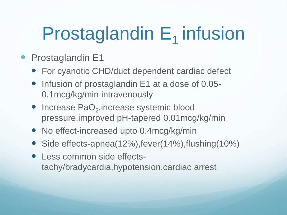
Prostaglandin E1 infusion Prostaglandin E1
For cyanotic CHD/duct dependent cardiac defect
Infusion of prostaglandin E1 at a dose of 0.05-
0.1mcg/kg/min intravenously
Increase PaO2,increase systemic blood
pressure,improved pH-tapered 0.01mcg/kg/min
No effect-increased upto 0.4mcg/kg/min
Side effects-apnea(12%),fever(14%),flushing(10%)
Less common side effects-
tachy/bradycardia,hypotension,cardiac arrest

Cyanosis
Pulmonary
Central CNS depression
Local
Ventilation-perfusion mismatch
Alveolar hypoventilation
Diffuse impairment
Cardiac
Increased pulmonary vascularity
Decreased pulmonary vascularity
Hemoglobi-nopathies

System Causes Clinical findings
CNS depression Perinatal asphyxia
Heavy maternal sedation
Intra uterine fetal distress
• Shallow irregular respiration
• Poor muscle tone
• Cyanosis disappears when
patient is stimulated or O2
given
Pulmonary disease Parenchyma
Pneumothorax or pleural
effusion
Diaphragmatic hernia
PPHN
• Tachypnea, respiratory
distress with retraction and
expiratory grunt
• Crackles or decreased
breath sounds
• X-ray findings
• Improve/abolish with oxygen
inhalation
Cardiac disease Cyanotic CHD with R-L shunt • Tachypnea without
retractions
• lack of crackles/abnormal
breath sounds
• Continuous murmur(PDA)
• X-ray findings
• Little/no increase with O2

Conclusion Central cyanosis in a newborn is an abnormal finding and
one must consider all of the possible etiologies with a complete history, physical examination and relevant investigations.
Remember to think about the various mechanisms causing cyanosis and go through each systematically until you have your diagnosis.
Prompt management should be undertaken while you are trying to figure out your diagnosis.
For ductal dependent lesion, start prostaglandin E1 and early referral

Thank You