Cyanoacrylate for Intraoral Wound Closure: A Possibility? · 2 InternationalJournalofBiomaterials...
6
Research Article Cyanoacrylate for Intraoral Wound Closure: A Possibility? Parimala Sagar, Kavitha Prasad, R. M. Lalitha, and Krishnappa Ranganath Department of Oral & Maxillofacial Surgery, Faculty of Dental Sciences, MSRUAS, MSRIT Post, MSR Nagar, Bangalore 560054, India Correspondence should be addressed to Parimala Sagar; [email protected] Received 24 July 2015; Revised 13 October 2015; Accepted 28 October 2015 Academic Editor: Ravin Narain Copyright © 2015 Parimala Sagar et al. is is an open access article distributed under the Creative Commons Attribution License, which permits unrestricted use, distribution, and reproduction in any medium, provided the original work is properly cited. Wound closure is a part of any surgical procedure and the objective of laceration repair or incision closure is to approximate the edges of a wound so that natural healing process may occur. Over the years new biomaterials have been discovered as an alternate to conventional suture materials. Cyanoacrylate bioadhesives are one among them. ey carry the advantages of rapid application, patient comfort, resistance to infection, hemostatic properties, and no suture removal anxiety. Hence this study was undertaken to study the effect of long chain cyanoacrylate as an adhesive for intraoral wound closure and also to explore its hemostatic and antibacterial effects. Isoamyl-2-cyanoacrylate (AMCRYLATE) was used as the adhesive in the study. In conclusion isoamyl cyanoacrylate can be used for intraoral wound closure, as an alternative to sutures for gluing the mucoperiosteum to bone, for example, aſter impaction removal, periapical surgeries, and cleſt repair. Its hemostatic and antibacterial activity has to be further evaluated. 1. Introduction Wound closure is a part of any surgical procedure. e objec- tive of laceration repair or incision closure is to approximate the edges of a wound so that natural healing process may occur. To promote healing one should achieve precise wound approximation, reduction of patient discomfort, easy han- dling of the working properties of wound closure materials, and low infection rates [1]. ere has been a search for new biomaterials over a few decades to serve as an alternate to conventional suture materials. Conventional suture materials have disadvantages like scar formation, need for dressing to protect the wound and suture, and revisit for suture removal [1]. One among the various biomaterials developed is the cyanoacrylate group. Cyanoacrylates have been found to be a good alternative to sutures in extraoral wound closure, as they carry advantages of rapid application, patient comfort, resistance to infection, hemostatic properties, and no suture removal anxiety [2]. Cyanoacrylates were first recognized to have adhesive properties by Coover in 1959. eir general formula is CNCH 2 =COO-R where R is side chain [2](Scheme 1). ey belong to the family of polymers whose monomer is formed by reversible condensation of formaldehyde with a cyanoacrylate ester. ese adhesives polymerize in the presence of anions especially hydroxyl ions. is indicates that it forms a firm adhesive bond on coming in contact with water or tissue moisture, by undergoing exothermic poly- merization. e number of alkyl groups in the side chain of cyanoacrylate can be increased from one (methyl cyanoacry- late), to two (ethyl), to four (butyl), and to five (isoamyl) but usually not more than eight (octyl cyanoacrylate). Isoamyl- 2-cyanoacrylate contains 5 carbon alkyl constituents of the carboxyl group (COO-R). ese adhesives differ physically to meet the defined applications. e main distinguishing feature between the esters is the size of molecule. Methyl, the original cyanoacry- late ester, is the smallest. Consequently, a large number of polymer chains can be formed resulting in bonds with high tensile strength and also molecules, which are less toxic [3]. As these long chain acrylates degrade at a slower rate, they permit the degradation products to be more safely metabolized resulting in less intense inflammatory response. Cyanoacrylates are maintained in a liquid state by an acidic stabilizer, which has the action of inhibiting the molecules from cross-linking. Partially ionized molecules of water, normally found on all surfaces exposed to the atmosphere, have the action of neutralizing the inhibitor. Hindawi Publishing Corporation International Journal of Biomaterials Volume 2015, Article ID 165428, 6 pages http://dx.doi.org/10.1155/2015/165428
Transcript of Cyanoacrylate for Intraoral Wound Closure: A Possibility? · 2 InternationalJournalofBiomaterials...
Research ArticleCyanoacrylate for Intraoral Wound Closure: A Possibility?
Parimala Sagar, Kavitha Prasad, R. M. Lalitha, and Krishnappa Ranganath
Department of Oral &Maxillofacial Surgery, Faculty of Dental Sciences, MSRUAS, MSRIT Post, MSR Nagar, Bangalore 560054, India
Correspondence should be addressed to Parimala Sagar; [email protected]
Received 24 July 2015; Revised 13 October 2015; Accepted 28 October 2015
Academic Editor: Ravin Narain
Copyright © 2015 Parimala Sagar et al.This is an open access article distributed under the Creative Commons Attribution License,which permits unrestricted use, distribution, and reproduction in any medium, provided the original work is properly cited.
Wound closure is a part of any surgical procedure and the objective of laceration repair or incision closure is to approximate theedges of a wound so that natural healing process may occur. Over the years new biomaterials have been discovered as an alternateto conventional suture materials. Cyanoacrylate bioadhesives are one among them.They carry the advantages of rapid application,patient comfort, resistance to infection, hemostatic properties, and no suture removal anxiety. Hence this study was undertakento study the effect of long chain cyanoacrylate as an adhesive for intraoral wound closure and also to explore its hemostaticand antibacterial effects. Isoamyl-2-cyanoacrylate (AMCRYLATE) was used as the adhesive in the study. In conclusion isoamylcyanoacrylate can be used for intraoral wound closure, as an alternative to sutures for gluing the mucoperiosteum to bone, forexample, after impaction removal, periapical surgeries, and cleft repair. Its hemostatic and antibacterial activity has to be furtherevaluated.
1. Introduction
Wound closure is a part of any surgical procedure.The objec-tive of laceration repair or incision closure is to approximatethe edges of a wound so that natural healing process mayoccur. To promote healing one should achieve precise woundapproximation, reduction of patient discomfort, easy han-dling of the working properties of wound closure materials,and low infection rates [1].
There has been a search for new biomaterials over afew decades to serve as an alternate to conventional suturematerials. Conventional suture materials have disadvantageslike scar formation, need for dressing to protect the woundand suture, and revisit for suture removal [1]. One among thevarious biomaterials developed is the cyanoacrylate group.Cyanoacrylates have been found to be a good alternative tosutures in extraoral wound closure, as they carry advantagesof rapid application, patient comfort, resistance to infection,hemostatic properties, and no suture removal anxiety [2].
Cyanoacrylates were first recognized to have adhesiveproperties by Coover in 1959. Their general formula isCNCH
2=COO-R where R is side chain [2] (Scheme 1).
They belong to the family of polymers whose monomeris formed by reversible condensation of formaldehyde with
a cyanoacrylate ester. These adhesives polymerize in thepresence of anions especially hydroxyl ions. This indicatesthat it forms a firm adhesive bond on coming in contact withwater or tissue moisture, by undergoing exothermic poly-merization. The number of alkyl groups in the side chain ofcyanoacrylate can be increased from one (methyl cyanoacry-late), to two (ethyl), to four (butyl), and to five (isoamyl) butusually not more than eight (octyl cyanoacrylate). Isoamyl-2-cyanoacrylate contains 5 carbon alkyl constituents of thecarboxyl group (COO-R).
These adhesives differ physically to meet the definedapplications. The main distinguishing feature between theesters is the size of molecule. Methyl, the original cyanoacry-late ester, is the smallest. Consequently, a large number ofpolymer chains can be formed resulting in bonds with hightensile strength and also molecules, which are less toxic[3]. As these long chain acrylates degrade at a slower rate,they permit the degradation products to be more safelymetabolized resulting in less intense inflammatory response.
Cyanoacrylates are maintained in a liquid state by anacidic stabilizer, which has the action of inhibiting themolecules from cross-linking. Partially ionized moleculesof water, normally found on all surfaces exposed to theatmosphere, have the action of neutralizing the inhibitor.
Hindawi Publishing CorporationInternational Journal of BiomaterialsVolume 2015, Article ID 165428, 6 pageshttp://dx.doi.org/10.1155/2015/165428

2 International Journal of Biomaterials
H2C
CN
O
O
H2
H2
CC
CH
CH3
CH3
Isopentyl-2-cyanoacrylateIsoamyl-2-cyanoacrylate
Scheme 1: Structure of isoamyl-2-cyanoacrylate.
When applied on tissue surface the inhibitor is eliminatedand the chemical action allows the molecule to polymerizein 10 seconds. The adhesive strength depends on relation tothe approximation of the molecules attraction to each other.Physical locking is also a factor by virtue of the penetrationof the adhesive into irregularities of the tissue surfaces [3].
Cyanoacrylates are biodegradable and their rate ofremoval from the application site is by polymer degradationand surface sloughing. They undergo hydrolytic attack ofcarbon-carbon bond to produce formaldehyde and cyanoac-etate.
Wide applications have been cited in the literature fromsealing of CSF leaks encountered during orbital surgery andrepair of tympanic membrane to microvascular anastomosis[2]. Very few studies have been done about the intraoral useof cyanoacrylates. This study was undertaken to explore thevarious uses of cyanoacrylates intraorally.
In the present study isoamyl-2-cyanoacrylate was usedafter various surgical procedures intraorally. Isoamyl-2-cyanoacrylate was the only cyanoacrylate, which could beused intraorally and cost-effective when compared to others.Octyl cyanoacrylate could not be used as it is not indicatedin areas subject to frequent moisture; hence isoamyl-n-cyanoacrylate was chosen for the study [4].
The aim of this study was to explore the various usesof isoamyl cyanoacrylates intraorally and also to study itshemostatic and antimicrobial effects.
2. Materials and Methods
The study conducted was a prospective observational invivo study of patients attending the Department of Oral& Maxillofacial Surgery for various surgical procedures.Isoamyl-2-cyanoacrylate was used for wound closure. It wasavailable from CONCORD DRUGS LIMITED under thetrade name of “AMCRYLATE,” manufactured at Chennai,India.
10 patients reported to the Department of Oral & Max-illofacial Surgery at M. S. Ramaiah.
Dental College&Hospital for various surgical proceduresfrom November 2005 to August 2007 were selected for thestudy. Isoamyl-2-cyanoacrylate was used intraorally for vari-ous indications/applications. Data was recorded according tothe pro forma. After obtaining clearance from ethics com-mittee, study was begun. Patient selection was based on thefollowing inclusion and exclusion criteria.The study included
patientswith fracturemandible, cleft palate,mandibular thirdmolar impaction, laceration, periapical cysts, and need forbiopsy. Patients with infected wounds, patients with woundswhere tension-free closure was not possible, and medicallycompromised patients were excluded from the study.
Cases selected for the study were to glue the mucoperios-teum to the underlying bone, following impactedmandibular3rd molar removal, cases of periapical surgeries, or cases toglue periosteum to the palate in cleft patients. It was alsoused for gluing of collagen over the surgical wound, closurefollowing incisional biopsy in case of carcinoma of alveolus,pyogenic granuloma, and gluing of a lacerated wound.
After completion of the procedure, AMCRYLATE avail-able as (0.25mL or 0.5mL) ampule was loaded in a syringeprovided along with the adhesive. The incised margins werethen checked for approximation. Cyanoacrylate loaded in thesyringe was applied over the bone and inner surface of themucoperiosteal flap and also applied over the incisedmarginsand then approximated into the desired position. It was heldthere under pressure for a few seconds till the polymerizationprocess was complete to ensure firm adhesion of the flap tothe underlying bone.Woundwas observed on the 1st, 7th, and15th postoperative day for presence/absence of inflammation,edema, approximation, or dehiscence. The findings wererecorded and assessment was done based on the results.
3. Case Details and Results
A total of 10 patients were enrolled in the study and resultstabulated (Table 1).
In case 1, a case of pleomorphic adenoma of palate locatedat the junction of hard and soft palate surgical excisionwas done. Overlying mucosa was also excised along withthe tumor. Here cyanoacrylate was used to glue collagenmembrane over the surgical wound to promote healing. Onthe 2nd postoperative day collagen membrane got dislodged;however, wound later healed without any complications.
Case 2 was a patient with fracture of left angle andright parasymphysis of mandible. Here in the parasymphysisregion fracture site was exposed, reduced, and fixed with tita-nium plates and screws. Submucosal suturing with VICRYL(Figure 1(a)) was done for layered closure; mucosal adhesionwas achieved with cyanoacrylate (Figure 1(b)). Postoperativehealing (Figure 1(c)) was uneventful.
In case 3, following incisional biopsy in case of carcinomaof left alveolus, cyanoacrylate was used to seal the biopsy site.The tissue was very fragile and suturing could have resultedin further trauma. Hemostasis was readily achieved.
In case 4, cyanoacrylate was used to cover the mucosalincision line over the bone graft in addition to sutures forairtight closure. This was a case of fracture of angle ofmandible on right side. ORIF under GA was done, usingtitanium plates and screws. Postoperatively after one monthpatient returned back with nonhealing of fracture due tobroken titanium plate. So fracture was fixed again withreconstruction plate over the angle region and iliac bone graftwas used to promote healing as the defect was wide. Mattresssuture was given intraorally over the mucosal surface. Onthe first postoperative day intraoral wound at the third molar
International Journal of Biomaterials 3
Table 1
Results tabulationInflammation Edema Dehiscence Wound seal/closure
1st POD 7th POD 15th POD 1st POD 7th POD 15th POD 1st POD 7th POD 15th POD 1st POD 7th POD 15th PODCase 1 √ × × √ × × × × × √ × ×
Case 2 √ × × √ × × × × × √ √ √
Case 3 √ × × √ × × × × × √ √ √
Case 4 √ × × √ × × × × × √ √ √
Case 5 √ × × √ × × × × × √ √ √
Case 6 √ × × √ × × × × × √ √ √
Case 7 √ × × √ × × × × × √ √ √
Case 8 — — — — — — — — — — — —Case 9 √ × × √ × × × × × √ √ √
Case 10 √ × × √ × × √ × × × √ √
×: absent,√: present, and —: could not be assessed.Case 1: case of pleomorphic adenoma.Case 2: fracture of right parasymphysis of mandible. Mucosal closure done.Case 3: case of carcinoma of left alveolus. Hemostasis achieved at biopsy site with acrylic.Case 4: case of fracture of right angle of mandible. Acrylate used to cover the incision line over the bone graft.Case 5: case of periapical cyst. Mucoperiosteum glued with acrylate.Case 6: case of impacted 48. Ward’s incision closed with acrylate.Case 7: case of periapical cyst. Mucoperiosteum glued with acrylate.Case 8: case of pyogenic granuloma. Acrylate used to achieve hemostasis.Case 9: case of cleft palate. Mucoperiosteum glued with acrylate.Case 10: case of laceration of angle of mouth. Wound glued with acrylate.
(a) (b)
(c)
Figure 1: (a) Incision after submucosal closure. (b) Cyanoacrylate application over incision line. (c) Seventh postoperative day.
4 International Journal of Biomaterials
(a) (b) (c)
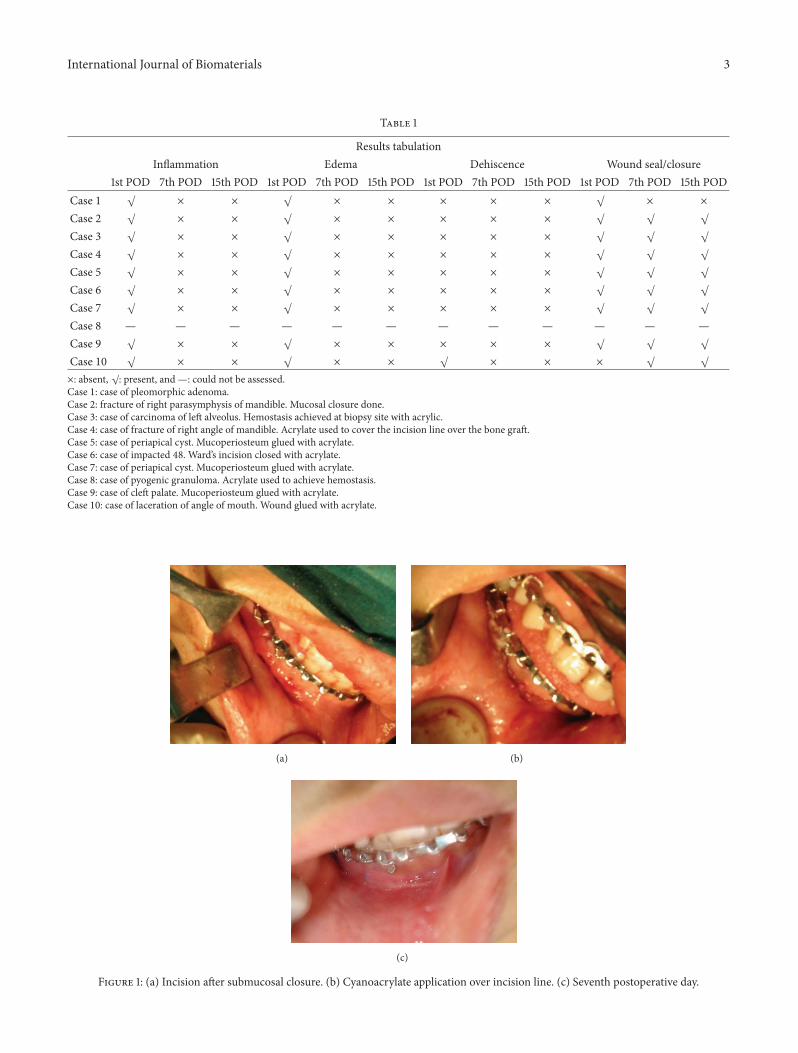
Figure 2: (a) Lacerated wound on lower lip. (b) Wound glued with cyanoacrylate. (c) 15th postoperative day.
region showed slight gaping at the graft site. So cyanoacrylatewas used over the incision line, after, approximating the flapto cover the bone graft. Wound healing was successfullyachieved later without any complication.
In cases 5 and 7, periapical surgery was performed forcuretting apical cysts of maxillary anterior. Triangular shapeflap was designed. Full thickness mucoperiosteum flap wasreflected. After the surgical procedure mucoperiosteum flapwas glued back to the underlying bone in its position withAMCRYLATE. In case 5 intraoral sinus discharging pus waspresent at the surgical site on the 15th postoperative day. Case7 did not come back for review.
In case 6, cyanoacrylate was used to glue the mucope-riosteum and close the incision line (Ward’s incision) aftercoronectomy of mandibular third molar (48) was done. Nocomplications were noted and wound healed uneventfully.
In case 8, it was also used as a hemostatic agent in a patientwith an exophytic growth over themaxillary canine-premolarregion where biopsy was planned initially. Hemostasis couldnot be achieved in this case so complete excision was thendone.
Case 9 was a patient with the midline cleft of the palate.Cyanoacrylate was used to affix the mucoperiosteum to thehard palate.The nasal mucosal layer was closed with VICRYLsutures beginning fromuvula to alveolar ridges.Oralmucosallayer was then closed in the same manner. Instead of placingsling suture, to hold the mucoperiosteum to the underlyingbone, cyanoacrylate was spread over mucoperiosteal flap andalso on the hard palate. Gentle pressure for 1min was appliedover the hard palate to affix the flap. Postoperative woundhealing was uneventful.
Case 10 was a 4-year-old boy who presented with lac-eration at lip commissure (Figure 2(a)). Suturing was doneunder local anesthesia at the initial appointment. Woundgaped on the 2nd postoperative day. Cyanoacrylate was thenused to glue the lacerated margin (Figure 2(b)), but againwound gaping was seen the next day. Cyanoacrylate was
reapplied to the lacerated margins and dressing was given toprotect the lacerated site. Wound later healed (Figure 2(c))without complications.
Summary. Marginal seal/wound seal was acceptable as theincised margins were in close apposition with each other.Inflammation and edema were not present in maximumnumber of cases at the 15th postoperative day. One case of pusdischarge was recorded after 15 days of surgery and one caseof wound dehiscence was noted and gluing of the collagenmembrane to the surgical site was not successful.
4. Discussion
The aim of all wound closure techniques is to approximatethe edges of the wound so that natural process of uneventfulhealing takes place. In wound closure the primary focusshould be on relieving tension on the wound and bring-ing the tissue edges together. Precise approximation of theincised/lacerated margins is critical to favorable cosmeticand functional results. The most commonly used methodfor closing lacerations is suturing [1]. This is the classicmethod of wound closure which carries the advantages ofcareful closure, good tissue approximation, highest resilientstrength, and lowest dehiscence rate. However it has severaldisadvantages like requirement of anesthesia, greatest tissuereactivity, higher cost, more time consuming, need for sutureremoval, and associated risk of needle stick injury [1]. Overthe past 3 decades new biomaterials have been discovered asan alternative to conventional suture material. These includestaples, adhesive tapes, and tissue adhesives.
Cyanoacrylates include short chain (methyl and ethylcyanoacrylates) and longer chain (butyl, isobutyl, isoamyl,and octyl cyanoacrylates) derivatives [2]. The long chainderivatives are the least histotoxic. Some authors havereported the clinical use of cyanoacrylates with severaladvantages including the ease of use with shorter operative

International Journal of Biomaterials 5
time, formation of protective barrier, and painless appli-cation [2, 5]. The major disadvantage is reduced tensilestrength (they should not be used in high tension areas) [5].They also should be avoided over areas subject to frequentmoisture and friction such as the hands and feet (unlesssplinted and dressed appropriately) [5]. These adhesives arecontraindicated in infected wounds, immunocompromisedpatients, and patients with known allergy to cyanoacrylatesand formaldehyde [5].
After discovery of cyanoacrylates in 1949, there hasbeen interest in their use for various surgical procedures.These include traumatic laceration repair, bronchopleuralfistula repair, repair of myocardial tears, mesh fixation foringuinal hernia repair, cosmetic rhinoplasty, embolization ofintracranial AV malformations, and sealing of CSF leaks [6].After use of these adhesives in various tissue organs it wasfound to be a valuable adjunct in intraoperative managementof difficult problems and no case of general or local toxicreaction to this material was observed [6]. Initially it wasused as an alternative to sutures only in superficial smalland tension-free skin incisions or lacerations extraorally[7]. Now it has wide applications intraorally, for example,for promoting of healing in extraction sockets [8], as localhemostatic agent in warfarin treated patients undergoing oralsurgery [9], for promoting of healing of recurrent aphthousulcers [10], in cleft palate surgery [2], and for closing sinusmembrane perforations during sinus lifts [11].
Numerous studies have been conducted on extraoral usesof cyanoacrylates in oral and maxillofacial region. Octylcyanoacrylate was used to close neck incisions in cancerpatients and has been found to be an effective alternate tosutures [12]. Another comparative study between isoamylcyanoacrylate and butyl cyanoacrylate on healing of surgicalincisions over the neck was done and isoamyl cyanoacrylatewas found to be better than butyl acrylate [13]. However, veryfew studies have been conducted on intraoral applicationsof these cyanoacrylates. So this study was undertaken toexplore the various uses of these acrylates intraorally. Animalstudies on intraoral application of cyanoacrylates have beensuccessful and are safe and suitable without any significantdifferences in liver and kidney functions [14]. In the presentstudy, it was found that, in cases 5 and 7, cyanoacrylatecould be successfully used in closure of mucoperiosteumin periapical surgery (triangular), in case 6, after impactionremoval (Ward’s incision), and, in case 9, for cleft palateclosure (gluing mucoperiosteum). Results obtained in cleftpatient are comparable to the results obtained in a previ-ous study where butyl cyanoacrylate was used for gluingmucoperiosteum to palate [2]. Further comparative studiesbetween the use of sutures and cyanoacrylate adhesive formucoperiosteal closure in a larger sample of patients canclearly establish its usefulness. In our study cyanoacrylatewas used as adjuvant in closing the mucosa in fracture casesas in cases 2 and 4. In case 2, wherein patient had fractureparasymphysis mucosal closure with acrylate was done aftersubmucosal closurewith sutures. In case 4 it was used to coverthe bone graft site intraorally, in addition to sutures at theangle region of mandible. In both the cases successful closure
was achieved with acrylate. Further comparative studies witha larger sample need to be conducted so that it replacessutures successfully in closing the incision lines intraorally.
In a study conducted in the year 2003, butyl cyanoacrylatewas effectively used as local hemostatic agent; warfarintreated patients underwent extraction of teeth without achange in their anticoagulation level [9]. In the present study,hemostasis was successfully achieved in case 3, when usedat biopsy site of one patient (later reported as squamouscell carcinoma of left buccal mucosa), but was unsuccessfulin case 8. In case 8, patient with exophytic growth in themaxilla, acrylate was applied over the bleeding site; thoughmucosal seal was obtained, bleeding continued submucosally.Hemostasis was achieved only after complete excision of thelesion. It was reported as pyogenic granuloma. Its use as alocal hemostatic agent therefore needs further evaluation.Collagen glued to the underlying surgical site in case 1 (atthe junction of hard and soft palate) got dislodged on the 2ndpostoperative day.Thismay be due to friction created by foodmaterials while eating and swallowing. Hence, cyanoacrylatein such situations can be used as an adjuvant to sutures ratherthan alternate to sutures. Use of cyanoacrylate will help inadhesion to the underlying arched (curved) palate as seenin case 9. Wound dehiscence was seen on the second dayin the 4-year-old kid, that is, case 10 with lip lacerationat the angle of mouth. Sutures also were not successful inapproximating the margins at this site. This may be due topatient being uncooperative/unable to follow instructions.Also constant movement of the sutured/glued site duringopening and closing of mouth and excessive moisture due todrooling of saliva could have been further hindrances. So useof adhesives in this region is comparable to sutures. Howeverthe second application of cyanoacrylate resulted in woundclosure and better patient compliance with cyanoacrylate wasseen as it was painless and less time consuming.
In our study group 9 patients had no significant signsof infection at the 15th postoperative day. Pus discharge wasrecorded in one case with periapical surgery after 15 days.Symptomatic treatment with antibiotics and analgesics wasdone and patient reviewed after a week. Sinus opening inrelation to canine and first premolar was still persistent.Vitality test and IOPA radiograph confirmed nonvital firstpremolar. RCTwas done and later wound healed successfully.So probably pus discharge may not be related as reaction tocyanoacrylate but due to nonvital first premolar. Inflamma-tory edema seen in all cases on the first postoperative day canbe related to surgical trauma. Successful mucosal wound sealin all cases was obtained.
Future of tissue adhesives can be as broad as imagination.New ways to manufacture these agents more safely are beingexplored by better defining the synthetic additions. Thebiodegradable adhesives can be engineered for slow releaseof medications as delivery of medications to a specific targetsite is very desirable [4]. Closure of wounds without the needfor sutures will be a major advancement, an opportunity toimprove care for patients, especially children, and reduce painand anxiety caused by the treatment.

6 International Journal of Biomaterials
5. Conclusion
Isoamyl-2-cyanoacrylate tissue adhesive was equivalent tosuture and other traditional epidermal wound closure devicesin providing adequate wound healing after closure of surgicalincisions. There are several advantages of these adhesives;some of them are as follows: they have easy and painlessapplication, need to remove sutures is eliminated, also theyact as a hemostatic agent, andmost important is better patientcooperation compared to sutures for the procedures doneunder local anesthesia. Cyanoacrylates can be used as analternative to sutures for gluing themucoperiosteum to bone,for example, after impaction removal, periapical surgeries,cleft closure, or biopsy. It can be used as an adjuvant to suturesin many situations to offer wound protection and reducedehiscence and infection rates, for example, at bone graftsites, for skin grafts or collagen retention, and for mucosalclosure at fracture sites. Its hemostatic, antimicrobial, andanalgesic properties require further evaluation. Its cost-effectiveness is still debatable but it is an adhesive worthconsidering for intraoral closure. Further randomized clinicaltrials should definitely highlight its distinction from suturesand help in evolving a set of guidelines for intraoral use so asto achieve maximum results of this revolutionary product.
Conflict of Interests
The authors declare that there is no conflict of interestsregarding the publication of this paper.
References
[1] R. Gassner, “Wound closure materials,” Oral and MaxillofacialSurgery Clinics of North America, vol. 14, no. 1, pp. 95–104, 2002.
[2] T. Turkaslan and D. Dayicioglu, “Use of adhesive in cleftpalate surgery: a new flap fixation technique,” The Journal ofCraniofacial Surgery, vol. 16, no. 4, pp. 719–722, 2005.
[3] P. R. Samuel, A. C. Robert, and A. Nigam, “The use of Indermil(n-butyl cyanoacrylate) in otorhinolaryngology and head andneck surgery. A preliminary report on the first 33 patients,”TheJournal of Laryngology & Otology, vol. 111, no. 6, pp. 536–540,1997.
[4] T. B. Reece, T. S. Maxey, and I. L. Kron, “A prospectus on tissueadhesives,”The American Journal of Surgery, vol. 182, no. 2, pp.40S–44S, 2001.
[5] A. J. Singer and H. C. Thode, “A review of the literature onoctylcyanoacrylate tissue adhesive,” The American Journal ofSurgery, vol. 187, no. 2, pp. 238–248, 2004.
[6] D. T. Tse, W. R. Panje, and R. L. Anderson, “Cyanoacrylateadhesive used to stopCSF leaks during orbital surgery,”Archivesof Ophthalmology, vol. 102, no. 9, pp. 1337–1339, 1984.
[7] G. E. Amiel, I. Sukhotnik, B. Kawar, and L. Siplovich, “Use ofN-butyl-2-cyanoacrylate in elective surgical incisions—Longtermoutcomes,” Journal of the American College of Surgeons, vol. 189,no. 1, pp. 21–25, 1999.
[8] S. N. Bhaskar, D. E. Cutright, J. Frisch, and P.Margetis, “Effect ofbutyl cyanoacrylate on the healing of extraction wounds,” OralSurgery, Oral Medicine, Oral Pathology, vol. 24, no. 5, pp. 604–616, 1967.
[9] F. A. Al-Belasy and M. Z. Amer, “Hemostatic effect of n-butyl-2-cyanoacrylate (histoacryl) glue in warfarin-treated patientsundergoing oral surgery,” Journal of Oral & MaxillofacialSurgery, vol. 61, no. 12, pp. 1405–1409, 2003.
[10] J. B. Ludlow, M. J. Kutcher, A. Samuelson, and C. Hill, “Intrao-ral digital imaging documenting recurrent aphthous ulcerhealing in 2-octyl cyanoacrylate versus sham-treated lesions,”Oral Surgery, Oral Medicine, Oral Pathology, Oral RadiologyEndodontics, vol. 89, no. 4, pp. 425–431, 2000.
[11] B.-H. Choi, B.-Y. Kim, J.-Y. Huh et al., “Cyanoacrylate adhesivefor closing sinus membrane perforations during sinus lifts,”Journal of Cranio-Maxillofacial Surgery, vol. 34, no. 8, pp. 505–509, 2006.
[12] C. A. Ashoka,Evaluation of octyl-2-cyanoacrylate tissue adhesiveas an acceptable alternative to sutures in head & neck surgery[Ph.D. dissertation], Rajiv Gandhi University of Health SciencesKarnataka, Bengaluru, India, 2005.
[13] L. Babu, A comparative clinical study of isoamyl-2-cyanoacrylate& butyl-2-cyanoacrylate on healing of surgical wounds [Ph.D.dissertation], Rajiv Gandhi University of Health Sciences Kar-nataka, Bengaluru, India, 2005.
[14] S. Inal, N. Yilmaz, C. Nisbet, and T. Guvenc, “Biochemical andhistopathological findings of N-Butyl-2-Cyanoacrylate in oralsurgery: an experimental study,” Oral Surgery, Oral Medicine,Oral Pathology, Oral Radiology and Endodontology, vol. 102, no.6, pp. e14–e17, 2006.


![smart2 introduction [호환 모드] - ZIVELAB · Total Electrochemistry Since 1991 Ch4 Ch3 Ch2 Ch1 MFC MFC H2 detector COdetector Fuel Heater power control Differential Pressure Guage](https://static.fdocuments.in/doc/165x107/5e9fd956a740a0272e49ba23/smart2-introduction-eeoe-total-electrochemistry-since-1991-ch4-ch3.jpg)