CY TOTOXIt:lTya'r TUMOUR (IN - Information and...
27
CY TOTOXIt:lTya'r TO TUMOUR CELLS AND CEL.LS lPI1 CULrrTJIltE (IN VITRO) -m . - . "- . "-n" . - . " . -" . "-
Transcript of CY TOTOXIt:lTya'r TUMOUR (IN - Information and...

CY TOTOXIt:lTya'r TO TUMOUR CELLS AND CEL.LS lPI1 CULrrTJIltE (IN VITRO)
-m...-."-."-n".--..".-""".."--

3.1. INTRODUCTION
Traditional and folk medicine have long used plant products for
therapeutic purposes. But it is only since 1959 that a concerted
systematic effort has been made to screen crude plant extracts for
their inhibitory activity against animal tumour systems. In the
Indian system of medicine many of the plant products are used for the
treatment of various diseases including cancer (285).
In this study we have investigated the cytotoxic effect of
Saraca asoca bark, flower, and leaves extract to various tumour cells
in vitro and also the growth inhibitory effect of these extracts to - - cultured cell S.
3.2. Materials and Methods:
DLA, EAC, S-180 and P-388 tumour cells were obtained from
tumour bearing mice as described in 2.1.4. Radioactive isotopes,
trypan blue and culture medium was obtained from sources mentioned in
Chapter-2.
3.2.1. Differential extractions:
Preliminary experiment was carried out by extracting Saraca
asoca bark, flower and leaves, powder with petroleum - ether,
choroform, 90% acetone, Ethylacetate, Ethanol (95x1, methanol and
water were used. The extract was evaporated under reduced pressure
and active pfinciple was isolated as stated in 2.2.2. This was used
for cytotoxicity studies.
3.2.2. Determination of cytotoxicity to tumour cells:
The cytotoxic effect of Saraca asoca bark,flower and leaves
extract was evaluated using murine tumour cells like DLA, EAC, S-180
and P-388 (2.2.5). Brief1 y 1 X 10⁶ cells were incubated with
various drug concentrations at 37'~ for 3 hour. Cell viability was
ascertained by the trypan blue exclusion method.
3.2.3. Determination of cytotoxicity to isolated Iymphocytes:
Lymphocytes were is01 ated from the peripheral blood of heal thy
normal persons and patients with leukaemia (acute lymphocytic
leukaemia and chronic myeloid leukaemia) by the Ficol Hypaque Method.
(273) Chronic myelogenous leukemia (CML) and Acute Lymphoblastic
Leukaemia (ALL) cells were obtained from a 52 and a 9 year old
patient admitted in our oncology department. The blood was obtained
from these patients before the commencement of any kind of treatment.
The is01 ated l ymphocyte/ml PBS a1 ong with various extract
concentrations was incubated at 37'~ for 3 hour and the viability of
lymphocytes was determined by trypan blue exclusion method.

3.2.4. Cytotoxicity of extracts in in vitro cultures:
5x10~ K-562 cell S were added to each of the culture bottles
containing 10 m1 RPMI medium, different concentrations of extracts
were then added. Controls were maintained without the drug. All the
culture bottles were incubated at 37'~ in 5% CO2 and the inhibition
in the growth of cells was assessed as detailed in (2.2.4.1)
4 5x10 CH0,KB or Vero cells were added to tissue culture bottles
containing 10 m1 Eagle's minimum essential medium and incubated for
24 hour at 37'~. Drugs at different concentration was added after 24
hours and the bottles incubated for seven days (37'~) After
incubation, the cells were harvested and the growth inhibitory effets
ascertained (2.2.4.1)
3.2.5. Determination of intracellul ar enzyme l eve1 of S-180
tumour cel l S in cul ture :
lxlo7 Sarcoma-180 tumour cell S were cultured in 10 m1 RPMI
medium containing 10% calf serum at 3 7 O ~ in CO2 for 48 hours in the
presence of various concentrations of Saraca asoca bark and flower
extract and assay was carried out as described in 2.2.13.4
Glutathione reductase (GSH-R) was determined from the tumour
cells and glutathione S-transferase (GSH-S) levels using 2,4 -

dinitro - chl or benzene as substrate. Cell suspensions (2x10⁷/ml )
were sonicated on ice (90s) using a sonicator and the resultant
sonicated rn'ixture was spun at 1500 rev/min for 6 min. The protein
content in the supernatent was also determined (283).
3.2.6. Inhibition of the synthesis of macromolecules:
The effect of Saraca asoca bark, flowers and leaves extracts on
DNA synthesis was studied using radio labelled precursors by the
method developed by Pavlik et a1 (1903). Briefly 3(H) thymidine was
added (2p ci/ml) to the tubes containing one million tumour cells/ml
and incubated with various drug concentrations in a total of 2 m1
MEM(+10% FCS) for four hour at 37'~. The precipitated DNA was
dissolved and radio activity measured.
Tritiated uridine and leucine were used to study the effect of
Saraca asoca bark, flowers and leaves extracts on the synthesis of
RNA and protein respectively. Details of the assay procedure are
given in 2.2.7 and 2.2.8.
3 . 3 RESULTS
The extraction of Saraca W, bark,flowers and leaves with
solvents of different polarity and their cytotoxic study indicated
maximum extraction at the active principle with 95% methanol in case

of flowers, Ethyl acetate in Bark and 90% acetone as in leaves
(Tab1 e-20) ,-
3 . 3 . 1 . Cytotoxicity of Saraca asoca active principle.
In vitro studies indicated that the active principle of ethyl - - acetate extract of Saraca asoca bark, methanolic extract of flower
which furthur purified as stated in 2.2.2 and was highly active
against .bDCA, S-180, and P-388 tumour cells. The concentrations
of the "Ashoka" bark needed to produce 50% cytotoxicity were 16
yg/ml, 23 pg/ml, 47 pg/ml and 55 pg/ml respectively for DLA, 5-180,
EAC, P-388 tumour cells Table(21). In the case of flower extract the
corresponding figures were 24pg,37pg/ml and 52pg as in the case of
DLA, S-180, and P-388,Table(22). Extracts of leaves required higher
concentrations 65 and 73yg/ml to produce 50% cytotoxic effect
Table(23). While the flower and leaves extract were insensitive to
Ehrl ich ascites carcinoma cel l S.
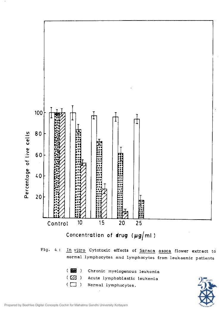
3.3.2. Effect of extracts on lyrnphocytes:
It was found that normal lymphocytes were insensitive to drug
while lymphocytes from leukaemic patients were acted upon hy bark
and flower extract. 50% cytotoxicity was obtained at a concentration
of 11 and 22pg to ALL and CML in the case of flower extract (Fig 4)
and it was 9 pg and 16 pg in the case of bark extract (Fig 5)

Table 21: in v i t ro cytotoxicity of the act ive pr inciple of Saraca asoca ba rk -- extracts on tumour cell l i nes
Percentage of Live cells
=onckntration of ................................................................................. extract ( ~ g ) DLA S-180 EAC cells P-388 cells
.............................................................................................................. 80 ND ND 10.25+1.37 - 13.6 - +1.79
Values a re average of mean - + SD from 4 separate studies
N.D. - Not determined
N . A . - No activity
DLA - Dalton's 1yrnphQha a s d t e s ..
5-180 - Sarcoma - 180
DAC - Dalton's lymphoma ascites Carcinoma
Amount of extract needed for 50% Cytotoxicity was determined from graph by plotting concentration
against percent of l ive cells.

Table 22: -- i n vitro cytotoxidty of the active principle of 'Ashoka' (Saraca asoca) flower
extract on tumour cell l ines
Percentage of l ive cells Concentratiori of .......................................................................................
extracts ( ~ g ) DLA S-180 E . A C P 388
Values are average of mean + S.D. from 4 separate determination5
N D - Not determined
DLA - Dalton's lymphoma ascites
EAC - Ehrlich a s d t e s carcinoma
Amount of extract needed for 50% cytotoxicity was determined from graph by plotting
concentration against percent of live cells.
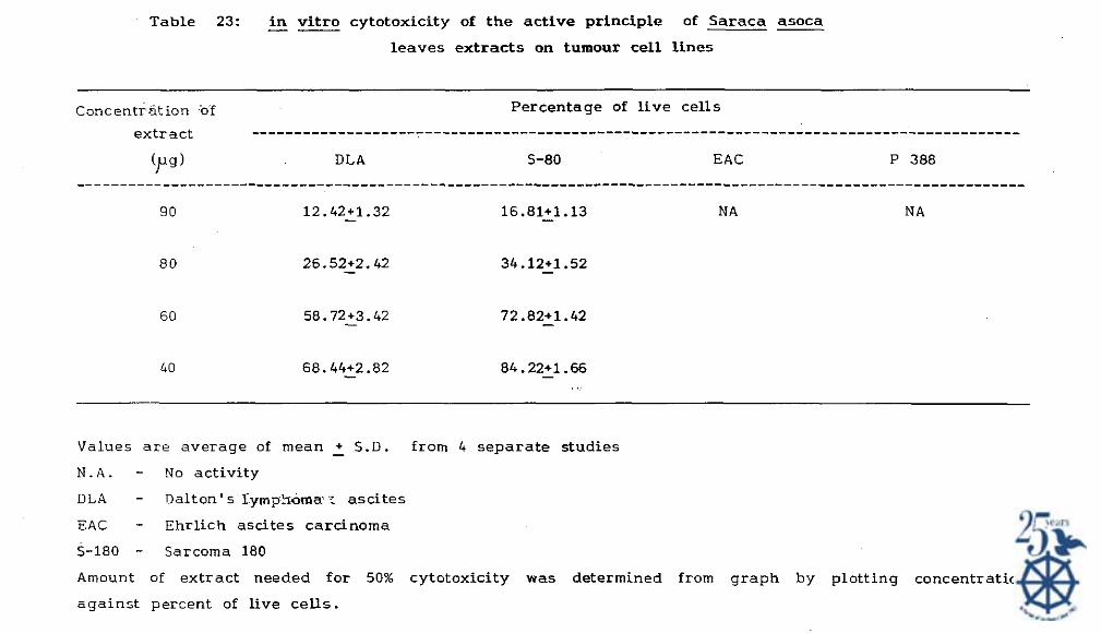
Table 23: i n v i t ro cytotoxicity of t h e active principle of Saraca asoca -- leaves extracts on tumour cell l ines
Concentrdtion of Percentage of l ive cells
extract ......................................................................................... (p) DLA S-80 EAC P 388
.............................................................................................................. 9 0 12.42+1.32 - 16.81+1.13 - NA N A
Values a r e average of mean - + S.D. from 4 separa te studies
N . A . - No activity
D L A - Dalton's lym??iama : ascites
EAC - Ehrlich a s d t e s carcinoma
S-180 - Sarcoma 180
Amount of extract needed for 50% cytotoxicity w a s determined from graph by plotting concentration
against percent of live cel ls .
Concentrat ion o f drug (pg/ml I
Fig. 4 : I n vi t ro Cytotoxic effects of Saraca asoca flower extract to -- P
normal lymphocytes and lymphocytes from leukaemic patients
( ) Chronic myelogenous leukemia
( Acute lymphoblastic leukemia
n 1 Normal lymphocytes.
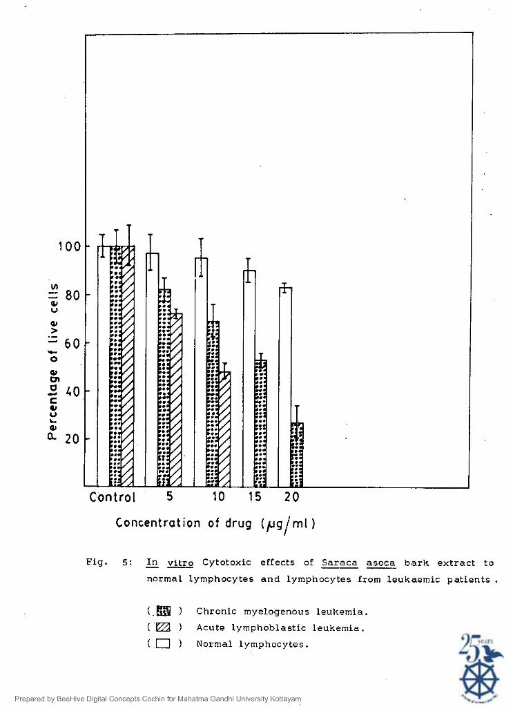
Concentration o f drug ( p g / m l )
Fig. 5: I n vi t ro Cytotoxic effects of Saraca asoca ba rk extract to
normal lymphocytes and lymphocytes from leukaemic patient*
) Chronic myelogenous leukemia.
-m Acute lymphoblastic leukernia.
( 0 ) Normal lymphocytes.

3 . 3 . 3 . Inhibitory effect of active principle of Saraca asoca
on the growth of K-562 c e l l s in culture.
The active principle of Saraca asoca bark and flower inhibited
the growth of K-562 cells in culture. The percentage of cell survival
0.5 ~ ~ / m l and 1.5 pg/ml active principle of bark after 24 hour of
treatment was found to be 16% and 11%. The corresponding figures
were 36% and 22% after 72 hour. While in presence of flower extract
the percentage of cell survival was 31% and 17% (day-l) and 52% and
39% (day-3) at a concentration of 3 and 41~glml respectievely (Table-
24)
3 . 3 . 4 . Effect of Saraca aeoca (Aehoka) on the growth of K B , CH0
and vero cell S in culture:
The growth of KB, CH0 and Vern cells in culture was inhibited
by active principle of Saraca asoca bark and flower extracts. The
concentration required to produce 50% growth inhibition in the case
of Ashoka bark, flower and leaves were found to be 0.6 yg/ml, 1.3
pg/ml and 2.4 )lg/mI respectively when cultured with KB cell S (Fig.6).
Vero cells were found to be more sensitive to bark and flower
extracts in culture (fig.7,8). The 50% growth inhibition to vero
cells waso.16pg and 0.34 pg of Ashoka bark and flower respectively.
While vero cells were insensitive to leaves extracts.


0.5 1.0 1.5 2 .0 2.5
Concentration o f drug ( ~ g l r n l 1
Fig. 6: Effect of Saraca asoca active principle on KB cells i n
culture.
Different concentrations of Saraca asoca bark or flower or -- - leaf extract were added to t issue culture bottles, 24 h after
cell inoculation. After incubation for 6 more d-lys cells
were detached and viable cells counted. Cell . surv iva l
was 100% i n the control (without drug)
(U) Saraca 3 bark
(W) Saraca asoca flower. -- (0-0) Saraca leaf .

Concentration of drug ( p g/ml )
Fig. 7 : Effect of Saraca asoca active principle on Vero cells i n -- - culture. Different concentrations of Saraca asoca bark or
flower or leaf extract were added to t issue culture bottles
24 h a f te r cell inoculation. After incubation for 6 more
days cells were detached and viable cells counted. Cell
surv iva l was 100% i n control bottles (without d r u g ) .
(M) Saraca asoca bark .
(U) Saraca asoca flower.
(0-0) Saraca asoca leaf.

Fig. 8: Effect of Saraca active principle on CH0 cells i n cu l ture .
Different centrations of Saraca asoca b a r k or flower or leaf extract were added to t issue culture bottles 24 h a f te r cell inoculation. After incubation for 6 more days cells were detached and viable cells counted. Cell surv iva l was 100% i n control (without drug)
(U) Saraca asoca Dark.
( 0 - 4 -- Saraca -. asoca flclwer.
(M) Saraca .- asocc l ea f .

3.3.5 Effect of Ashoka extracts on the intracellular glutathione
l eve1 s of S-180 tumour cell s . 3.
The studies on the effect of the purified compound from the
bark and flower extract of ashoka on the intracellular glutathione
levels of S-180 tumour cells indicated a concentration dependent
increase in the levels of intracellular reduced glutathione (GSH)
glutathione reductase (GSH-R) and glutathione S-transferase (GSH-S).
The elevation (P < 0.001) in GSH was almost 4-fold and 3-fold in bark
and flower respectively whereas GSH-R and GSH-S were increased by
near1 y 2-fold in the presence of 1.0 ,ug/ml of ashoka bark and 1.5
fold in the presence of 1.0 pg/ml of flower as compared to control s
(Table-25).
3.3.6. Effect of active principle of Saraca asoca on the
synthesis of macro m01 ecul es: -
The effect of Saraca asoca bark, flower and leaves active --
principle on DNA synthesis was studied by incorporating tritiated
thymidine into Dalton's lymphoma ascites cells. The results showed a
concentration dependent decrease in thymidine incorporation into
cellular DNA, as 50% inhibition in incorporation was achieved at a
concentration of 4.7~glrn1, 6.5 pg/ml and 17 pg/ml as in the case of
bark, flower and leaves extracts respectively (Table 26,27,28).

Table 25: Effect of Saraca asoca bark and flower extract on the in t race l lu la r glutathione a n d glutathione related enzyme
activity i n Sarcoma-180 tumour cells ( in vitro) ..............................................................................................................
Saraca Saraca Saraca -- Saraca asoca asoca asoca asoca - -
Control Bark Bark Flower Flower 0.25 1.0 0.25 1.0 pg/ml pg/ml p s / m l pg/ml ..............................................................................................................
Reduced glutathione l .82+0.61 - (GSHI -
- 6 (nmol/lO cells)
Glutathione 18.58+1.71 - 23.41+0.99 - 38.22+4.0*** - 20.36+1.76 - 32.26+4.65 reductase (GSH-R) (nmol NADPH oxidised/min/mg protein)
Glutathione S-transferase (GSH-S) (nmol CDNB conjugated/min/ mg protein) ..............................................................................................................
Values represented a r e mean + SD from three separate experiments i n duplicate significantly - different from control.
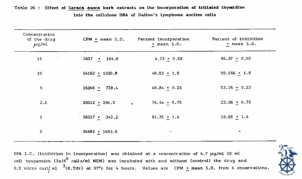
Table 26 : Effect of Saraca asoca ba rk extracts on the incorporation of t r i t ia ted thymidine
into the cellulose DNA of Dalton's lymphoma ascites cells
Concentration of the drug CPM + mean S.D. Percent incorporation - Percent of inhibition
pg/ml - + mean S.D. + mean S.D. -
50% I . C . (inhibition in incorporation) was obtained a t a concentration of 4.7 pg/ml 10 m1
cell suspension ( 2 x 1 0 ~ cells/rnl MEM) was incubated with and without (control) the drug and
0.5 micro curi m 1 3 ( ~ . ~ d r ) at 37Oc for 4 hours. Values a r e CPM + mean S.D. from 4 observations. I -

Table: 27.
Effect of Saraca asoca flowers on the DNA bio-synthesis of DLA tumour cells (in m -- ..................................................................... Concentration CPM - + Mean S.D. Percent Percent of the drug incorporation inhibition (pg/ml) Mean+ - Mean - + S.D. .....................................................................
50% IC (Inhibition in incorporation was obtained at a concentration
of 6 . 5 ) ~ g / m l . Values are CPM+Mean - S.D. from 4 observations.
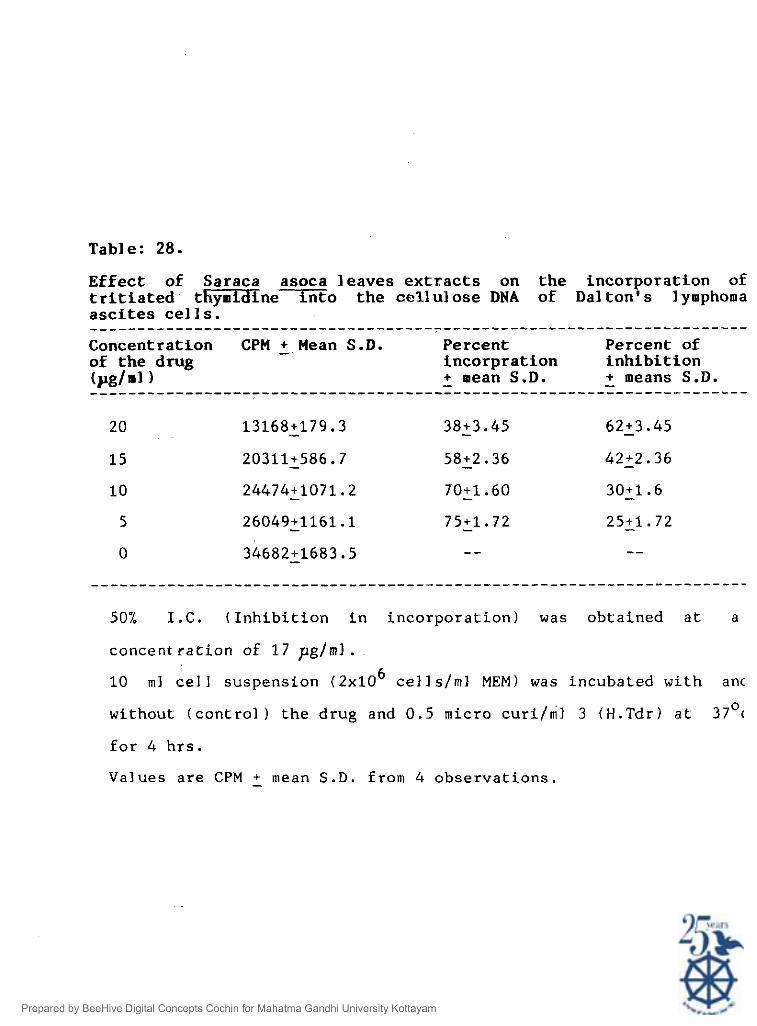
Table: 28.
Effect of Saraca asoca leaves extracts on the incorporation of tritiated t-nemo the cellulose DNA of Dal ton's l ymphoma ascites cell S. ..................................................................... Concentration CPM - + Mean S.D. Percent Percent of of the drug incorpration inhibition (pg/ml) - + mean S.D. - + means S.D. .....................................................................
50% I.C. (Inhibition in incorporation) was obtained at a
concent ration of 17 pglml . 10 m1 cell suspension ( 2 x 1 0 ~ cell s/ml MEM) was incubated with anc
without (control ) the drug and 0.5 micro c u r i / i l 3 (H.Tdr) at 37O(
for 4 hrs.
Values are CPM - + mean S.D. from 4 observations

Saraca asoca a c t i v e pr inci -ples of ba rk , f lower and l eaves
f a i l e d t o i n h i b i t t h e i n c o r p o r a t i o n a t Ur id ine and l e u c i n i n t o
c e l l u l a r RNA "and p r o t e i n r e spec t ive1 y ( F i g . 9 , 1 0 )
3 . 4 . DISCUSSION:
Saraca asoca i s widely used i n Ayurvedic Medicines ( 1 8 5 ) . But
very l i t t l e has been r epo r t ed on i t s a n t i c a n c e r a c t i v i t y . I t i s
c l e a r l y e s t a b l i s h e d from t h e above s t u d i e s t h a t t h e p l a n t Saraca
asoca con ta in c y t o t o x i c p r i n c i p l e s . The d i f f e r e n t i a l e x t r a c t i o n
procedure adopted i n d i c a t e d maximum e x t r a c t i o n of t h e a c t i v e
component from bark wi th e thy l a c e t a t e , from f lowers 95% methanol and
from l eaves 90% ace tone . I t was no t i ced t h a t a comparatively
increased a c t i v i t y i n t h e a c t i v e p r i n c i p l e of bark than o t h e r p a r t of
t h e p l a n t .
I n a d d i t i o n t o DLA, a c t i v i t y was t e s t e d wi th P-388, Eh r l i ch
a s c i t e s carcinoma and S-180 grown i n t h e p e r i t o n i a l c a v i t y of mice.
Grea t e r a c t i v i t y was observed i n DLA system probably due t o t h e
s p e c i f i c n a t u r e of t h e drug. E h r l i c h a s c i t e s tumour c e l l s were l e s s
s e n s i t i v e t o e i t h e r of t h e e x t r a c t s . Another p o i n t of i n t e r e s t was
t h a t t h e e x t r a c t s were i n s e n s i t i v e t o normal lymphocytes while
lymphocytes from leukaemia p a t i e n t s were ac t ed upon by t h e bs rk and
f lower e x t r a c t . Acute lymphoblas t ic leukaemia ( A L L ) c e l l s were more
s e n s i t i v e t o t h e acc ive p r i n c i p l e a s compared t o ch ron ic myelogenous
leukaemia c e l l s ( C M L ) . The p r e f e r e n t i a l a c t i o n no t having any e f f e c t

Fig. 9: Effect of active principle of Saraca on 3H-Uridine incorporation into cel lular macromolecules. 3FI-Uridine ( 2 U d / m l ) were incubated with different
loo
C .-O 80 .d
g P
6 0 - C .-
% A O - - C Y Y 2 2 0 -
.. concentrations of active principle for 4 h a t 37Oc. After precipitation, radioactivity was measured in a Liquid scinti l lat ion counter.
-4 ,. W = - 8 ,. - - -
I I I
0 5 10 l5 2 0 2 5
(W ) Saraca asoca bark
on cent rat ion of drug [pg/ rnl 1
( W 3 Saraca -- asoca flower.
(U) Saracs asoc'3 leaf. -- --

Concentration of drug ( pg/ml 1
Fig. 10: Effect of active principle of Saraca asoca on 3 -1eucine H incorporation into cel lular macromolecules. 3 -1eucine H ( 2 u ci'./ml) were incubated with different concentrations of
active principle for 4 h at 37Oc; a f t e r precipitation, radioactivity measured i n a l iquid scinti l lat ion counter.
( W ) Saraca W bark .
(M ) Saraca 5 flower.
( ) Saraca leaf

on normal lymphocytes clearly shows the added benefit of the drug in
case it is used as a mode of treatment in future.
The growth inhibitory effects and cytotoxicity of the extracts
was further evaluated using K-562, KB, CH0 and Vero cells. Active
principles of bark and flower was found to inhibit the growth of
above tumour cell S. CH0 and Vero cells were found to be the most
sensitive towards active principles. The enhancement of reduced
glutathione (GSH), glutathione reductase (GSH-R) and g1utathione-S-
transferase (GSH-S) levels in cultured tumour cells may be attributed
to the cytotoxic potentials of Saraca asoca bark and flower extracts.
Results of thymidine incorporation studies indicated that DNA
synthesis was inhibited by active principles of Saraca -- asoca
bark,flower and leaves extracts in a dose dependent manner. The
concentration required to produce 50% inhibition of thymidine
incorporation was lower than that required for 50% cytotoxicity.
Therefore it seems possible that the mechanism of action of the drug
is the inhibition of DNA synthesis. On the other hand, the active
principles failed to inhibit the incorporation of Uridine and leucine
into cellular RNA and protein respectively. Thus active principles
(bark, flower, leaves) may not have any effect on RNA or protein
biosynthesis.
The exact nature of the active components responsible for
cytotoxicity is not known now. Chemical analysis of Saraca asoca
bark active principle yielded (-)-Epicatechin, Proanthocyanidin B2

and a new Proanthocyanidin. The extract of flower showed the
presence of (-)-epicatechin, leucopelargonidin and dihydro
chalcone. Whether the cytotoxic action is due to these
compounds alone or in combination with' other compounds remains
to be investigated.