CXR NOT111
38
CXR NOT111
description
CXR NOT111
Transcript of CXR NOT111

CXR
NOT111
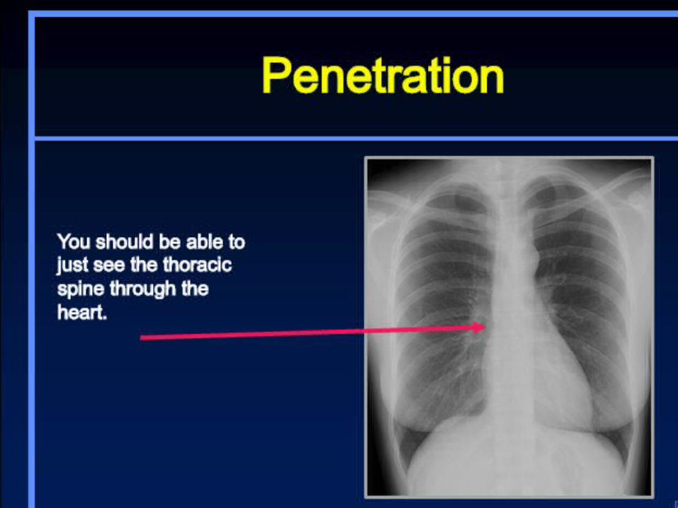


Lobes and Fissures
pleural effusion extending into the fissure

Mediastinum and Lungs

Pulmonary Vasculature

How to Read a Chest X-Ray• Patient Data (name history #, age, sex, old films)• Routine Technique: AP/PA, exposure, rotation, supine or erect• Trachea: midline or deviated, caliber, mass• Lungs: abnormal shadowing or lucency• Pulmonary vessels: artery or vein enlargement• Hila: masses, lymphadenopathy• Heart: thorax: heart width > 2:1 ? Cardiac configuration?• Mediastinal contour: width? mass?• Pleura: effusion, thickening, calcification• Bones: lesions or fractures• Soft tissues: don’t miss a mastectomy• ICU Films: identify tubes first and look for pneumothorax
http://www.med-ed.virginia.edu/courses/rad/cxr/interpretation1chest.html

Silhouette sign

air bronchogram
• 6 causes : lung consolidation, pulmonary edema, nonobstructive atelectasis,• severe interstitial disease, neoplasm, and normal expiration.

Solitary Pulmonary Nodule

Atelectasis
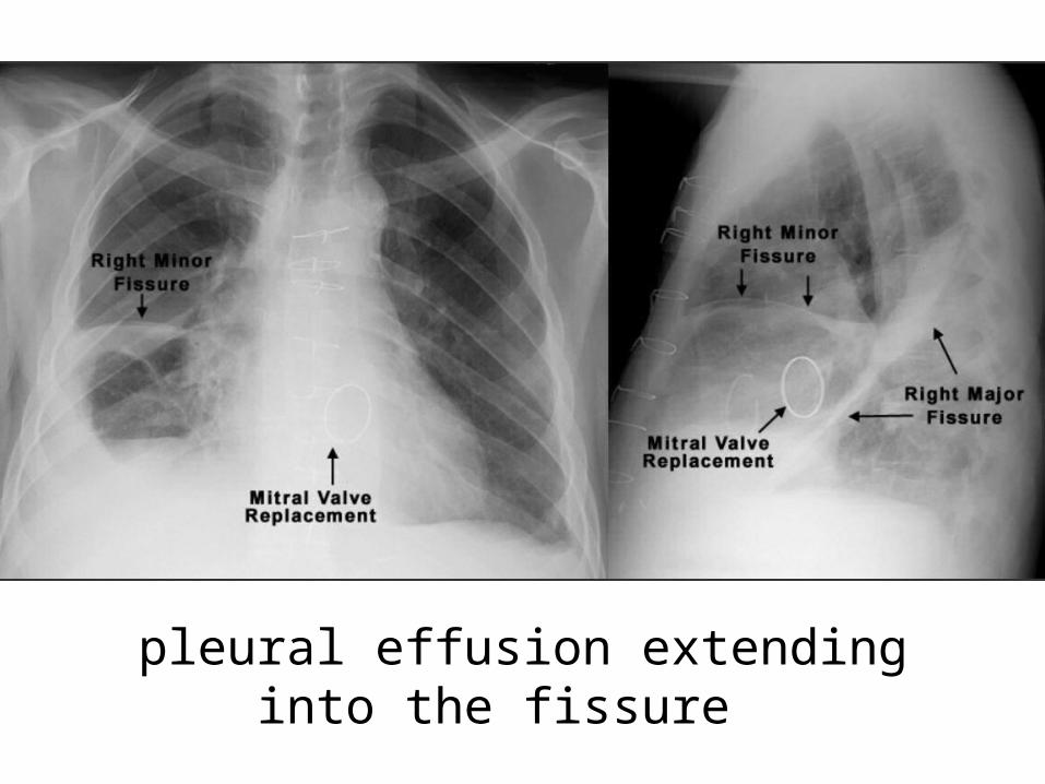
LUL atelectasis
Luftsichel Sign =‘air crescent’ at aortic knob
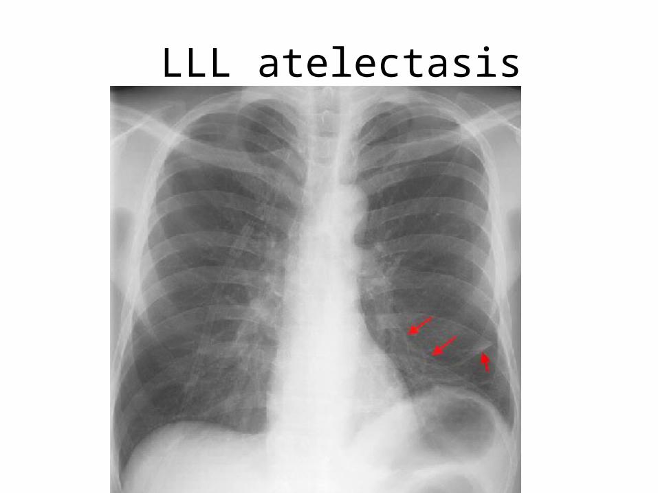
LLL atelectasis

RUL atelectasis
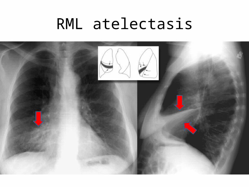
RML atelectasis
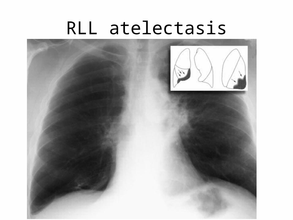
RLL atelectasis

Pul. Edema :cardiogenic , noncardiogenic
• cephalization of pulmonary vessels, Kerley B lines or septal lines, peribronchial cuffing, "bat wing" pattern, patchy shadowing with air bronchograms,fluid in fissure , pleural effusion and increased cardiac size.

Kerley B lines

Pneumonia

Pneumonia

???
TB

??
RUL hemorrhage
left pleural effusion


Pleural Effusion : 200ml vs 75ml

Pneumothorax

hydropneumothorax

Emphysema
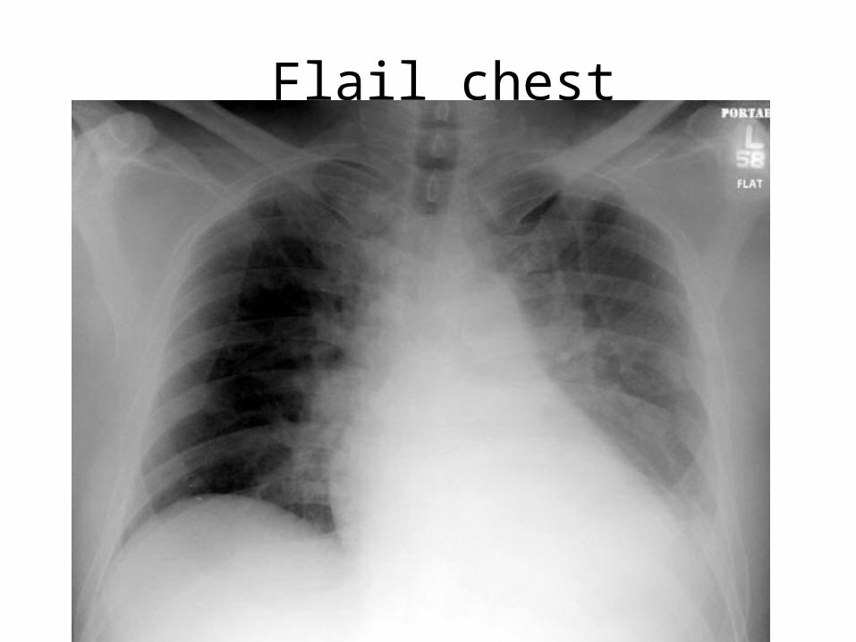
Flail chest

Anterior Mediastinal Mass : 3T1L

Middle Mediastinal Mass : Arch,Eso.

Posterior Mediastinal Mass
• ddx : neoplasm, lymphadenopathy,aneurysm, lung mass, neurenteric cyst,meningocele, and extramedullary hematopoiesis
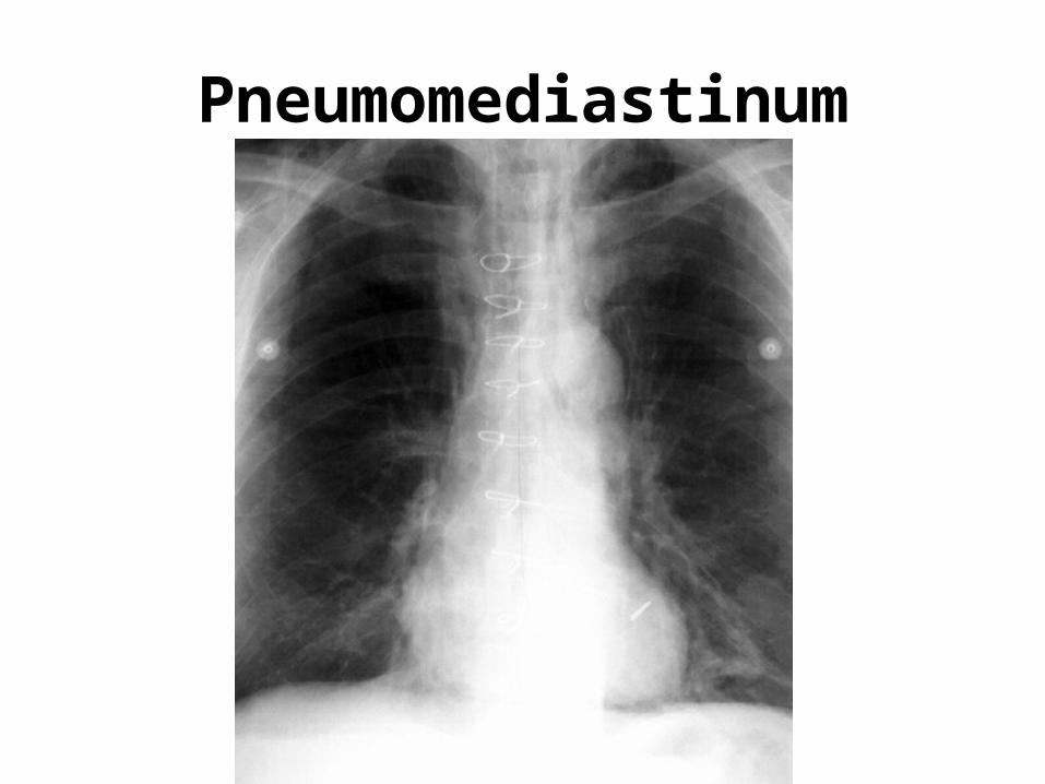
Pneumomediastinum

Diaphragmatic hernia :hiatal,Boch.,Morg.
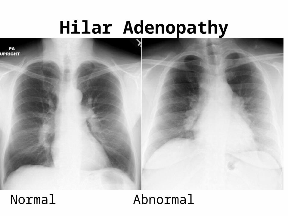
Hilar Adenopathy
Normal Abnormal

CA• cavitation, which
is found more characteristically in squamous cell CA