Cuticular characteristics of Neuralethopteris jongmansii ...
10
2017 | 70/1 | 1–10 | 2 Figs. | 2 Tabs. | 4 Pls. | www.geologia-croatica.hr Journal of the Croatian Geological Survey and the Croatian Geological Society 1. INTRODUCTION The fossil-genus Neuralethopteris CREMER is characterised by neuropteroid pinnules that have cordate bases and alethopteroid venation (GOUBET et al., 2000). Representatives of Neuralethop- teris are widespread in the Euramerican Realm, and are impor- tant biostratigraphic indicators in upper Namurian and lower Westphalian strata. Neuralethopteris schlehanii (STUR) CRE- MER, the type and most common species of Neuralethopteris, occurs in Euramerica, although 10 other fossil-species are known (five each from Europe and North America) (CLEAL & SHUTE, 1995, GOUBET et al., 2000 TENCHOV & CLEAL, 2010). To date, N. schlehanii is the only member of the fossil-genus for which cuticles have been studied ( CLEAL & SHUTE, 1992). During a visit to Wrocław University in Poland to study the type material of Cordaites palmaeformis (GÖPPERT) WEISS (ŠIMŮNEK, 2015), some pinnules of Neuralethopteris were found on the same slab as one of the syntypes of C. palmaeformis and sampled for maceration. The pinnules were identified as Neu- ralethopteris jongmansii LAVEINE, which is relatively rare in the Intrasudetic Basin and has yet to be determined in the Czech part of the basin. In this paper, a detailed cuticular analysis is pre- sented for N. jongmansii, which represents only the second spe- cies of Neuralethopteris for which cuticles are known. 2. MATERIAL AND METHOD The syntype of Cordaites palmaeformis, from which the pinnules of Neuralethopteris jongmansii were collected, came from the Wałbrzych locality, Intrasudetic Basin, Poland. The syntype slab is stored in Wrocław University under no. 1667p. Although the exact horizon and stratigraphic level from which the syntype slab Cuticular characteristics of Neuralethopteris jongmansii LAVEINE (medullosalean foliage, Westphalian, Intrasudetic Basin, Poland) Zbyněk Šimůnek Czech Geological Survey, Klárov 3/131, 118 21 Praha 1, Czech Republic; ([email protected]) doi: 10.4154/gc.2017.02 Abstract Foliar cuticles are described for the first time from the medullosalean pteridosperm Neuralethop- teris jongmansii LAVEINE. This species is not as common, either in abundance or distribution, as Neuralethopteris schlehanii (STUR) CREMER, the abaxial cuticle of which has paracytic (in morphological sense) and anomocytic stomata. In contrast, the stomata of Neuralethopteris jongmansii occur on both abaxial and adaxial cuticles and are anomocytic, monocyclic with prominent proximal papillae and sometimes paracytic. The epidermal cells of N. jongmansii are papillate and partly differ from N. schlehanii cuticles. However, there is a striking similarity between the foliar cuticles of N. jongmansii and Neurodontopteris auriculata (BRONGNIART) POTONIÉ. Although N. auriculata is a much younger fossil-species with pinnules that are more robust and of a different shape than N. jongmansii, their epidermal structures are practically the same – polygonal papillate cells, anomocytic, monocyclic stomata with prominent proximal papillae. Even their stomatal densities are equivalent. Despite this, there is a marked difference in the frond architecture of both genera. Neuralethopteris has „alethopterid“ bifurcate-pinnate frond architecture, Neurodontopteris has quite a different bifurcate-semi-pinnate frond architec- ture. Based on comparison of the cuticles, Neuralethopteris jongmansii and Neurodontopteris auriculata appear very similar and it may reflect common palaeoenvironmental demands. was collected is not known, the fact that other occurrences of N. jongmansii in Europe are in Langsettian (Lower Pennsylvanian) strata ( CLEAL et SHUTE, 1995) suggests that it probably came from the lower part of the Žacléř Formation (Table 1). The cuticles were prepared by a standard maceration method using Schulze’s Reagent, as described by KERP (1990), KRINGS & KERP (1997) and KERP & KRINGS (1999). 3. GEOLOGICAL SETTING The Intrasudetic Basin extends from the north-eastern Czech Re- public into south-western Poland, where the Wałbrzych locality is located (Fig. 1). Mississippian strata of the Intrasudetic Basin are largely coarse-grained siliciclastics that reach 5 km in thick- ness. Sedimentation changed at the end of the Mississippian, af- ter which the coal-bearing Wałbrzych Formation was deposited. It reaches up to 250 m thickness, and is of Serpukhovian and ear- liest Bashkirian (lower Namurian) age. The overlying coal-barren Biały Kamień Formation is up to 380 m thick and largely com- prises sandstones and conglomerates. In the Polish part of the Intrasudetic Basin, the lower part of the overlying Žacléř Forma- tion (upper Bashkirian, Langsettian and Duckmantian) is coal- bearing. The study samples probably come from this stratigraphic level, a conclusion supported by the fact that coals of the Žacléř Formation were mined in the vicinity of Wałbrzych. The upper - most Bashkirian (Duckmantian) part of the formation only con- tains a few workable coal seams in the Polish part of the basin. The Moscovian and Kasimovian (upper Westphalian and lower Stephanian) Glinik Formation, which reaches 850 m in thickness, is practically devoid of workable coal seams. The uppermost for- mation in the Polish part of the basin is the Ludwikowice Forma- Article history: Manuscript received May 30, 2016 Revised manuscript accepted November 23, 2016 Available online February 20, 2017 Keywords: Neuralethopteris, cuticular analysis, Westphalian, Poland, Intrasudetic Basin
Transcript of Cuticular characteristics of Neuralethopteris jongmansii ...

2017 | 70/1 | 1–10 | 2 Figs. | 2 Tabs. | 4 Pls. | www.geologia-croatica.hr Journal of the Croatian Geological Survey and the Croatian Geological Society
1. INTRODUCTIONThe fossil-genus Neuralethopteris CREMER is characterised by neuropteroid pinnules that have cordate bases and alethopteroid venation (GOUBET et al., 2000). Representatives of Neuralethop-teris are widespread in the Euramerican Realm, and are impor-tant biostratigraphic indicators in upper Namurian and lower Westphalian strata. Neuralethopteris schlehanii (STUR) CRE-MER, the type and most common species of Neuralethopteris, occurs in Euramerica, although 10 other fossil-species are known (five each from Europe and North America) (CLEAL & SHUTE, 1995, GOUBET et al., 2000 TENCHOV & CLEAL, 2010). To date, N. schlehanii is the only member of the fossil-genus for which cuticles have been studied (CLEAL & SHUTE, 1992). During a visit to Wrocław University in Poland to study the type material of Cordaites palmaeformis (GÖPPERT) WEISS (ŠIMŮNEK, 2015), some pinnules of Neuralethopteris were found on the same slab as one of the syntypes of C. palmaeformis and sampled for maceration. The pinnules were identified as Neu-ralethopteris jongmansii LAVEINE, which is relatively rare in the Intrasudetic Basin and has yet to be determined in the Czech part of the basin. In this paper, a detailed cuticular analysis is pre-sented for N. jongmansii, which represents only the second spe-cies of Neuralethopteris for which cuticles are known.
2. MATERIAL AND METHODThe syntype of Cordaites palmaeformis, from which the pinnules of Neuralethopteris jongmansii were collected, came from the Wałbrzych locality, Intrasudetic Basin, Poland. The syntype slab is stored in Wrocław University under no. 1667p. Although the exact horizon and stratigraphic level from which the syntype slab
Cuticular characteristics of Neuralethopteris jongmansii LAVEINE (medullosalean foliage, Westphalian, Intrasudetic Basin, Poland)Zbyněk Šimůnek
Czech Geological Survey, Klárov 3/131, 118 21 Praha 1, Czech Republic; ([email protected])
doi: 10.4154/gc.2017.02
AbstractFoliar cuticles are described for the first time from the medullosalean pteridosperm Neuralethop-teris jongmansii LAVEINE. This species is not as common, either in abundance or distribution, as Neuralethopteris schlehanii (STUR) CREMER, the abaxial cuticle of which has paracytic (in morphological sense) and anomocytic stomata. In contrast, the stomata of Neuralethopteris jongmansii occur on both abaxial and adaxial cuticles and are anomocytic, monocyclic with prominent proximal papillae and sometimes paracytic. The epidermal cells of N. jongmansii are papillate and partly differ from N. schlehanii cuticles. However, there is a striking similarity between the foliar cuticles of N. jongmansii and Neurodontopteris auriculata (BRONGNIART) POTONIÉ. Although N. auriculata is a much younger fossil-species with pinnules that are more robust and of a different shape than N. jongmansii, their epidermal structures are practically the same – polygonal papillate cells, anomocytic, monocyclic stomata with prominent proximal papillae. Even their stomatal densities are equivalent. Despite this, there is a marked difference in the frond architecture of both genera. Neuralethopteris has „alethopterid“ bifurcate-pinnate frond architecture, Neurodontopteris has quite a different bifurcate-semi-pinnate frond architec-ture. Based on comparison of the cuticles, Neuralethopteris jongmansii and Neurodontopteris auriculata appear very similar and it may reflect common palaeoenvironmental demands.
was collected is not known, the fact that other occurrences of N. jongmansii in Europe are in Langsettian (Lower Pennsylvanian) strata (CLEAL et SHUTE, 1995) suggests that it probably came from the lower part of the Žacléř Formation (Table 1).
The cuticles were prepared by a standard maceration method using Schulze’s Reagent, as described by KERP (1990), KRINGS & KERP (1997) and KERP & KRINGS (1999).
3. GEOLOGICAL SETTINGThe Intrasudetic Basin extends from the north-eastern Czech Re-public into south-western Poland, where the Wałbrzych locality is located (Fig. 1). Mississippian strata of the Intrasudetic Basin are largely coarse-grained siliciclastics that reach 5 km in thick-ness. Sedimentation changed at the end of the Mississippian, af-ter which the coal-bearing Wałbrzych Formation was deposited. It reaches up to 250 m thickness, and is of Serpukhovian and ear-liest Bashkirian (lower Namurian) age. The overlying coal-barren Biały Kamień Formation is up to 380 m thick and largely com-prises sandstones and conglomerates. In the Polish part of the Intrasudetic Basin, the lower part of the overlying Žacléř Forma-tion (upper Bashkirian, Langsettian and Duckmantian) is coal-bearing. The study samples probably come from this stratigraphic level, a conclusion supported by the fact that coals of the Žacléř Formation were mined in the vicinity of Wałbrzych. The upper-most Bashkirian (Duckmantian) part of the formation only con-tains a few workable coal seams in the Polish part of the basin. The Moscovian and Kasimovian (upper Westphalian and lower Stephanian) Glinik Formation, which reaches 850 m in thickness, is practically devoid of workable coal seams. The uppermost for-mation in the Polish part of the basin is the Ludwikowice Forma-
Article history:
Manuscript received May 30, 2016 Revised manuscript accepted November 23, 2016 Available online February 20, 2017
Keywords: Neuralethopteris, cuticular analysis, Westphalian, Poland, Intrasudetic Basin
ide cb
Geo
logi
a C
roat
ica
Geologia Croatica 70/12
tion, which reaches 440 m thickness and largely comprises coarse-grained redbeds of Gzhelian (late Stephanian) and Asse-lian (early Permian) age (Table 1.) (BOSSOWSKI et al., in ZDAN-OWSKI & ŻAKOWA, 1995).
4. SYSTEMATICSOrder Medullosales CORSIN, 1960Family Alethopteridaceae CORSIN, 1960*Fossil-genus Neuralethopteris CREMER, 1893 emend. LAVEINE,
1967
Type species: Neuralethopteris schlehanii (STUR, 1877) CREMER, 1893
*Family Alethopteridaceae is based on frond architecture that is an ancillary feature, however a natural botanical system should be based on fructifications. Such a family is the Trigonocarpaceae SEWARD, 1917 family, but plants with different frond architec-tures can have trigonocarpalean seeds. So the Alethopteridaceae family is an artificial family from this point of view.
Neuralethopteris jongmansii LAVEINE, 19671967 Neuralethopteris jongmansii LAVEINE: p. 107–112, pls. 2–4.2010 Neuralethopteris jongmansii LAVEINE; TENCHOV & CLEAL:
2010, p. 303, pls. 1, figs. 5, 6.
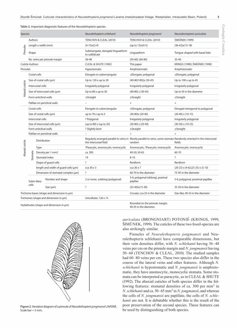
Description: Two pinna fragments up to 35 mm long, together with several isolated pinnules are preserved on the edge of spec-imen 1667p. Pinnules are small, being 5–8 mm long and usually 2–3 mm wide, and have a cordate, asymmetric base and a thick midvein that nearly extends to the pinnule apex. Lateral veins arise oblique to the midvein and arch broadly to meet the pinnule margin at 80–90°. Each lateral vein generally forks twice, result-ing in a high vein density of 60–80 veins per cm on the pinnule margin.
Adaxial cuticle: Stomata are present in the intercostal fields but absent in the costal fields. Ordinary cells are polygonal, 40–80 (100) mm long and 30–45 mm wide, and slightly elongated in the costal fields. A small papilla, up to 10 mm in diameter, is present in the middle of each cell. Stomatal complexes are 60–70 mm in diameter, and comprise two guard cells and five or six small po-
lygonal subsidiary cells that are 25–40 mm long and 15–30 mm wide. Cutinisation of papillae on the subsidiary cells is stronger than Cutinisation of papillae on ordinary cells. These papillae are also longer, up to 15 mm wide, and oriented (bent) towards the guard cells (proximal papillae). Hydathodes (Pl. I/j) are 40–45 mm in diameter and situated at the edge of pinnules. Stomatal density varies from 40–50 stomata per mm2 and the stomatal index ranges from 8–10.
Abaxial cuticle: Abaxial cuticles are very similar to adaxial cuticles. Stomatal complexes are generally monocyclic, anomo-cytic, but some stomata can be also paracytic in a morphological sense (Pl. II/i). Polygonal cells are 30–80 mm long and 20–40 mm wide, and stomata are practically the same as those on adax-ial cuticles. Stomatal density varies from 50–65 stomata per mm2 and the stomatal index is between 8–10, similar to the values for adaxial cuticles. Trichome bases, surrounded by six or seven or-dinary cells, occur in the area that is presumed to correspond to the midvein (Pl. II/b,c,g,h). Trichome bases are circular to slightly oval and their margins are strongly cutinised. No hairs have been observed. Also occurring in these areas are stomata-like features (Pl. II/g,h), which are oval and surrounded by eight small cells, although no guard cells have been observed.
5. COMPARISONSThe first descriptions of cuticles of the fossil-genus Neuralethop-teris were made by CLEAL & SHUTE (1992), who noted that cuticles of the type species Neuralethopteris schlehanii have an unusual type of paracytic stomatal structure that was previously unknown in the medullosaleans. But CLEAL (pers. com. 2016) admitted that the abaxial cuticle of Neuralethopteris schlehanii was fragmentary in preservation, and some stomata can be inter-preted as anomocytic, the others as paracytic. A stoma of Neura-lethopteris jongmansii figured on Pl. II/i resembles the paracytic type. Nevertheless, the cuticles of Neuralethopteris jongmansii differ from those of N. schlehanii (Table 2.), namely by the ad-axial cuticle. In contrast to Neuralethopteris schlehanii, N. jong-mansii also has stomata on the adaxial cuticle which represents amphistomatic foliage, a feature that, among the medullosaleans, is only known for the monotypic fossil-species Neuro dontopteris
Table 1. Stratigraphy of the polish part of the Intrasudetic Basin with emphasize to the Pennsylvanian strata.
GLOBAL SCALE REGIONAL SCALE FORMATION
PERMIAN Asselian Ludwikowice
PEN
NSY
LVAN
IAN
Gzhelian
Stephanian
C
Kasimovian
B
GlinikBarruelian
Cantabrian
Moscovian
Westphalian
Asturian
BolsovianŽacléř
Bashkirian
Duckmantian
Langsettian
Biały Kamień
Namurian
Yeadonian
Marsdenian
Kinderscoutian
WałbrzychAlportian
Chokierian
MISSISSIPPIAN
SerpukhovianArnsbergianPendleian
Viséan
Tournaisian
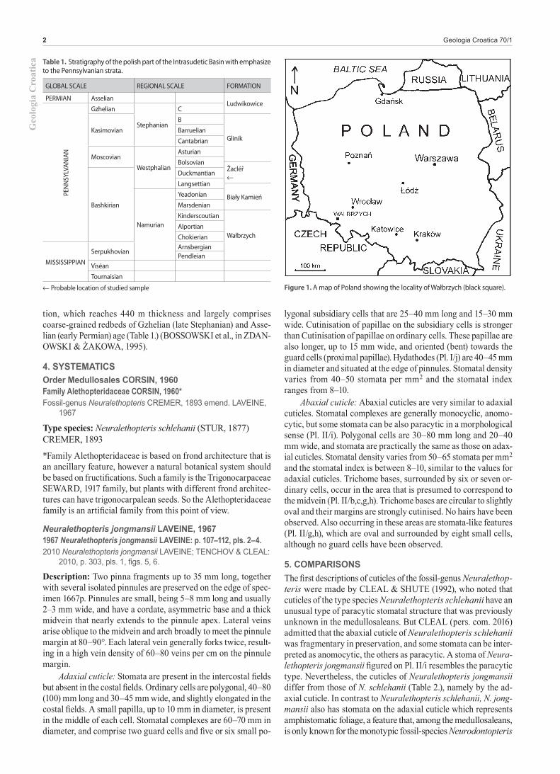
Probable location of studied sample Figure 1. A map of Poland showing the locality of Wałbrzych (black square).
Geologia C
roaticaZbyněk Šimůnek: Cuticular characteristics of Neuralethopteris jongmansii Laveine (medullosalean foliage, Westphalian, Intrasudetic Basin, Poland) 3
auriculata (BRONGNIART) POTONIÉ (KRINGS, 1999, ŠIMŮNEK, 1999). The cuticles of these two fossil-species are also strikingly similar.
Pinnules of Neuralethopteris jongmansii and Neu-ralethopteris schlehanii have comparable dimensions, but their vein densities differ, with N. schlehanii having 36–48 veins per cm on the pinnule margin and N. jongmansii having 50–60 (TENCHOV & CLEAL, 2010). The studied samples had 60–80 veins per cm. These two species also differ in the course of the lateral veins and other features. Although N. schlehanii is hypostomatic and N. jongmansii is amphisto-matic, they have anomocytic, monocyclic stomata. Some sto-mata can be interpreted as paracytic, as in CLEAL & SHUTE (1992). The abaxial cuticles of both species differ in the fol-lowing features: stomatal densities of ca. 300 per mm2 in N. schlehanii and ca. 50–65 mm2 in N. jongmansii, and whereas the cells of N. jongmansii are papillate, the cells of N. schle-hanii are not. It is debatable whether this is the result of the poor preservation of the second species. These features can be used by distinguishing of both species.
Figure 2. Venation diagram of a pinnule of Neuralethopteris jongmansii LAVEINE. Scale bar = 5 mm.
Table 2. Important diagnostic features of the Neuralethopteris species.
Species Neuralethopteris schlehanii Neuralethopteris jongmansii Neurodontopteris auriculata
Pinn
ules
Authors TENCHOV & CLEAL (2010) TENCHOV & CLEAL (2010) ŠIMŮNEK (1999)
Length x width (mm) (4-15)x(2-4) (up to 13)x(4-5) (36-42)x(15-18)
Shape Subtriangular, elongate linguaeform to subfalcate Linguaeform Tongue-shaped with basal lobe
No. veins per pinnule margin 36-48 (50-60); (60-80) 35-40
Cuticle Authors CLEAL & SHUTE (1992) This paper KRINGS (1999); ŠIMŮNEK (1999)
Pinnules Hypostomatic Amphistomatic Amphistomatic
Adax
ial c
utic
le
Costal cells Elongate to subrectangular ±Elongate, polygonal ±Elongate, polygonal
Size of costal cells (µm) Up to 120 x up to 20 [40-80(100)]x (30-45) Up to 100 x up to 45
Intercostal cells Irregularly polygonal Irregularly polygonal Irregularly polygonal
Size of intercostal cells (µm) Up to 80 x up to 30 (40-80) x (30-45) Up to 45 in the diameter
Form anticlinal walls ±Straight ±Straight ±Straight
Palillae on periclinal walls – + +
Abax
ial c
utic
le
Costal cells Elongate to subrectangular ±Elongate, polygonal Elongate tetragonal to polygonal
Size of costal cells (µm) up to 70 x up to 5 (30-80)x (20-40) (20-40) x (10-15)
Intercostal cells ? Polygonal Irregularly polygonal Irregularly polygonal
Size of intercostal cells (µm) (up to 80) x (up to 20) (30-80) x (20-40) (30-50) x (10-25)
Form anticlinal walls ? Slightly bent ±Straight ±Straight
Palillae on periclinal walls – + +
Stom
ata
Distribution Regularly arranged parallel to veins in the intercostal field
Mostly parallel to veins, some stomata random
Randomly oriented in the intercostal fields
Type ?Paracytic, anomocytic; monocyclic Anomocytic, ?Paracytic; monocyclic Anomocytic; monocyclic
Density per 1 mm2 ca. 300 40-50; 50-65 60-70
Stomatal index 19 8-10 ?
Shape of guard cells ? Reniform Reniform
length and width of guard cells (µm) cca 30 x 3 cca 30 x 7 (20-25) x (4-6);(25-35) x (5-10)
Dimension of stomatal complex (µm) ? 60-70 in the diameter 75-95 in the diameter
Subsi-diary cells
Number and shape 2 or more; ±oblong (polygonal) 5-6; polygonal (oblong), poximal papilae 5-6; polygonal, proximal papillae
Size (µm) ? (25-40)x(15-30) 35-50 in the diameter
Trichome bases (shape and dimension in µm) Circular; cca 25 in the diameter Star-like; 40-55 in the diameter
Trichomes (shape and dimension in µm) Unicellular; 120 x 15
Hydathodes (shape and dimension in µm) Rounded on the pinnule margin; 40-45 in the diameter.

Geo
logi
a C
roat
ica
Geologia Croatica 70/14
Even though the morphology of Neuralethopteris jong-mansii and Neurodontopteris auriculata differ, their cuticles are surprisingly remarkably similar. Neuralethopteris fronds have an „alethopterid“ bifurcate-pinnate architecture (LAVEINE et al., 1992, fig. 3). The fronds can be several metres long and do not have intercalary elements. The fronds of Neurodontopteris auriculata are about 0.5 m long (LANGIAUX, 1984, fig. 233) and have a bifurcate-semi-pinnate architecture. Pinnules of N. au-riculata have a basal lobe and are much larger and wider than pinnules of N. jongmansii (Table 2). Furthermore, N. auriculata occurs in much younger strata of Stephanian B to early Permian age. Nonetheless, these two fossil-species are the only amphisto-matic taxa known to date in the Order Medullosales. Both have anomocytic, monocyclic stomata, strikingly similar papillate po-lygonal cells, stomata with proximal papillae, and similar adaxial and abaxial cuticles. Even though occurrences of the two fossil-species in the fossil record are separated by some 8 Myr, their stomatal densities are comparable: 50–65 stomata per cm for the abaxial cuticle of N. jongmansii and 60–70 for N. auriculata (Ta-ble 2.). In fact, the only distinguishing feature between the fossil-species is the presence of star-like trichome bases (ŠIMŮNEK, 1999) in N. auriculata, although the feature only occurs on a sin-gle (?adaxial) cuticle. On the whole, distinguishing these fossil-taxa would be very difficult if only dispersed cuticles were availa-ble and the age of the strata were not known. However, both genera differ in gross-morphology. The frond architecture of Neuralethopteris is mainly based on Neuralethopteris schlehanii (LAVEINE et al., 1992), because the occurrences of Neuralethop-teris jongmansii specimens are rare and fragmentary. Neurodon-topteris auriculata belongs to plants with small stature (ŠI MŮ-NEK, 1999). The dimensions of the Neuralethopteris jongmansii plant are not known. Due to the similarity of the cuticles with those of Neurodontopteris auriculata¸ it probably was also of small stature (similar palaeoecological demands of both plants) in contrast to Neuralethopteris schlehanii.
6. PALAEOBIOLOGYThe presence of amphistomatic pinnules in medullosaleans is ex-ceptional. ŠIMŮNEK (1999) speculated that this feature, found in Neurodontopteris auriculata, could point to a small-statured plant, having fronds that were protected from direct sunshine.
Neuralethopteris jongmansii also had some adaptation to living in the understory (stomata on both pinnule sides). Proximal pa-pillae on subsidiary cells around guard cells could prevent watter drops to enter the stoma opening. The adaxial and abaxial cuticles have approximately the same stomatal densities. Both species are amphistomatic and have small papillae on periclinal cell walls. This probably helped the plant to get rid of water drops from the pinnule surface. Neuralethopteris jongmansii and Neurodontop-teris auriculata probably lived in similar habitats.
7. CONCLUSION(1) The morphology of Neuralethopteris jongmansii pinnules
is similar to those of Neuralethopteris schlehanii, The latter is the type species of the fossil-genus Neuralethopteris, although N. jongmansii has denser lateral veins that follow a slightly dif-ferent course.
(2) The pinnules of Neuralethopteris jongmansii are amphis-tomatic and the pinnules of Neuralethopteris schlehanii are hy-postomatic. Abaxial cuticles of both species are similar in sto-matal types. N. jongmansii have anomocytic, monocyclic stomata, some stomata morphologically resemble the paracytic type and resemble those of N. schlehanii stomata.
(3) Based on cuticles, Neuralethopteris jongmansii and Neu-rodontopteris auriculata are strikingly similar, and are the only medullosaleans known to date with amphistomatic foliage. Both fossil-species have the same type of stomata, cell shapes and sto-matal densities, indicating life in similar environments. Both plants were probably of small stature and probably lived in un-dergrowth.
ACKNOWLEDGEMENTThis paper was written with grant support from the Grant Agency of the Czech Republic, no. P210-12-2053. The author thanks the workers from the Collections of the Wrocław University in Poland, Dr. A. SETLIK, and Dr. P. RACZYŃSKI, for help with studying GÖPPERT’s collection. The author thanks Dr. C.J. CLEAL (Cardiff) for his advice concerning neuropteroids and his critical review of the text; and Dr. A.R. BASHFORTH for English editing of the text. W.A. DIMICHELE gave as a second reviewer productive remarks that improved the manuscript.
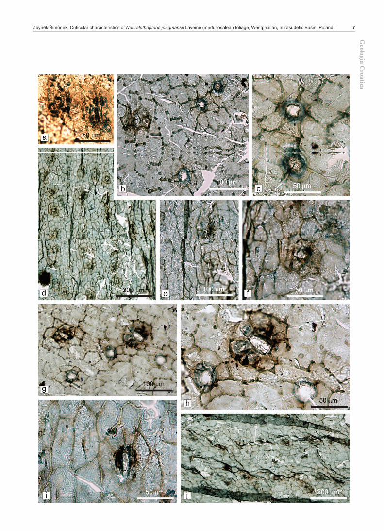
Plate 1 Neuralethopteris jongmansii LAVEINE. loc. Wałbrzych, „Langsettian?“, coll. GÖPPERT, Wrocław Univ., Inv. No. 1667 p.
a – Pinnule margin with adaxial cuticle on the left and abaxial cuticle on the right. Slide 603/8.b – Detail of abxial cuticle with stomata from Fig. a.c – Close up of Fig. b with detail of stomata.d – Adaxial cuticle on the left and abaxial cuticle on the right. Slide 602/10.e – Detail of abaxial cuticle with stomata from Fig. d.f – Adaxial cuticle with stomata. Slide 456/4.g – Close up of Fig. f with stomata and cells with very small papillae.h – Close up of Fig. f with details of stomata and cells of intercostal field with very small papillae.i – Detail of s stoma from Fig. h. Note the papillae arising from subsidiary cells.j – Margin of adaxial cuticle with hydathodes. Slide 456/5.k – Close up of two hydathodes from Fig. j.

Geologia C
roaticaZbyněk Šimůnek: Cuticular characteristics of Neuralethopteris jongmansii Laveine (medullosalean foliage, Westphalian, Intrasudetic Basin, Poland) 5

Geo
logi
a C
roat
ica
Geologia Croatica 70/16
Plate 2 Neuralethopteris jongmansii LAVEINE. loc. Wałbrzych, „Langsettian“, coll. GÖPPERT, Wrocław Univ., Inv. No. 1667 p. Abaxial cuticles.
a – Two stomata with papillae on subsidiary cells. Slide 602/9.b – ? probably an area of midrib with ?trichome basis. Slide 602/10c – Close up of two trichome bases from Fig. b.d – Abaxial cuticle with stomata. Slide 603/8 (cuticle from Pl. I, a).e – Close up of two stomata from Fig. d.f – Close up of a stomata from Fig. d. Note the papillae on subsidiary cells.g – ? Probably midrib with trichome bases. Slide 603/9.h – Close up of trichome bases? from Fig. g.i – Close up of a stoma resembling the paracytic stomatal type. Slide 602/11.j – Abaxial cuticle with stomata. Slide 603/7.
REFERENCESBOSSOWSKI, A., IHNATOWICZ, A., MASTALERZ, K. KUROWSKI, L. NOWAK,
G.J. (1995): Intra-Sudetic Depression.– In: ZDANOWSKI, A. & ŻAKOWA, H. (eds.): The Carboniferous System in Poland. Prace Państwowego Instytutu Geo-logicznego, 124–146, Warszawa.
CLEAL, C.J. & SHUTE, C.H. (1992): Epidermal features of some Carboniferous neu-ropteroid fronds.– Review of Palaeobotany and Palynology, 71, 191–206. doi: 10.1016/0034-6667(92)90162-A
CLEAL, C.J. & SHUTE, C.H. (1995): A synopsis of neuropteroid foliage from the Car-boniferous and Lower Permian of Europe. Bulletin of the British Museum (Natu-and Lower Permian of Europe. Bulletin of the British Museum (Natu-ral History).– Geology Series, 51, 1–52.
CORSIN, P. (1960): Classification des Ptéridophytes et des Ptéridospermophytes du Car-bonifère. Bulletin de la Société Géologique de France 2 (Série 7), 566–572.
CREMER, L. (1893): Über die Fossilen Farne des Westfälischen Carbons und ihre Be-deutung für eine Gliederung des letzeren. Mitteilungen aus dem Geologischen Museum der Westfälischen Berggewerkschaftskasse, 1, 1–49.
GOUBET, P., PFEFFERKORN, H.W. & GILLESPIE, W.H. (2000): Neuralethopterids (trigonocarpalean pteridosperms) from the Early Pennsylvanian of eastern North America.– PaleoBios, 20, 11–37.
KERP, H. (1990): The study of fossil gymnosperms by means of cuticular analysis.– Palaios, 5, 548–569. doi: 10.2307/3514861
KERP, H. & KRINGS, M. (1999): Light microscopy of cuticles.– In: JONES, T.P. & ROWE, N.P. (eds.): Fossil Plants and Spores: Modern Techniques. Geological So-ciety, London, 52–56.
KRINGS, M. (1999): Zum Bau der Spaltöffnungsapparate von Neurodontopteris auricu-lata (Brongniart) Potonié, einer Pteridosperme aus dem Stefan von Blanzy-Mont-ceau (Zentralfrankreich).– M�nstersche Forschungen zur Geologie und Paläonto-(Zentralfrankreich).– M�nstersche Forschungen zur Geologie und Paläonto-logie, 86, 69–78.
KRINGS, M. & KERP, H.(1997): An improved method for obtaining large pteridosperm cuticles.– Rev. Palaeobot. Palynol., 96, 453–456. doi: 10.1016/S0034-6667(96)00059-0
LANGIAUX, J. (1984): Flores et Faunes des formations supérieurs du Stéphanien de Blanzy-Montceau (Massif Central français). Stratigraphie et paléoécologie. Revue Périodique de ‚La Physiophyle‘, Societé d‘ Études des Sciences Naturelles et His-toriques de Montceau-les-Mines, 100 (Supplément), 1–270.
LAVEINE, J.-P.(1967): Contribution a l‘étude de la flore du terrain houiller. Les Neu-roptéridées du Nord de la France.– Études Géologiques pour l‘Atlas Topographie Souterraine, 1/5, 1–344, pls A–P, 1–84.
LAVEINE, J.-P., BELHIS, A., LEMOIGNE, Y. & ZHANG, S. (1992): Frond architectu-re in the genera Neuralethopteris CREMER, Alethopteris STERNBERG and Lon-chopteris BRONGNIART (Carboniferous Pteridosperms).– Revue de Paléobiolo-gie, Volume spécial, Geneva, 6, 149–166.
SEWARD, A.C. (1917): Fossil plants. Volume III. Pteridospemae, Cycadofilices, Cordaitales, Cycadophyta.– Hafner Publishing Company, New York & London (reprint 1963), 656 p.
ŠIMŮNEK, Z. (1999): Cuticles of Neuropteris auriculata (BRONGNIART) POTONIÉ from the Stephanian B of the Czech Republic. Acta Universitatis Carolinae.– Geo-logica, 43, 4, 625–631.
ŠIMŮNEK, Z. (2015): Cuticles of the Polish type material of Cordaites palmaeformis (GÖPPERT)WEISS and a Cordaites principalis-like form from Germany, Penn-sylvanian.– Review of Palaeobotany and Palynology, 223, 50–70.
STUR, D. (1877): Die Culm-Flora der Ostrauer und Waldenburger Schichten.– Abhan-dlungen der Kaiserlich-Königlichen Geologischen Reichsanstalt, 8/2, 107–472.
TENCHOV, Y. & CLEAL, C.J. (2010): Neuralethopteris foliage (Medullosales) in the Carboniferous of the Dobrudzha Coalfield, Bulgaria.– Review of Palaeobotany and Palynology, 158, 298–307. doi: 10.1016/j.revpalbo.2009.10.001
Geologia C
roaticaZbyněk Šimůnek: Cuticular characteristics of Neuralethopteris jongmansii Laveine (medullosalean foliage, Westphalian, Intrasudetic Basin, Poland) 7

Geo
logi
a C
roat
ica
Geologia Croatica 70/18
Plate 3 Neuralethopteris jongmansii LAVEINE. loc. Wałbrzych, „Langsettian“, coll. GÖPPERT, Wrocław Univ., Inv. No. 1667 p. Abaxial cuticles and cuticles in SEM.
a – Abaxial cuticle with costal and intercostal fields; intercostal fields with stomata and small papillae on epidermal cells. Slide 603/6.b – Close up of three stomata from Fig. b. Note the papillae on the subsidiary cells and normal epidermal cells.c – Abaxial cuticle with stomata. Slide 456/3d – Close up of abaxial cuticle with stomata and papillae on epidermal cells from Fig. c.e – Close up of a stoma from Fig. d. Note the papillae on the subsidiary cells.f – Cuticle under SEM. Stomata are not discernible. SEM stub 39.g – Abaxial cuticle in SEM. SEM stub 84.h – Close up of abaxial cuticle from Fig. g.

Geologia C
roaticaZbyněk Šimůnek: Cuticular characteristics of Neuralethopteris jongmansii Laveine (medullosalean foliage, Westphalian, Intrasudetic Basin, Poland) 9
Geo
logi
a C
roat
ica
Geologia Croatica 70/110
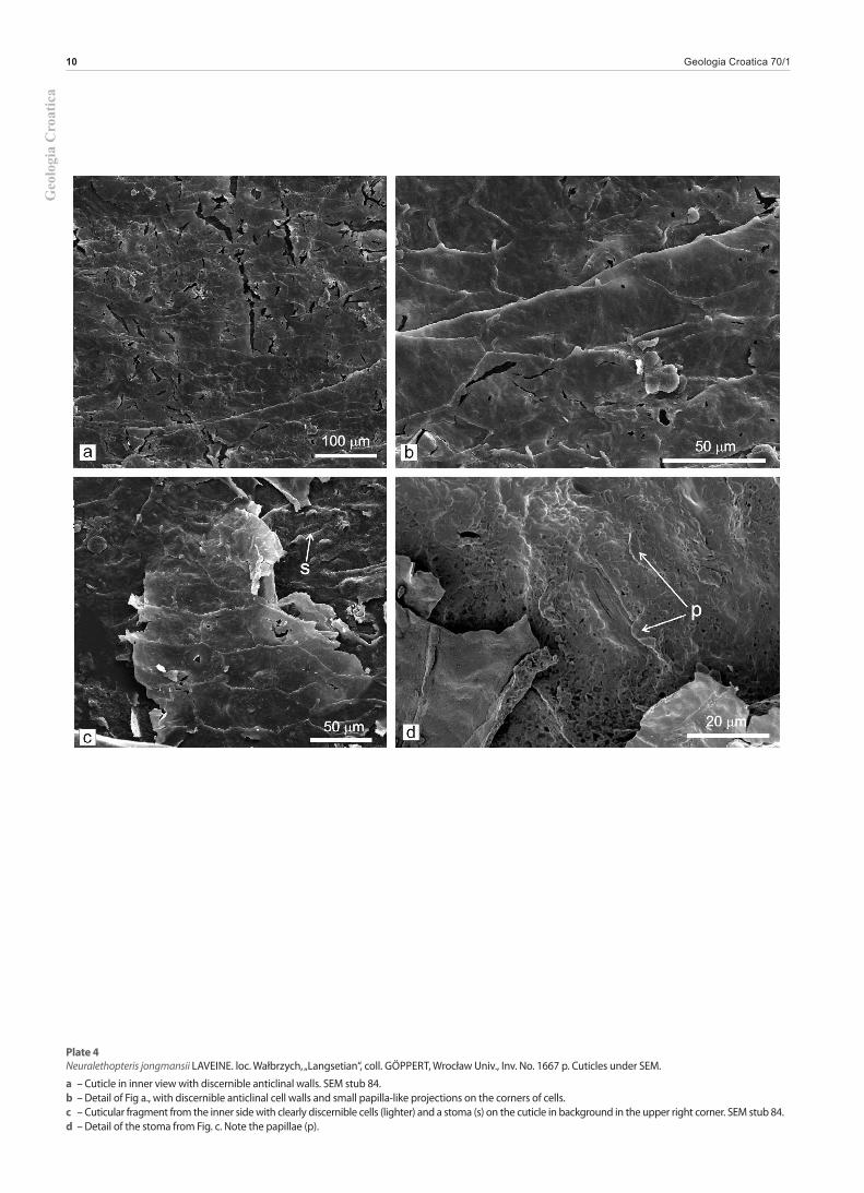
Plate 4Neuralethopteris jongmansii LAVEINE. loc. Wałbrzych, „Langsetian“, coll. GÖPPERT, Wrocław Univ., Inv. No. 1667 p. Cuticles under SEM.
a – Cuticle in inner view with discernible anticlinal walls. SEM stub 84.b – Detail of Fig a., with discernible anticlinal cell walls and small papilla-like projections on the corners of cells.c – Cuticular fragment from the inner side with clearly discernible cells (lighter) and a stoma (s) on the cuticle in background in the upper right corner. SEM stub 84.d – Detail of the stoma from Fig. c. Note the papillae (p).


![THE CUTICULAR PATTERN IN AN INSECT, RHODNIUS ...[ 45 ]9 THE CUTICULAR PATTERN IN AN INSECT,RHODNIUS PROLIXUS STAL BY M. LOCKE Department of Zoology, University College of the West](https://static.fdocuments.in/doc/165x107/60d8dfdd6bafa25aa5444dad/the-cuticular-pattern-in-an-insect-rhodnius-45-9-the-cuticular-pattern-in.jpg)