Mazin Barry, MD, FRCPC, ABIM, DTM&H Division of Infectious Diseases King Saud University.
Upload
ferdinand-fieldsCategory
view
216download
1
Cutaneous manifestations of systemic disease
Mohammed Al-Haddab,MD,FRCPC,DABDAssistant Professor, Dept. of DermatologyCollege of Medicine, King Saud University
Objectives
• To highlight the relation between skin manifestations and common systemic disorders.
• To understands various skin clues and their importance in investigating and managing different systemic diseases.

Skin and endocrine system
• Diabetes mellitus.
• Thyroid diseases.
• Cushing’s syndrome.
• Addison’s disease.

Diabetes mellitus
• Skin tags.• Acanthosis nigricans.• Diabetic dermopathy.• Bullous diabeticorum.• Thickening of skin.• Necrobiosis lipoidica diabeticorum. • Bacterial and fungal infections.• Perforating dermatosis.
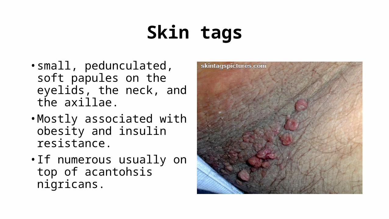
Skin tags
• small, pedunculated, soft papules on the eyelids, the neck, and the axillae.• Mostly associated with obesity
and insulin resistance.• If numerous usually on top of
acantohsis nigricans.
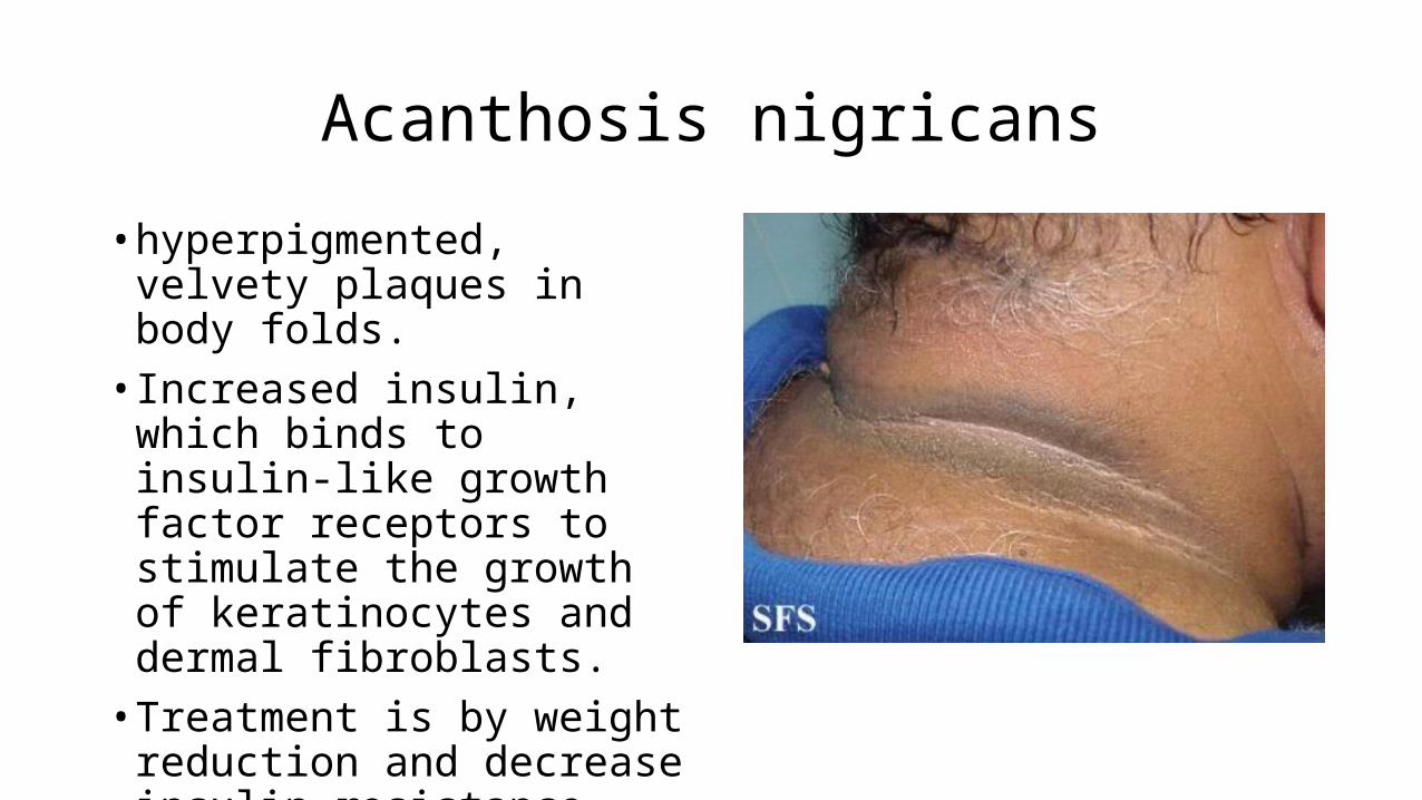
Acanthosis nigricans
• hyperpigmented, velvety plaques in body folds.• Increased insulin, which binds to
insulin-like growth factor receptors to stimulate the growth of keratinocytes and dermal fibroblasts.• Treatment is by weight reduction
and decrease insulin resistance.
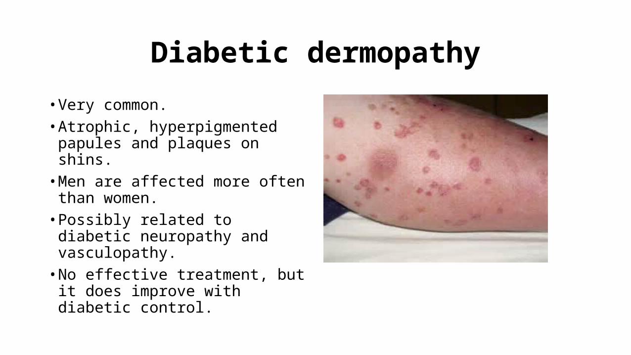
Diabetic dermopathy
• Very common.• Atrophic, hyperpigmented
papules and plaques on shins.• Men are affected more often
than women.• Possibly related to diabetic
neuropathy and vasculopathy.• No effective treatment, but it
does improve with diabetic control.
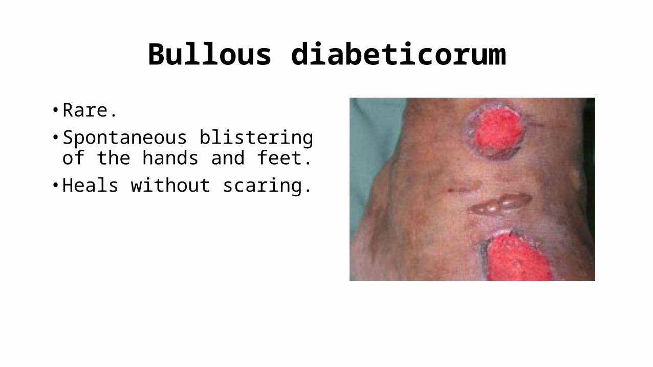
Bullous diabeticorum
• Rare.• Spontaneous blistering of the
hands and feet.• Heals without scaring.
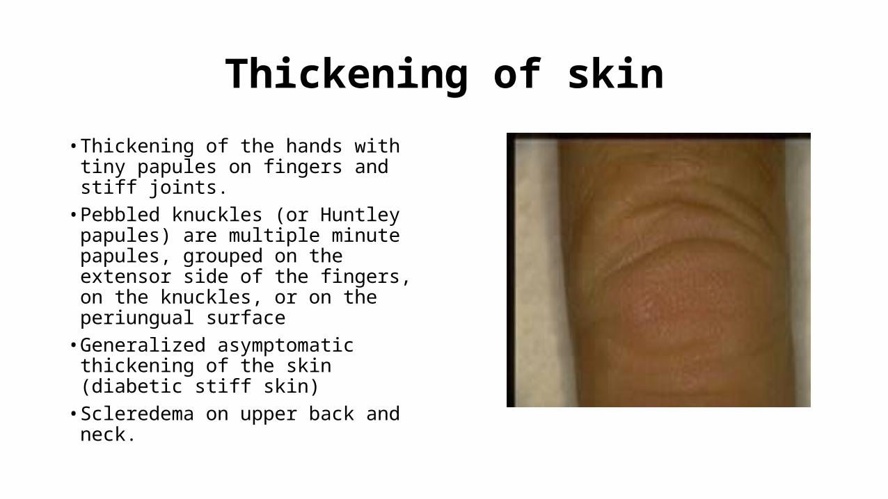
Thickening of skin
• Thickening of the hands with tiny papules on fingers and stiff joints.• Pebbled knuckles (or Huntley
papules) are multiple minute papules, grouped on the extensor side of the fingers, on the knuckles, or on the periungual surface• Generalized asymptomatic
thickening of the skin (diabetic stiff skin)• Scleredema on upper back and neck.

Necrobiosis lipoidica diabeticorum
• Yellow atrophic plaques on the shins.• Sometimes they ulcerate.• Histopathology shows tiered
granulomatous reaction.• Treatment with topical,
intralesional steroids, tacrolimus, phototherapy, cyclosporine, and rarely surgery.

Bacterial and fungal infections
• Pyodermic infections such as impetigo, folliculitis, carbuncles, furunculosis, ecthyma, and erysipelas can be more severe and widespread in diabetic patients.• Erythrasma, caused by
Corynebacterium minutissimum mostly on axillae and groin.• malignant otitis externa, often
caused by Pseudomonas aeruginosa.
• Tinea pedis and onychomycosis.• Candidal infections like perleche
on corners of mouth, and on vulva.• Rare infections like
mucormycosis by Phycomycetes and anaerobic cellulitis by Clostridium species.

Perforating dermatosis
• Pruritic hyperkeratotic papules on the legs and trunk.• Histopathology shows
transepidermal elimination of collagen and/or elastin.• Common in patients with diabetes
and renal failure.• treatments include topical
keratolytics, phototherapy, topical and systemic retinoids, topical and intralesional steroids, oral antihistamines, and cryotherapy.

Hyperthyroidism
• Pretibial myxedema: is the most characteristic features of thyrotoxicosis appearing as shiny waxy papules and plaques having orange-skin appearance on the chin of the tibia.• Warm skin and increased
sweating.• Pruritus
• Premature hair graying.• Alopecia with fine soft thinned
scalp hair.• Hyperpigmentation or vitiligo.• Brittle nails.

Hyperthyroidism
Hypothyroidism
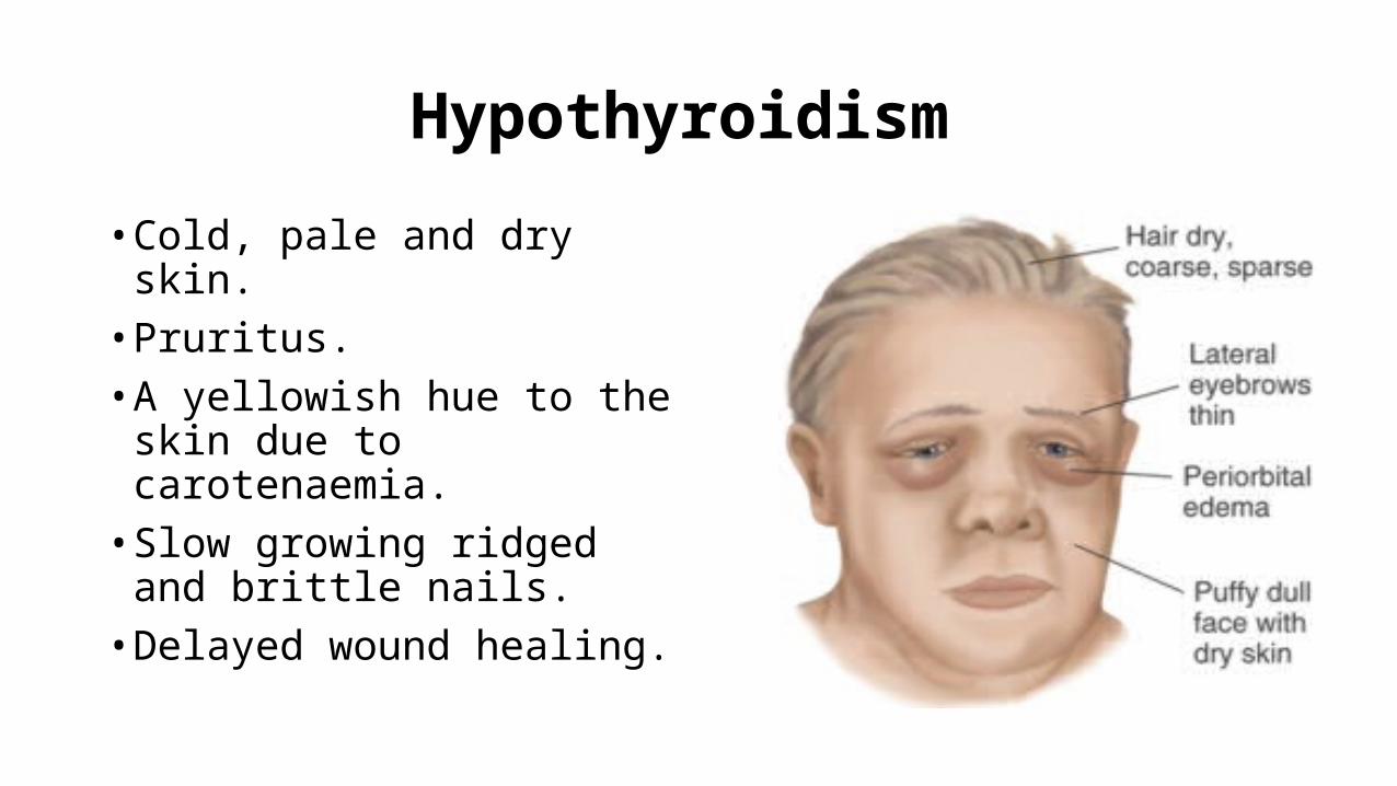
• Cold, pale and dry skin.• Pruritus.• A yellowish hue to the skin due
to carotenaemia.• Slow growing ridged and brittle
nails.• Delayed wound healing.

Cushing’s syndrome
• caused by prolonged exposure to high levels of plasma glucocorticoid.• Adrenocortical hyperplasia.• Benign or malignant adrenal
tumours.• Ectopic ACTH syndrome – secretion
of ACTH by malignant or benign tumours arising in structures other than the pituitary or adrenal glands.• Exogenous steroid administration
• Acne and hirsutism.• Clitromegaly and male pattern
alopecia.• Striae.• Easy bruising and purpura.• Moon face and buffalo hump
with fat redistribution.• Telangectasia on face.• Poor wound healing.

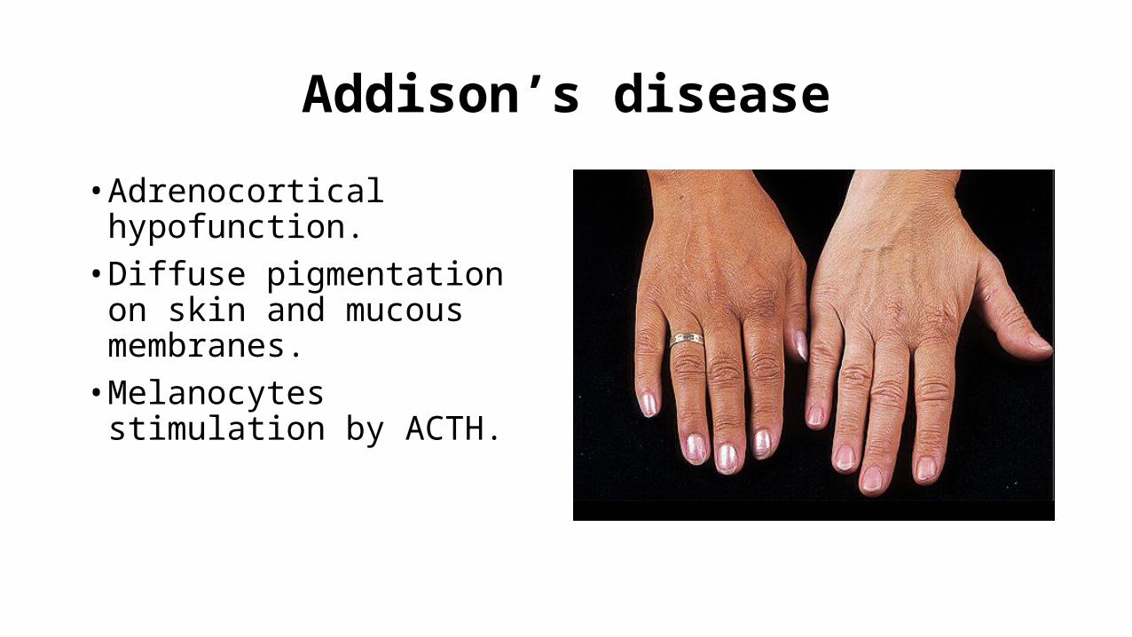
Addison’s disease
• Adrenocortical hypofunction.• Diffuse pigmentation on skin and
mucous membranes.• Melanocytes stimulation by
ACTH.

Gastrointestinal disease
• Dermatitis herpetiformis.• Acrodermatits enteropathica.• Pyoderma gangrenosum.• Peutz Jeghers syndrome.• Porphyria cutanea tarda.• Hemochromatosis.• Liver cirrhosis.

Dermatitis herpetiformis
• Small severely pruritic vesicular lesions found in a symmetric distribution of both upper and lower extensor surfaces, buttocks and the scalp.
• direct immunofluorescence finding is granular deposition of IgA within the dermal papillae.
• celiac disease (also known as gluten-sensitive enteropathy and celiac sprue) are caused by the inability to absorb gluten from the diet.
• Treatment: gluten-free diet and dapsone.

Acrodermatits enteropathica
• a rare autosomal recessive disorder that impairs dietary zinc absorption in the jejunum and ileum.
• presents in infants several weeks after breastfeeding is discontinued.
• characterized by diarrhea, inflammatory rash, and hair loss.
• scaly, erythematous patches and plaques similar to atopic dermatitis, but progress to vesicles, crusts, erosions, and pustules on acral areas, perioral and perianal areas.
• Treatment by zinc supplementation for life.

Pyoderma gangrenosum
• a painful, ulcerative lesion with a well-defined, undermined violaceous border.• start as small pustules, which
subsequently burst and expand to form the larger noninfectious ulcer.• Positive pathergy test.• Mostly associated with ulcerative
colitis. Also with Crohn’s disease, rheumatoid arthritis, and leukemia.• Surgery is contraindicated.

Peutz Jeghers syndrome
• autosomal dominant disorder.• mucocutaneous hyperpigmentation
together with GI polyposis.• The skin findings first appear in
infancy or early childhood and involve brown macules on the lips and buccal mucosa.• multiple hamartomatous polyps
occurring most commonly in the jejunum.• 2-3% of patients develop GI carcinoma
during their lifetimes.

Porphyria cutanea tarda
• most common porphyria occurring in adults.• skin photosensitivity with increased skin
fragility, facial hypertrichosis, blisters, scarring with milia formation, and skin hyperpigmentation on the hands and other sun-exposed areas.
• results from the decreased activity of the enzyme uroporphyrinogen decarboxylase.
• Associated with Hep C virus.• Treatment by removal of possible triggers,
including iron supplementation, alcohol, and estrogens. Also by phlebotomy and hydroxycholorquine.

Hemochromatosis
• a disorder of iron overload leading to excess deposition in multiple body organs.• metallic gray or bronze-brown color
that is generally diffuse.• skin atrophy, ichthyosis, partial hair
loss (most often in the pubic region), and koilonychia.• cirrhosis may develop, and might lead
to hepatocellular carcinoma.• treatment involves phlebotomy and
chelating agents.
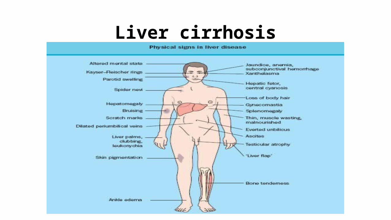
Liver cirrhosis

Renal diseases
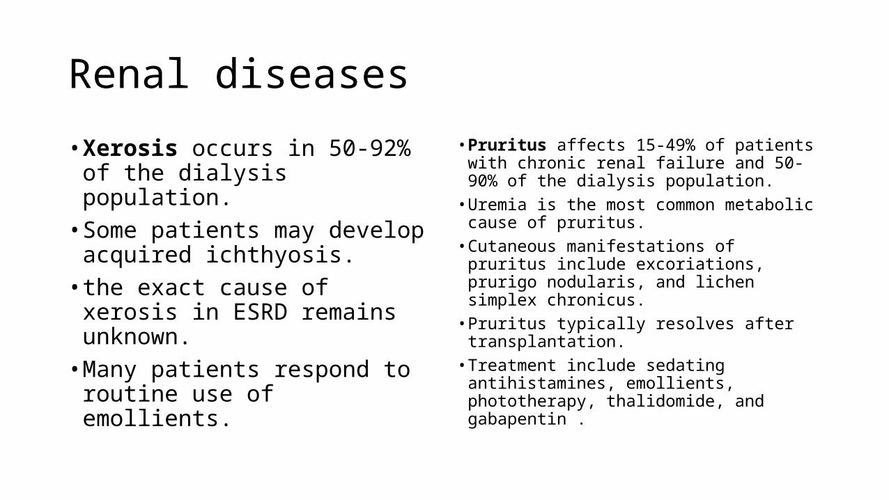
• Xerosis occurs in 50-92% of the dialysis population.
• Some patients may develop acquired ichthyosis.
• the exact cause of xerosis in ESRD remains unknown.
• Many patients respond to routine use of emollients.
• Pruritus affects 15-49% of patients with chronic renal failure and 50-90% of the dialysis population.
• Uremia is the most common metabolic cause of pruritus.
• Cutaneous manifestations of pruritus include excoriations, prurigo nodularis, and lichen simplex chronicus.
• Pruritus typically resolves after transplantation.
• Treatment include sedating antihistamines, emollients, phototherapy, thalidomide, and gabapentin .
Renal diseases
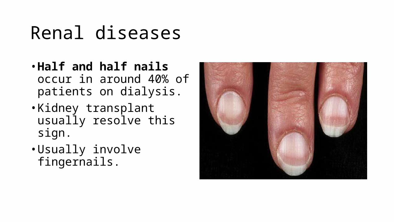
• Half and half nails occur in around 40% of patients on dialysis.• Kidney transplant usually resolve
this sign.• Usually involve fingernails.

Renal diseases
• Nephrogenic systemic fibrosis mostly seen in ESRD and dialysis patients.• Presents as thick, indurated plaques
on the extremities and the trunk similar to scleroderma.• gadolinium might have a role in the
pathogenesis of this condition.• Treatment includes
immunosuppressive agents, phototherapy, topical steroids, retinoids, and photophoresis.

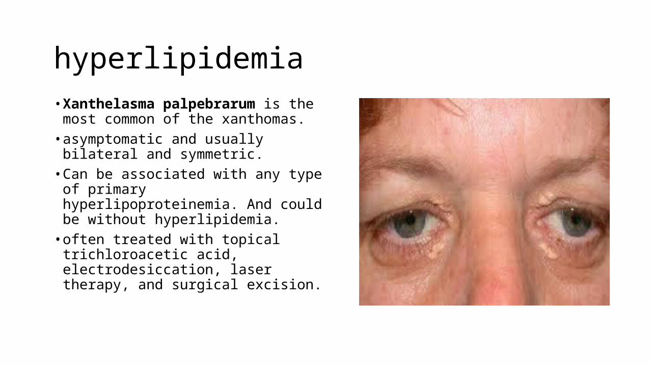
hyperlipidemia• Xanthelasma palpebrarum is the
most common of the xanthomas.• asymptomatic and usually bilateral
and symmetric.• Can be associated with any type of
primary hyperlipoproteinemia. And could be without hyperlipidemia. • often treated with topical
trichloroacetic acid, electrodesiccation, laser therapy, and surgical excision.

Hyperlipidemia
• Tendinous xanthomas commonly seen on the Achilles tendon followed by the hands, feet, elbows, and knees.• The least responsive xanthoma
to treatment.• Mostly seen in patients with
familial hypercholesterolemia.

hyperlipidemia
• Tuberous xanthomas are firm and nontender cutaneous and subcutaneous yellowish nodules on extensor surfaces.• Mostly associated with familial
dysbetalipoproteinemia.• May resolve after months of
treatment with lipid lowering agents.

Hyperlipidemia
• Eruptive xanthomas are painless, yellowish papules on an erythematous base that present as grouped lesions on trunk, elbows and buttocks.• Usually associated with
hypertriglyceridemia.• Could be seen in poorly controlled
diabetes and acute pancreatitis.• Usually resolve in few weeks after
therapy.
Hyperlipidemia
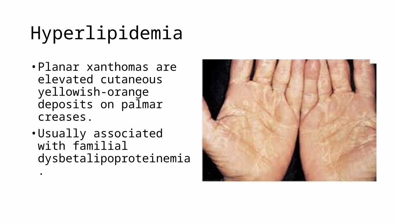
• Planar xanthomas are elevated cutaneous yellowish-orange deposits on palmar creases.• Usually associated with familial
dysbetalipoproteinemia.