Current status of L. infantum infection in stray cats in the · PDF fileRESEARCH Open Access...
7
RESEARCH Open Access Current status of L. infantum infection in stray cats in the Madrid region (Spain): implications for the recent outbreak of human leishmaniosis? Guadalupe Miró 1* , Cristina Rupérez 1 , Rocío Checa 1 , Rosa Gálvez 1 , Leticia Hernández 1 , Manuel García 2 , Isabel Canorea 1 , Valentina Marino 1 and Ana Montoya 1 Abstract Background: Since 2009, the incidence of human leishmaniosis in the SW of the Madrid region has been unusually high. Although dogs are the main reservoir for this disease, a role played by dogs in this outbreak has been ruled out and investigators are now considering other hosts (eg. cats, rabbits, hares) as possible alternative reservoirs. This study was designed to examine the Leishmania infantum status of stray cats in Madrid to assess its possible implications in the human leishmaniosis outbreak. Methods: 346 captured stray cats were tested for antibodies against L. infantum by the indirect fluorescent antibody technique (IFAT) and nested-PCR methods were used to detect Leishmania DNA in blood samples of cats testing seropositive for L. infantum and/or retroviruses infection. Cats were also tested for Toxoplasma gondii using the direct agglutination test (DAT) and feline leukemia virus (FeLV) antigen and feline immunodeficiency virus (FIV) antibodies (PetChek* FIV/FeLV). The presence of intestinal parasites was determined using a routine coprological method. Results: The seroprevalence of L. infantum infection (cut off ≥ 1/100) was 3.2% (11/346). However, it was not possible to amplify Leishmania DNA in any of the blood samples. Seropositivity was not associated with sex, age, capture site, clinical status, retrovirus infection or T. gondii seropositivity. Of the 11 cats seropositive for L. infantum, 3 also tested positive for FIV, none for FeLV and 6 for T. gondii. It should be mentioned that the prevalence of FeLV p27 antigen was 4% and of FIV antibody was 9.2%. Although the seroprevalence of T. gondii was quite high at 53.5%, no T. gondii oocysts were found in any of the faeces samples analysed (n = 287). In contrast, intestinal parasites were detected in 76 (26.5%) samples, Toxocara cati being the most prevalent. Conclusions: Our results suggest a stable L. infantum infection situation among the stray cats of the Madrid area; the disease is uncommon and no clinical cases have been reported to date. The detection of other zoonotic parasites such as T. gondii and T. cati in stray cats indicates a need to adopt strict control measures in this population. Keywords: Cat, L. infantum, Toxoplasma gondii, Feline retroviruses, Toxocara cati, IFAT, PCR, Spain * Correspondence: [email protected] 1 Departamento de Sanidad Animal, Facultad de Veterinaria, Universidad Complutense de Madrid Full list of author information is available at the end of the article © 2014 Miró et al.; licensee BioMed Central Ltd. This is an Open Access article distributed under the terms of the Creative Commons Attribution License (http://creativecommons.org/licenses/by/2.0), which permits unrestricted use, distribution, and reproduction in any medium, provided the original work is properly credited. The Creative Commons Public Domain Dedication waiver (http://creativecommons.org/publicdomain/zero/1.0/) applies to the data made available in this article, unless otherwise stated. Miró et al. Parasites & Vectors 2014, 7:112 http://www.parasitesandvectors.com/content/7/1/112
Transcript of Current status of L. infantum infection in stray cats in the · PDF fileRESEARCH Open Access...
Miró et al. Parasites & Vectors 2014, 7:112http://www.parasitesandvectors.com/content/7/1/112
RESEARCH Open Access
Current status of L. infantum infection in straycats in the Madrid region (Spain): implications forthe recent outbreak of human leishmaniosis?Guadalupe Miró1*, Cristina Rupérez1, Rocío Checa1, Rosa Gálvez1, Leticia Hernández1, Manuel García2,Isabel Canorea1, Valentina Marino1 and Ana Montoya1
Abstract
Background: Since 2009, the incidence of human leishmaniosis in the SW of the Madrid region has been unusuallyhigh. Although dogs are the main reservoir for this disease, a role played by dogs in this outbreak has been ruledout and investigators are now considering other hosts (eg. cats, rabbits, hares) as possible alternative reservoirs.This study was designed to examine the Leishmania infantum status of stray cats in Madrid to assess its possibleimplications in the human leishmaniosis outbreak.
Methods: 346 captured stray cats were tested for antibodies against L. infantum by the indirect fluorescentantibody technique (IFAT) and nested-PCR methods were used to detect Leishmania DNA in blood samples of catstesting seropositive for L. infantum and/or retroviruses infection. Cats were also tested for Toxoplasma gondii usingthe direct agglutination test (DAT) and feline leukemia virus (FeLV) antigen and feline immunodeficiency virus (FIV)antibodies (PetChek* FIV/FeLV). The presence of intestinal parasites was determined using a routine coprologicalmethod.
Results: The seroprevalence of L. infantum infection (cut off≥ 1/100) was 3.2% (11/346). However, it was notpossible to amplify Leishmania DNA in any of the blood samples. Seropositivity was not associated with sex, age,capture site, clinical status, retrovirus infection or T. gondii seropositivity. Of the 11 cats seropositive for L. infantum,3 also tested positive for FIV, none for FeLV and 6 for T. gondii. It should be mentioned that the prevalence of FeLVp27 antigen was 4% and of FIV antibody was 9.2%. Although the seroprevalence of T. gondii was quite high at53.5%, no T. gondii oocysts were found in any of the faeces samples analysed (n = 287). In contrast, intestinalparasites were detected in 76 (26.5%) samples, Toxocara cati being the most prevalent.
Conclusions: Our results suggest a stable L. infantum infection situation among the stray cats of the Madrid area;the disease is uncommon and no clinical cases have been reported to date. The detection of other zoonoticparasites such as T. gondii and T. cati in stray cats indicates a need to adopt strict control measures in thispopulation.
Keywords: Cat, L. infantum, Toxoplasma gondii, Feline retroviruses, Toxocara cati, IFAT, PCR, Spain
* Correspondence: [email protected] de Sanidad Animal, Facultad de Veterinaria, UniversidadComplutense de MadridFull list of author information is available at the end of the article
© 2014 Miró et al.; licensee BioMed Central Ltd. This is an Open Access article distributed under the terms of the CreativeCommons Attribution License (http://creativecommons.org/licenses/by/2.0), which permits unrestricted use, distribution, andreproduction in any medium, provided the original work is properly credited. The Creative Commons Public DomainDedication waiver (http://creativecommons.org/publicdomain/zero/1.0/) applies to the data made available in this article,unless otherwise stated.
Miró et al. Parasites & Vectors 2014, 7:112 Page 2 of 7http://www.parasitesandvectors.com/content/7/1/112
BackgroundLeishmaniosis is a zoonotic disease caused by the proto-zoan Leishmania infantum. In Spain, the disease is trans-mitted by the female sandflies, Phlebotomus perniciosusand P. ariasi. Although dogs are considered the main res-ervoir, L. infantum has been detected in a wide range ofmammalian species, including cats [1-4].Because of its zoonotic nature, leishmaniosis has been a
notifiable disease in Spain since 1982. Before 2009, themean reported annual incidence of human leishmaniosisin the Madrid Autonomous Community (CM) was 1.12cases/100,000 inhabitants [5]. However, this incidence in-creased abruptly to 22.2 cases per 100,000 inhabitantsfrom mid-2009 to the end of 2012 in the southwest regionof Madrid. In total, 446 cases were reported: 6 in 2009, 97in 2010, 196 in 2011 and 147 in 2012 [6]. Entomologicaland serological surveys in the area have surprisingly de-tected an infection rate in dogs of 1.6-2% [7]. Other animalsincluding cats, rats, rabbits, and hares from the affectedarea have also been examined. Preliminary results confirmthe role that hares and rabbits could serve as wild reservoirsof leishmaniosis for the recent outbreak of visceral leishma-niosis in Madrid [8,9]. Nevertheless, antibodies against L.infantum have also been detected in four cats [9] and bloodfrom cats was found in a gravid female P. perniciosus col-lected from the area affected by the outbreak [10].In the past decade, several studies have been conducted
in cats, and seroprevalences ranging from 0.9 to 59% havebeen reported in Mediterranean countries and Brazil [11].In Spain, L. infantum seroprevalence data on cat popula-tions is still scarce and ranges from 1.3% in the central re-gion [4] to 28% in the South ((cut off ≥ 1/40); the realseroprevalence being 12.2% using a cut off ≥ 1:80) [1].Prevalences detected by PCR have ranged from 0.3 to 26%for blood samples from cats in the Mediterranean area[11-13] and from 5.8 to 9.9% for bone marrow and lymphnode samples from cats in Brazil [14,15].Despite the high prevalence of canine leishmaniosis in
endemic areas, feline leishmaniosis is frequently subclin-ical. Over 40 clinical cases in cats have been described inthe literature in Europe, South America and Texas. Thesecases were characterized by cutaneous lesions (nodularand ulcerative lesions), enlarged lymph nodes, weight lossand ocular lesions [11,16-26]. Xenodiagnosis studies per-formed in two chronically infected cats have shown that acat can be infectious to a competent L. infantum vector[22,27]. However, the role of the cat in the transmissioncycle of leishmaniosis is not clear [28], highlighting a needfor further studies.The present study was designed to determine the status
of L. infantum infection among the stray cats inhabitingthe central region of Spain (Madrid and two of its border-ing provinces Toledo and Guadalajara) and to identify riskfactors associated with the presence of infection/disease.
These factors include coinfection with other pathogensmainly affecting outdoor cats: feline immunodeficiencyvirus (FIV), feline leukemia virus (FeLV), T. gondii andother intestinal parasites.
MethodsPopulation studiedEvery year in spring, the Animal Protection Society ALBAin Madrid undertakes a health control programme forstray cats. A large number of cats are captured to assesstheir health state and FeLV-FIV status and then healthycats are sterilized before they are returned to their site ofcapture.Clinical assessment consisted of sedation with a combin-
ation of 80 micrograms medetomidine/kg-5 mg ketamine/kg [29] followed by a thorough physical exam and the col-lection of samples: blood (serum and EDTA) by jugularvenipuncture, rectum and ear swabs, and skin scrapings ifcutaneous lesions were observed. Samples were kept at 4°Cuntil processed at the laboratory.
Leishmania infectionSerum antibody testingFor serological tests, specific antibodies to L. infantum weredetected using the indirect immunofluorescence antibodytest (IFAT) against in-house cultured promastigotes. TheIFAT for anti-Leishmania-specific immunoglobulin G (IgG)antibodies was performed as described previously using acut-off ≥ 1:100 to define seropositivity [4].
Molecular analysisDNA was extracted from the blood samples of cats thathad been identified as L. infantum seropositive and/orFeLV/FIV positive (n = 57) using the QIAamp DNA Mikrokit (QIAGEN). Extracted DNA was stored at −20°C untilPCR was performed.The parasite was detected using two nested PCR proto-
cols. One was a nested-PCR targeting the LeishmaniaSSUrRNA gene (LnPCR) as described by Cruz et al. [30].This protocol is Leishmania genus specific and uses theprimer pair R221 (5’-GGTTCCTTTCCTGATTTACG-3’)and R332 (5’-GGCCGGTAAAGGCCGAATAG-3’) in thefirst reaction. In this second mixture, the starting primerswere replaced with the primers R223 (5’-TCCCATCGCAACCTCGGTT-3’) and R333 (5’-AAAGCGGGCGCGGTGCTG-3’). The PCR product amplification size was603 bp in the first reaction and 353 bp in the nested PCR.The second protocol was PCR amplification of a portion
of the ITS-1n gene according to the protocol described bySchönian et al. [31] but briefly modified. In this PCR, theregion of the tandem ribosomal RNA genes (ITS-1) wasamplified using the primers SAC (5'-CATTTTCCGATGATTACACC-3') and VAN2 (5'-GCGACACGTTATGTGAGCCG-3'). L. infantum DNA amplification was carried
Miró et al. Parasites & Vectors 2014, 7:112 Page 3 of 7http://www.parasitesandvectors.com/content/7/1/112
out in a 25 μl reaction volume containing 2.5 ul Buffer10X (Biotools), 2 mM MgCl2; 0.5 ul dNTP mix 10 mM(Biotools), 0.5 ul of each primer (15 pmol/uL), 0.7 ul TthDNA polymerase (1U/ul) (Biotools) and 10 ul of DNA.PCR amplification was performed in a thermal cycler at80°C for 2 min, 94°C for 5 min, and 40 temperature cycles(94°C for 30 s, 57°C for 30 s, and 72°C for 30 s); this wasfollowed by an extension step of 5 min at 72°C. The sizeof the PCR amplification product was 280–330 bp.
Toxoplasma gondii infectionCat sera were tested for antibodies against T. gondii usinga direct agglutination test (DAT) kit (Toxo-Screen DA;Biomerieux) as described by Desmonts and Remington(1980). An antibody titre of 1:40 was considered indicativeof exposure of the cat population to T. gondii [32].
FeLV-FIV infectionCats were tested for FeLV antigen (p27) and FIV anti-bodies using a commercial ELISA kit (PetChek* FIV/FeLV; IDEXX Laboratories) [33].
Other parasitological methodsFaeces samples were collected using a rectal swab. Forcoprological analysis, we used the modified Telemannsedimentation method plus merthiolate-iodine-formalinstaining, followed by examination under a light micro-scope [34].Ear swabs were also collected to determine the pres-
ence of ectoparasites, mainly Otodectes cynotis. Auricu-lar secretions were examined under the microscope.Whole skin was explored to detect the presence of
skin lesions and during this process other ectoparasiteswere sometimes detected. When these were detected,they were stained in 70° alcohol until their identificationunder the microscope and/or magnification using identi-fication keys [35,36].
Statistical analysisCorrelations between all the variables examined wereidentified by the Chi-square test (SPSS 17.0). Signifi-cance was set at p < 0.05.
Ethical considerationsThe study was carried out in accordance with Spanish Le-gislation guidelines (Ley 1/1990, Comunidad de Madrid)and the International Guiding Principles for BiomedicalResearch Involving Animals, issued by the Council for theInternational Organizations of Medical Sciences.
ResultsOf the 346 stray cats (146 male, 200 female) included inthe study, 181 were enrolled in 2012 and 165 in 2013.Cats were grouped according to age: 6.3% (22/346) were
kittens (under 6 months), 25.7% (89/346) were young(6 months to 1 year) and 67.9% (235/346) were esti-mated to be older than one year.The cats were captured throughout the Madrid region
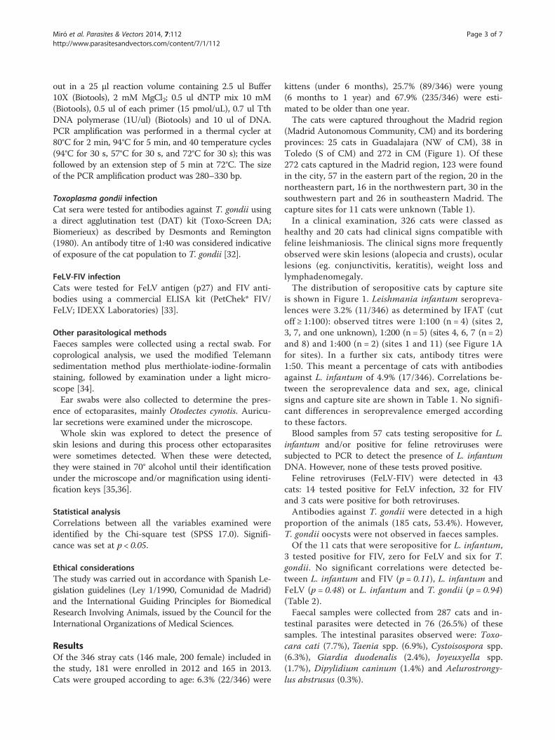
(Madrid Autonomous Community, CM) and its borderingprovinces: 25 cats in Guadalajara (NW of CM), 38 inToledo (S of CM) and 272 in CM (Figure 1). Of these272 cats captured in the Madrid region, 123 were foundin the city, 57 in the eastern part of the region, 20 in thenortheastern part, 16 in the northwestern part, 30 in thesouthwestern part and 26 in southeastern Madrid. Thecapture sites for 11 cats were unknown (Table 1).In a clinical examination, 326 cats were classed as
healthy and 20 cats had clinical signs compatible withfeline leishmaniosis. The clinical signs more frequentlyobserved were skin lesions (alopecia and crusts), ocularlesions (eg. conjunctivitis, keratitis), weight loss andlymphadenomegaly.The distribution of seropositive cats by capture site
is shown in Figure 1. Leishmania infantum seropreva-lences were 3.2% (11/346) as determined by IFAT (cutoff ≥ 1:100): observed titres were 1:100 (n = 4) (sites 2,3, 7, and one unknown), 1:200 (n = 5) (sites 4, 6, 7 (n = 2)and 8) and 1:400 (n = 2) (sites 1 and 11) (see Figure 1Afor sites). In a further six cats, antibody titres were1:50. This meant a percentage of cats with antibodiesagainst L. infantum of 4.9% (17/346). Correlations be-tween the seroprevalence data and sex, age, clinicalsigns and capture site are shown in Table 1. No signifi-cant differences in seroprevalence emerged accordingto these factors.Blood samples from 57 cats testing seropositive for L.
infantum and/or positive for feline retroviruses weresubjected to PCR to detect the presence of L. infantumDNA. However, none of these tests proved positive.Feline retroviruses (FeLV-FIV) were detected in 43
cats: 14 tested positive for FeLV infection, 32 for FIVand 3 cats were positive for both retroviruses.Antibodies against T. gondii were detected in a high
proportion of the animals (185 cats, 53.4%). However,T. gondii oocysts were not observed in faeces samples.Of the 11 cats that were seropositive for L. infantum,
3 tested positive for FIV, zero for FeLV and six for T.gondii. No significant correlations were detected be-tween L. infantum and FIV (p = 0.11), L. infantum andFeLV (p = 0.48) or L. infantum and T. gondii (p = 0.94)(Table 2).Faecal samples were collected from 287 cats and in-
testinal parasites were detected in 76 (26.5%) of thesesamples. The intestinal parasites observed were: Toxo-cara cati (7.7%), Taenia spp. (6.9%), Cystoisospora spp.(6.3%), Giardia duodenalis (2.4%), Joyeuxyella spp.(1.7%), Dipylidium caninum (1.4%) and Aelurostrongy-lus abstrusus (0.3%).
Figure 1 Geocoded sites where the cats were captured, shown on a digital elevation model. A. Distribution of cats seropositive for L.infantum (IFAT titre≥ 1:50) in the study area. B. Detailed distribution of cats seropositive for L. infantum (IFAT titre≥ 1:50) in the Madrid city area.1: Uceda (Guadalajara); 2: Guadalajara city; 3: Fresno del Torote (CM); 4: Camarma de Esteruelas (CM); 5: San Sebastián de los Reyes (CM); 6: Alcaláde Henares (CM); 7: Torrejón de Ardoz (CM); 8: Las Rosas (Madrid city); 9: Casa de Campo (Madrid city); 10: Orcasur (Madrid city); 11: Fuenlabrada(CM); 12: Carranque (Toledo). **For 3 seropositive cats (two showing a titre of 1:50 and 1 of 1:100), the capture site was unknown so they do notappear on the map.
Miró et al. Parasites & Vectors 2014, 7:112 Page 4 of 7http://www.parasitesandvectors.com/content/7/1/112
In ear swab samples collected from 281 cats, 36 (12.8%)showed the presence of Otodectes cynotis. In some cats,other ectoparasites were also detected during physicalexamination: Ctenocephalides felis (n = 13), Felicola sub-rostratus (n = 2), Rhipicephalus sanguineus (n = 1) andDemodex spp (n = 1).
Table 1 L. infantum infected cats according to sex, age,capture site and clinical signs compatible with leishmaniosis
Variables Total L. infantum IFAT p-value
Positive (%) Negative (%)
Sex 0.14
Male 146 7 (4.8) 137 (95.2)
Female 200 4 (2) 196 (98)
Age 0.52
< 6 months 22 0 (0) 22 (100)
6 months- 1 year 89 2 (2.2) 87 (97.8)
> 1 year 235 9 (3.8) 226 (96.2)
Clinical signs 0.63
Absence 326 10 (3.1) 316 (96.9)
Presence 20 1 (5) 19 (95)
Capture site 0.13
Toledo 37 0 (0) 37 (100)
Guadalajara 25 2 (8) 23 (92)
Madrid
City 123 2 (1.6) 121 (98.4)
Northeast 20 1(5) 19 (95)
East 57 5 (8.8) 52 (91.2)
Southeast 26 0 (0) 26 (100)
Southwest 30 1 (3.3) 29 (96.7)
Northwest 16 0 (0) 16 (100)
Unknown 11 0 (0) 11(100)
Total 346 11 (3.2) 335(96.8)
DiscussionIn this study, the seroprevalence of L. infantum in stray catsin Madrid and its neighbouring provinces was estimated at3.2%. Prior studies have indicated similar seroprevalencesfor Madrid (1.3-3.7%) [3,4] and other Mediterranean re-gions such as Greece [37], Portugal [38,39], southern Italy[40] and Israel (Jerusalem) [41]. In contrast, reported sero-prevalences for Ibiza (13.2%) [42] and southern Spain(28.3%) [1] have been notably higher. These two regions areendemic for canine leishmaniosis and seroprevalences of L.infantum in dogs are also much higher than in other Span-ish regions [43-45]. We should, however, mention thatthese rates are difficult to compare due to differences in thediagnostic techniques used (IFAT, ELISA), the cut-offsestablished, the sizes and origins of samples, and the seasonof study. There is a clear need to standardize protocols, es-tablish cut-offs for the different techniques and define agold standard procedure, as in the case of canine leishma-niosis [46,47].Despite these differences among the seroprevalence
studies performed to date, the seroprevalence of L. infan-tum among cats observed here and in other studies hasnot substantially changed since the first studies conducted
Table 2 L. infantum infected cats and co-infection withfeline immunodeficiency virus (FIV), feline leukemia virus(FeLV) or Toxoplasma gondii
L. infantum IFAT p- value
Positive (%) Negative (%)
FIV Positive 3 (27.3) 30 (9) 0.11
Negative 8 (72.7) 306 (91)
FeLV Positive 0 (0) 14 (4.2) 0.48
Negative 11 (100) 321 (95.8)
T. gondii Positive 6 (54.5) 179 (53.4) 0.94
Negative 5(45.5) 156 (46.6)
Miró et al. Parasites & Vectors 2014, 7:112 Page 5 of 7http://www.parasitesandvectors.com/content/7/1/112
in Madrid [3,4]. This trend has been paralleled by the L.infantum situation in dogs in Madrid, where the sero-prevalence over the past 15 years has been stable at 6.4 to8.1% [43,48,49], including the time period correspondingto an unusual outbreak of human leishmaniosis in SWMadrid [6].Although PCR is a sensitive method [50], we were un-
able to detect L. infantum DNA in any of the blood sam-ples testing seropositive for the pathogen. One possiblereason for this inconsistency is that blood is not a goodsample for this purpose or that the sample volume gen-erally used is insufficient (100 μl) [2,51]. Similar studieshave also reported a low prevalence of Leishmania posi-tivity detected by PCR in blood samples (0.3-0.6%) col-lected from cats in Madrid and N Portugal, where theseroprevalence is also lower (2.8-3.7%) [3,12,52]. Surveysin the Balearic Islands, S Spain and S Portugal have de-tected higher percentages of cats testing positive forLeishmania infection by PCR on blood samples (8.7-26%) [13,38,42,53]. In these studies, however, seropreva-lences were also higher (13.2-26%) [1,42]. The findingsof molecular studies from Brazil are not comparable tothese data because the samples used for PCR were bonemarrow, spleen and lymph nodes [14,15,54]. This typeof sample probably should be the sample of choice yet hasthe drawback that invasive methods are needed for theircollection. Also, with cats being smaller and more difficultto handle than dogs, samples are difficult to obtain. Theselimitations prompt a need to assess the sensitivity of the useof non invasive samples (eg. conjunctival swabs), as reportedfor canine leishmaniosis [55], for the PCR detection ofLeishmania. Further molecular studies need to determinewhether samples such as bone marrow, lymph nodes, con-junctival/oral swabs and/or skin will improve the detectionof Leishmania DNA and diagnosis of the parasite [14,42,51].In the current study, no significant correlation was
found between L. infantum infection and sex, age or clin-ical status, as reported in similar studies [1,41,56]. Accord-ing to the capture site, seroprevalences were higher in theE (8.8%) and NE of Madrid (10%) and in Guadalajara (8%),though differences were not significant (p = 0.13). Catsseropositive for L. infantum showed a homogenous distri-bution in the area of the human leishmaniosis (SW) out-break, indicating that cats are not playing a role as areservoir for L. infantum infection in this area.Reported clinical cases of feline leishmaniosis are fre-
quently associated with skin lesions as well as other lessspecific clinical signs (eg, lymph node enlargement,weight loss, ocular involvement), which can also be ob-served in other infectious diseases such as those pro-duced by retroviruses. Accordingly, most cats developingclinical signs are suspected of having an impaired im-mune system [11]. In our study, 5.7% (20/346) of thecats showed clinical signs compatible with feline
leishmaniosis, of which, only one was seropositive for L.infantum antibodies. These results are in line with thoseof prior studies in which high proportions of seropositivedogs and/or cats showed no clinical signs of leishmanio-sis [13,14,42,48] and suggest the need for tests (e.g. ser-ology, molecular diagnosis, etc.) in companion animalsregardless of the presence of clinical signs, since in manycases the disease is underdiagnosed.The prevalence of feline retroviruses (FeLV/FIV) in stray
cats of our study area was consistent with the rates de-tected in previous studies [2,42]. FeLV and FIV infectionsare frequently associated with opportunistic infectionscaused by protozoans, fungi, bacteria or viruses since theseviruses may compromise the cellular immune response[57]. Sobrinho et al. [14], and Pennisi et al. [40] reportedfeline leishmaniosis in association with FIV, while in astudy performed by our group in Ibiza (Spain) associationbetween L. infantum and FeLV was detected [42]. In thepresent study, certain positive association was observed be-tween Leishmania and retroviruses as reported in Jerusalem(Israel) [41] and Brazil [56]. However, since leishmaniosis isan immunomediated disease [58], future studies need to ad-dress possible correlations with immunosuppressive diseasesor states (eg, retrovirus infections, neoplasias, treatmentwith corticosteroids, etc.) [2,11].Antibodies against T. gondii were detected in 53.4% of
the stray cat population studied. This seroprevalence issimilar to that reported by Aparicio et al. [59] and Alonsoet al. [60] for stray cats in Madrid, though we previouslyobserved a lower seroprevalence over the period of 2003–2007 [61,62]. The presence of cats testing seropositive forT. gondii is significant because, despite not detecting theoocysts of T. gondii in any faeces sample, these cats musthave excreted oocysts sometime during their life, mainlywhen they were young. This could not be confirmed, how-ever, due to the low numbers of kittens and young cats in-cluded in our study. Thus, the oocysts of T. gondii andother zoonotic parasites such as Giardia duodenalis andToxocara cati [63], also detected here, could be present inthe soils of public parks. In effect,Toxocara cati was the in-testinal parasite appearing at the highest prevalence. Toxo-cara spp. (Toxocara cati and T. canis) are responsible forvisceral larvae migrans (VLM) in humans and although,Toxocara canis is often more recognized as a zoonoticagent, Toxocara cati should not be overlooked [64,65].Moreover, some patients with ocular larva migrans (OLM)due to Toxocara spp. show a stronger reaction to the T. catithan T. canis antigen in the Ouchterlony test [64]. It ap-pears that T. cati may contribute to OLM and VLM in lar-ger measure than was previously thought [64,65]. Theprevalence of the other intestinal parasites was similar tothat detected in stray cats from other major cities such asLisbon [39] or Milan [66] or even in other studies per-formed in Madrid [61,62].
Miró et al. Parasites & Vectors 2014, 7:112 Page 6 of 7http://www.parasitesandvectors.com/content/7/1/112
Prevalences of the ectoparasites Otodectes cynotis, Cte-nocephalides felis, Rhipicephalus sanguineus and Felicolasubrostratus were similar to those reported by others[39,67]. As vectors for zoonotic diseases (eg, dipylidiosis,ricketsiosis, bartonellosis, etc.), many of these ectopara-sites will have implications for public health.
ConclusionsIn conclusion, the results of this study point to the stablesituation of Leishmania infantum infection among thestray cats of Madrid and neighbouring areas (Toledo andGuadalajara), with clinical cases being rare. Further mo-lecular and xenodiagnosis studies are, however, needed toclarify the epidemiological role of this alternative host. Wesuggest that veterinarians in canine leishmaniosis endemicareas should include leishmaniosis in the differential diag-nosis of cats with compatible cutaneous or systemic clin-ical signs and inform cat owners of measures to preventor reduce disease transmission. In addition, efficient repel-lents/insecticides to prevent sandfly bites need to be devel-oped for use in cats.The detection of other important zoonotic parasites
such as Toxoplasma gondii, Toxocara cati, Giardia duode-nalis, etc. in the stray cats analyzed identifies a need forappropriate control measures in this population.
Competing interestsThe authors declare they have no competing interests.
Authors’ contributionsGM conceived and coordinated the study, and participated in its design, thefield study and drafted the manuscript. CR and MG participated in thephysical exam and collection of blood samples from cats. RC participated incollecting samples and carrying out the molecular technique. RGparticipated in data elaboration and helped to draft the manuscript. LH andVM participated in the diagnostic assays. IC participated in the molecularassays. AM participated in the field studies and the diagnostic assays,performed the statistical analysis, and helped draft the manuscript.All authors read and approved the final manuscript.
AcknowledgementsWe would like to thank all the veterinarians of ALBA for their collaboration inthis study and the volunteers who captured the cats. The authors are alsoindebted to ALBA for their initiative to control cat colonies in Madrid.This study was funded by Bayer HealthCare (Animal Health) and IDEXX (Spain).
Author details1Departamento de Sanidad Animal, Facultad de Veterinaria, UniversidadComplutense de Madrid. 2Asociación para la Liberación y el BienestarAnimal, ALBA, Madrid.
Received: 17 January 2014 Accepted: 4 March 2014Published: 24 March 2014
References1. Martín-Sánchez J, Acedo C, Muñoz-Pérez M, Pesson B, Marchal O, Morillas-
Márquez F: Infection by Leishmania infantum in cats: epidemiologicalstudy in Spain. Vet Parasitol 2007, 145(3–4):267–273.
2. Solano-Gallego L, Rodríguez-Cortés A, Iniesta L, Quintana J, Pastor J, Espada Y,Portús M, Alberola J: Cross-sectional serosurvey of feline leishmaniasis inecoregions around the Northwestern Mediterranean. Am J Trop Med Hyg2007, 76(4):676–680.
3. Ayllón T, Diniz PP, Breitschwerdt EB, Villaescusa A, Rodríguez-Franco F, SainzA: Vector-borne diseases in client-owned and stray cats from Madrid,Spain. Vector Borne Zoonotic Dis 2012, 12(2):143–150.
4. Ayllon T, Tesouro MA, Amusategui I, Villaescusa A, Rodriguez-Franco F, Sainz A:Serologic and molecular evaluation of Leishmania infantum in cats fromCentral Spain. Ann N Y Acad Sci 2008, 1149:361–364.
5. Valcárcel Y, Bastero R, Anegón M, González S, Gil A: The epidemiology ofhospital admissions due to leishmaniasis in Spain (1999–2003).Enferm Infecc Microbiol Clin 2008, 26(5):278–281.
6. Arce A, Estirado A, Ordobas M, Sevilla S, García N, Moratilla L, de la Fuente S,Martínez AM, Pérez AM, Aránguez E, Iriso A, Sevillano O, Bernal J, Vilas F:Re-emergence of leishmaniasis in Spain: community outbreak in Madrid,Spain, 2009 to 2012. Euro Surveill 2013, 18(30):20546.
7. Carrillo E, Moreno J, Cruz I: What is responsible for a large and unusualoutbreak of leishmaniasis in Madrid? Trends Parasitol 2013, 29(12):579–580.
8. Molina R, Jiménez MI, Cruz I, Iriso A, Martín-Martín I, Sevillano O, Melero S,Bernal J: The hare (Lepus granatensis) as potential sylvatic reservoir ofLeishmania infantum in Spain. Vet Parasitol 2012, 190(1–2):268–271.
9. Moreno I, Alvarez J, García N, de la Fuente S, Martínez I, Marino E, Toraño A,Goyache J, Vilas F, Domínguez L, Domínguez M: Detection of anti-Leishmania infantum antibodies in sylvatic lagomorphs from an epi-demic area of Madrid using the indirect immunofluorescence antibodytest. Vet Parasitol 2014, 19(3-4):264–267.
10. Jiménez M, González E, Iriso A, Marco E, Alegret A, Fúster F, Molina R:Detection of Leishmania infantum and identification of blood meals inPhlebotomus perniciosus from a focus of human leishmaniasis inMadrid, Spain. Parasitol Res 2013, 112(7):2453–2459.
11. Pennisi MG, Hartmann K, Lloret A, Addie D, Belák S, Boucraut-Baralon C, Egber-ink H, Frymus T, Gruffydd-Jones T, Hosie MJ, Lutz H, Marsilio F, Möstl K, RadfordAD, Thiry E, Truyen U, Horzinek MC: Leishmaniosis in cats: ABCD guidelineson prevention and management. J Feline Med Surg 2013, 15(7):638–642.
12. Vilhena H, Martinez-Díaz VL, Cardoso L, Vieira L, Altet L, Francino O, Pastor J,Silvestre-Ferreira AC: Feline vector-borne pathogens in the north andcentre of Portugal. Parasit Vectors 2013, 6:99.
13. Millán J, Zanet S, Gomis M, Trisciuoglio A, Negre N, Ferroglio E: An investigationinto alternative reservoirs of canine leishmaniasis on the endemic island ofMallorca (Spain). Transbound Emerg Dis 2011, 58(4):352–357.
14. Sobrinho LS, Rossi CN, Vides JP, Braga ET, Gomes AA, de Lima VM, Perri SH,Generoso D, Langoni H, Leutenegger C, Biondo AW, Laurenti MD,Marcondes M: Coinfection of Leishmania chagasi with Toxoplasmagondii, Feline Immunodeficiency Virus (FIV) and Feline Leukemia Virus(FeLV) in cats from an endemic area of zoonotic visceral leishmaniasis.Vet Parasitol 2012, 187(1–2):302–306.
15. Coelho WM, Richini-Pereira VB, Langoni H, Bresciani FD: Molecular detec-tion of Leishmania sp. in cats (Felis catus) from Andradina Municipality,São Paulo State, Brazil. Vet Parasitol 2011, 176(2–3):281–282.
16. Hervás J, Chacón-M De Lara F, Sánchez-Isarria MA, Pellicer S, Carrasco L,Castillo JA, Gómez-Villamandos JC: Two cases of feline visceral andcutaneous leishmaniosis in Spain. J Feline Med Surg 1999, 1(2):101–105.
17. Hervás J, Chacón-Manrique Delara F, López J, Gómez-Villamandos JC,Guerrero MJ, Moreno A: Granulomatous (pseudotumoral) iridociclitisassociated with leishmaniasis in a cat. Vet Rec 2001, 149(20):624–625.
18. Ozon C, Marty P, Pratlong F, Breton C, Blein M, Lelièvre A, Haas P:Disseminated feline leishmaniosis due to Leishmania infantum inSouthern France. Vet Parasitol 1998, 75(2–3):273–277.
19. Poli A, Abramo F, Barsotti P, Leva S, Gramiccia M, Ludovisi A, Mancianti F:Feline leishmaniosis due to Leishmania infantum in Italy. Vet Parasitol2002, 106(3):181–191.
20. Grevot A, Jaussaud Hugues P, Marty P, Pratlong F, Ozon C, Haas P, Breton C,Bourdoiseau G: Leishmaniosis due to Leishmania infantum in a FIV and FelVpositive cat with a squamous cell carcinoma diagnosed with histological,serological and isoenzymatic methods. Parasite 2005, 12(3):271–275.
21. Rüfenacht S, Sager H, Müller N, Schaerer V, Heier A, Welle MM, Roosje PJ:Two cases of feline leishmaniosis in Switzerland. Vet Rec 2005,156(17):542–545.
22. Maroli M, Pennisi MG, Di Muccio T, Khoury C, Gradoni L, Gramiccia M:Infection of sandflies by a cat naturally infected with Leishmaniainfantum. Vet Parasitol 2007, 145(3–4):357–360.
23. Navarro JA, Sánchez J, Peñafiel-Verdú C, Buendía AJ, Altimira J, Vilafranca M:Histopathological lesions in 15 cats with leishmaniosis. J Comp Pathol2010, 143(4):297–302.
Miró et al. Parasites & Vectors 2014, 7:112 Page 7 of 7http://www.parasitesandvectors.com/content/7/1/112
24. Verneuil M: Ocular leishmaniasis in a cat: case report. J Fr Ophtalmol 2013,36(4):e67–e72.
25. Rougeron V, Catzeflis F, Hide M, De Meeûs T, Bañuls AL: First clinical caseof cutaneous leishmaniasis due to Leishmania (Viannia) braziliensis in adomestic cat from French Guiana. Vet Parasitol 2011, 181(2–4):325–328.
26. Trainor KE, Porter BF, Logan KS, Hoffman RJ, Snowden KF: Eight cases offeline cutaneous leishmaniasis in Texas. Vet Pathol 2010, 47(6):1076–1081.
27. da Silva SM, Rabelo PF, Gontijo NF, Ribeiro RR, Melo MN, Ribeiro VM,Michalick MS: First report of infection of Lutzomyia longipalpis byLeishmania (Leishmania) infantum from a naturally infected cat of Brazil.Vet Parasitol 2010, 174(1–2):150–154.
28. Maia C, Campino L: Can domestic cats be considered reservoir hosts ofzoonotic leishmaniasis? Trends Parasitol 2011, 27(8):341–344.
29. Verstegen J, Fargetton X, Ectors F: Medetomidine/ketamine anaesthesia incats. Acta Vet Scand Suppl 1989, 85:117–123.
30. Cruz I, Cañavate C, Rubio JM, Morales MA, Chicharro C, Laguna F, Jiménez-Mejías M, Sirera G, Videla S, Alvar J: A nested polymerase chain reaction(Ln-PCR) for diagnosing and monitoring Leishmania infantum infectionin patients co-infected with human immunodeficiency virus. Trans R SocTrop Med Hyg 2002, 96(Suppl 1):S185–S189.
31. Schönian G, Nasereddin A, Dinse N, Schweynoch C, Schallig HD, Presber W,Jaffe CL: PCR diagnosis and characterization of Leishmania in local andimported clinical samples. Diagn Microbiol Infect Dis 2003, 47(1):349–358.
32. Desmonts G, Remington JS: Direct agglutination test for diagnosis ofToxoplasma infection: method for increasing sensitivity and specificity.J Clin Microbiol 1980, 11(6):562–568.
33. Tonelli QJ: Enzyme-linked immunosorbent assay methods for detectionof feline leukemia virus and feline immunodeficiency virus. J Am Vet MedAssoc 1991, 199(10):1336–1339.
34. Thiepont D, Rochette F, Vanparijs F: Diagnostic de verminose par examencoprologique. In Edited by Janssen Research Foundation. Beerse, Belgique:Belgique; 1979:35–36.
35. Krämer F, Mencke N: Flea Biology and Control. Germany: Springer; 2001.36. Miró G: Atlas de Dermatología del Perro y el Gato. Enfermedades Infecciosas y
Parasitarias, vol. I. Madrid, Spain; 2004.37. Diakou A, Papadopoulos E, Lazarides K: Specific anti-Leishmania spp. anti-
bodies in stray cats in Greece. J Feline Med Surg 2009, 11(8):728–730.38. Maia C, Gomes J, Cristóvão J, Nunes M, Martins A, Rebêlo E, Campino L:
Feline Leishmania infection in a canine leishmaniasis endemic region,Portugal. Vet Parasitol 2010, 174(3–4):336–340.
39. Duarte A, Castro I, da Fonseca IM P, Almeida V, de Carvalho LM M, MeirelesJ, Fazendeiro MI, Tavares L, Vaz Y: Survey of infectious and parasiticdiseases in stray cats at the Lisbon Metropolitan Area, Portugal. J FelineMed Surg 2010, 12(6):441–446.
40. Pennisi M, Lupo T, Malara D, Massuci M, Migliazzo A, Lombardo G:Serological and molecular prevalence of Leishmania infantum infectionin cats from Southern Italy. J Feline Med Surg 2012, 14:656–657.
41. Nasereddin A, Salant H, Abdeen Z: Feline leishmaniasis in Jerusalem:serological investigation. Vet Parasitol 2008, 158(4):364–369.
42. Sherry K, Miró G, Trotta M, Miranda C, Montoya A, Espinosa C, Ribas F, FurlanelloT, Solano-Gallego L: A serological and molecular study of Leishmania infantuminfection in cats from the Island of Ibiza (Spain). Vector Borne Zoonotic Dis 2011,11(3):239–245.
43. Miró G, Montoya A, Roura X, Gálvez R, Sainz A: Seropositivity rates foragents of canine vector-borne diseases in Spain: a multicentre study.Parasit Vectors 2013, 6:117.
44. Martín-Sánchez J, Morales-Yuste M, Acedo-Sánchez C, Barón S, Díaz V,Morillas-Márquez F: Canine leishmaniasis in southeastern Spain.Emerg Infect Dis 2009, 15(5):795–798.
45. Suárez Rodríguez B, Isidoro Fernández B, Santos Sanz S, Sierra Moros MJ,Molina Moreno R, Astray Mochales J, Amela Heras C: Review of the currentsituation and the risk factors of Leishmania infantum in Spain. Rev EspSalud Publica 2012, 86(6):555–564.
46. Solano-Gallego L, Koutinas A, Miró G, Cardoso L, Pennisi MG, Ferrer L,Bourdeau P, Oliva G, Baneth G: Directions for the diagnosis, clinicalstaging, treatment and prevention of canine leishmaniosis. Vet Parasitol2009, 165(1–2):1–18.
47. Solano-Gallego L, Miró G, Koutinas A, Cardoso L, Pennisi MG, Ferrer L,Bourdeau P, Oliva G, Baneth G, The LeishVet Group: LeishVet guidelinesfor the practical management of canine leishmaniosis. Parasit Vectors2011, 4:86.
48. Miró G, Montoya A, Mateo M, Alonso A, García S, García A, Caballero MJ,Molina R: A leishmaniosis surveillance system among stray dogs in theregion of Madrid: ten years of serodiagnosis (1996–2006). Parasitol Res2007, 101(2):253–257.
49. Gálvez R, Miró G, Descalzo MA, Nieto J, Dado D, Martín O, Cubero E, MolinaR: Emerging trends in the seroprevalence of canine leishmaniasis in theMadrid region (central Spain). Vet Parasitol 2010, 169(3–4):327–334.
50. Cruz I, Millet A, Carrillo E, Chenik M, Salotra P, Verma S, Veland N, Jara M,Adaui V, Castrillón C, Arévalo J, Moreno J, Cañavate C: An approach forinterlaboratory comparison of conventional and real-time PCR assays fordiagnosis of human leishmaniasis. Exp Parasitol 2013, 134(3):281–289.
51. Vita S, Santori D, Aguzzi I, Petrotta E, Luciani A: Feline leishmaniasis andehrlichiosis: serological investigation in Abruzzo region. Vet Res Commun2005, 29(Suppl 2):319–321.
52. Cardoso L, Lopes AP, Sherry K, Schallig H, Solano-Gallego L: Low seropreva-lence of Leishmania infantum infection in cats from northern Portugalbased on DAT and ELISA. Vet Parasitol 2010, 174(1–2):37–42.
53. Maia C, Nunes M, Campino L: Importance of cats in zoonoticleishmaniasis in Portugal. Vector Borne Zoonotic Dis 2008, 8(4):555–559.
54. Vides JP, Schwardt TF, Sobrinho LS, Marinho M, Laurenti MD, Biondo AW,Leutenegger C, Marcondes M: Leishmania chagasi infection in cats withdermatologic lesions from an endemic area of visceral leishmaniosis inBrazil. Vet Parasitol 2011, 178(1–2):22–28.
55. Strauss-Ayali D, Jaffe CL, Burshtain O, Gonen L, Baneth G: Polymerase chainreaction using noninvasively obtained samples, for the detection ofLeishmania infantum DNA in dogs. J Infect Dis 2004, 189(9):1729–1733.
56. Coelho WM, Do Amarante AF, Apolinário JC, Coelho NM, de Lima VM, Perri SH,Bresciani KD: Seroepidemiology of Toxoplasma gondii, Neospora caninum,and Leishmania spp. infections and risk factors for cats from Brazil.Parasitol Res 2011, 109(4):1009–1013.
57. Mancianti F: Feline leishmaniasis: what's the epidemiological role of thecat? Parassitologia 2004, 46(1–2):203–206.
58. Baneth G, Koutinas AF, Solano-Gallego L, Bourdeau P, Ferrer L: Canineleishmaniosis - new concepts and insights on an expanding zoonosis:part one. Trends Parasitol 2008, 24(7):324–330.
59. Aparicio Garrido J, Cour Boveda I, Berzosa Aguilar AMJPM: Study on theepidemiology of toxoplasmosis. Infection in the domestic cat in suburbsof Madrid. Serological and copro-parasitological study. Med Trop (Madr)1972, 48:24–39.
60. Alonso A, Quintanilla-Gozalo A, Rodríguez M, Pereira-Bueno J, Ortega-MoraL, Miró G: Seroprevalencia de la infección por Toxoplasma gondii engatos vagabundos en el área de Madrid. Acta Parasitológica Portuguesa1997, 12.
61. Miró G, Montoya A, Jiménez S, Frisuelos C, Mateo M, Fuentes I: Prevalenceof antibodies to Toxoplasma gondii and intestinal parasites in stray,farm and household cats in Spain. Vet Parasitol 2004, 126(3):249–255.
62. Montoya A: La infección por Toxoplasma gondii en el gato: aspectosepidemiológicos, diagnóstico y caracterización de aislados autóctonos. Madrid:Universidad Complutense de Madrid; 2007.
63. Dado D, Izquierdo F, Vera O, Montoya A, Mateo M, Fenoy S, Galván AL, GarcíaS, García A, Aránguez E, López L, del Águila C, Miró G: Detection of zoonoticintestinal parasites in public parks of Spain, Potential epidemiological roleof microsporidia. Zoonoses Public Health 2012, 59(1):23–28.
64. Fisher M: Toxocara cati: an underestimated zoonotic agent. TrendsParasitol 2003, 19(4):167–170.
65. Lee AC, Schantz PM, Kazacos KR, Montgomery SP, Bowman DD:Epidemiologic and zoonotic aspects of ascarid infections in dogs andcats. Trends Parasitol 2010, 26(4):155–161.
66. Spada E, Proverbio D, Della Pepa A, Perego R, Baggiani L, DeGiorgi GB,Domenichini G, Ferro E, Cremonesi F: Seroprevalence of felineimmunodeficiency virus, feline leukaemia virus and Toxoplasma gondiiin stray cat colonies in northern Italy and correlation with clinical andlaboratory data. J Feline Med Surg 2012, 14(6):369–377.
67. Salant H, Mumcuoglu KY, Baneth G: Ectoparasites in urban stray cats inJerusalem, Israel: differences in infestation patterns of fleas, ticks andpermanent ectoparasites. Med Vet Entomol 2013: doi: 10.1111/mve.12032.
doi:10.1186/1756-3305-7-112Cite this article as: Miró et al.: Current status of L. infantum infection instray cats in the Madrid region (Spain): implications for the recentoutbreak of human leishmaniosis?. Parasites & Vectors 2014 7:112.