
Current Micropatterning, Microfluidics and Nerve Guidance ... · structural proteins, this process...
9
Biomaterials Research (2011) 15(4) : 159-167 159 Biomaterials Research C The Korean Society for Biomaterials Current Micropatterning, Microfluidics and Nerve Guidance Conduit for Nerve Regeneration and Future Recommendations MinJung Song* Department of Biomedical Engineering, Rutgers University, 599 Taylor Road, Piscataway, NJ 08854 (Received October 26, 2011/Acccepted November 7, 2011) Each year, approximately 200,000 patients suffer from peripheral nerve injuries, resulting in tremendous medical expense in the United States. 1) The peripheral nerves are damaged by injury, disease, trauma, or other medical dis- orders. After the injury, peripheral nerves can regenerate, but when the injury is too severe, nerves degenerate and lose functionality. This review focuses on improving peripheral nerve regeneration with microfabrication techniques and biomaterials. In this review, several concepts will be introducing relating to the peripheral nervous system, microfabrication techniques (e.g., micropatterning and microfluidics) and biomaterials (e.g., drug-containing polymer) to improve nerve regeneration in the cellular and tissue levels. Key words: peripheral nerve regeneration, micropatterns, microfluidics, gradient, polyNSAID Peripheral Nervous System he nervous system is a communicating network to inte- grate and monitor the actions throughout the body. This system is classified as the peripheral nervous system (PNS) and central nervous system (CNS). The PNS senses a variety of environmental reactions with sensory neurons and transmits the information to the CNS. Central neurons in the brain and spi- nal cord generate motor commands and peripheral neurons execute them. 2,3) Peripheral Nervous System Structure The nervous system has two main cell classes: neurons and glial cells. Neurons are the functional unit of the nervous sys- tem and glial cells support neuronal functions in numerous ways. Neurons consist of three parts: cell body (soma), axon (nerve fiber) and dendrites as described in Figure 1. 2-4) Den- drites receive signals from neighboring cells, whereas the neu- ron cell body (soma) and axon send information by electrical impulses. The axon terminal and dendrites form synapses to transmit impulses to another neuron, which allows the signal to continue on to another neuron. The signals pass only in the forward direction from dendrite to axon. 2) Representative glial cells in the PNS are Schwann cells. The primary function of Schwann cells is to produce a myelin which surrounds and insulates axons that conduct electrical impulses (Figure 1). 5,6) In addition, Schwann cells perform vari- ous roles to support neurons. First, they produce biomolecules such as laminin, cell adhesion molecules, integrins, and neu- rotrophins, which give growth signals to provide guidance for regenerating axons and control cellular differentiation and sur- vival. Second, Schwann cells are involved in removing cell debris after nerve injury. 7-11) Several nerve fibers constitute a single nerve, and each nerve fiber is composed of Schwann cells, neurons, endoneurium, perineurium and epineurium (Figure 2). Endoneurium consists of single neurons and Schwann cells, perineurium includes sev- eral endoneurium with connective tissues, and epineurium contains multiple perineurium with fibrous connective tissue, fibroblasts and blood vessels. 12,13) The nerve injury types related with fiber structure are described in the next section. Peripheral Nerve Injury and Regeneration In most cases, injuries in the nervous system divide the axons into a proximal segment that is intact with the cell body and a distal segment that is detached from cell body. 1) T *Corresponding author: [email protected] Figure 1. Structure of a typical neuron and Schwann cells.
Transcript of Current Micropatterning, Microfluidics and Nerve Guidance ... · structural proteins, this process...
Biomaterials Research (2011) 15(4) : 159-167
159
Biomaterials
Research
C The Korean Society for Biomaterials
Current Micropatterning, Microfluidics and Nerve Guidance Conduit for Nerve Regeneration and Future Recommendations
MinJung Song*
Department of Biomedical Engineering, Rutgers University, 599 Taylor Road, Piscataway, NJ 08854(Received October 26, 2011/Acccepted November 7, 2011)
Each year, approximately 200,000 patients suffer from peripheral nerve injuries, resulting in tremendous medicalexpense in the United States.1) The peripheral nerves are damaged by injury, disease, trauma, or other medical dis-orders. After the injury, peripheral nerves can regenerate, but when the injury is too severe, nerves degenerate andlose functionality. This review focuses on improving peripheral nerve regeneration with microfabrication techniquesand biomaterials. In this review, several concepts will be introducing relating to the peripheral nervous system,microfabrication techniques (e.g., micropatterning and microfluidics) and biomaterials (e.g., drug-containingpolymer) to improve nerve regeneration in the cellular and tissue levels.
Key words: peripheral nerve regeneration, micropatterns, microfluidics, gradient, polyNSAID
Peripheral Nervous System
he nervous system is a communicating network to inte-
grate and monitor the actions throughout the body. This
system is classified as the peripheral nervous system (PNS) and
central nervous system (CNS). The PNS senses a variety of
environmental reactions with sensory neurons and transmits the
information to the CNS. Central neurons in the brain and spi-
nal cord generate motor commands and peripheral neurons
execute them.2,3)
Peripheral Nervous System StructureThe nervous system has two main cell classes: neurons and
glial cells. Neurons are the functional unit of the nervous sys-
tem and glial cells support neuronal functions in numerous
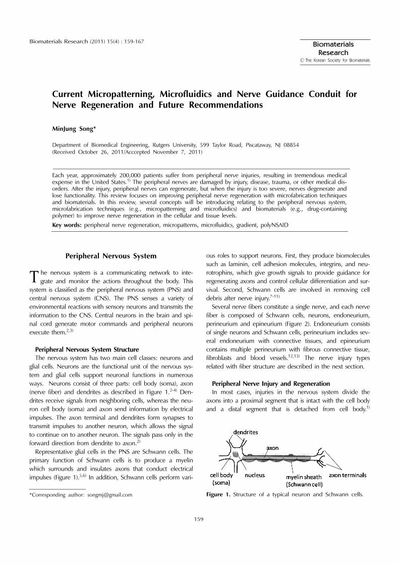
ways. Neurons consist of three parts: cell body (soma), axon
(nerve fiber) and dendrites as described in Figure 1.2-4) Den-
drites receive signals from neighboring cells, whereas the neu-
ron cell body (soma) and axon send information by electrical
impulses. The axon terminal and dendrites form synapses to
transmit impulses to another neuron, which allows the signal
to continue on to another neuron. The signals pass only in the
forward direction from dendrite to axon.2)
Representative glial cells in the PNS are Schwann cells. The
primary function of Schwann cells is to produce a myelin
which surrounds and insulates axons that conduct electrical
impulses (Figure 1).5,6) In addition, Schwann cells perform vari-
ous roles to support neurons. First, they produce biomolecules
such as laminin, cell adhesion molecules, integrins, and neu-
rotrophins, which give growth signals to provide guidance for
regenerating axons and control cellular differentiation and sur-
vival. Second, Schwann cells are involved in removing cell
debris after nerve injury.7-11)
Several nerve fibers constitute a single nerve, and each nerve
fiber is composed of Schwann cells, neurons, endoneurium,
perineurium and epineurium (Figure 2). Endoneurium consists
of single neurons and Schwann cells, perineurium includes sev-
eral endoneurium with connective tissues, and epineurium
contains multiple perineurium with fibrous connective tissue,
fibroblasts and blood vessels.12,13) The nerve injury types
related with fiber structure are described in the next section.
Peripheral Nerve Injury and Regeneration In most cases, injuries in the nervous system divide the
axons into a proximal segment that is intact with the cell body
and a distal segment that is detached from cell body.1)
T
*Corresponding author: [email protected] Figure 1. Structure of a typical neuron and Schwann cells.

160 MinJung Song
Biomaterials Research 2011
Sunderland classified nerve injury into five degrees based on
nerve histologic structure changes (Figure 3).12) First degree
injury (1) is the mildest one that involves local ion-induced
conduction blockage at the injury site with possible segmental
demyelination, a degenerative process removing the myelin
sheath that normally protects nerve fibers. Second degree injury
(2) is a complete interruption of nerve axon and surrounding
myelin, while the surrounding endoneurial sheath is preserved.
In third and fourth degree injuries (3 and 4), nerves lose the
continiuity of the endoneurium and perineurium, respectively,
with the disruption of the axon, but with an intact epineurium.
Fifth degree injury (5) is the complete loss of continuity of the
epineurium. The lack of surrounding extracellular protein
connections ensure that nerve regeneration will not occur.
In the first and second degree injuries, if the environment
allows, the neuron attempts to repair the axon by making new
structural proteins, this process is called axonal reaction.14)
Peripheral nerve regeneration processes are described in sev-
eral reviews.2,14,15) In specific, nerve regeneration initiates
when growth factors, adhesion molecules and structural com-
ponents arrive at the injury site for axonal elongation. The
proximal stump of the damaged axon develops sprouts and
the Schwann cells in the distal stump of the nerve proliferate
and form routes along the course previously taken by the
axons. Axon sprouts elongate and grow along the path of the
original nerve to the distal stump if this route is available.
Growth cones of the sprouting axons find their way along the
routes of Schwann cells and eventually reinnervate the original
peripheral target structures. Once they return to their targets,
the regenerated axons can form new functional nerve endings.
At the distal segments in injured nerve. A Wallerian degen-
eration which is the process of degeneration at the distal part
of neurons, occurs.16,17) Axon degeneration and disintegration
from calcium influx and axonal proteases initiated a Wallerian
degeneration. As a response of axonal breakdown, the myelin
sheath produced by Schwann cells degenerates. Degeneration
products and other debris are removed by phagocytic cells and
macrophages.14,18) In the case of Sunderland’s third, fourth and
fifth degree injuries (Figure 3 - 3, 4, 5), a Wallerian degeneration
occurs at the distal segments. In the third degree injury, axons
undergo a distal Wallerian degeneration and the endoneurium
is disconnected. In the fourth degree injury, all portions of the
nerve are disrupted except the epineurium and the fifth degree
injury is the complete severance of the nerve trunk. In these
injury degrees, a functional recovery is rarely obtained in a
natural environment. This article will focus on the third, fourth
and fifth degree injury types.
Improving Nerve Regeneration at The CellLevel with Micropatterned Surfaces
Micropatterning is a method to create patterns on both
organic and inorganic surfaces that can provide physical and/or
chemical cues for cellular guidance. Various cell types including
osteoblast, peripheral and central neurons, smooth muscle cells
and endothelial cells have been succesfully guided on micropat-
terned surfaces.19-23) Neural cells are particularly sensitive to their
geometrical environment, and guidance cues are really impor-
tant in neural cell growth. Therefore, micropatterned surfaces
have been extensively used in nerve regeneration studies.24-33)
Figure 2. Nerve bundle cross-section structure comprised of neu-ron, Schwann cells and endoneurium, perineurium and epineu-rium.
Figure 3. Sunderland’s five nerve injury classification from firstdegree to fifth degree injury.

Biomaterials and microfabrication for nerve regeneration 161
Vol. 15, No. 4
Micropatterning Methods Micropatterned surfaces are generated using various methods
including photolithography, microcontact printing (µCP), microf-
luidic patterning,34) micromolding in capillaries, stencil pattern-
ing and microscale plasma-initiated patterning (µPIP).35-38) Pho-
tolithography uses a projection-printing system in which light
passes through a photomask and selectively exposes a spin-
coated photoresist (photosensitive polymer). Following develop-
ment, this method generates a patterned photoresist.37,38) This
method has the advantage of generating patterns at the nanos-
cale level, whereas it has many disadvantages in its application
to biology; photolithography requires an “expensive” clean-
room facility and the processing are not necessary amenable
to biological systems.36,37,39)
Microcontact printing (µCP) (Figure 4) and plasma-initiated
patterning (µPIP) (Figure 5) methods utilize photolithography
to generate masters, but the micropatterns are then generated
with soft lithography. Microcontact printing utilizes poly
(dimethylsiloxane) (PDMS) stamp to transfer biomolecules to
the more hydrophilic surfaces (Figure 4).27,40) This method is
simple and does not require expensive or complex instruments.
The µCP method was used to generate micropatterns on
inorganic surfaces such as glass.24,41) Further studies generated
micropatterns on organic surfaces (e.g., polymers), which in-
creased applications of patterned surfaces.27,42) Generating
micropatterns on polymer surfaces is unique because polymers
can be implanted following patterning. µCP has many advan-
tages,26-28) but a few issues regarding regarding biomolecule
transferring limit further applications. Because biomolecule
transfer depends on hydrophilic differences between the
PDMS stamp and polymer surfaces, two major issues are: (i)
ink affinity to the PDMS is sometimes too strong to transfer
proteins from the PDMS stamp to the surface;43) and (ii) the
aqueous solution containing the biomolecules may dry up.39)
The µPIP method was developed in our laboratory by selec-
tively exposing surfaces to oxygen plasma to temporarily pro-
mote hydrophilicity (Figure 5).39) This method is relatively
simple and generates patterns consistently and reproducibly. In
addition, it allows complicated or gradient protein micropat-
terns to be generated. This article will describe both microcon-
tact printing and microscale plasma-initiated methods to pattern
protein on biocompatible polymer surfaces.
Micropatterning and Nerve RegenerationExtracellular matrix and oriented tissue structures influence
cell migration and neural cell guidance during nerve regenera-
tion and development,3,44,45) Micropatterned surfaces may
mimic the in vivo microenvironment system at in vitro level by
Figure 4. Microcontact printing method (µCP) to generate micro-patterns on PMMA surfaces.
Figure 6. Connecting nerve injured site with a nerve guidanceconduit.
Figure 5. Microscale plasma initiated patterning (µPIP) methodto generate micropatterns on PMMA surfaces.

162 MinJung Song
Biomaterials Research 2011
providing guidance cues (i.e., chemical, physical and biological
cues). Neurons or glial cells recognize, adhere or extend based
on these guidance cues. Micropatterned surfaces provide chem-
ical and physical cues for neural cell guidance, thus, it has
been extensively applied in nerve regeneration study.24-33)
Dorsal root ganglia (DRG) neurons and Schwann cells pref-
erentially adhered to, and subsequently aligned on, laminin
micropatterned polymer26,27,46) and glass24,31,41) surfaces. The
studies demonstrate that neural cell behavior can be controlled
with micropatterning. Aligned Schwann cell surfaces can be fur-
ther used to guide neurons as a biological cue.25,28) Protein
micropatterned surfaces are also utilized to generate central
neuron guidance, for example, brain stem neurons and hippoc-
ampal neurons form the neural network.30,32,47-49) Furthermore,
the patterned surfaces successfully guide neural progenitor cells
and show the possibility of the surface applications onto
progenitor cell differentiation.45)
Nerve Regeneration at The Cell Level with Microfluidics
Microfluidics is a newly developed technique that has many
applications in biological fields such as tissue engineering,50)
biological and cell-based assays.51) This micro- level system uti-
lizes a small amount of liquid, reagents and cells in short reac-
tion times, at low cost and power. Therefore, multiple biological
assays can be conducted, and many parallel operations are pos-
sible.51-54) Microfluidic applications have been reviewed in cel-
lular biology including immunoassay, protein and DNA
separation, cell sorting and manipulation.51,53)
Another primary advantage of microfluidics is the ability to
create gradients. Many cell types respond to a molecular or
extracellular matrix gradient, and the microfluidic system enables
the systemic analysis of cell-biomolecular gradient interac-
tions.51) Epithelial cells,55) neutrophils56-58) endothelial cell59)
and neurons51,54,60) respond to molecular or extracellular gra-
dients. As a chemical gradient can be the guidance cue to
neurons, a gradient is closely related to nerve regeneration.
Many studies have demonstrated that neuron growth cone can
be guided by gradient both in vitro44,61,62) and in vivo.63)
Gradient Generation via MicrofluidicsIn microfluidic systems, most fluids undergo laminar flow
because the channel diameter is relatively small. Fluid ten-
dency is described with Reynolds number (Re = dρυ/µ) when
d is the channel diameter and ρ, υ, µ are the density, velocity
and viscosity of fluid, respectively. Laminar flow is a fluid
stream (Re << 2000) and turbulent flow is chaotic and unpre-
dictable (Re >> 2000).36,52) In most cases, the channel diame-
ter (d) is less than 500 µm and flow rate (υ) is slow such that
the flow is typically laminar with Re values, around 0.1~ 1.53)
When two or more laminar flow streams from independent
inlets join at a single stream, the combined streams flow paral-
lel. Mixing of streams occurs only by diffusion across the inter-
face, which produces gradients in microfluidic systems.52,54,64)
Diffusion is the process in which molecules spread from areas
of high concentration to areas of low concentration to create
a concentration gradient. Therefore, gradients created by
microfluidics are well defined.52,54)
Microfluidics and Nerve RegenerationGradients of substrate-bound substances (haptotaxis), mech-
anical rigidity (durotaxis) or diffusible substances (chemotaxis)
play an important role in neuron guidance during nervous
system development.3,61-63) Axons reach their target by re-
sponding to repulsive or attractive molecular cues and follow
the molecule gradient.65,66) For example, semaphorin, netrins
and slits are neuron-attractive molecules and ephrin is a
neuron-repulsive molecule.66,67) Because the molecular gradient
is known as the main guidance cue to neurons during devel-
opment,3) a molecular gradient in nerve regeneration may be
potentially significant. Therefore, the microfluidic system is an
attractive tool for nerve regeneration. In recent studies,
microfluidic systems successfully generated diverse biomolecular
gradients and maintained the constant gradient over long
periods of time; the gradient systems were then used to
manipulate cellular microenvironments.51,54,55,58-60)
The influence of a diffusible substance gradient (i.e., chemo-
taxis) has also been extensively studied and used to elucidate
the molecular and cellular mechanisms of axon guidance.
However, neuron chemotaxis by microfluidics has not been
reported. By comparison, haptotaxis has been investigated; it is
the cells motility or outgrowth guided by a gradient of mem-
brane-bound ligand. A laminin-based protein gradient was gen-
erated with microfluidics and studied for haptotatic influence
on neuron guidance.60) In addition, axon outgrowth can be
guided by a gradient of rigidity (durotaxis).68,69) The rigid gradi-
ent is established by controlling the cross-linking degree on a
substrate. Lo et al., showed cell migration on collagen-coated
polyacrylamide substrates with rigidity gradients.70) In addition,
the rate of neurite extension was correlated to the mechanical
stiffness of agarose gels.69) No study has yet shown durotatic
generation through microfluidics. This article also focuses on
generating an adhesive peptide bound gradient in a three-
dimensional cell culture system using microfluidics.
Nerve Regeneration in Tissue byNerve Guidance Conduit
When the nerve injury become severe to the level of
extracellular component disconnection, (Sunderland’s injury
degree III, IV and V), implanting a nerve guidance conduit is
necessary to reconnect the injured site. By bridging a nerve
defect region with a guidance channel, improved nerve

Biomaterials and microfabrication for nerve regeneration 163
Vol. 15, No. 4
regeneration is expected in the peripheral nervous system
(Figure 6).14) As a nerve guidance channel, the autologous nerve
graft is considered the gold standard.71,72) The autologous nerve
tissue contains growth factors and cytokines for regeneration,
without a foreign body response. However, autologous nerve
graft supply is a significant problem; because a healthy nerve is
utilized, a second surgery is required and causes additional
injury.
Various natural-based and synthetic materials have been
synthesized or modified as alternative nerve guidance systems.
To be a nerve guidance conduit, materials should possess
specific properties: (i) biocompatible; (ii) biodegradable, is other-
wise, the remaining graft must be removed from the injury site;
(iii) retain proper mechanical properties, they must strong
enough to resist collapse during implantation and regeneration
yet be flexible enough to handle; (iv) easily fabricated and
modified with desired dimensions; and (v) sterilizable and tear-
resistant.14,73)
Collagen and laminin as natural-based materials, and poly
(glycolic acid), poly (D, L-lactide-co-glycolide) (PLGA) and poly
(lactide-co-caprolactone) as synthetic materials are current
examples of nerve guidance materials.74-77) Particularly, PLGA
is biodegradable and approved by a Food and Drug Adminis-
tration (FDA) for implantation. All materials demonstrated
improved nerve regeneration upon grafting, however full func-
tional recovery is limited compared to the autologous
grafting.74,78,79) One of the main reasons for limited regener-
ation is inflammation at the nerve injury site. This article will
discuss this issue and describe novel materials to address the
current inflammation issue.
Drug-based Poly(anhydride-esters)Poly (anhydride-ester) is a biodegradable polymer that has
been applied to drug delivery, tissue engineering and medical
devices.80) Previously, Erdmann et al., synthesized and charac-
terized salicylic acid-based poly (anhydride-esters) (1).81) This
polymer (1) is unique in that the drugs are incorporated into
the polymer backbone, not attached as a side group or phys-
ically admixing (Figure 7). These polymers (1) degraded into
salicylic acid (2) and sebacic acid (3) upon hydrolysis due to
the instability of the anhydride and ester bonds. Salicylic acid
(2) is generated upon hydrolysis of acetylsalicylic acid (aspirin)
and sebacic acid (3) is the linker group that connects the sali-
cylic acid unit. Other non-steroidal anti-inflammatory drugs
(NSAIDs) such as thiosalicylic acid, diflunisal and salicylsalicylic
acid were also incorporated into polymer backbone82-85) and
different types of linker groups, (e.g., diethylmalonic and adi-
pic acid), have been used to connect the drug molecules.84)
From the unique properties described above, these poly
(anhydride-esters) have many advantages. First, the polymer
contains high drug amounts (e.g., 62 wt% of polymer 1 is sali-
cylic acid). Second, polymer properties such as drug release
rate, and melting temperature can be regulated by controlling
the polymer structure.84) Third, the polymer is biocompatible
and the degradation products are nontoxic.83) These polymers
have shown reduced inflammation, inhibition of bone resorp-
tion, prevented bacterial contamination and reduced foreign-
body responses.86-88) Based upon these characteristics, the
drug-based poly(anhydride-ester) is a good candidate as a
nerve guidance material. This article will further discuss the
possibility of applying this polymer to nerve guidance conduit.
Future Work and Recommendations
Micropatterned Surfaces in Nerve RegenerationWe have developed protein micropatterning systems to
guide neural cells on biocompatible polymer substrates. For in
Figure 7. Hydrolytic degradation of poly(anhydride ester) (1) into salicylic acid (2) and sebacic acid (3).
Figure 8. Protein micropatterned surfaces can be rolled up intoconduits.

164 MinJung Song
Biomaterials Research 2011
vivo use, generating micropatterns and guiding cells on biode-
gradable polymer drug substrates such as salicylic acid-based
poly(anhydride-esters) should be attempted. Micropatterned
surfaces in biodegradable polymer can be rolled up as a con-
duit shape to implant in animals (Figure 8). As this conduit
contains an inner surface with laminin micropatterns, improved
nerve regeneration is expected.
In stem cells, physical cues as well as biomolecular cues play
important roles in cell differentiation.89) As micropatterned
surfaces can provide physical and biomolecular cues, studying
stem cell differentiation with these cues will be an interesting
topic. Figure 6.2 shows embryonic stem cells on laminin micro-
patterned surfaces; the cells recognized and aligned on
patterns, indicating that a patterned surface may influence a
stem cell differentiation (Figure 9). Differentiated stem cells will
bring various potential therapies methods for nerve regenera-
tion.90,91)
Microfluidic Applications in Nerve RegenerationWe have successfully generated an adhesive gradient in three
dimensions. Neuron guidance by the gradient can be assessed
after immunostaining and image analysis. Simple measurement
of neurite movement, either positive or negative to the gradi-
ent, will describe the impact of the adhesive gradient (Figure
10). As a future study, gradients of neuron-specific peptides
such as YIGSR and IKVAV can be generated with a current
microfluidic system, then their influence on neuron guidance
elucidated in vitro.
To use the biomolecular gradient collagen gel as a biomate-
rial in vivo, detaching the gel from a PDMS network remains
as an issue. To perform this function, a freeze-drying method
may be utilized. After the gradient is generated in a collagen
gel with an open microfluidic system, the gel may be frozen at
−20oC (Figure 11). After freeze-drying, the collagen gel may
be detached from the network.
Nerve Guidance ConduitsNerve guidance conduits can be fabricated as described in
Figure 12. For example, SAA polymer can be used as an outer
layer to reduce acute inflammation and an inner layer based
upon diflunisal polymer can reduce a chronic inflammation
upon implantation. Filling the current hollow conduit with col-
lagen gel may further improve nerve regeneration.92)
Iodinated salicylate-based poly(anhydride-esters) (SAA) can
be a novel material for nerve guidance conduits. Iodinated SAA
was synthesized in our laboratory (Figure 13), and showed X-
ray opacity in clinical X-ray techniques with good biocom-
Figure 9. Embryonic stem cell guidance on micropatterned sur-faces on day 3.
Figure 10. Neuron guidance analysis to a biomolecular gradient(negative or positive).
Figure 11. A gradient collagen gel detachment from the PDMSnetwork using freeze-drying method.
Figure 12. Further modified nerve guidance conduit consistingof salicylic acid- and diflunisal-based polymers.
Figure 13. Iodinated salicylate-based poly(anhydride-ester) struc-tures.93)

Biomaterials and microfabrication for nerve regeneration 165
Vol. 15, No. 4
patibility.93) If the nerve guidance conduit is fabricated with the
polymer and implanted in vivo, observing regeneration pro-
cesses can be easily monitored without sacrificing animals.
References
1. Yannas, I.V., Hill, B.J., “Selection of biomaterials for peripheralnerve regeneration using data from the nerve chamber model,”Biomaterials. 25, 1593-1600 (2004).
2. Willis, W.D., The Nervous System. in Physiology, Berne, R.M.,Levy, M.N., Koeppen, B.M. and Stanton, B.A. (ed) St. Louis: Mosby,1998, pp. 81-226.
3. Thomas M, J., Sanes., J.R., The Development of the Nervous Sys-tem. in Principles of Neural Science, Kandel, E.R., Schwartz, J.H.and Jessell, T.M.(ed). by New York: McGraw-Hill Health Profes-sions Division, 2000, pp. 1019-1161.
4. Guyton, A.C., Hall, J.E., Textbook of Medical Physiology. 10th edn(Philadelphia: Saunders, 2000), pp. xxxii, 1064.
5. Bhatheja, K., Field, J., “Schwann cells: origins and role in axonalmaintenance and regeneration,” Int J Biochem Cell Biol. 38,1995-1999 (2006).
6. Bunge, R.P., “The role of the Schwann cell in trophic support andregeneration,” J Neurol. 242, S19-21 (1994).
7. Mirsky, R., Jessen, K.R., Brennan, A., Parkinson, D., Dong, Z.,Meier, C., Parmantier, E., Lawson, D., “Schwann cells as regulatorsof nerve development,” J Physiol Paris. 96, 17-24 (2002).
8. Ard, M.D., Bunge, R.P., Bunge, M.B., “Comparison of the Schwanncell surface and Schwann cell extracellular matrix as promoters ofneurite growth,” J Neurocytol. 16, 539-555 (1987).
9. Bunge, M.B., Williams, A.K., Wood, P.M., Uitto, J., Jeffrey, J.J.,“Comparison of nerve cell and nerve cell plus Schwann cell cul-tures, with particular emphasis on basal lamina and collagen forma-tion,” J Cell Biol. 84, 184-202 (1980).
10. Jessen, K.R., Mirsky, R., “Schwann cells and their precursors emergeas major regulators of nerve development,” Trends Neurosci. 22,402-410 (1999).
11. Morrissey, T.K., Kleitman, N., Bunge, R.P., “Isolation and functionalcharacterization of Schwann cells derived from adult peripheralnerve,” J Neurosci. 11, 2433-2442 (1991).
12. Sunderland, S., “The anatomy and physiology of nerve injury,”Muscle Nerve. 13, 771-784 (1990).
13. Flores, A.J., Lavernia, C.J., Owens, P.W., “Anatomy and physiologyof peripheral nerve injury and repair,” Am J Orthop. 29, 167-173(2000).
14. Schmidt, C.E., Leach, J.B., “Neural tissue engineering: strategies forrepair and regeneration,” Annu Rev Biomed Eng. 5, 293-347(2003).
15. Makwana, M., Raivich, G., “Molecular mechanisms in successfulperipheral regeneration,” FEBS J. 272, 2628-2638 (2005).
16. Johnson, E.O., Zoubos, A.B., Soucacos, P.N., “Regeneration andrepair of peripheral nerves,” Injury. 36 Suppl 4, S24-29 (2005).
17. Richardson, P.M., Lu, X., “Inflammation and axonal regenera-tion,” J Neurol. 242, S57-60 (1994).
18. Stoll, G., Jander, S., Myers, R.R., “Degeneration and regenera-tion of the peripheral nervous system: from Augustus Waller'sobservations to neuroinflammation,” J Peripher Nerv Syst. 7, 13-27(2002).
19. Hasenbein, M.E., Andersen, T.T., Bizios, R., “Micropatterned sur-faces modified with select peptides promote exclusive interactionswith osteoblasts,” Biomaterials. 23, 3937-3942 (2002).
20. Kaji, H., Takii, Y., Nishizawa, M., Matsue, T.,“Pharmacological char-
acterization of micropatterned cardiac myocytes,” Biomaterials. 24,4239-4244 (2003).
21. Kulkarni, S.S., Orth, R., Ferrari, M., Moldovan, N.I., “Micropattern-ing of endothelial cells by guided stimulation with angiogenic fac-tors,” Biosens Bioelectron. 19, 1401-1407 (2004).
22. Thakar, R.G., Ho, F., Huang, N.F., Liepmann, D., Li, S., “Regula-tion of vascular smooth muscle cells by micropatterning,” Bio-chem. Biophys. Res. Commun. 307, 883-890 (2003).
23. Glawe, J.D., Hill, J.B., Mills, D.K., McShane, M.J., “Influence ofchannel width on alignment of smooth muscle cells by high-aspect-ratio microfabricated elastomeric cell culture scaffolds,” J.Biomed. Mater. Res. A. 75, 106-114 (2005).
24. Thompson, D.M., Buettner, H.M., “Schwann cell response tomicropatterned laminin surfaces,” Tissue Eng. 7, 247-265 (2001).
25. Thompson, D.M., Buettner, H.M., “Oriented Schwann cell mono-layers for directed neurite outgrowth,” Ann Biomed Eng. 32, 1120-1130 (2004).
26. Schmalenberg, K.E., Uhrich, K.E., “Micropatterned polymer sub-strates control alignment of proliferating Schwann cells to directneuronal regeneration,” Biomaterials. 26, 1423-1430 (2005).
27. Schmalenberg, K.E., Buettner, H.M., Uhrich, K.E., “Microcontactprinting of proteins on oxygen plasma-activated poly(methyl meth-acrylate),” Biomaterials. 25, 1851-1857 (2004).
28. Miller, C., Jeftinija, S., Mallapragada, S., “Micropatterned Schwanncell-seeded biodegradable polymer substrates significantly enhanceneurite alignment and outgrowth,” Tissue Eng. 7, 705-715 (2001).
29. Miller, C., Shanks, H., Witt, A., Rutkowski, G., Mallapragada, S.,“Oriented Schwann cell growth on micropatterned biodegrad-able polymer substrates,” Biomaterials. 22, 1263-1269 (2001).
30. Chang, J.C., Brewer, G.J., Wheeler, B.C., “A modified micros-tamping technique enhances polylysine transfer and neuronalcell patterning,” Biomaterials. 24, 2863-2870 (2003).
31. Tai, H.C., Buettner, H.M., “Neurite outgrowth and growth conemorphology on micropatterned surfaces,” Biotechnol Prog. 14,364-370 (1998).
32. Vogt, A.K., Stefani, F.D., Best, A., Nelles, G., Yasuda, A., Knoll,W., Offenhausser, A., “Impact of micropatterned surfaces on neu-ronal polarity,” J Neurosci Methods. 134, 191-198 (2004).
33. Oliva, A.A., Jr., James, C.D., Kingman, C.E., Craighead, H.G.,Banker, G.A., “Patterning axonal guidance molecules using a novelstrategy for microcontact printing,” Neurochem Res. 28, 1639-1648 (2003).
34. Tan, W., Desai, T.A., “Microfluidic patterning of cells in extracellu-lar matrix biopolymers: effects of channel size, cell type, and matrixcomposition on pattern integrity,” Tissue Eng. 9, 255-267 (2003).
35. Park, T.H., Shuler, M.L., “Integration of cell culture and microfabri-cation technology,” Biotechnol Prog. 19, 243-253 (2003).
36. Whitesides, G.M., Ostuni, E., Takayama, S., Jiang, X., Ingber, D.E.,“Soft lithography in biology and biochemistry,” Annu Rev BiomedEng. 3, 335-373 (2001).
37. Xia, Y.W., George M., “Soft Lithography,” Angew. Chem. Int. Ed.37, 550-575 (1998).
38. Schmalenberg, K.E., Thompson, D.M., H.M., B., K.E., U., L.F.,G., “In situ stepwise surface analysis of micropatterned glass sub-strates in liquids using functional near-field scanning opticalmicroscopy,” Langmuir. 18, 8593-8600 (2002).
39. Langowski, B.A., Uhrich, K.E., “Microscale plasma-initiated pat-terning (µPIP),” Langmuir. 21, 10509-10514 (2005).
40. Langowski, B.A., Uhrich, K.E., “Oxygen plasma-treatment effectson Si transfer,” Langmuir. 21, 6366-6372 (2005).
41. Clark, P., Britland, S., Connolly, P., “Growth cone guidance andneuron morphology on micropatterned laminin surfaces,” J Cell

166 MinJung Song
Biomaterials Research 2011
Sci. 105 ( Pt 1), 203-212 (1993).42. Csucs, G., Michel, R., Lussi, J.W., Textor, M., Danuser, G., “Micro-
contact printing of novel co-polymers in combination with pro-teins for cell-biological applications,” Biomaterials. 24, 1713-1720(2003).
43. Tan, J.T., J; Chen CS, “Microcontact printing of proteins on mixedself-assembled monolayers,” Langmuir. 18, 519-523 (2002).
44. Gurdon, J.B., Bourillot, P.Y., “Morphogen gradient interpretation,”Nature. 413, 797-803 (2001).
45. Recknor, J.B., Sakaguchi, D.S., Mallapragada, S.K., “Directedgrowth and selective differentiation of neural progenitor cells onmicropatterned polymer substrates,” Biomaterials. 27, 4098-4108 (2006).
46. Song, H.K., Toste, B., Ahmann, K., Hoffman-Kim, D., Palmore,G.T., “Micropatterns of positive guidance cues anchored to poly-pyrrole doped with polyglutamic acid: a new platform for charac-terizing neurite extension in complex environments,” Biomaterials.27, 473-484 (2006).
47. Kam, L., Shain, W., Turner, J.N., Bizios, R., “Axonal outgrowth ofhippocampal neurons on micro-scale networks of polylysine-con-jugated laminin,” Biomaterials. 22, 1049-1054 (2001).
48. Saneinejad, S., Shoichet, M.S., “Patterned glass surfaces direct celladhesion and process outgrowth of primary neurons of the cen-tral nervous system,” J Biomed Mater Res. 42, 13-19 (1998).
49. Lauer, L., Vogt, A., Yeung, C.K., Knoll, W., Offenhausser, A., “Elec-trophysiological recordings of patterned rat brain stem slice neu-rons,” Biomaterials. 23, 3123-3130 (2002).
50. Khademhosseini, A., Langer, R., Borenstein, J., Vacanti, J.P.,“Microscale technologies for tissue engineering and biology,”Proc Natl Acad Sci U S A. 103, 2480-2487 (2006).
51. Sia, S.K., Whitesides, G.M., “Microfluidic devices fabricated inpoly(dimethylsiloxane) for biological studies,” Electrophoresis. 24,3563-3576 (2003).
52. Beebe, D.J., Mensing, G.A., Walker, G.M., “Physics and applica-tions of microfluidics in biology,” Annu Rev Biomed Eng. 4, 261-286 (2002).
53. Kenis, P.J., Ismagilov, R.F., Whitesides, G.M., “Microfabricationinside capillaries using multiphase laminar flow patterning,” Sci-ence. 285, 83-85 (1999).
54. Dertinger, S.K., Chiu, D.L., Li Jeon, N., Whitesides, G.M., “Gener-ation of gradients having complex shapes using microfluidic net-works,” Anal. Chem. 73, 1240-1246 (2001).
55. Gunawan, R.C., Choban, E.R., Conour, J.E., Silvestre, J., Schook,L.B., Gaskins, H.R., Leckband, D.E., Kenis, P.J., “Regiospecific con-trol of protein expression in cells cultured on two-componentcounter gradients of extracellular matrix proteins,” Langmuir. 21,3061-3068 (2005).
56. Lin, F., Nguyen, C.M., Wang, S.J., Saadi, W., Gross, S.P., Jeon, N.L.,“Effective neutrophil chemotaxis is strongly influenced by mean IL-8 concentration,” Biochem Biophys Res Commun. 319, 576-581(2004).
57. Lin, F., Nguyen, C.M., Wang, S.J., Saadi, W., Gross, S.P., Jeon, N.L.,“Neutrophil migration in opposing chemoattractant gradients usingmicrofluidic chemotaxis devices,” Ann Biomed Eng. 33, 475-482(2005).
58. Li Jeon, N., Baskaran, H., Dertinger, S.K., Whitesides, G.M., Van deWater, L., Toner, M., “Neutrophil chemotaxis in linear and com-plex gradients of interleukin-8 formed in a microfabricated device,”Nat Biotechnol. 20, 826-830 (2002).
59. Burdick, J.A., Khademhosseini, A., Langer, R., “Fabrication of gradi-ent hydrogels using a microfluidics/photopolymerization process,”Langmuir. 20, 5153-5156 (2004).
60. Dertinger, S.K., Jiang, X., Li, Z., Murthy, V.N., Whitesides, G.M.,“Gradients of substrate-bound laminin orient axonal specificationof neurons,” Proc Natl Acad Sci USA. 99, 12542-12547 (2002).
61. Messersmith, E.K., Leonardo, E.D., Shatz, C.J., Tessier-Lavigne, M.,Goodman, C.S., Kolodkin, A.L., “Semaphorin III can function as aselective chemorepellent to pattern sensory projections in the spi-nal cord,” Neuron. 14, 949-959 (1995).
62. Ming, G.L., Wong, S.T., Henley, J., Yuan, X.B., Song, H.J., Spitzer,N.C., Poo, M.M., “Adaptation in the chemotactic guidance ofnerve growth cones,” Nature. 417, 411-418 (2002).
63. Isbister, C.M., Mackenzie, P.J., To, K.C., O'Connor, T.P., “Gradientsteepness influences the pathfinding decisions of neuronal growthcones in vivo,” J Neurosci. 23, 193-202 (2003).
64. Atencia, J., Beebe, D.J., “Controlled microfluidic interfaces,”Nature. 437, 648-655 (2005).
65. Song, H., Poo, M., “The cell biology of neuronal navigation,” NatCell Biol. 3, E81-88 (2001).
66. Dickson, B.J., “Molecular mechanisms of axon guidance,” Sci-ence. 298, 1959-1964 (2002).
67. von Philipsborn, A.C., Lang, S., Loeschinger, J., Bernard, A., David,C., Lehnert, D., Bonhoeffer, F., Bastmeyer, M., “Growth cone nav-igation in substrate-bound ephrin gradients,” Development. 133,2487-2495 (2006).
68. Dubey, N., Letourneau, P.C., Tranquillo, R.T., “Neuronal contactguidance in magnetically aligned fibrin gels: effect of variation ingel mechano-structural properties,” Biomaterials. 22, 1065-1075(2001).
69. Balgude, A.P., Yu, X., Szymanski, A., Bellamkonda, R.V., “Agar-ose gel stiffness determines rate of DRG neurite extension in 3Dcultures,” Biomaterials. 22, 1077-1084 (2001).
70. Lo, C.M., Wang, H.B., Dembo, M., Wang, Y.L., “Cell move-ment is guided by the rigidity of the substrate,” Biophys J. 79,144-152 (2000).
71. Brunelli, G.A., Brunelli, G.R., “Restoration of walking in paraple-gia by transferring the ulnar nerve to the hip: A report on thefirst patient,” Microsurgery. 19, 223-226 (1999).
72. Carlstedt T, G.P., Hallin RG, Noren G, “Return of function afterspinal cord implantation of avulsed spinal nerve roots,” Lancet.346, 1323-1325 (1995).
73. Kannan, R.Y., Salacinski, H.J., Butler, P.E., Seifalian, A.M., “Artificialnerve conduits in peripheral-nerve repair,” Biotechnol Appl Bio-chem. 41, 193-200 (2005).
74. Evans, G.R., Brandt, K., Katz, S., Chauvin, P., Otto, L., Bogle, M.,Wang, B., Meszlenyi, R.K., Lu, L., Mikos, A.G., Patrick, C.W., Jr.,“Bioactive poly(L-lactic acid) conduits seeded with Schwann cellsfor peripheral nerve regeneration,” Biomaterials. 23, 841-848(2002).
75. Ceballos, D., Navarro, X., Dubey, N., Wendelschafer-Crabb, G.,Kennedy, W.R., Tranquillo, R.T., “Magnetically aligned collagen gelfilling a collagen nerve guide improves peripheral nerve regenera-tion,” Exp Neurol. 158, 290-300 (1999).
76. Hadlock, T., Sundback, C., Hunter, D., Cheney, M., Vacanti, J.P.,“A polymer foam conduit seeded with Schwann cells promotesguided peripheral nerve regeneration,” Tissue Eng. 6, 119-127(2000).
77. Taras, J.S., Nanavati, V., Steelman, P., “Nerve conduits,” J HandTher. 18, 191-197 (2005).
78. Evans, G.R., Brandt, K., Niederbichler, A.D., Chauvin, P., Her-rman, S., Bogle, M., Otta, L., Wang, B., Patrick, C.W., Jr., “Clinicallong-term in vivo evaluation of poly(L-lactic acid) porous conduitsfor peripheral nerve regeneration,” J Biomater Sci Polym Ed. 11,869-878 (2000).

Biomaterials and microfabrication for nerve regeneration 167
Vol. 15, No. 4
79. Yoshii, S., Oka, M., Shima, M., Taniguchi, A., Akagi, M., “Bridginga 30-mm nerve defect using collagen filaments,” J Biomed MaterRes A. 67, 467-474 (2003).
80. Kumar, N., Langer, R.S., Domb, A.J., “Polyanhydrides: an over-view,” Adv Drug Deliv Rev. 54, 889-910 (2002).
81. Erdmann, L., Uhrich, K.E., “Synthesis and degradation characteris-tics of salicylic acid-derived poly(anhydride-esters),” Biomaterials.21, 1941-1946 (2000).
82. Schmeltzer, R.A., T.; Uhrich, K, “Optimized synthesis of salicylate-based poly(anhydride-esters),” Polymer Bulletin. 49, 441-448 (2003).
83. Schmeltzer, R.C., Schmalenberg, K.E., Uhrich, K.E., “Synthesis andcytotoxicity of salicylate-based poly(anhydride esters),” Biomacro-molecules. 6, 359-367 (2005).
84. Prudencio, A., Schmeltzer, R. C., Uhrich K. E., “Effect of linkerstructure on salicylic acid-derived poly(anhydride-esters),” Macro-molecules. 38, 6895-6901 (2005).
85. Schmeltzer, R.C., Uhrich, K.E., “Synthesis and characterization ofsalicylic acid-based poly(anhydride-ester) copolymers,” J of Bioac-tive Compatible Poly. 21, 123-133 (2006).
86. Erdmann, L., Macedo, B., Uhrich, K.E., “Degradable poly(anhy-dride ester) implants: effects of localized salicylic acid release onbone,” Biomaterials. 21, 2507-2512 (2000).
87. Bryers, J.D., Jarvis, R.A., Lebo, J., Prudencio, A., Kyriakides, T.R.,
Uhrich, K.E., “Biodegradation of poly(anhydride-esters) into non-steroidal anti-inflammatory drugs and their effect on Pseudomo-nas aeruginosa biofilms in vitro and on the foreign-body responsein vivo,” Biomaterials. 27, 5039-5048 (2006).
88. Harten, R.D., Svach, D.J., Schmeltzer, R., Uhrich, K.E., “Salicylicacid-derived poly(anhydride-esters) inhibit bone resorption andformation in vivo,” J. Biomed. Mater. Res. A. 72, 354-362 (2005).
89. Philp, D., Chen, S.S., Fitzgerald, W., Orenstein, J., Margolis, L.,Kleinman, H.K., “Complex extracellular matrices promote tissue-specific stem cell differentiation,” Stem Cells. 23, 288-296 (2005).
90. Myckatyn, T.M., Mackinnon, S.E., McDonald, J.W., “Stem celltransplantation and other novel techniques for promoting recoveryfrom spinal cord injury,” Transpl Immunol. 12, 343-358 (2004).
91. Tohill, M., Terenghi, G., “Stem-cell plasticity and therapy for inju-ries of the peripheral nervous system,” Biotechnol Appl Biochem.40, 17-24 (2004).
92. Schmidt, C.E., Leach, J.B., “Neural tissue engineering: strategies forrepair and regeneration,” Annu. Rev. Biomed. Eng. 5, 293-347(2003).
93. Carbone, A.L., Song, M.J., Uhrich, K.E., Melt-condensation andsolution polymerization of iodinated salicylate-based poly(anhy-dride-esters) as radiopaque biomaterials, Biomacromolecules.9(6),1604-12 (2008).


![Insulin-Ameliorated Peripheral Motor Neuropathy in ...by nerve fiber loss, axonal degeneration and segmental demyelination with a slowing of nerve conduction velocity [8]. Rodent models](https://static.fdocuments.in/doc/165x107/5ed9d9a884a4f7018c1d9492/insulin-ameliorated-peripheral-motor-neuropathy-in-by-nerve-fiber-loss-axonal.jpg)


