Current Biology, Vol. 12, 813–824, May 14, 2002, 2002 ... · Summary Stbm, which has yet to be...
12
Current Biology, Vol. 12, 813–824, May 14, 2002, 2002 Elsevier Science Ltd. All rights reserved. PIIS0960-9822(02)00841-2 Asymmetric Localization of Frizzled and the Determination of Notch-Dependent Cell Fate in the Drosophila Eye diverse tissues, such as the eye, wing, leg, and notum. A similar core set may also operate in vertebrates [3]. The core genes include frizzled (fz), which encodes a seven-pass transmembrane protein of the Wnt receptor family, dishevelled (dsh), which encodes a multidomain David Strutt, 1,3 Ruth Johnson, 2 Katherine Cooper, 1 and Sarah Bray 2,3 1 Centre for Developmental Genetics School of Medicine and Biomedical Science University of Sheffield Western Bank, Sheffield S10 2TN cytoplasmic/cortical protein required for Fz signal trans- duction, flamingo (fmi, also known as starry night), which 2 Department of Anatomy University of Cambridge encodes a seven-pass transmembrane cadherin, prickle- spiny-legs (pk-sple), which encodes a LIM domain pro- Downing Street, Cambridge CB2 3DY United Kingdom tein, and strabismus (stbm, also known as Van Gogh), which encodes a predicted four-pass transmembrane protein [2]. The protein products of all these loci (except Stbm, which has yet to be examined) adopt asymmetric Summary subcellular localizations in the pupal wing during cell polarization [4–8]. The product of the diego locus (an Background: During patterning of the Drosophila eye, a critical step is the Notch-mediated cell fate decision ankyrin repeat protein) also adopts an asymmetric local- ization in wing cells and may be part of the core group that determines the identities of the R3/R4 photorecep- tor pair in each ommatidium. Depending on the decision of genes [9]. During patterning of the compound eye, the planar taken, the ommatidium adopts either the dorsal or ven- tral chiral form. This decision is directed by the activity polarity genes ensure the correct orientation of the om- matidial units which make up the facets [10]. Differentia- of the planar polarity genes, and, in particular, higher activity of the receptor Frizzled confers R3 fate. tion begins in the third instar eye disc and proceeds in a wave from posterior to anterior. The front of the wave Results: We present evidence that Frizzled does not modulate Notch activity via Rho GTPases and a JNK is marked by a band of contracted cells known as the morphogenetic furrow. Ommatidia are born behind the cascade as previously proposed. We find that the planar polarity proteins Frizzled, Dishevelled, Flamingo, and furrow in rows polarized in the anteroposterior axis and in about row 6 begin to rotate relative to the dorsoventral Strabismus adopt asymmetric protein localizations in the developing photoreceptors. These protein localiza- midline, such that ommatidia dorsal to the midline rotate 90 clockwise (in the left eye) and those ventral to the tions correlate with the bias of Notch activity between R3/R4, suggesting that they are necessary to modulate midline rotate 90 anticlockwise. Rotation is accompa- nied by cell movements that break the symmetry of Notch activity between these cells. Additional data sup- port a mechanism for regulation of Notch activity that the ommatidium on the dorsoventral axis, such that the dorsoventral midline of the eye (the equator) becomes could involve direct interactions between Dishevelled and Notch at the cell cortex. a line of mirror-image symmetry, with ommatidia on each side having opposite chirality (Figure 1, reviewed in [11]). Conclusions: In the light of our findings, we conclude that Rho GTPases/JNK cascades are not major effectors The direction in which each ommatidium rotates and the chirality it adopts is preceded by a fate decision in of planar polarity in the Drosophila eye. We propose a new model for the control of R3/R4 photoreceptor fate the two most anterior cells of the five-cell ommatidium, the R3/R4 pair. In the wild-type, the cell of this pair by Frizzled, whereby asymmetric protein localization is likely to be a critical step in modulation of Notch activity. closest to the equator (the “equatorial cell”) takes the R3 fate and is sometimes referred to as the “presumptive This modulation may occur via direct interactions be- tween Notch and Dishevelled. R3,” with the “polar cell” or “presumptive R4” taking R4 fate. The correct spatial choice of R3/R4 fate is con- trolled by the planar polarity genes—this being an exam- Introduction ple of these genes directing cell fate as well as polarity. Mosaic analysis has shown that the cell of the R3/R4 The polarization of cells in more than one axis is an pair with highest fz activity becomes R3 [12, 13]. The important process that permits greater complexity of mechanism by which higher fz activity is generated in pattern in multicellular organisms. An example of this is this cell is unknown, but it is generally supposed that “planar polarity” in which cells that are already in an an extracellular gradient of a Fz activating ligand exists epithelium with apical-basal polarity become polarized in the eye, which is highest at the equator and lowest in a second axis in the plane of the epithelium. The at the poles [14, 15]. In the absence of fz function in genetic dissection of this process in Drosophila has both R3/R4, orientation of cell fate in the R3/R4 pair led to the identification of several genes involved in is no longer correctly determined, and the ommatidia mediating planar polarity decisions [1, 2]. Among these acquire random chiralities or remain symmetrical (re- are a “core” set that are thought to act together as a sulting in “achiral” ommatidia) [12]. common cassette to implement polarity decisions in R3/R4 fate specification also requires a Notch (N)/ Delta (Dl) feedback loop that is coordinated by polarity 3 Correspondence: [email protected] (D.S.), [email protected]. cam.ac.uk (S.B.) gene function [13, 16, 17]. This results in high N activity
Transcript of Current Biology, Vol. 12, 813–824, May 14, 2002, 2002 ... · Summary Stbm, which has yet to be...

Current Biology, Vol. 12, 813–824, May 14, 2002, 2002 Elsevier Science Ltd. All rights reserved. PII S0960-9822(02)00841-2
Asymmetric Localization of Frizzled andthe Determination of Notch-DependentCell Fate in the Drosophila Eye
diverse tissues, such as the eye, wing, leg, and notum.A similar core set may also operate in vertebrates [3].The core genes include frizzled (fz), which encodes aseven-pass transmembrane protein of the Wnt receptorfamily, dishevelled (dsh), which encodes a multidomain
David Strutt,1,3 Ruth Johnson,2
Katherine Cooper,1 and Sarah Bray2,3
1Centre for Developmental GeneticsSchool of Medicine and Biomedical ScienceUniversity of SheffieldWestern Bank, Sheffield S10 2TN cytoplasmic/cortical protein required for Fz signal trans-
duction, flamingo (fmi, also known as starry night), which2 Department of AnatomyUniversity of Cambridge encodes a seven-pass transmembrane cadherin, prickle-
spiny-legs (pk-sple), which encodes a LIM domain pro-Downing Street, Cambridge CB2 3DYUnited Kingdom tein, and strabismus (stbm, also known as Van Gogh),
which encodes a predicted four-pass transmembraneprotein [2]. The protein products of all these loci (exceptStbm, which has yet to be examined) adopt asymmetricSummarysubcellular localizations in the pupal wing during cellpolarization [4–8]. The product of the diego locus (anBackground: During patterning of the Drosophila eye,
a critical step is the Notch-mediated cell fate decision ankyrin repeat protein) also adopts an asymmetric local-ization in wing cells and may be part of the core groupthat determines the identities of the R3/R4 photorecep-
tor pair in each ommatidium. Depending on the decision of genes [9].During patterning of the compound eye, the planartaken, the ommatidium adopts either the dorsal or ven-
tral chiral form. This decision is directed by the activity polarity genes ensure the correct orientation of the om-matidial units which make up the facets [10]. Differentia-of the planar polarity genes, and, in particular, higher
activity of the receptor Frizzled confers R3 fate. tion begins in the third instar eye disc and proceeds ina wave from posterior to anterior. The front of the waveResults: We present evidence that Frizzled does not
modulate Notch activity via Rho GTPases and a JNK is marked by a band of contracted cells known as themorphogenetic furrow. Ommatidia are born behind thecascade as previously proposed. We find that the planar
polarity proteins Frizzled, Dishevelled, Flamingo, and furrow in rows polarized in the anteroposterior axis andin about row 6 begin to rotate relative to the dorsoventralStrabismus adopt asymmetric protein localizations in
the developing photoreceptors. These protein localiza- midline, such that ommatidia dorsal to the midline rotate90� clockwise (in the left eye) and those ventral to thetions correlate with the bias of Notch activity between
R3/R4, suggesting that they are necessary to modulate midline rotate 90� anticlockwise. Rotation is accompa-nied by cell movements that break the symmetry ofNotch activity between these cells. Additional data sup-
port a mechanism for regulation of Notch activity that the ommatidium on the dorsoventral axis, such that thedorsoventral midline of the eye (the equator) becomescould involve direct interactions between Dishevelled
and Notch at the cell cortex. a line of mirror-image symmetry, with ommatidia on eachside having opposite chirality (Figure 1, reviewed in [11]).Conclusions: In the light of our findings, we conclude
that Rho GTPases/JNK cascades are not major effectors The direction in which each ommatidium rotates andthe chirality it adopts is preceded by a fate decision inof planar polarity in the Drosophila eye. We propose a
new model for the control of R3/R4 photoreceptor fate the two most anterior cells of the five-cell ommatidium,the R3/R4 pair. In the wild-type, the cell of this pairby Frizzled, whereby asymmetric protein localization is
likely to be a critical step in modulation of Notch activity. closest to the equator (the “equatorial cell”) takes theR3 fate and is sometimes referred to as the “presumptiveThis modulation may occur via direct interactions be-
tween Notch and Dishevelled. R3,” with the “polar cell” or “presumptive R4” taking R4fate. The correct spatial choice of R3/R4 fate is con-trolled by the planar polarity genes—this being an exam-Introductionple of these genes directing cell fate as well as polarity.Mosaic analysis has shown that the cell of the R3/R4The polarization of cells in more than one axis is anpair with highest fz activity becomes R3 [12, 13]. Theimportant process that permits greater complexity ofmechanism by which higher fz activity is generated inpattern in multicellular organisms. An example of this isthis cell is unknown, but it is generally supposed that“planar polarity” in which cells that are already in anan extracellular gradient of a Fz activating ligand existsepithelium with apical-basal polarity become polarizedin the eye, which is highest at the equator and lowestin a second axis in the plane of the epithelium. Theat the poles [14, 15]. In the absence of fz function ingenetic dissection of this process in Drosophila hasboth R3/R4, orientation of cell fate in the R3/R4 pairled to the identification of several genes involved inis no longer correctly determined, and the ommatidiamediating planar polarity decisions [1, 2]. Among theseacquire random chiralities or remain symmetrical (re-are a “core” set that are thought to act together as asulting in “achiral” ommatidia) [12].common cassette to implement polarity decisions in
R3/R4 fate specification also requires a Notch (N)/Delta (Dl) feedback loop that is coordinated by polarity3 Correspondence: [email protected] (D.S.), [email protected].
cam.ac.uk (S.B.) gene function [13, 16, 17]. This results in high N activity

Current Biology814
similar asymmetric protein complexes to those seen inthe pupal wing, and are these involved in regulating Nactivity?
Results
Rho/Rac GTPases and the JNK Pathway Are NotSufficient to Regulate Notch in R3/R4Initially, we investigated the role of the Rho/RacGTPases and the JNK cascade in the regulation of N/Dlactivity in R3/R4. As a reporter for N activation, we usedthe m�0.5-lacZ construct, which provides a readout ofN-dependent activation of E(spl) transcription [16]. Inthe wild-type, the initial activation of its expression inommatidial rows 4/5 in both R3/R4 quickly resolves tohigh expression in the polar cell which subsequentlytakes on the R4 fate. This resolution into a single cell
Figure 1. Summary of Early Events in Eye Patterning in the Third occurs by row 6 and shortly preceeds the first 45� omma-Instar Imaginal Disc tidial rotation.Anterior left, posterior right, dorsal up, and ventral down in this and We generated loss-of-function clones for null allelesall subsequent figures. Differentiation proceeds from posterior to of the JNK pathway components basket (bsk, whichanterior in a wave marked by the passage of the morphogenetic
encodes the Drosophila JNK homolog [20, 21]) and Djunfurrow. Behind the furrow, clusters of photoreceptors (the omma-(encoding the homolog of the Jun transcription factortidia) differentiate in ordered rows following a precise sequence
(reviewed in [11]). The R8 photoreceptor is the first to express neural [22–25]) and hypomorphic alleles of RhoA p21 GTPaseantigens (as determined by anti-HRP staining [42]) in row 2, followed (clones of null alleles fail to proliferate [26]). We alsoby the R2/5 pair in rows 3/4 and the R3/4 pair in rows 5/6. Beginning altered RhoA and Rac1 activity by expression of domi-in about row 6, the ommatidia rotate, initially 45� and then a further nant-negative and dominant-active forms [18]. Surpris-45�. Ommatidia in the dorsal half of the disc rotate in the opposite
ingly, although all of these genotypes result in polaritydirection to those in the ventral half, such that the dorsoventraldefects in the adult eye (except bsk and Djun, whichmidline (known as the equator) becomes a line of mirror-image
symmetry. Rotation is accompanied by the ommatidium breaking are believed to act redundantly in this process), we sawits symmetry and forming two characteristic chiral forms in the two no effect on m�0.5 expression, with normal activationhalves of the disc and adult eye (far right). Rotation and chiral form in the polar cell at the appropriate stage of developmentis under the control of the Fz pathway, with the cell of the R3/R4 (Figures 2C–2H).pair with highest Fz activity taking the R3 fate (green shading in row
A possible explanation for the lack of effects on m�0.54)—this being the cell closest to the equator in the wild-type eye.is that in these genotypes there is still a low level ofThe proposed mechanism for this is a gradient of Fz ligand which
is high at the equator and low at poles of the disc. This imbalance differential Fz/Dsh activity between the equatorial andin Fz activity in R3/R4 results in higher N activity in R4 (blue shading polar cells, which is amplified by the N/Dl feedbackin row 5), which is critical in conferring R4 fate. loop. However, if the initial difference in Fz/Dsh signaling
between the R3 and R4 is small (as predicted by mostmodels), then any mutation that compromised the sig-naling activity in only the equatorial cell (i.e., the cellin the polar cell of the R3/R4 pair, which specifies R4
fate, and high Dl activity in the equatorial cell, which where Fz/Dsh activity is normally highest) would causea switch in cell fate because the polar cell would nowbecomes R3. The current model is that an initially weak
bias of Fz signaling between R3/R4 leads to a weak bias have higher signaling. Therefore, we examined mosaicommatidia in which either of the R3/R4 pair were defi-in N/Dl, which is then amplified. As fz activity is required
in R3, this predicts that Fz should either activate Dl or cient in bsk, RhoA, or Rac activity to see whether thedirection of signaling was ever reversed. Again, we didrepress N in this cell, although the mechanistic details
of how Fz modulates N/Dl activity are still uncertain. not see any change in m�0.5 expression, even whenonly the equatorial (presumptive R3) cell was mutantOne proposal is that Fz signals via Dsh and Rho family
GTPases to activate a MAPK cascade of the JNK class, (Figure 2J). Similarly, mosaic analysis of ommatidia inthe adult eye with different levels of RhoA activity in R3/leading to the transcription factor Djun upregulating Dl
transcription [17–19]. However, evidence in favor of this R4 did not reveal an effect on cell fate consistent withRhoA activity promoting R3 fate (Figure 2L, Table 1).comes primarily from overexpression of dominant-nega-
tive or dominant-active components of the proposed The RhoA and Rac genotypes tested neverthelesselicit polarity phenotypes in adult eyes, even thoughcascade, and the effect of loss of function mutations
on N activity in R3/R4 has not yet been examined. there is no disruption to N signaling in R3/R4. For thisreason, we reexamined the adult eye phenotypes. Muta-Here we have further investigated the relationship be-
tween the polarity genes and N activity in the developing tions in core planar polarity genes result in high ratesof both ommatidial rotation and chirality defects: indeed,eye. Specifically, we investigate two questions. First,
can the activity of the Rho/Rac GTPases and JNK path- even weak mutations result in a high rate of chiralitydefects (as a proportion of all polarity defects) (Table 1,way account for the effects of Fz on N activity and R3/
R4 fate? Second, do polarity decisions in the eye involve see also Figure 4G). However, in RhoA and Rac back-
Cell Fate in the Drosophila Eye815
grounds, chirality defects are exceeding rare or absent,although defects in ommatidial rotation are relativelycommon. In particular, the rates of chirality defects com-pared to rotation defects were 10 to 30 times lower inthese genotypes than in mutations for planar polarityloci (Figures 2K and 2L and Table 1). This difference inphenotypes indicates that Rho GTPases are unlikely tobe general effectors of core polarity gene function duringdetermination of R3/R4 fate and ommatidial chirality.
Expression of the activated form of Jun (JunAsp; [19])does have some variable effects on m�0.5 expression(data not shown). However, the significance of this isunclear, since ectopic expression of Dl, its presumptivetarget in R3/R4, is not sufficient to compromise the di-rection of signaling (Figure 2I), although it does some-times disrupt photoreceptor recruitment. Hence, the lev-els of Dl transcription do not appear to be a criticaldeterminant of R3/R4 fate.
Thus, we conclude that it is highly unlikely that Rho/Rac GTPases or the JNK cascade play a primary roledownstream of Fz/Dsh in modulating N/Dl activity anddetermining R3/R4 fate, although they do influence rota-tion. Furthermore, the orientation of the R3/R4 fate deci-sion does not appear to be dependent on transcriptionalregulation of Dl.
Fz, Dsh, and Stbm Localize Asymmetricallyat the R3/R4 BoundaryAn alternative mechanism for modulation of N/Dl activityis that Dsh could interact with N directly, as is proposedto occur during sensory bristle patterning [27, 28].Hence, lower N activity in R3 would be achieved bystronger Dsh repressive function in this cell. One mecha-nism could involve differential subcellular localization ofDsh, such that N is downregulated by Dsh binding onthe R3 side of the R3/R4 border but not on the R4 side.As Fz and Dsh both become asymmetrically localizedin cells of the pupal wing [5–7], we analyzed the localiza-tion of these proteins in the eye to see whether theyhave similar distinct subcellular localizations.
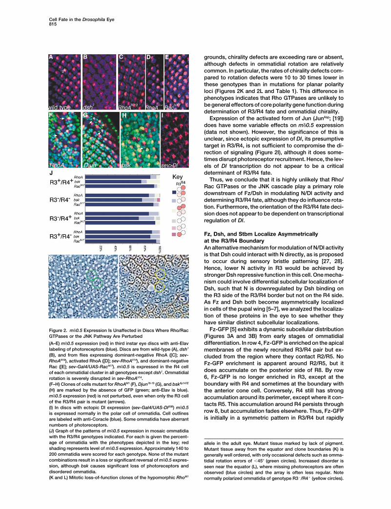
Fz-GFP [5] exhibits a dynamic subcellular distributionFigure 2. m�0.5 Expression Is Unaffected in Discs Where Rho/RacGTPases or the JNK Pathway Are Perturbed (Figures 3A and 3B) from early stages of ommatidial
differentiation. In row 4, Fz-GFP is enriched on the apical(A–E) m�0.5 expression (red) in third instar eye discs with anti-Elavlabeling of photoreceptors (blue). Discs are from wild-type (A), dsh1 membranes of the newly recruited R3/R4 pair but ex-(B), and from flies expressing dominant-negative RhoA ([C]; sev- cluded from the region where they contact R2/R5. NoRhoAN19), activated RhoA ([D]; sev-RhoAV14), and dominant-negative Fz-GFP enrichment is apparent around R2/R5, but itRac ([E]; sev-Gal4/UAS-RacN17). m�0.5 is expressed in the R4 cell
does accumulate on the posterior side of R8. By rowof each ommatidial cluster in all genotypes except dsh1. Ommatidial6, Fz-GFP is no longer enriched in R3, except at therotation is severely disrupted in sev-RhoAV14.boundary with R4 and sometimes at the boundary with(F–H) Clones of cells mutant for RhoAM1 (F), Djun76-19 (G), and bskflp147E
(H) are marked by the absence of GFP (green; anti-Elav is blue). the anterior cone cell. Conversely, R4 still has strongm�0.5 expression (red) is not perturbed, even when only the R3 cell accumulation around its perimeter, except where it con-of the R3/R4 pair is mutant (arrows). tacts R5. This accumulation around R4 persists through(I) In discs with ectopic Dl expression (sev-Gal4/UAS-Dl30B) m�0.5
row 8, but accumulation fades elsewhere. Thus, Fz-GFPis expressed normally in the polar cell of ommatidia. Cell outlinesis initially in a symmetric pattern in R3/R4 but rapidlyare labeled with anti-Coracle (blue). Some ommatidia have aberrant
numbers of photoreceptors.(J) Graph of the patterns of m�0.5 expression in mosaic ommatidiawith the R3/R4 genotypes indicated. For each is given the percent-age of ommatidia with the phenotypes depicted in the key; red allele in the adult eye. Mutant tissue marked by lack of pigment.shading represents level of m�0.5 expression. Approximately 140 to Mutant tissue away from the equator and clone boundaries (K) is200 ommatidia were scored for each genotype. None of the mutant generally well ordered, with only occasional defects such as omma-combinations result in a loss or significant reversal of m�0.5 expres- tidial rotation errors of �45� (green circles). Increased disorder ission, although bsk causes significant loss of photoreceptors and seen near the equator (L), where missing photoreceptors are oftendisordered ommatidia. observed (blue circles) and the array is often less regular. Note(K and L) Mitotic loss-of-function clones of the hypomorphic RhoM1 normally polarized ommatidia of genotype R3�/R4� (yellow circles).

Current Biology816
Table 1. Quantitation of Adult Eye Polarity Phenotypes
Ommatidial Polarity DefectChirality Defects Ommatidia
Normal Rotation Chirality Achiral Unscoreable as % of Total ExaminedGenotype (%) (%) (%) (%) (%) Polarity Defects (Eyes Examined)
RhoM1 (mitotic clones) 70.8 17.1 0.2 0.3 11.6 2.9 994 (16)sev-RhoN19 (dominant-negative RhoA) 95.4 3.8 0 0.1 0.5 2.5 988 (9)sev-RhoV14 (activated RhoA) 40.8 3.3 0 0 55.9 0 213 (3)sev-RacN17 (dominant-negative Rac1) 77.6 18.1 0.5 0.7 3.1 6.2 1249 (11)fz25 (strong fz) 26.8 28.6 37.0 7.2 0.3 60.7 332 (3)fz19/fz20 (weak fz) 84.8 6.3 6.1 2.5 0.3 57.8 608 (4)dsh3 (mitotic clones) 34.0 16.3 38.3 11.3 0 75.3 141 (3)fmiE45/fmiE59 (rescued in embryo) 36.8 20.2 26.1 15.9 1.0 67.5 410 (4)stbmVang-A3/Df(2R)w45-30n 48.0 14.1 36.8 0.9 0.2 72.9 427 (6)sple1/pk-sple13 57.8 0.6 41.6 0 0 98.6 166 (2)pk-sple13 53.0 4.6 41.9 0.5 0 90.0 415 (3)RhoM1 Mosaic Ommatidia
R3�/R4� 88.1 11.9 0 0 0 0 84R3�/R4� 94.3 5.7 0 0 0 0 87R3�/R4� 81.0 17.6 1.4 0 0 7.4 74R3�/R4� 94.2 5.8 0 0 0 0 86
Polarity of ommatidia was scored in eye sections crossing the equator from homozygous mutant individuals. dsh3 and RhoM1 were scored inmitotic clones, counting nonmosaic ommatidia away from the clone boundaries. Bottom row shows polarity defects associated with mosaicRhoM1 ommatidia at clone boundaries: only one chirality defect was observed in an ommatidium of genotype R3�/R4� (which due to thepolarity reversal indicates that the presumptive R3 cell in the disc was Rho� and the presumptive R4 was Rho�). It was noticed that equatorformation was often disrupted in RhoM1 clones, and many abnormal ommatidia were seen in this region (see Figure 2L), making accuratescoring difficult; so in this genotype, the two ommatidial rows straddling the equator were not scored. Ommatidia that showed both rotationand chirality defects are counted in the “chirality” column. Unscoreable ommatidia have too many or too few photoreceptors.
resolves into an asymmetric pattern which is visible by Fz-GFP. In row 4, a symmetric pattern is observed, withStbm-YFP around R3/R4, except where they contactthe time ommatidial rotation occurs in row 6. Using anti-
bodies against Dsh and Fmi [4, 7], we found that these R2/R5, and enriched on the posterior face of R8. Thissymmetric pattern is maintained until the ommatidia areproteins colocalized with Fz and showed the same dy-
namic distribution (Figures 3A and 3B). already rotated in row 6 and more posteriorly. Stainingthen fades around R3, except where it contacts R4.We found that N was also at highest levels in apical
membranes of cells posterior to the furrow and in rows Mosaic analysis revealed that, in contrast to Fz-GFP,Stbm-YFP is enriched on the R4 side of the R3/R44 through 6 overlapped with Fz-GFP at the R3/R4 bound-
ary (but showed no asymmetry, Figure 3C). The localiza- boundary from row 4 onward, i.e., Stbm is on the oppo-site side of the boundary to Fz (Figure 3F).tion of Fz-GFP (and Fmi/Dsh) to the R3/R4 boundary is
therefore consistent with Fz/Dsh being able to directlymodulate N activity in this location. However, if Fz/Dsh Regulation of Asymmetric Protein Localization
by the Planar Polarity Genesare differentially regulating N activity, a crucial require-ment is that these complexes should be preferentially To investigate the relationship between the asymmetric
localization of planar polarity proteins and the regulationlocalized on one side of the R3/R4 boundary. As thiscannot be distinguished by light microscopy, we created of R3/R4 fate, we analyzed the evolution of the protein
complexes in planar polarity mutants. Looking at Fz-genetic mosaics in which both R3/R4 had sufficient fzactivity for normal signaling and fate determination, but GFP, three different phenotypes are seen. (1) Removal
of fmi results in a failure of Fz-GFP to accumulate api-only one of the pair carried the Fz-GFP transgene. Usingthis approach, we found that Fz-GFP is more highly cally with only diffuse staining remaining (Figure 4A). (2)
Removal of dsh does not block general apical localiza-enriched on the R3 side of the R3/R4 boundary in row4 and more posteriorly is found exclusively on the R3 tion, but no asymmetric pattern is established (Figure
4B). (3) Removal of stbm, sple, or pk-sple also does notside of the boundary (Figure 3D). Thus, about two rowsprior to ommatidial rotation, Fz-GFP is asymmetrically block initial symmetric apical accumulation but leads to
a delay in the establishment of the asymmetric patterndistributed across the R3/R4 boundary. As studies inthe wing demonstrate that Dsh adopts the identical of Fz-GFP by about one to two ommatidial rows, with
a concomitant delay in the onset of ommatidial rotationasymmetric localization to Fz (and indeed their asym-metric localization is mutually dependent) [6], we infer (Figures 4C–4E). Furthermore, the polarity of the asym-
metric distribution is largely random, correlating withthat Dsh is also differentially localized on the R3 sideof the R3/R4 boundary. the random polarity decisions in these backgrounds.
Mosaic analysis with Fz-GFP in a stbm background con-The polarity gene stbm is required for R4 fate [29],so we investigated whether this protein also shows an firmed that Fz-GFP did indeed eventually adopt its nor-
mal asymmetric distribution, being localized on the R3/asymmetrical localization in R3/R4 using a Stbm-YFPtransgene. Stbm-YFP is apically localized in cells poste- R4 boundary in the cell taking the R3 fate and on the
outer boundary of the cell taking the R4 fate (as judgedrior to the furrow (Figure 3E), and, subsequently, itsdistribution is similar but distinct from that exhibited by by the associated ommatidial rotation, Figure 4F).

Cell Fate in the Drosophila Eye817
Figure 3. Planar Polarity Gene Products Show Dynamic Patterns of Subcellular Localization in Developing Photoreceptors
(A–F) Apical confocal sections (of about 1 �m depth), with anti-HRP labeling (blue) of neuronal membranes. (A–D) Fz-GFP (green) and Fmi(red) in (A), Dsh (red) in (B), and N (red) in (C). (D) Non Fz-GFP expressing cells labeled for lacZ (red). (E and F) Stbm-YFP (green).(A and B) Rows 4 to 8 of a wild-type eye.(C) Rows 4, 6, and 8 of a wild-type eye.(D) Mosaic Fz-GFP expression in R3/R4, generated by mitotic recombination. All cells have endogenous fz activity and patterning is normal,but lacZ-expressing cells (red) lack Fz-GFP transgene. Bottom panels show cartoon representations. In row 4, Fz-GFP is more stronglylocalized on R3 membranes at the R3/4 boundary and in row 6 Fz-GFP is exclusively on R3 membranes. We have not definitively determinedwhether expression around the outer boundary of R4 is in the R4 cell or in neighboring cells (or both).(E) Rows 4 to 7 of a wild-type eye.(F) Mosaic Stbm-YFP expression in R3/R4 generated by random excision of the FRT cassette from an Actin5C�stop�Stbm-YFP transgene(all cells have endogenous stbm activity, and patterning is normal). Stbm-YFP is more strongly localized on R4 membranes at the R3/4boundary.(G) Summary of Fz and Stbm localization in photoreceptors of nascent ommatidia. Localization on the R3/4 boundary is determined frommosaic experiments; localization on other boundaries may be in either R3/4 or in neighboring cells (or both).

Current Biology818
Figure 4. The Effect of Mutations in PlanarPolarity Genes on Planar Polarity Protein Lo-calization
(A–F and H–K) Apical confocal sections (ofabout 1 �m depth), with anti-HRP labeling(blue) of neuronal membranes. (A–F) Fz-GFP/YFP (green) and lacZ (red) in (A)–(C) and (E),Dsh (red) in (F). (H–J) Dsh (red) and lacZ(green). (K) Stbm-YFP (green) and lacZ (red).(G) Semi-thin section of adult eye at R7 level.Mitotic loss-of-function clones (A–C, E, andH–K) are marked by lack of lacZ expression.(A) fmiE59 clone. Apical accumulation of Fz-GFP is only seen at boundaries between fmi�
cells. Arrowhead indicates fmi� ommatidiumdelayed in rotation.(B) dshV26 clone. Some apical accumulationof Fz-GFP is seen in dsh� tissue, but highlevels of accumulation and asymmetric loca-tion are not observed. Arrowhead indicatesdsh� ommatidium exhibiting symmetric Fz-GFP localization.(C) stbmVang-A3 clone. Apical accumulation ofFz-GFP is approximately normal, but acquisi-tion of asymmetry in R3/4 appears to be de-layed by about one row, becoming pro-nounced in rows 7/8 rather than 6/7. Arrowmarks mosaic ommatidium with stbm� polarcell and reversed asymmetric localization ofFz-GFP.(D) sple1/pk-sple13 disc. Apical accumulationof Fz-GFP is approximately normal, but ac-quisition of asymmetry in R3/R4 is delayedby about one row relative to wild-type. Arrow-heads indicate ommatidia with asymmetriclocalization in row 8.(E) pk-sple13 clone. Apical accumuluation ofFz-GFP is approximately normal, but acquisi-tion of asymmetry in R3/R4 is delayed byabout one row relative to wild-type.(F) Mosaic Fz-YFP expression in a stbmVang-A3/Df(2R)w45-30n disc. Second panel of eachpair shows same ommatidium without Fz-YFP expression. Mosaic Fz-YFP expressiongenerated by random excision of the FRTcassette from an Actin5C�stop�Fz-YFPtransgene. Normal Fz/Dsh asymmetry is gen-erated in a stbm� background, with Fz-GFPenriched on the R3 side of the R3/R4 bound-
ary. However, Dsh localization on the R3/R4 boundary is significantly reduced relative to wild-type. Cartoons below show Fz-YFP-expressingcells of the R3/4 pair (pale green), Fz localization (green), and Dsh localization (red).(G) fmiE45/fmiE59. Diagram shows ommatidial polarities, with dorsal forms in red and ventral in green. Both chirality and rotation appearrandomized.(H) fz25 clone. Apical accumulation of Dsh is not seen in fz� cells.(I) fmiE59 clone. Apical accumulation of Dsh is not seen in fmi� cells.(J) stbm6 clone. Apical accumulation of Dsh does occur in this background, albeit slightly more weakly than in wild-type. Acquisition ofasymmetry is delayed by about one to two rows. Arrowheads indicate stbm� ommatidia which still show symmetric Dsh, when nearby stbm�
ommatidia show asymmetric expression. Arrow marks mosaic ommatidium with stbm� polar cell and reversed asymmetric localization of Dshand rotation.(K) fz25 clone. Apical localization is observed, but asymmetric localization does not occur. Arrowhead marks rotated ommatidium still exhibitingsymmetric Stbm-YFP localization.
The distribution of Dsh was found to be similar to Fz- that the initial symmetric distribution was established,but this never resolved to an asymmetric pattern (FigureGFP in the backgrounds tested (fmi, stbm, sple, pk-sple;
Figures 4F, 4I, and 4J and data not shown), although 4K), indicating that fz function is required for the asym-metric localization of Stbm.we did find that levels of Dsh localization at the R3/R4
boundary were consistently weaker in stbm� than inwild-type (Figures 4F and 4J). In a fz background, Dsh Expression of m�0.5 Is Affected by Polarity
Gene Mutationsdid not accumulate apically (Figure 4H), suggesting thatFz is required to recruit Dsh to the apical cell cortex If asymmetric polarity protein localization is required for
the specification of R3/R4 fate and ommatidial polarity,(as is observed in the wing [6, 7]). Similarly, when weanalyzed Stbm-YFP distribution in fz clones, we found then we would predict that mutations in polarity genes
Cell Fate in the Drosophila Eye819
ity in one or other cell, with 32% showing higher levelsin the polar cell (as in the wild-type) and 19% showinghigher levels in the equatorial cell (n � 134).
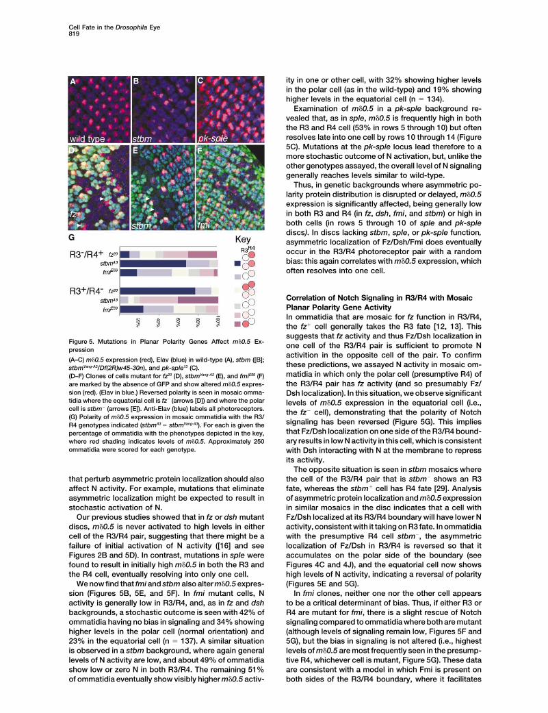
Examination of m�0.5 in a pk-sple background re-vealed that, as in sple, m�0.5 is frequently high in boththe R3 and R4 cell (53% in rows 5 through 10) but oftenresolves late into one cell by rows 10 through 14 (Figure5C). Mutations at the pk-sple locus lead therefore to amore stochastic outcome of N activation, but, unlike theother genotypes assayed, the overall level of N signalinggenerally reaches levels similar to wild-type.
Thus, in genetic backgrounds where asymmetric po-larity protein distribution is disrupted or delayed, m�0.5expression is significantly affected, being generally lowin both R3 and R4 (in fz, dsh, fmi, and stbm) or high inboth cells (in rows 5 through 10 of sple and pk-splediscs). In discs lacking stbm, sple, or pk-sple function,asymmetric localization of Fz/Dsh/Fmi does eventuallyoccur in the R3/R4 photoreceptor pair with a randombias: this again correlates with m�0.5 expression, whichoften resolves into one cell.
Correlation of Notch Signaling in R3/R4 with MosaicPlanar Polarity Gene ActivityIn ommatidia that are mosaic for fz function in R3/R4,the fz� cell generally takes the R3 fate [12, 13]. Thissuggests that fz activity and thus Fz/Dsh localization in
Figure 5. Mutations in Planar Polarity Genes Affect m�0.5 Ex- one cell of the R3/R4 pair is sufficient to promote Npression
activition in the opposite cell of the pair. To confirm(A–C) m�0.5 expression (red), Elav (blue) in wild-type (A), stbm ([B];
these predictions, we assayed N activity in mosaic om-stbmVang-A3/Df(2R)w45-30n), and pk-sple13 (C).matidia in which only the polar cell (presumptive R4) of(D–F) Clones of cells mutant for fz20 (D), stbmVang-A3 (E), and fmiE59 (F)the R3/R4 pair has fz activity (and so presumably Fz/are marked by the absence of GFP and show altered m�0.5 expres-
sion (red). (Elav in blue.) Reversed polarity is seen in mosaic omma- Dsh localization). In this situation, we observe significanttidia where the equatorial cell is fz� (arrows [D]) and where the polar levels of m�0.5 expression in the equatorial cell (i.e.,cell is stbm� (arrows [E]). Anti-Elav (blue) labels all photoreceptors. the fz� cell), demonstrating that the polarity of Notch(G) Polarity of m�0.5 expression in mosaic ommatidia with the R3/
signaling has been reversed (Figure 5G). This impliesR4 genotypes indicated (stbmA3 � stbmVang-A3). For each is given thethat Fz/Dsh localization on one side of the R3/R4 bound-percentage of ommatidia with the phenotypes depicted in the key,ary results in low N activity in this cell, which is consistentwhere red shading indicates levels of m�0.5. Approximately 250
ommatidia were scored for each genotype. with Dsh interacting with N at the membrane to repressits activity.
The opposite situation is seen in stbm mosaics wherethe cell of the R3/R4 pair that is stbm� shows an R3that perturb asymmetric protein localization should also
affect N activity. For example, mutations that eliminate fate, whereas the stbm� cell has R4 fate [29]. Analysisof asymmetric protein localization and m�0.5 expressionasymmetric localization might be expected to result in
stochastic activation of N. in similar mosaics in the disc indicates that a cell withFz/Dsh localized at its R3/R4 boundary will have lower NOur previous studies showed that in fz or dsh mutant
discs, m�0.5 is never activated to high levels in either activity, consistent with it taking on R3 fate. In ommatidiawith the presumptive R4 cell stbm�, the asymmetriccell of the R3/R4 pair, suggesting that there might be a
failure of initial activation of N activity ([16] and see localization of Fz/Dsh in R3/R4 is reversed so that itaccumulates on the polar side of the boundary (seeFigures 2B and 5D). In contrast, mutations in sple were
found to result in initially high m�0.5 in both the R3 and Figures 4C and 4J), and the equatorial cell now showshigh levels of N activity, indicating a reversal of polaritythe R4 cell, eventually resolving into only one cell.
We now find that fmi and stbm also alter m�0.5 expres- (Figures 5E and 5G).In fmi clones, neither one nor the other cell appearssion (Figures 5B, 5E, and 5F). In fmi mutant cells, N
activity is generally low in R3/R4, and, as in fz and dsh to be a critical determinant of bias. Thus, if either R3 orR4 are mutant for fmi, there is a slight rescue of Notchbackgrounds, a stochastic outcome is seen with 42% of
ommatidia having no bias in signaling and 34% showing signaling compared to ommatidia where both are mutant(although levels of signaling remain low, Figures 5F andhigher levels in the polar cell (normal orientation) and
23% in the equatorial cell (n � 137). A similar situation 5G), but the bias in signaling is not altered (i.e., highestlevels of m�0.5 are most frequently seen in the presump-is observed in a stbm background, where again general
levels of N activity are low, and about 49% of ommatidia tive R4, whichever cell is mutant, Figure 5G). These dataare consistent with a model in which Fmi is present onshow low or zero N in both R3/R4. The remaining 51%
of ommatidia eventually show visibly higher m�0.5 activ- both sides of the R3/R4 boundary, where it facilitates

Current Biology820
marked dominant effect on the resolution of signalingbetween R3/R4. More ommatidia showed poor resolu-tion, such that m�0.5 expression was detected in bothR3/R4 (20%), and in a few cases signaling had resolvedin the incorrect direction, altering chirality and directionof rotation (Figures 6B and 6D). Nmcd8 hemizygotes(Nmcd8/Y) also showed delayed discrimination, with moreommatidia exhibiting m�0.5 expression in R3 as well asR4 in rows 5 through 12 (older rows having more normalexpression) and occasional ommatidia exhibiting re-versed chirality (Figures 6C and 6D). Much weaker ef-fects were seen if the level of Notch was simply reduced(N55e11/�, Figures 6A and 6D). These data therefore areconsistent with asymmetrically localized Dsh directlyinfluencing N activity via its C-terminal region.
A direct interaction is further supported by the effectsof overexpressing some Dsh dominant-negative con-structs. In the wing, overexpression of a form of Dshlacking the PDZ domain (DshPDZ) gives strong effectson planar polarity and disrupts the apical localizationof Fz/Dsh, whereas a form lacking the C-terminal DEPdomain (DshDEP) causes weaker effects [6]. Con-versely, in the eye, the strongest phenotypes are seenwith DshDEP, resulting in a high frequency of achiralommatidia in the adult [32] and in low levels of m�0.5in the disc (data not shown). We attribute the greaterseverity of the eye phenotypes seen with DshDEP tothis molecule retaining its N-terminal domain that caninteract with N [27] and so interfering more potentlywith N signaling and R3/R4 fate decisions than doesDshPDZ (which lacks a large part of its N terminus).
Figure 6. Effects of Mutations that Truncate the Dsh-Interacting In the wing, Dsh apparently does not interact with NDomain of Notch during polarity decisions, and so DshDEP cannotm�0.5 expression (red), Elav (blue) in N55e11/� (A), N60g11/� ([B], raised cause strong polarity phenotypes via this N-dependentat 21�C), and NMcd8/Y (C). In a significant number of ommatidia in (B) mechanism.and (C), m�0.5 is detectable in R3 as well as R4. A few ommatidiahave reversed polarity of signaling (arrow [B]). In (D), these effectsare represented graphically as the percentage of ommatidia show-
Discussioning the phenotypes indicated in the key. Approximately 350 omma-tidia in rows 5 through 12 were scored for each genotype. Diagramsat bottom indicate extent of deletions in each allele (N55e11 is a puta- Interactions between N/Dl-expressing cells are criticaltive protein null). Note that N60g11 also contains a point mutation in many contexts for determining cell fate. One example(S2257G), which is thought to be phenotypically silent [30]. of this is in the fly eye, where N/Dl interactions between
a single pair of cells (R3/R4) lead to a binary fate choicewhich then determines the future polarity of the omma-
Fz/Dsh function but does not impose any bias in signal- tidium. The choice of whether to take on R3 or R4 fateing itself. is governed by the activity of the planar polarity genes.
In particular, the cell with the highest activity of fz be-comes R3. Here we have examined possible mecha-Mutations in Notch that Disrupt Dsh Binding Affect
the Resolution of Signaling in R3/R4 nisms by which Fz might regulate N activity and socontrol the spatial acquisition of cell fate.Direct interactions between Dsh and N have been re-
ported, which map to the C-terminal region of the N Our major findings are as follows. First, the primarymechanism used by fz to control N activity does notintracellular domain [27]. If the asymmetric distribution
of Fz/Dsh is responsible for biasing N signaling, muta- appear to involve Rho/Rac GTPases and the JNK cas-cade as previously proposed. Second, we find that Fz,tions that disrupt the binding of Dsh to N (but not neces-
sarily other aspects of N activity) might be anticipated Dsh, Fmi, and Stbm localize to the apical region of theR3/R4 cell boundary, where they become asymmetri-to alter this regulation. We therefore tested two alleles
of N, N60g11 and Nmcd8, both of which have C-terminal cally distributed prior to or concomitant with R3/R4 fatedetermination. Normally, Fz/Dsh are enriched on onetruncations that remove all or part of the Dsh interacting
domain, N60g11 being the more severe (Figure 6D) [28, 30, particular side of the cell boundary, in the presumptiveR3 cell. However, in mosaic ommatidia where one or31]. These were compared to a putative protein null
allele (N55e11). other cell is mutant for polarity genes, the assemblyof the asymmetrical complexes can be reversed. In allAs N60g11 is lethal in hemizygotes, we examined this
allele in heterozygotes. In N60g11/� flies, there was a conditions examined, the polarity of Notch signaling be-

Cell Fate in the Drosophila Eye821
tween R3/R4 is consistent with the polarity of the asym- pupal wing [4–8]. Thus, the R3/R4 cell boundary appearsmetric complexes, with Notch activity being lowest in analogous to the proximodistal wing cell boundaries,the cell where Fz/Dsh accumulate. Finally, we provide with the R3 side of the boundary, where Fz and Dsh areevidence that the domain of N, which is known to interact localized, being equivalent to the wing cell distal edge.directly with Dsh, is required for efficient R3/R4 fate Another of the polarity gene products, Stbm, is localizeddecisions. on the R4 side of the boundary, which is consistent with
Considering these results together, we propose that the requirement for stbm function in R4 [29]. By analogyan extracellular polarity signal leads to the asymmetric to the wing, it is likely that Fmi is present on both sidesassembly of a complex of planar polarity proteins at the of the R3/R4 boundary and Pk-Sple/Sple on the R4 side.boundary between the R3/R4 cell pair. This asymmetric The adoption of the asymmetric pattern occurs in twocomplex then leads to asymmetric N activity between phases. The first involves symmetric apicolateral local-the cell pair and thus determines cell fate. Since we find ization of Fz, Dsh, Fmi, and Stbm in R3/R4 (and in allno evidence that this regulation occurs via the proposed other cells except R2/R5), which is evident in ommatidialsignaling cascade downstream of Fz/Dsh (i.e., Rho row 4. As in the wing, the initial apical recruitment of FzGTPases/JNK) and as manipulation of Dl transcription is dependent on Fmi [5], and the recruitment of Dshdoes not perturb polarity of Notch signaling, we con- is in turn dependent on Fz [6, 7]. Subsequently, theclude that there must be an alternative pathway by which distribution evolves rapidly into an asymmetric pattern.asymmetrical Fz/Dsh affects Notch activity. Adoption of asymmetry requires the function of dsh,
One favored mechanism for the modulation of N/Dl stbm, and pk-sple, and if any of these are missing, Fzactivity is via local interactions between N and asymmet- distribution remains symmetric in ommatidial rows 5/6,rically localized proteins and, in particular, between the and ommatidial rotation is delayed. It is likely that theintracellular domain of N and Dsh. Four lines of evidence asymmetry evolves through the same mechanisms assupport the proposal that the regulation occurs at the in the wing, where it has been proposed that an extrinsiccell cortex. First, Fz/Dsh are in the same subcellular signal leads to a small bias in Fz/Dsh signaling on eitherdomain as N at the apical R3/R4 boundary during the side of the cell boundary, which subsequently becomescritical stages of development when the cell fate deci- amplified by feedback loops that lead to Fz/Dsh becom-sion is made. Second, the appearance of the asymmetric
ing concentrated on one side of the interface and Pk-Fz/Dsh complexes is shortly prior to or concomitant with
Sple/Stbm on the other [5, 6, 8].the appearance of a bias in N/Dl activity and ommatidial
One notable difference between the eye and the wingrotation. Third, direct interactions between N and Dshis that asymmetric Fz/Dsh distribution is eventually ob-have been previously demonstrated and proposed toserved in stbm and pk-sple eye discs, but in both casesbe important for patterning in other tissues [27, 28], andit occurs with a random bias and is delayed by aboutthese interactions have been found to be repressive,one to two ommatidial rows. This correlates well withconsistent with Fz/Dsh being required in R3, where Nthe fact that the adult phenotypes of stbm and pk-spleactivity is lowest. Finally, deletion of the domain of Nexhibit a low incidence of achiral ommatidia. Con-required for interactions with Dsh leads to less-efficientversely, in fmi, fz and dsh, negligible asymmetric proteinR3/R4 fate decisions.localization occurs, and there is a relatively high propor-tion of “achiral” ommatidia in the adult eye (Table 1),Regulation of N/Dl Activity by the Planarsuggesting that achirality is a result of poor asymmetricPolarity Genescomplex formation. In general, the aquisition of asym-We note that our model whereby asymmetric Fz/Dshmetry also correlates with m�0.5 activity, particularly inlocalization leads to downregulation of N activity on thepk-sple and sple mutations where its expression usuallyR3 side of the R3/R4 boundary is similar to one firstresolves into a single cell by row 10.suggested by Tomlinson and Struhl [13]. It is further
supported by studies in the Drosophila leg, where lossof planar polarity gene activity leads to ectopic activity
Both Asymmetric Protein Localization and N/Dlof Notch ([33] and data not shown). However, there areSignaling Are Required for R3/R4 Fate Choicestill unexplained observations: if the only role of theIn the pupal wing, asymmetric localization of Fz/Dsh/polarity genes is to inhibit N in R3, mutations in fmi, fz,Pk-Sple is proposed to involve a signaling feedbackor dsh (which result in no apical Dsh localization) shouldloop that amplifies an initially small bias in Fz/Dsh activ-have high N activity in both R3/R4, not the reducedity across the axis of each cell [5, 6, 8]. In the eye, theactivity that we detect.N/Dl feedback loop was proposed to perform a similarThis discrepancy might be explained if there are twofunction, amplifying an initially small difference in Fz/Dshphases to polarity gene regulation of N activity. Oneactivity between R3/R4 [13, 16, 17]. With the observationwould be an activation/derepression of N, which wouldthat Fz/Dsh are also distributed in asymmetric com-require symmetric protein localization of Fz/Dsh in R3/plexes in the eye, it appears that both mechanisms areR4. The second would be linked to asymmetric proteinoperating in R3/R4, although it is not clear why bothlocalization, when Fz/Dsh would in turn become repres-would be required, since either alone should be suffi-sors of N activity in R3.cient to amplify small biases in signaling activity.
One possible explanation is that use of both mecha-Asymmetric Localization of Planar Polaritynisms increases the speed and robustness of the R3/Proteins in R3/R4R4 fate decision. A fast fate decision may be necessaryThe asymmetric localization of Fz, Dsh, and Fmi in the
eye develops in a similar manner to that seen in the because of the dynamic nature of eye patterning, in

Current Biology822
which the R3/R4 decision is only part of a complex series of function analysis of msn did reveal some ommatidialchirality defects, albeit rarely [36]. More compellingly,of events involving cell recruitment and movement to
generate the final polarized ommatidium. It is also possi- mosaic analysis suggested that if one cell of the R3/R4 pair was msn�, this cell would generally (but notble that a rapid decision is required because the extrin-
sic polarity cue is transient in nature. We note that the exclusively) take the R4 fate. This suggests a role forMsn in repression of N in R3, although the apparent rarityrapidity of the decision would be further enhanced if
N/Dl signaling also influenced Fz/Dsh localization. While of chirality defects in ommatidia lacking msn functionsuggests this is a nonessential pathway.we have no direct evidence for this, it could explain
the eventual, randomly polarized, asymmetric protein Taken together, the phenotypic evidence from loss-of-function studies does not support a primary role forlocalization seen in stbm and pk-sple backgrounds in
the eye. In this case, the inability of Fz/Dsh to efficiently Rho GTPases/JNK cascades in the R3/R4 fate decision.But the weight of genetic evidence does support a sec-localize asymmetrically in the absence of Stbm/Pk-Sple
might lead to N/Dl making a stochastic decision that ondary role for some of the proposed pathway compo-nents, possibly in the augmentation of polarity decisionsthen leads to Fz/Dsh asymmetry. Conversely, in the pu-
pal wing, where N/Dl are not active in planar polarity driven largely by asymmetric localization of polarity pro-teins and direct repression of N activity. In addition,decisions, Stbm/Pk-sple activity would be absolutely
required, as their absence would not be compensated the observation that RhoA mutations result largely indefects in ommatidial rotation supports the hypothesisfor by the N/Dl feedback loop.that RhoA acts downstream of the planar polarity genesin regulating this aspect of ommatidial polarity.
Rho GTPases, JNK Cascades,and Ommatidial Polarity ConclusionsA number of lines of evidence previously suggested that For some years, the standard view of planar polarityRho/Rac GTPases and the JNK cascade were required gene function has been that this is mediated by Rhofor ommatidial polarity decisions and, in particular, the GTPases and a JNK kinase cascade, most likely leadingR3/R4 fate decision. These include the following. Over- to a transcriptional response [17–19, 26, 32]. In particu-expression of Fz or Dsh in the eye gives a polarity pheno- lar, it was thought that this pathway controlled R3/R4type that is dominantly suppressed by RhoA, bsk, hep, photoreceptor fate in the developing eye, via transcrip-and Djun [26, 32]. RhoA clones or expression of domi- tional activation of Dl. We now show that although Rhonant-active/negative RhoA or Rac1 gives ommatidial GTPases do regulate one aspect of planar polarity inpolarity phenotypes [18, 26]. Also, overexpression of the eye (ommatidial rotation), they do not appear to bedominant-active/negative JNK pathway components the primary determinant of R3/R4 fate. Therefore, weand human Jun elicits ommatidial polarity defects [19]. conclude that Rho GTPases/JNK cascades are not theFurthermore, expression of a Dl enhancer trap is altered major effectors of planar polarity activity in this context.by overexpression of either fz or dsh or by activated Furthermore, we demonstrate that several planar polar-human Jun, Hep, RhoA, or Rac1 [17–19]. These observa- ity proteins adopt asymmetric subcellular localizationstions led to the hypothesis that higher levels of Fz/Dsh in the eye that correlate with N activation and R3/R4signaling in R3 resulted in higher activation of Dl tran- fate. We propose an alternative model for modulationscription in R3 via a Rho GTPase/JNK cascade, biasing of N activity via direct interactions with planar polaritythe N/Dl feedback loop to produce high N in R4. proteins (and most probably Dsh) at the cell cortex.
However, our data do not support the hypothesis thatactivation of Dl transcription via Rho GTPases/JNK cas-
Experimental Procedurescade is the primary mechanism for biasing N activity inR3/R4 during normal eye development. Reexamination Genetics
Except where otherwise stated, fly stocks used are described inof RhoA phenotypes indicates that these rarely affectFlyBase. NMcd8 is as described [28]. Note that flamingo (fmi) is alsoR3/R4 fate and ommatidial chirality, and RhoA activityknown as starry night (stan) and that strabismus (stbm) is also knownis not required for N repression in R3. We also find noas Van Gogh (Vang). Transgenic insertions of m�0.5-lacZ [16], Fz-role for Rac in this process, a finding confirmed by theGFP [5], or Stbm-YFP on chromosomes II and III were introduced
recent report that deletion of all three Rac homologs in into different mutant backgrounds and balanced over the compoundthe Drosophila genome has no effect on planar polarity chromosome SM5a:TM6b to enable selection of larvae of the correct
genotype. Mitotic clones were generated using the FLP/FRT [37][34]. In addition, notwithstanding the observation thatsystem and marked in eye discs using Armadillo-lacZ [38] or Ubi-loss of Djun activity can result in ommatidial polarityGFP [39] and in adult eyes using P[w�]. The size of RhoM1 clonesdefects in 2%–3% of ommatidia [19], mosaic ommatidiawas increased by use of a Minute mutation on the marker chromo-where the presumptive R3 lacks Djun activity have wild-some. UAS-RacN17 overexpression clones in eye discs were gener-
type levels and polarity of Notch signaling, so we find ated using Actin�y��GAL4, UAS-GFP [40]. Clones were inducedno evidence that Djun is directly modulating N activity by 30 min to 2 hr heat shocks at 38�C at 24–48 and 48–72 hr of
development, except when using Actin5C�stop�Fz-YFP or Stbm-in R3/R4. It is also interesting to note that although aYFP, in which case a 30 min heat shock at 38�C at 48–72 hr wasdouble mutant combination of the JNK pathway compo-used. Adults lacking fmi activity were generated by crossing fmiE59/nents hemipterous and puckered produces an ommatid-CyO; UAS-Fmi flies to fmiE45 GAL4/CyO flies as previously describedial polarity phenotype, this apparently consists entirely[4]. One of the stbm alleles which we used (stbmVang-A3) was reported
of rotation and not chirality defects [19]. to be amorphic in the wing [41]; however, experiments in the eyeOne factor which we have not directly investigated is demonstrated this allele to be a strong hypomorph, as the pheno-
type of homozygotes was slightly weaker than the phenotype overthe STE20 kinase homolog encoded by msn [35]. Loss

Cell Fate in the Drosophila Eye823
a deficiency (which was approximately identical to the phenotype T. (2001). Asymmetric co-localisation of Flamingo, a seven-passtransmembrane cadherin, and Dishevelled in planar cell polari-of homozygotes for the null allele stbm6 [29]). As N60g11 is temperature
sensitive, embryos were cultured at the permissive temperature sation. Curr. Biol. 11, 859–863.8. Tree, D.R.P., Shulman, J.M., Rousset, R., Scott, M.P., Gubb, D.,(29�C) and then shifted to the restrictive temperature (21�C) in the
first instar. and Axelrod, J.D. (2002). Prickle mediates feedback amplifica-tion to generate asymmetric planar cell polarity signaling. Cell109, 371–381.Histology
9. Feiguin, F., Hannus, M., Mlodzik, M., and Eaton, S. (2001). TheSemi-thin sections of adult eyes were made as previously describedankyrin-repeat protein Diego mediates Frizzled-dependent pla-[42].nar polarisation. Dev. Cell 1, 93–101.Eye discs were dissected from wandering third instar larvae. Indi-
10. Strutt, H., and Strutt, D. (1999). Polarity determination in therect immunofluorescence was carried out as previously describedDrosophila eye. Curr. Opin. Genet. Dev. 9, 442–446.[5, 16]. Primary antibodies were 1:4000 rabbit anti--galactosidase
11. Wolff, T., and Ready, D.F. (1993). Pattern formation in the Dro-(Cappel), 1:4000 rabbit anti-GFP (Molecular Probes), 1:1000 rat anti-sophila retina. In The Development of Drosophila melanogaster,Dsh [7], 1:20 anti-Fmi mAb [4], 1:20 anti-NIC mAb ([43]; Develop-M. Bate and A. Martinez-Arias, eds. (Cold Spring Harbor, NY:mental Studies Hybridoma Bank), 1:200 anti-Elav mAb ([44]; Devel-Cold Spring Harbor Press), pp. 1277–1326.opmental Studies Hybridoma Bank), 1:1000 guinea-pig anti-coracle
12. Zheng, L., Zhang, J., and Carthew, R.W. (1995). frizzled regulates[45]. For detection of polarity protein localizations, confocal sectionsmirror-symmetric pattern formation in the Drosophila eye. De-are of the most apical regions of eye disc cells, generally represent-velopment 121, 3045–3055.ing the average of several confocal image planes for a total image
13. Tomlinson, A., and Struhl, G. (1999). Decoding vectorial informa-depth of about 1 �m.tion from a gradient: sequential roles of the receptors Frizzledand Notch in establishing planar polarity in the Drosophila eye.Molecular BiologyDevelopment 126, 5725–5738.Stbm-YFP is a fusion at the C-terminal end of the full-length Stbm
14. Ma, C., and Moses, K. (1995). Wingless and patched are nega-ORF to the enhanced GFP variant EYFP (Clontech), cloned down-tive regulators of the morphogenetic furrow and can affect tis-stream of the Actin5C promoter and an FRT-PolyA-FRT cassette assue polarity in the developing Drosophila compound eye. Devel-described [5]. Use of the Actin5C promoter to drive Fz-YFP or Stbm-opment 121, 2279–2289.YFP expression has not been found to result in polarity patterning
15. Wehrli, M., and Tomlinson, A. (1998). Independent regulation ofdefects in the eye, even when expressed in mosaic fashion. Thisanterior/posterior and equatorial/polar polarity in the Drosophilasuggests that expression levels are relatively close to normal physio-eye; evidence for the involvement of Wnt signaling in the equato-logical levels and below the level at which aberrant cellular re-rial/polar axis. Development 125, 1421–1432.sponses might occur due to overexpression of these gene products.
16. Cooper, M.Y., and Bray, S.J. (1999). Frizzled regulation of Notchsignalling polarizes cell fate in the Drosophila eye. Nature 397,Acknowledgments526–530.
17. Fanto, M., and Mlodzik, M. (1999). Asymmetric Notch activationWe thank P. Adler, R. Fehon, D. Gubb, P. Heitzler, T. Uemura, T.specifies photoreceptors R3 and R4 and planar polarity in theWolff, the Bloomington Stock Centre, and the Developmental Stud-Drosophila eye. Nature 397, 523–526.ies Hybridoma Bank for fly stocks and antibodies; J. Axelrod and
18. Fanto, M., Weber, U., Strutt, D.I., and Mlodzik, M. (2000). NuclearB. Dickson for communicating results prior to publication; N. Brown,
signalling by Rac and Rho GTPases is required in the establish-H. Strutt, and R. White for comments on the manuscript. R.J. and
ment of epithelial planar polarity in the Drosophila eye. Curr.K.C were supported by scholarships/studentships from Fuchs and
Biol. 10, 979–988.Merck Sharpe and Dohme and the Biotechnology and Biological
19. Weber, U., Paricio, N., and Mlodzik, M. (2000). Jun mediatesSciences Research Council, respectively. Yorkshire Cancer Re-
Frizzled-induced R3/R4 cell fate distinction and planar polaritysearch is acknowledged for provision of confocal microscopy facili-
determination in the Drosophila eye. Development 127, 3619–ties. This work was supported by grants from the Medical Research 3629.Council (S.B. and D.S.) and Wellcome Trust (D.S.). D.S. is Lister 20. Riesgo-Escovar, J.R., Jenni, M., Fritz, A., and Hafen, E. (1996).Institute-Jenner Research Fellow. The Drosophila Jun-N-Terminal kinase is required for cell mor-
phogenesis but not for DJun-dependent cell fate specificationReceived: March 20, 2002 in the eye. Genes Dev. 10, 2759–2768.Revised: April 3, 2002 21. Sluss, H.K., Han, Z., Barrett, T., Davis, R.J., and Ip, Y.T. (1996). AAccepted: April 3, 2002 JNK signal transduction pathway that mediates morphogenesisPublished: May 14, 2002 and an immune response in Drosophila. Genes Dev. 10, 2745–
2758.References 22. Glise, B., and Noselli, S. (1997). Coupling of Jun amino-terminal
kinase and Decapentaplegic signalling pathways in Drosophila1. Shulman, J.M., Perrimon, N., and Axelrod, J.D. (1998). Frizzled morphogenesis. Genes Dev. 11, 1738–1747.
signaling and the developmental control of cell polarity. Trends 23. Hou, X.S., Goldstein, E.S., and Perrimon, N. (1997). DrosophilaGenet. 14, 452–458. Jun relays the Jun amino-terminal kinase signal transduction
2. Adler, P.N., and Lee, H. (2001). Frizzled signalling and cell-cell pathway in regulating epithelial cell sheet movement. Genesinteractions in planar polarity. Curr. Opin. Cell Biol. 13, 635–640. Dev. 11, 1728–1737.
3. Park, M., and Moon, R.T. (2002). The planar cell-polarity gene 24. Kockel, L., Zeitlinger, J., Staszewski, L.M., Mlodzik, M., andstbm regulates cell behaviour and cell fate in vertebrate em- Bohmann, D. (1997). Jun in Drosophila development: redundantbryos. Nat. Cell Biol. 4, 20–25. and nonredundant functions and regulation by two MAPK signal
4. Usui, T., Shima, Y., Shimada, Y., Hirano, S., Burgess, R.W., transduction pathways. Genes Dev. 11, 1748–1758.Schwarz, T.L., Takeichi, M., and Uemura, T. (1999). Flamingo, 25. Riesgo-Escovar, J.R., and Hafen, E. (1997). Drosophila Jun ki-a seven-pass transmembrane cadherin, regulates planar cell nase regulates expression of decapentaplegic via the ETS-polarity under the control of Frizzled. Cell 98, 585–595. domain protein Aop and the Ap-1 transcription factor DJun
5. Strutt, D.I. (2001). Asymmetric localisation of Frizzled and the during dorsal closure. Genes Dev. 11, 1717–1727.establishment of cell polarity in the Drosophila wing. Mol. Cell 26. Strutt, D.I., Weber, U., and Mlodzik, M. (1997). The role of RhoA7, 367–375. in tissue polarity and Frizzled signalling. Nature 387, 292–295.
6. Axelrod, J.D. (2001). Unipolar membrane association of Dishev- 27. Axelrod, J.D., Matsuno, K., Artavanis-Tsakonas, S., and Perri-elled mediates Frizzled planar cell polarity signalling. Genes mon, N. (1996). Interaction between Wingless and Notch signal-Dev. 15, 1182–1187. ling pathways mediated by Dishevelled. Science 271, 1826–
1832.7. Shimada, Y., Usui, T., Yanagawa, S., Takeichi, M., and Uemura,

Current Biology824
28. Ramain, P., Khechumian, K., Seugnet, L., Arbogast, N., Acker-mann, C., and Heitzler, P. (2001). Novel Notch alleles reveal aDeltex-dependent pathway repressing neural cell fate. Curr.Biol. 11, 1729–1738.
29. Wolff, T., and Rubin, G. (1998). strabismus, a novel gene thatregulates tissue polarity and cell fate decisions in Drosophila.Development 125, 1149–1159.
30. Lyman, D., and Young, M.W. (1993). Further evidence for func-tion of Drosophila Notch protein as a transmembrane receptor.Proc. Natl. Acad. Sci. USA 90, 10395–10399.
31. Brennan, K., Tateson, R., Lewis, K., and Martinez-Arias, A.(1997). A functional analysis of Notch mutations in Drosophila.Genetics 147, 177–188.
32. Boutros, M., Paricio, N., Strutt, D.I., and Mlodzik, M. (1998).Dishevelled activates JNK and discriminates between JNK path-ways in planar polarity and wingless signaling. Cell 94, 109–118.
33. Bishop, S.A., Klein, T., Martinez-Arias, A., and Couso, J.P.(1999). Composite signalling from Serrate and Delta establishesleg segments in Drosophila through Notch. Development 126,2993–3003.
34. Hakeda-Suzuki, S., Ng, J., Tzu, J., Dietzl, G., Sun, Y., Harms,M., Nardine, T., Luo, L., and Dickson, B.J. (2002). Rac functionand regulation during Drosophila development. Nature 416,438–442.
35. Treisman, J.E., Ito, N., and Rubin, G.M. (1997). Misshapen en-codes a protein kinase involved in cell shape control in Drosoph-ila. Gene 186, 119–125.
36. Paricio, N., Feiguin, F., Boutros, M., Eaton, S., and Mlodzik, M.(1999). The Drosophila STE20-like kinase misshapen is requireddownstream of the Frizzled receptor in planar polarity signalling.EMBO J. 18, 4669–4678.
37. Xu, T., and Rubin, G.M. (1993). Analysis of genetic mosaicsin developing and adult Drosophila tissues. Development 117,1223–1237.
38. Vincent, J.P., Girdham, C.H., and O’Farrell, P.H. (1994). A cell-autonomous, ubiquitous marker for the analysis of Drosophilagenetic mosaics. Dev. Biol. 164, 328–331.
39. Davis, I., Girdham, C.H., and O’Farrell, P.H. (1995). A nuclearGFP that marks nuclei in living Drosophila embryos: Maternalsupply overcomes a delay in the appearance of zygotic fluores-cence. Dev. Biol. 170, 726–729.
40. Ito, K., Awano, W., Suzuki, K., Hiromi, Y., and Yamamoto, D.(1997). The Drosophila mushroom body is a quadruple structureof clonal units each of which contains a virtually identical setof neurones and glial cells. Development 124, 761–771.
41. Taylor, J., Abramova, N., Charlton, J., and Adler, P.N. (1998).Van Gogh: a new Drosophila tissue polarity gene. Genetics 150,199–210.
42. Tomlinson, A., and Ready, D.F. (1987). Neuronal differentiationin the Drosophila ommatidium. Dev. Biol. 120, 366–376.
43. Fehon, R.G., Johansen, K., Rebay, I., and Artavanis-Tsakonas,S. (1991). Complex cellular and subcellular regulation of Notchexpression during embryonic and imaginal development of Dro-sophila: implications for Notch function. J. Cell Biol. 113,657–669.
44. Robinow, S., and White, K. (1991). Characterisation and spatialdistribution of the ELAV protein during Drosophila melanogasterdevelopment. J. Neurobiol. 22, 443–461.
45. Fehon, R.G., Dawson, I.A., and Artavanis-Tsakonas, S. (1994).A Drosophila homologue of membrane-skeleton protein 4.1 isassociated with septate junctions and is encoded by the coraclegene. Development 120, 545–557.