Current applications of multiparameter flow cytometry in ... · Current applications of...
12
OPEN REVIEW Current applications of multiparameter flow cytometry in plasma cell disorders This article has been corrected since Online Publication and an Erratum has also been published T Jelinek 1,2,3 , R Bezdekova 4 , M Zatopkova 2 , L Burgos 3 , M Simicek 2 , T Sevcikova 2 , B Paiva 3 and R Hajek 1,2 Multiparameter flow cytometry (MFC) has become standard in the management of patients with plasma cell (PC) dyscrasias, and could be considered mandatory in specific areas of routine clinical practice. It plays a significant role during the differential diagnostic work- up because of its fast and conclusive readout of PC clonality, and simultaneously provides prognostic information in most monoclonal gammopathies. Recent advances in the treatment and outcomes of multiple myeloma led to the implementation of new response criteria, including minimal residual disease (MRD) status as one of the most relevant clinical endpoints with the potential to act as surrogate for survival. Recent technical progress led to the development of next-generation flow (NGF) cytometry that represents a validated, highly sensitive, cost-effective and widely available technique for standardized MRD evaluation, which also could be used for the detection of circulating tumor cells. Here we review current applications of MFC and NGF in most PC disorders including the less frequent solitary plasmocytoma, light-chain amyloidosis or Waldenström macroglobulinemia. Blood Cancer Journal (2017) 7, e617; doi:10.1038/bcj.2017.90; published online 20 October 2017 INTRODUCTION Plasma cell (PC) dyscrasias are a heterogeneous group of blood disorders characterized by the detection of a monoclonal paraprotein in serum or urine, which is often associated with the presence of clonal PCs in the bone marrow (BM) and eventually other tissues. 1 With an estimated incidence of 6 cases per 100 000 persons per year, multiple myeloma (MM) represents the second most common hematologic malignancy and ~ 1% of all malignant tumors. 2 MM is characterized by the presence of 410% of clonal BM PCs (based on morphological assessment) or biopsy-proven plasmocytoma, together with at least one of the so called ‘myeloma defining events’. 3 Myeloma is virtually always preceded by an asymptomatic premalignant stage termed monoclonal gammopathy of undetermined significance (MGUS) that is present in about 3–4% of normal individuals over the age of 50 years. 4,5 The risk of progression of MGUS to MM or related disorders is about 1% per year. 6,7 Smoldering multiple myeloma (SMM) represents an intermediate clinical stage between MGUS and MM in which the risk of progression to malignant disease in the first 5 years after diagnosis is much higher, about 10% per year. 3,8 Amongst other, less frequent, PC dyscrasias there are: (i) plasma cell leukemia (PCL), (ii) AL amyloidosis (AL), (iii) Waldenström macroglobulinemia (WM), (iv) POEMS syndrome and (v) solitary plasmocytoma with or without minimal BM involvement (Table 1). Multiparameter flow cytometry (MFC) immunophenotyping has been a mainstay in the diagnosis and monitoring of most hematologic malignancies. 9–13 Together, with the patient’s clinical history, analytic results and morphological assessment of blood smears, MFC is also part of the initial diagnostic work-up, mainly because of its capacity to typically provide conclusive results within a few hours. As the importance of MFC has progressively increased in PC dyscrasias (Figure 1), its utility will be thoroughly reviewed in this manuscript. DETECTION OF NORMAL AND PATHOLOGICAL PLASMA CELLS Identification and enumeration of the PC compartment The first step during the analysis of patients with PC dyscrasias at diagnosis and during follow-up is represented by the identifica- tion and enumeration of the PC compartment. PCs are end-stage antibody producing B-cells that are derived from antigen- activated B-cells generated in secondary lymphoid tissues. Early-stage PCs (generally called plasmablasts) can be found in peripheral blood (PB) during their recirculation from the tissues of origin seeking for survival niches (for example, in BM) where they evolve to long-living PCs. Plasmablasts lose CD20, express CD19, CD38 high , CD45 and approximately half of them show reactivity for CD138. 14 Although CD38 is a very promiscuous antigen ubiquitously expressed on all immune cells, its intensity is uniquely high on PCs, 15 making it a reliable marker for PC gating. Conversely, CD138 (Syndecan-1) is specific to PCs (within hematopoietic cells) and, accordingly, has been found very useful in their identification. These two markers together with CD45, sideward (SSC) and forward (FSC) light scatter are recommended for accurate identification and enumeration of PCs. 16,17 1 Department of Haematooncology, University Hospital Ostrava and Faculty of Medicine, University of Ostrava, Ostrava, Czech Republic; 2 Faculty of Science, University of Ostrava, Ostrava, Czech Republic; 3 Clinica Universidad de Navarra, Centro de Investigacion Medica Aplicada (CIMA), IDISNA, Pamplona, Spain and 4 Department of Clinical Haematology, University Hospital Brno, Brno, Czech Republic. Correspondence: Dr T Jelinek, Department of Haematooncology University Hospital Ostrava, 17.listopadu 1790, Ostrava 708 52, Czech Republic. E-mail: [email protected] Received 11 June 2017; revised 30 July 2017; accepted 7 August 2017 Citation: Blood Cancer Journal (2017) 7, e617; doi:10.1038/bcj.2017.90 www.nature.com/bcj
Transcript of Current applications of multiparameter flow cytometry in ... · Current applications of...
OPEN
REVIEW
Current applications of multiparameter flow cytometryin plasma cell disordersThis article has been corrected since Online Publication and an Erratum has also been published
T Jelinek1,2,3, R Bezdekova4, M Zatopkova2, L Burgos3, M Simicek2, T Sevcikova2, B Paiva3 and R Hajek1,2
Multiparameter flow cytometry (MFC) has become standard in the management of patients with plasma cell (PC) dyscrasias, and couldbe considered mandatory in specific areas of routine clinical practice. It plays a significant role during the differential diagnostic work-up because of its fast and conclusive readout of PC clonality, and simultaneously provides prognostic information in most monoclonalgammopathies. Recent advances in the treatment and outcomes of multiple myeloma led to the implementation of new responsecriteria, including minimal residual disease (MRD) status as one of the most relevant clinical endpoints with the potential to act assurrogate for survival. Recent technical progress led to the development of next-generation flow (NGF) cytometry that represents avalidated, highly sensitive, cost-effective and widely available technique for standardized MRD evaluation, which also could be used forthe detection of circulating tumor cells. Here we review current applications of MFC and NGF in most PC disorders including the lessfrequent solitary plasmocytoma, light-chain amyloidosis or Waldenström macroglobulinemia.
Blood Cancer Journal (2017) 7, e617; doi:10.1038/bcj.2017.90; published online 20 October 2017
INTRODUCTIONPlasma cell (PC) dyscrasias are a heterogeneous group of blooddisorders characterized by the detection of a monoclonalparaprotein in serum or urine, which is often associated withthe presence of clonal PCs in the bone marrow (BM) andeventually other tissues.1 With an estimated incidence of 6 casesper 100 000 persons per year, multiple myeloma (MM) representsthe second most common hematologic malignancy and ~ 1% ofall malignant tumors.2 MM is characterized by the presence of410% of clonal BM PCs (based on morphological assessment) orbiopsy-proven plasmocytoma, together with at least one of the socalled ‘myeloma defining events’.3 Myeloma is virtually alwayspreceded by an asymptomatic premalignant stage termedmonoclonal gammopathy of undetermined significance (MGUS)that is present in about 3–4% of normal individuals over the age of50 years.4,5 The risk of progression of MGUS to MM or relateddisorders is about 1% per year.6,7 Smoldering multiple myeloma(SMM) represents an intermediate clinical stage between MGUSand MM in which the risk of progression to malignant disease inthe first 5 years after diagnosis is much higher, about 10% peryear.3,8 Amongst other, less frequent, PC dyscrasias there are:(i) plasma cell leukemia (PCL), (ii) AL amyloidosis (AL),(iii) Waldenström macroglobulinemia (WM), (iv) POEMS syndromeand (v) solitary plasmocytoma with or without minimal BMinvolvement (Table 1).Multiparameter flow cytometry (MFC) immunophenotyping has
been a mainstay in the diagnosis and monitoring of mosthematologic malignancies.9–13 Together, with the patient’s clinicalhistory, analytic results and morphological assessment of blood
smears, MFC is also part of the initial diagnostic work-up, mainlybecause of its capacity to typically provide conclusive resultswithin a few hours. As the importance of MFC has progressivelyincreased in PC dyscrasias (Figure 1), its utility will be thoroughlyreviewed in this manuscript.
DETECTION OF NORMAL AND PATHOLOGICAL PLASMA CELLSIdentification and enumeration of the PC compartmentThe first step during the analysis of patients with PC dyscrasias atdiagnosis and during follow-up is represented by the identifica-tion and enumeration of the PC compartment. PCs are end-stageantibody producing B-cells that are derived from antigen-activated B-cells generated in secondary lymphoid tissues.Early-stage PCs (generally called plasmablasts) can be found inperipheral blood (PB) during their recirculation from the tissuesof origin seeking for survival niches (for example, in BM) wherethey evolve to long-living PCs. Plasmablasts lose CD20, expressCD19, CD38high, CD45 and approximately half of them showreactivity for CD138.14 Although CD38 is a very promiscuousantigen ubiquitously expressed on all immune cells, its intensityis uniquely high on PCs,15 making it a reliable marker for PCgating. Conversely, CD138 (Syndecan-1) is specific to PCs (withinhematopoietic cells) and, accordingly, has been found very usefulin their identification. These two markers together with CD45,sideward (SSC) and forward (FSC) light scatter are recommendedfor accurate identification and enumeration of PCs.16,17
1Department of Haematooncology, University Hospital Ostrava and Faculty of Medicine, University of Ostrava, Ostrava, Czech Republic; 2Faculty of Science, University of Ostrava,Ostrava, Czech Republic; 3Clinica Universidad de Navarra, Centro de Investigacion Medica Aplicada (CIMA), IDISNA, Pamplona, Spain and 4Department of Clinical Haematology,University Hospital Brno, Brno, Czech Republic. Correspondence: Dr T Jelinek, Department of Haematooncology University Hospital Ostrava, 17.listopadu 1790, Ostrava 708 52,Czech Republic.E-mail: [email protected] 11 June 2017; revised 30 July 2017; accepted 7 August 2017
Citation: Blood Cancer Journal (2017) 7, e617; doi:10.1038/bcj.2017.90
www.nature.com/bcj
Immunophenotypic discrimination between normal vs pathologicplasma cellsIt should be noted that no single phenotypic marker is sufficient todistinguish between normal/reactive plasma cells vs tumor plasmacells. Most BM normal PCs do not express pan B-cell markers suchas CD20 or CD22, lack surface membrane immunoglobulins (smIg)and show polyclonal cytoplasmatic staining of light chains(cyKappa, cyLambda). Moreover, normal PCs show heterogeneousexpression of CD19, CD27, CD45 and CD81. Thus, among normalBM PCs there are several phenotypically distinct subpopulations
that display maturation-associated features according to the mostcommonly used markers CD19, CD27, CD45, CD56 and CD81. Themajority of normal BM PCs are CD19+, CD45dim, CD56− and CD81+,but430% of them are CD19−, CD45+, CD56+ or CD81−, in multiplepossible combinations.17,18
There is compelling evidence that tumor PCs display differentphenotypic features as compared to their normal counterparts. Itwas reported in 1998 a correlation between the presence ofabnormal phenotypes detectable by flow cytometry, and thepresence of tumor, clonal PCs; accordingly, fluorescent-activated
Table 1. Definitions of plasma cell related disorders (adopted from Rajkumar et al.,3)
Title Definition
MGUS Serum monoclonal protein (non-IgM type) o30 g/lClonal bone marrow plasma cells o10%Absence of end-organ damage such as hypercalcaemia, renal insufficiency, anaemia, and bone lesions (CRAB) or amyloidosis thatcan be attributed to the plasma cell proliferative disorder
SMM Both criteria must be met:• Serum monoclonal protein (IgG or IgA) ⩾ 30 g/l or urinary monoclonal protein ⩾ 500 mg per 24 h and/or clonal bone
marrow plasma cells 10–60%• Absence of myeloma defining events or amyloidosis
MM Clonal bone marrow plasma cells ⩾ 10% or biopsy-proven bony or extramedullary plasmacytomaEvidence of any of myeloma defining events
PCL Presence of 420% of clonal plasma cells in peripheral blood and/or the absolute number of circulating plasma cells exceeding2 ×109/l in peripheral blood
Solitary Biopsy-proven solitary lesion of bone or soft tissue with evidence of clonal plasma cellsPlasmacytoma Normal bone marrow with no evidence of clonal plasma cells
Normal skeletal survey and MRI (or CT) of spine and pelvis (except for the primary solitary lesion)Absence of end-organ damage such as hypercalcaemia, renal insufficiency, anaemia, or bone lesions (CRAB) that can beattributed to a lymphoplasma cell proliferative disorder
Light-chain Abnormal FLC ratio (o0·26 or 41·65)MGUS Increased level of the appropriate involved light chain (increased κ FLC in patients with ratio 41·65 and increased λ FLC in
patients with ratio o0·26)No immunoglobulin heavy chain expression on immunofixationAbsence of end-organ damage such as hypercalcaemia, renal insufficiency, anaemia, and bone lesions (CRAB) or amyloidosis thatcan be attributed to the plasma cell proliferative disorderClonal bone marrow plasma cells o10%Urinary monoclonal protein o500 mg/24 h
AL Presence of an amyloid-related systemic syndrome (eg, renal, liver, heart, gastrointestinal tract, or peripheral nerve involvement)Positive amyloid staining by Congo red in any tissue (eg, fat aspirate, bone marrow, or organ biopsy)Evidence that amyloid is light-chain-related established by direct examination of the amyloid using mass spectrometry-basedproteomic analysis, or immunoelectronmicroscopyEvidence of a monoclonal plasma cell proliferative disorder (serum or urine monoclonal protein, abnormal free light-chain ratio,or clonal plasma cells in the bone marrow)
IgM-MGUS Serum IgM monoclonal protein o30 g/lBone marrow lymphoplasmacytic infiltration o10%No evidence of anemia, constitutional symptoms, hyperviscosity, lymphadenopathy, hepatosplenomegaly or other end-organdamage that can be attributed to the underlying lymphoproliferative disorder
Smoldering WM Presence of serum IgM monoclonal proteinBone marrow lymphoplasmacytic infiltration 410%No evidence of anaemia, constitutional symptoms, hyperviscosity, lymphadenopathy, hepatosplenomegaly, or other end-organdamage that can be attributed to the underlying lymphoproliferative disorder
WM Presence of serum IgM monoclonal proteinBone marrow lymphoplasmacytic infiltration 410%Evidence of anaemia, constitutional symptoms, hyperviscosity, lymphadenopathy, hepatosplenomegaly, or other end-organdamage that can be attributed to the underlying lymphoproliferative disorder
POEMS PolyneuropathySyndrome Monoclonal plasma cell proliferative disorder (almost always λ)
Any one of the following three other major criteria:• Sclerotic bone lesions• Castleman’s disease• Elevated levels of VEGFA
Any one of the following six minor criteria:• Organomegaly (splenomegaly, hepatomegaly, or lymphadenopathy)• Extravascular volume overload (oedema, pleural eff usion, or ascites)• Endocrinopathy (adrenal, thyroid, pituitary, gonadal, parathyroid, pancreatic)• Skin changes (hyperpigmentation, hypertrichosis, glomeruloid haemangiomata, plethora, acrocyanosis, flushing, white
nails)• Papilloedema• Thrombocytosis/polycythaemia
Flow cytometry in plasma cell disordersT Jelinek et al
2
Blood Cancer Journal

cell sorting (FACS) of PCs with aberrant vs normal phenotypes (forexample, bright expression of CD56) correlated with clonal vspolyclonal PCR V–D–JH products.19 Additional studies confirmedthat tumor PCs typically show: (i) underexpression of CD19, CD27,CD38, CD45 and CD81, (ii) overexpression of CD28, CD33, CD56,CD117 and CD200 and (iii) asynchronous expression of CD20 andSmIg.17,20–22 Except for CD117 that is almost never expressed onnormal PCs, most of the expression patterns defined above can befound, individually (that is, not simultaneously), in small subsets ofnPCs. This has become particularly evident with the advent ofdigital flow cytometers allowing for multidimensional combina-tions together with faster acquisitions and the measurement ofhigher number of cells. For example, it has been recentlysuggested that long-lived PCs downregulated CD19, CD38,CD45, CD81 and upregulated CD28 and CD56, which are someof the phenotypic hallmarks of tumor PCs. In fact, it could be
speculated that such long-lived PCs could represent the normalcellular counterpart of many MM patients. Thus, it is currentlyrecommended that the clonal nature of tumor PCs as defined byMFC, should be confirmed by the presence of simultaneous andmultiple aberrant phenotypes together23 defined PC subsets withsuch aberrant phenotypes. Frequencies of these abnormalpatterns of expression are summarized in Table 2.
Practical considerationsIt is well-accepted that the percentage of PCs is usually under-represented by MFC as compared to other cytologic methods.While there are several factors that could be responsible for thisphenomenon, the most probable explanation is that tumor PCsare associated with lipid enriched BM spicules in the morphologyslides, as opposed to lipid-depleted liquid BM analyzed by MFC.24
That notwithstanding, higher PB hemodilution has typically
Figure 1. Time axis highlighting the most important discoveries concerning multiparameter flow cytometry and its use in plasma celldyscrasias.
Table 2. List of the most relevant antigens for the detection of aberrant plasma cells in multiple myeloma
Antigen Normal plasma cellimmunophenotype
Aberrant plasma cellimmunophenotype
Percentage of patients withaberrant phenotype
Reference
CD19 + − 95% 30
18
CD20 − Dim + 17–30% 30
22
CD27 ++ − or dim + 40–68% 22
CD28 − /weak + 15–45% 48
109
CD33 − + 15–18% 110
111
CD38 ++ Dim + 92% 62
26
CD45 + − 72–73% 62
112
CD54 + Dim + 60–80% 113
114
CD56 − ++ 60–76% 115
116
117
CD81 + − or dim + 45% 42
18
CD117 − + 30–37% 37
30
CD200 weak +/++ 65–86% 20
118
SmIg − + 30% 26
CD319 (SLAMF7, CS1) + + 90–97% 119
120
BCMA + + 100% 108
Flow cytometry in plasma cell disordersT Jelinek et al
3
Blood Cancer Journal

characterized BM samples devoted to MFC (and other laboratorytests), as the first BM pull has been dedicated to morphologyslides. Highly representative and non-diluted BM samples arecrucial for valid and precise results particularly during MRDevaluation, and it is highly recommendable to use the first BM pullfor flow-based MRD assessment. Another factor resulting in theunderestimation of BM PCs is the loss of PCs during samplepreparation due to the potentially higher susceptibility of PCs formechanical damage.22 Accordingly, we recommend the adoptionof methods that have been extensively validated and demon-strated superior PC recovery as compared to other methods.23 Itshould be noted though that most current applications of MFCtake place in disease stages or disease entities in which PCs areo1% of total nucleated BM cells and in which a mixture of normaland tumor PCs coexist, making it the only cytologic techniquewith enough sensitivity for accurate quantification and character-ization of tumor PCs.
THE TRANSITION FROM MGUS TO SMOLDERING AND ACTIVEMMRole of MFC in differential diagnosisFrom a clinical point of view, one of the most evident roles ofroutine MFC is during patients’ differential diagnostic work-up.First, to distinguish between prominent but reactive and benignplasmocytosis vs clonal and potentially malignant PC dyscrasias.25
Second, to recognize B-cell non-Hodgkin lymphomas (B-NHL) withextensive plasmacytic differentiation such as lymphoplasmacyticlymphoma (LPL, WM) or marginal zone lymphoma (MZL). Thedistinction can be made by careful identification of small B-cellclones that may be below the limit of detection of morphology orimmunohistochemistry (IHC), together with comprehensive eva-luation of the phenotypic profile of tumor PCs.26 Third, to confirmthe diagnosis of rare IgM myeloma cases based on distinct PCphenotype from other IgM producing B-cell disorders.25 Fourth, tohelp discriminate between MGUS vs SMM vs active MM based onthe percentage of nPCs within the bone marrow PC (BMPC)compartment. Twenty years have passed since Ocqueteau et al.described that MGUS is characterized by the co-existence ofnormal PCs and tumor PCs (100% of cases) whereas in MM thisfinding is less frequent (22% of cases). Moreover, only 1.5% ofMM patients had more than 3% of nPCs, whereas 98% ofMGUS patients had more than 3% of nPCs. Therefore, a proposedcut-off of 45% of residual normal PCs (within the BMPCcompartment) has been found to help in the discriminationbetween MGUS and active MM.19,27,28 That notwithstanding, oneof the most useful applications of this threshold has been foundin SMM, in which it would allow to discriminate patients withMGUS-like vs MM-like phenotypic profiles and different risk ofprogression.29,30
Role of MFC in providing prognostic informationPrognostic information provided by MFC-based evaluation of theBM PC compartment is not that widely used as other prognosticfactors such as International staging system (ISS), cytogeneticabnormalities or lactate dehydrogenase.31–33 Nevertheless, thereis growing evidence suggesting that MFC can be useful to: (i)predict the risk of transformation of MGUS and SMM into activeMM; (ii) identify a small subgroup of symptomatic MM patientswith an exceptionally favorable prognosis (that is, those withMGUS-like profile); and (iii) offer prognostic information based onthe immunophenotype of tumor PCs. The prognostic value ofcirculating PCs (CTCs) will be reviewed in a separate chapter.The model predicting the risk of transformation of MGUS and
SMM into symptomatic MM was designed by the Spanish groupand is based on the presence of 495% of tumor PCs within theBM PC compartment. Thus, MGUS patients who fulfill this criterion
had a cumulative probability of progression into symptomatic MMat 5 years of 25 vs 5% of those who had o95% of tumor PCs.Similarly, using the same criterion in SMM patients, a cumulativeprobability of progression into active MM at 5 years was 64% vsonly 8%.29,34 There are ongoing efforts to standardize MFC anddevelop automated models to predict risk of transformation inSMM.35 Conversely, a subgroup of newly diagnosed MM (NDMM)patients who have, at the time of diagnosis, more than 5% ofresidual normal PCs from all BM PCs, display unique clinical andbiological characteristics such as lower BM infiltration by PCs,higher hemoglobin levels, lower frequency of immune paresis andothers. Paiva et al.36 have shown in a cohort of 594 uniformlytreated NDMM patients, that this subgroup (14% of analyzedpatients) had significantly longer progression-free survival (med-ian PFS, 54 vs 42 months, P= .001) and overall survival (median OS,not reached vs 89 months, P= 0.04) than patients with ⩽ 5% ofnormal PCs in the BM PC compartment. Driven by theseobservations, the Spanish Myeloma Group subsequently devel-oped an automated flow cytometric algorithm that recognizes,amongst NDMM patients, those with the so-called MGUS-likephenotype that display a highly favorable prognosis. According tothis algorithm, 8% of NDMM patients were identified with anMGUS-like profile. MGUS-like cases had unprecedented longertime-to-progression (TTP) and OS (∼60% at 10 years; Po0.001)rates. Importantly, MGUS-like MM patients failing to achievecomplete remission (CR) showed similar TTP (P= 0.81) and OS(P= 0.24) vs cases attaining CR. In fact, identifying these patientscould be clinically relevant because they may experiencefavorable outcomes (may not progress despite evidence ofdisease, as MGUS patients do) in the absence of CR. This groupof patients may represent an exception to the rule: ‘the deeper theresponse, the longer the survival’ and should not be over-treatedin pursuit of reaching deep responses.30 That notwithstanding,MGUS-like patients reaching MRD-negativity after first-line treat-ment have the best outcomes of all MM patients (median PFS of12 years and a 10-year OS rate of 94%).37
Many studies have been conducted to evaluate the prognosticsignificance of the immunophenotype of myeloma PCs, but only afew antigens have shown to be of prognostic value. CD117 (proto-oncogene c-KIT) is a receptor tyrosine kinase normally expressedby mast cells and hematopoietic progenitors in the BM, but absentduring B-cell maturation from early precursors to PCs.38 Thisantigen is aberrantly over-expressed in ~ 70% of MGUS patientsand 30% of MM patients. Although its expression is related tooncogenic transformation in other malignancies, in MM it isassociated with favorable outcomes. This occurs hypotheticallybecause of the altered homing of clonal CD117+ PCs that areredirected towards the neutrophil precursor niches, thus givingspace for maintenance of residual nPCs.39,40 Conversely, adverseprognosis is associated with expression of CD28 (T-cell co-stimulatory receptor) that is positive in approximately 35% ofMM cases. This adverse effect was originally attributed to a strongassociation with high-risk cytogenetic abnormalities, but recentlyan alternative explanation has been proposed based on the pro-survival effect of plasma cell–dendritic cell interactions.39,41
According to Mateo et al., three separate risk groups can beidentified based on the cohort of 685 uniformly treated NDMMpatients: poor (CD28+/CD117−), intermediate (CD28+/CD117+ orCD28−/CD117−) and good (CD28−/CD117+) with correspondingmedian OS of 45 months, 68 months and not reached,respectively. This study also revealed an adverse prognostic roleof CD19 (part of the B-cell receptor, expressed only in B-celllineage) that is positive only in 5% of MM patients.39 The antigenCD19 is regulated by CD81 that is a glycoprotein from thetetraspanin family expressed in 45% of MM cases. Paiva et al.42
described and validated the expression of CD81 as a negativeprognostic marker for symptomatic MM patients as well as amarker for the risk of progression in SMM patients. Recently, a new
Flow cytometry in plasma cell disordersT Jelinek et al
4
Blood Cancer Journal

maturation axis of normal PCs, based on these two antigens—CD19 and CD81, was proposed. Three BM PC subsets withprogressively increased differentiation from CD19+/CD81+ intoCD19−/CD81+ and CD19−/CD81−PCs were identified. Authors alsodemonstrated that MM cells fit into such a model of normal BM PCdifferentiation and revealed that 59% of 225 NDMM patients hadfully differentiated (CD19−CD81−) clones, 38% intermediate-differentiated (CD19-CD81+), and 3% less-differentiated (CD19+CD81+) clones. The latter patients had a dismal outcome, and PCdifferentiation emerged as an independent prognostic marker forPFS and OS.18 The prognostic impact of other frequently usedmarkers such as CD20, CD45, CD56 or CD200 is not strong enoughto consider them to be independent prognostic markers.22,43
The assessment of PC ploidy and proliferation have been longshown to provide prognostic information in MM.44,45 However, itshould be noted that while the detection of both non-hyperdiploid DNA content and ⩾ 1% PCs in S-phase are ofindependent prognostic value for OS in newly diagnosed MMpatients, treatment with bortezomib-based regimens mightabrogate the inferior OS of patients with ⩾ 1% PCs in S-phase.46
Thus, the prognostic value of MFC-based DNA studies should berevisited in the era of modern treatment strategies.
Role of MFC in response assessmentTraditional response criteria in MM have been based on theevaluation of serum and urine monoclonal protein concentrationsby electrophoresis or immunofixation as a surrogate for tumorburden.47 The original definition of complete response (CR)required o5% of PCs in the BM, irrespective of their clonalnature, together with negative immunofixation and disappear-ance of any soft tissue plasmocytomas.48 This definition wasfurther refined to stringent complete response (sCR) by adding the
normalization of the serum free light chain (sFLC) ratio and theabsence of clonal PCs in BM assessed by IHC.49 Nowadays, there isa direct relationship between the depth of the response,particularly CR, and prolonged PFS and OS. This has beenconfirmed amongst NDMM transplant-eligible as well as elderlypatients and also in relapsed/refractory patients.50–52 This conceptof ‘the deeper the response, the longer the survival’ is valid for thevast majority of MM patients with the exception of specificmolecular subgroups or those with an MGUS-like phenotypicprofile.30,53 The recent progress in effective treatment strategiesfor MM was translated in considerably better outcomes withpractically 100% of patients responding to treatment and 450%reaching CR. These advances created an unmet need toimplement highly sensitive techniques able to determine thepresence of very low numbers of clonal PCs within the BM— thatis, minimal residual disease (MRD) (Table 3). In the most recentInternational Myeloma Working Group (IMWG) consensus guide-lines for response assessment, new MRD criteria were introducedand an MRD-negative status assessed by next-generation flow(NGF) cytometry was included.52
The concept of MFC-based monitoring of MRD and itsprognostic value was introduced in 2002 by Spanish and UKgroups.54,55 Paiva et al. demonstrated in 295 uniformly treatedNDMM patients receiving HDT/ASCT that MRD-negativity at day+100 after ASCT translated to significantly prolonged PFS (PFS;median 71 vs 37 months, Po0.001) and OS (OS; median notreached vs 89 months, P= 0.002).56 Similarly, Rawstron et al.reported that in 397 patients from the UK MRC Myeloma IX trial,MRD negativity at day +100 after ASCT is highly predictive forfavorable outcomes.57 Interestingly, the combined evaluation ofbaseline cytogenetics/FISH and flow MRD evaluation at day +100after ASCT provides powerful risk stratification and identifies asubgroup of patients with dismal outcomes of OS of only 2 years
Table 3. Results of the most relevant studies using multiparameter flow cytometry for detection of minimal residual disease in multiple myeloma
Setting Method LOD Number ofpatients
CR (%) MRD-negativity (%) PFS (MRD- vs MRD+) P-value OS (MRD- vs MRD+) P-value Reference
CT orASCT
4-colorMFC
10− 4 87 39/87 (45%) 23/87 (26%) 60 m vs 34 m 0.02 NA − 48
ASCT 3-colorMFC
10− 3–
10− 445 33/45 (73%) 24/45 (56%) 35 m vs 20 m 0.03 76 vs 64% at 5-years 0.28 49
ASCT 4-colorMFC
10− 4 295 147/295 (50%) 125/295 (42%) 71 m vs 37 m o0.001 NR vs 89 m 0.002 50
Elderly 4-colorMFC
10− 4–
10− 5102 44/102 (43%) 24/102 (24%) 90 vs 35% at 3-years o0.001 94 vs 70% at 3-years 0.08 90
ASCT 4-colorMFC
10− 4–
10− 5241, CR 241 (100%) 154/241 (64%) 86 vs 58% at 3-years o0.001 94 vs 80% at 3-years 0.001 52
ASCT 6-colorMFC
10− 4 397 214/394 (54%) 246/394 (62%) 29 m vs 14 m o0.001 81 m vs 59 m 0.02 51
ASCT 7-colorMFC
10− 5 31 18/31 (58%) 21/31 (68%) 100 vs 30% at 3-years NA NA − 121
R/R 4-colorMFC
10− 4 52, CR 52 (100%) 24/52 (46%) 75 m vs 14 m 0.03 NA − 122
Elderly 4 & 8-color
10− 5 162 81/162 (50%) 54/162 (34%) Median TTP: MRD-ve: NRCR & MRD+ve: 20 moCR & MRD+ve: 11 m
o0.001 3 year OS: MRD-ve:67%CR & MRD+ve: 53%oCR & MRD+ve: 60%
0.19 53
NA 4 & 6-color
10− 4 78, CR 78 (100%) 34/78 (44%) 29.2 m vs 13.8 m 0.009 110.7 m vs NR 0.94 123
Follow-up NGF 10− 5 110 ⩾VGPR 71/110 (64%) convent. flow: 37/110(34%) NGF: 52/110(47%)
75% NR vs 10 m 0.01 NA − 54
RVD+SCTRVD 7-color
MFC10− 4 350
350205/350 (59%)169/350 (48%)
220/278 (79%)a
171/265 (65%)aadjusted HR= 0.30 Po0.001 adjusted HR= 0.34 Po0.001 124
Abbreviations: ASCT autologous stem cell transplantation; CR, complete remission; CT, Chemotherapy; HR, hazard ratio; m, month; MFC, multiparameter flowcytometry; MRD, minimal residual disease; NA, data not available; NGF, next-generation flow; OS, overall survival; PFS, progression-free survival; R/R, relapse/refractory; RVD, lenalidomide, bortezomib and dexamethasone; SCT, stem cell transplantation; VGPR, very good partial response. aMRD evaluated in patientsreaching CR or VGPR.
Flow cytometry in plasma cell disordersT Jelinek et al
5
Blood Cancer Journal
(those with high-risk FISH and MRD positivity).58 These studieswere performed using 4- and 6-color (the first generation) MRDmethods reaching sensitivity of 10− 4 (ability to identify 1 PC in10 000 cells, that is 0.01%). On the basis of the second-generationflow MRD methods mostly defined by the usage of 8 colors andinterrogation of a higher numbers of cells, Paiva et al. showed thatthe MRD status is one of the most important and independentprognostic factors also in elderly transplant-ineligible patients(n= 162). The sensitivity of this method reached 10−5 (ability toidentify 1 PC in 100 000 cells, that is, 0.001%). This study alsoshowed that it is important to reach the sensitivity of 10−5 as thepatients being MRD positive, even at very low levels below 10−4,had significantly worse outcomes.59 A highly sensitive and fullystandardized approach called NGF for MRD detection in MM was
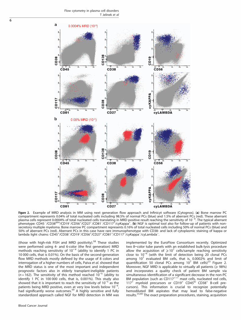
implemented by the EuroFlow Consortium recently. Optimizedtwo 8−color tube panels with an established bulk-lysis procedureallow the acquisition of ⩾ 107 cells/sample reaching sensitivityclose to 10−6 (with the limit of detection being 20 clonal PCsamong 107 evaluated BM cells, that is, 0.0002% and limit ofquantification 50 clonal PCs among 107 BM cells)23 Figure 2.Moreover, NGF MRD is applicable to virtually all patients (⩾ 98%)and incorporates a quality check of patient BM sample viasimultaneous identification of a significant decrease in the non-PCBM population (such as CD117+++ mast cells, nucleated red cells,117+ myeloid precursors or CD19+ CD45lo CD38+ B-cell pre-cursors). This information is crucial to recognize potentiallyhemodiluted BM aspirates that may lead to false-negativeresults.23,60 The exact preparation procedures, staining, acquisition
Figure 2. Example of MRD analysis in MM using next generation flow approach and Infinicyt software (Cytognos). (a) Bone marrow PCcompartment represents 0.04% of total nucleated cells including 98.5% of normal PCs (blue) and 1.5% of aberrant PCs (red). These aberrantplasma cells represent 0.0004% of total nucleated cells translating in MRD positive result reaching the sensitivity of 10− 6. The typical aberrantphenotype: CD45−/CD38dim/CD19−/CD56+/CD27−/CD81−/CD117+/cyKappa+. (b) NGF is optimal tool also for follow-up of patients with non-secretory multiple myeloma. Bone marrow PC compartment represents 0.16% of total nucleated cells including 50% of normal PCs (blue) and50% of aberrant PCs (red). Aberrant PCs in this case have rare immunophenotype with CD38- and lack of cytoplasmic staining of kappa orlambda light chains: CD45+/CD38−/CD19−/CD56+/CD27−/CD81+/CD117−/cyKappa−/cyLambda−.
Flow cytometry in plasma cell disordersT Jelinek et al
6
Blood Cancer Journal

and reporting of flow MRD are described in detail in recentlypublished guidelines.16,61 Flow MRD represents a fast, highlysensitive, standardized, cost-effective and widely available tech-nique for MRD evaluation in MM and is likely to soon beimplemented in routine clinical practice as one of the mostimportant clinical endpoints and sensitive readouts of treatmentefficacy.Other alternative to MFC-based method of MRD evaluation
represent molecular techniques, mainly ASO-PCR (allele-specificoligonucleotide PCR) and NGS (next-generation sequencing). Bothmethods reach high levels of sensitivity (down to 10−5–10−6) anddo not require immediate sample processing in contrary to MFCmethod (sample should be processed within 36–48 h after BMharvest). On the other hand, these methods have somedisadvantages: (i) lower applicability (ASO-PCR ~ 60–70%, NGS~90%), (ii) requirement of diagnostic sample to identify patient-specific clonotypic sequences, (iii) higher financial costs and (iv)methodologically complex and laborious methods difficult toimplement in routine clinical practice.62–64
CIRCULATING PLASMA CELLSLong-living BM PCs do not circulate under physiological condi-tions in PB, but in patients with certain monoclonal gammopa-thies (MG) tumor cells can egress from BM into PB as circulatingtumor cells (CTCs). Accordingly, the presence of CTCs wasdocumented not only in symptomatic MM, but also in SMM andMGUS.65,66 Paiva et al.67 demonstrated that in MM the number ofCTCs fluctuates throughout the day, following a circadian rhythmsimilar to CD34+ cells, suggesting that CTCs may egress to PB tocolonize other sites during the patients’ resting period.The detection of CTCs in NDMM patients by conventional
morphology is low (20% of all cases).68 By using more sensitive MFC(Figure 3), CTCs are detected in approximately 70–87% of all NDMMpatients65,69,70 and up to 60% of MGUS patients.71 The number ofCTCs is an independent prognostic factor in NDMM patients as wellas in AL amyloidosis, and their presence is associated with shortersurvival.66,72–74 CTCs are associated with an increased risk ofmalignant transformation in MGUS72 and SMM75,76 and they arealso a negative prognostic factor in RRMM.77 It is expected thatparticularly in SMM, MFC could become a convenient method (thatis, non-invasive) to identify patients with high-risk of progression to
MM before they develop end-organ damage.76 CTC FACS could alsobe used as a minimally-invasive method to interrogate patients’genomic landscape over time.78
From a biological standpoint, in comparison to BM PCs, CTCsshow down-regulation of some surface markers such as severalintegrins (CD11a/CD11c/CD29/CD49d/CD49e), adhesion molecules(CD33/CD56/CD117/CD138), and activation molecules (CD28/CD38/CD81).67 However, the cause and mechanism of PC egression fromBM remains poorly understood, as one of the hallmarks of aPCs inearly stages of the disease is their dependence on the BMmicroenvironment. Possible reasons for their migration/expansionmay include changes in angiogenesis and the subsequent increasein their proliferative rate, higher incidences of genetic abnormalitiesor changes in the expression profile of adhesion molecules.67,79,80
An important question remains unanswered: is the presence ofCTCs linked to the natural development of the disease or does itidentify a separate biological subgroup of patients?80–82 It wasshown that CTCs are mostly quiescent, but they could have a higherclonogenic potential to their paired BM counterparts. This fact couldexplain their potential ability of dissemination. In such cases, CTCswould represent a unique subpopulation of BM clonal PCs.67
Recently, Bretones et al. compared exomes of FASC sorted BM PCs,CTCs and PCs from extramedullary (EM) tissues in 6 MM patientswith EM disease and demonstrated the presence of systematicinter-tissue heterogeneity (though not for targetable mutations).CTCs displayed the highest frequency of shared mutations with thetwo other clones and the lowest number of private mutations,suggesting that while CTCs may bridge BM and EM myeloma, thereis continuous genomic evolution once different clones have seededin their respective niches.83
SOLITARY PLASMOCYTOMASolitary plasmocytomas (SPs) are rare PC dyscrasias (o5%) thatare characterized by the presence of bone (solitary boneplasmocytoma—SBP) or extramedullary soft tissue (extramedul-lary plasmocytoma—EMP) infiltrates of tumor PCs in the absenceof any clinical, laboratory and radiologic features of MM.84,85 Localradiotherapy, with or without surgical excision, is the recom-mended treatment of choice for these patients, and achieves highOS rates. Patients with SBP may progress to MM at a rate ofapproximately 40–50%, with lower rates of progression for
Figure 3. Next-generation flow approach used for identification of CTCs in MGUS, smoldering MM and active MM patients.
Flow cytometry in plasma cell disordersT Jelinek et al
7
Blood Cancer Journal
EMP.85,86 According to the 2014 IMWG criteria, SPs were dividedinto 2 entities: with or without minimal BM involvement based onthe evaluation of the presence of tumor PCs by MFC, Table 1. Infact, the presence of tumor PCs in patients with SP has becomethe most important prognostic marker defining the risk ofprogression. Paiva et al. have detected tumor PCs in 49% (17/35)of SBP patients and in 38% (11/29) of EMP patients. Seventy-onepercent of flow-positive vs only 8% of flow-negative SBP patientsevolved to MM (hazard ratio, 17.4; Po0.001). No significantdifferences were observed among EMP cases. Almost identicalresults were published by a UK group84 enhancing the importanceof MFC for the sub-classification of patients with SP and leading toits new classification as noted above.
AL AMYLOIDOSISImmunoglobulin light-chain amyloidosis (AL amyloidosis) is a rarePCD with an incidence of 10 patients per million cases per year butrepresents the most common of systemic amyloidoses.87 This oftenfatal disorder is characterized by the presence of a usually smallindolent clone of BM PCs that produce misfolded monoclonal lightchains of κ or most predominantly λ isotype formatting insolublefibrils causing damage to vital organs, Table 1.88 In AL, MFC can beuseful to confirm the presence of underlying tumor PCs responsiblefor the deposition of the amyloid light chains in tissue biopsies. Thisunderlying tumor PC clone is present and revealed by MFC invirtually all AL patients.89,90 There are only few MFC studies focusingon its prognostic value due to the rarity of this disease. Quantificationof the BM PC compartment by MFC was found to be a significantprognostic factor for OS (o1 vs 41% BMPC cutoff; 2-year OS ratesof 90 vs 44%, P=0.02) in the cohort of 35 newly diagnosed ALpatients. Moreover, detecting persistent nPCs at diagnosis identifieda subgroup of patients with prolonged OS (cut off 45 vs o5% ofnPCs/BMPC, 2−year rates of 88 vs 37%, P=0.01). In these series, 49%(17/35) of AL patients had 45% of nPCs/BMPC at diagnosis.91 Morerecently, a very similar study was published by the Mayo Clinic group(n=173) confirming some of the above mentioned results using adifferent cutoff for quantification of BM PCs (2.5%).92
WALDENSTRÖM MACROGLOBULINEMIAWaldenström macroglobulinemia (WM) was originally described byJan Gösta Waldenström in 1944.93,94 This disease is defined as alymphoplasmacytic lymphoma associated with the monoclonalimmunoglobulin IgM and BM infiltration by small IgM-producingclonal B-lymphocytes that may exhibit PC differentiation.95 Anincidence of 4 cases per million persons per year ranks this diseaseamongst rare disorders.93 The clinical presentation of patients withWM can be highly variable as the signs and symptoms are not onlydue to the infiltration of BM or lymphoid organs (anemia,hepatosplenomegaly and so on), but also due to the specificphysiochemical and immunological properties of monoclonal IgM(peripheral neuropathy, hyperviscosity and so on).96 Similarly toMM, it is supposed that virtually all cases of WM have gone throughthe benign stages of IgM MGUS and smoldering WM (SWM) beforedeveloping clinical symptoms. The hallmark of these three entities isthe presence of the monoclonal IgM protein. The differencebetween IgM MGUS and smoldering WM is in the BM infiltrationby B-lymphocytes (with a cutoff of 10% defined by morphology).Smoldering WM, in contrast to symptomatic WM, lacks any disease-associated clinical symptoms, thus not requiring any treatment.95,97
Advances in understanding the molecular pathogenesis of WMhave been paved by the discovery of a recurrent somatic mutationin MYD88L265P that is present in more than 90% of symptomaticWM patients, but also in at least half of IgM MGUS patientssuggesting its role as an early oncogenic event.98,99 The mostplausible normal counterpart of the WM clonal B-cell seems to be a
CD22low+/CD25+ memory B-cell as the cell of tumor origin,97 eventhough this question has still not been fully answered.100
MFC plays a significant role in the differential diagnostic work-up. It is particularly useful to distinguish WM from other B-celllymphoproliferative disorders as well as from related IgMmonoclonal gammopathies (that is, IgM MGUS, IgM myeloma)based on the specific immunophenotype of WM B-cells and PCs.96
The co-existence of a clonal B-cell population and the same light-chain-isotype clonal plasma cell population within the BMcompartment is usually accompanied by an increased numberof mast cells, thus representing typical findings.101 The mostcommon immunophenotype of WM clonal B-cells can bedescribed as follows: CD19+/CD22low+/CD23 −/CD25+/CD27+
/SmIgM+. In more detail, clonal B-cells systematically showSmIgM+ expression, with homogenously expressed CD22low+
(81% of cases) and CD25+ (89% of cases). CD79b and CD81 arepositive in all cases, whereas a heterogeneous bimodal pattern ofexpression can be observed for CD27 (51% of cases), CD38 (50%)and CD200 (62%). CD305 (LAIR1) is not expressed in up to 69% ofcases in contrast to its bimodal heterogeneous expression onnormal B-cells. The absence of the expression of CD5, CD10,CD11c and CD103 (in 95%, 100 96 and 100% of cases, respectively)is in contrast with most other mature lymphoid malignancies(chronic lymphocytic leukemia, mantle cell lymphoma, follicularlymphoma and hairy cell leukemia) except of marginal zonelymphoma (MZL), thus helping in their distinction. The most usefulmarkers to discriminate between WM and MZL, that possess theoverlapping phenotype in some cases, were SmIgM and CD79b,both over-expressed in WM, and CD305 upregulated in MZL.97,102
The antigenic profile of clonal PCs in WM resembles morethat of normal PCs and clearly differs from that of MM patients(for example, CD19−/CD27−/CD45− and/or CD56+). Bone marrowPCs from IgM MGUS to symptomatic WM patients show aprogressively increased frequency of CD19+/CD20+/CD45+ andSmIgM+ cells, together with a complete loss of CD56 reactivity.This suggests that the PC compartment is enriched in moreimmature clonal PCs with the plasmablastic phenotype.103 Thedifferent antigenic profile of clonal PCs can have a valuable inputto distinguish WM from IgM MM, together with the MYD88mutation status (not present in IgM MM) and the presence oft(11; 14) (does not occur in WM but with a high incidence inIgM MM).93,104,105
Flow cytometry may be a useful tool in the discriminationbetween IgM-related disorders. The thresholds of BM B-cellinfiltration (410%) and the degree of B-cell clonality (100%) arehighly specific to exclude the diagnosis of IgM-MGUS, but nototherwise to identify WM patients. However, flow cytometry canalso serve as a valuable prognostic tool in patients withsmoldering and symptomatic WM. Patients with smoldering WMwho have more than 10% of BM B-cell infiltration and whoshow full light-chain restriction of the B-cell compartment are at ahigher risk of progression to symptomatic disease (median TTP of26 vs 145 months, P⩽ 0.001). Similarly, patients with symptomaticWM and with 100% of light chain restricted B-cells showinferior survival vs those who maintain some polyclonal B-cells(median OS of 44 vs 78 months, P= 0.001). To note, full lightchain restriction of the PC compartment has no effect on both TTPand OS in smoldering and symptomatic WM patients,respectively.102
Finally, the role of MFC was also investigated during theresponse assessment. García-Sanz et al.106 described in 42 WMpatients that post-treatment BM residual disease status 45% ofmonoclonal B-cells was highly predictive of short PFS and OS,independently on hematological response. With the availability ofmore effective therapies for WM, it will be critical to combinesensitive and comprehensive monitoring of clonality in bothB-cells and PCs together with the evaluation of serum M-protein,since the complete eradication of only clonal B-cells but not PCs
Flow cytometry in plasma cell disordersT Jelinek et al
8
Blood Cancer Journal
(for example, through anti-CD20 immunochemotherapy) maytranslate into a favorable outcome despite sub-optimal responseaccording to current criteria (that is, persistent M-protein).
CONCLUSIONMFC immunophenotyping has become routinely used in themanagement of patients with PC dyscrasias. MFC plays asignificant role during the differential diagnostic work-up, whenthis technique can provide relatively fast and conclusive results,thus helping to distinguish between malignant and reactiveconditions as well as classifying different monoclonal gammopa-thies and other lymphoproliferative disorders. Flow cytometry canalso be useful in predicting outcomes not only in MGUS/SMM/MM,but also in patients with SWM/WM, AL and SP. The obtainedinformation could be particularly beneficial in maintaining a closersurveillance of patients at a higher risk of progression and inidentifying the subgroup of patients with exceptionally favorableoutcomes. Flow-based MRD monitoring in MM, particularly afterthe implementation of NGF, represents a fast, highly sensitive,standardized, cost-effective and widely available technique. Theimportance of defining the MRD status is indisputable in MMpatients and flow MRD is progressively being implemented inroutine clinical practice as one of the most relevant treatmentend-points. Immunophenotyping has greatly contributed also in theresearch of monoclonal gammopathies and may be of significantimportance in the upcoming era of immunotherapy, especially indefining and monitoring therapeutic targets such as CD38, SLAMF7,PD-L1, BCMA and many others that are yet to be discovered.107,108
CONFLICT OF INTERESTThe authors declare no conflict of interest.
ACKNOWLEDGEMENTSWe give special thanks to Shira Timilsina, MD for English language editing. This workwas supported by the MH CZ—DRO—FNOs/2017 and Ministry of Health (17-30089A);by the Institutional Development Plan of University of Ostrava (IRP201550), by SGS18/PrF/2017–2018 and by ‘Strengthening international cooperation in science, researchand education, projet ID: 01211/2016/RRC’. It was also supported by the InternationalMyeloma Foundation Black Swan Research Initiative and the European ResearchCouncil (ERC) 2015 Starting Grant (MYELOMA-NEXT).
AUTHOR CONTRIBUTIONSTJ—wrote the manuscript, concept, coordination. RB—wrote the part about
CTCs and PCL. MZ—took part in the preparation of the manuscript. LB—took
part in the preparation of the manuscript. MS—took part in the preparation of
the manuscript, overall proofread. TS—overall proofread. BP—concept, overall
proofread, work coordination. RH—work coordination, overall proofread.
REFERENCES1 Castillo JJ. Plasma cell disorders. Prim Care 2016; 43: 677–691.2 Sant M, Allemani C, Tereanu C, Angelis RD, Capocaccia R, Visser O et al. Incidence
of hematologic malignancies in Europe by morphologic subtype: results of theHAEMACARE project. Blood 2010; 116: 3724–3734.
3 Rajkumar SV, Dimopoulos MA, Palumbo A, Blade J, Merlini G, Mateos M-V et al.International Myeloma Working Group updated criteria for the diagnosis ofmultiple myeloma. Lancet Oncol 2014; 15: e538–e548.
4 Kyle RA, Therneau TM, Rajkumar SV, Larson DR, Plevak MF, Offord JR et al.Prevalence of monoclonal gammopathy of undetermined significance. N Engl JMed 2006; 354: 1362–1369.
5 Bladé J. Clinical practice. Monoclonal gammopathy of undetermined sig-nificance. N Engl J Med 2006; 355: 2765–2770.
6 Kyle RA, Rajkumar SV. Epidemiology of the plasma-cell disorders. Best Pract ResClin Haematol 2007; 20: 637–664.
7 Kyle RA, Therneau TM, Rajkumar SV, Offord JR, Larson DR, Plevak MF et al.A long-term study of prognosis in monoclonal gammopathy of undeterminedsignificance. N Engl J Med 2002; 346: 564–569.
8 Kyle RA, Remstein ED, Therneau TM, Dispenzieri A, Kurtin PJ, Hodnefield JM et al.Clinical course and prognosis of smoldering (asymptomatic) multiple myeloma.N Engl J Med 2007; 356: 2582–2590.
9 Kaleem Z, Crawford E, Pathan MH, Jasper L, Covinsky MA, Johnson LR et al. Flowcytometric analysis of acute leukemias. Diagnostic utility and critical analysisof data. Arch Pathol Lab Med 2003; 127: 42–48.
10 Braylan RC. Impact of flow cytometry on the diagnosis and characterization oflymphomas, chronic lymphoproliferative disorders and plasma cell neoplasias.Cytom Part J Int Soc Anal Cytol 2004; 58: 57–61.
11 Kwok M, Rawstron AC, Varghese A, Evans PAS, O’Connor SJM, Doughty C et al.Minimal residual disease is an independent predictor for 10-year survival in CLL.Blood 2016; 128: 2770–2773.
12 Theunissen P, Mejstrikova E, Sedek L, van der Sluijs-Gelling AJ, Gaipa G, Bartels Met al. Standardized flow cytometry for highly sensitive MRD measurements inB-cell acute lymphoblastic leukemia. Blood 2016; 129: 347–357.
13 Grimwade D, Freeman SD. Defining minimal residual disease in acute myeloidleukemia: which platforms are ready for ‘prime time’? Blood 2014; 124:3345–3355.
14 Caraux A, Klein B, Paiva B, Bret C, Schmitz A, Fuhler GM et al. Circulating human Band plasma cells. Age-associated changes in counts and detailed characteriza-tion of circulating normal CD138- and CD138+ plasma cells. Haematologica2010; 95: 1016–1020.
15 Jelinek T, Hajek R. Monoclonal antibodies - A new era in the treatment ofmultiple myeloma. Blood Rev 2016; 30: 101–110.
16 Arroz M, Came N, Lin P, Chen W, Yuan C, Lagoo A et al. Consensus guidelines onplasma cell myeloma minimal residual disease analysis and reporting. CytometryB Clin Cytom 2016; 90: 31–39.
17 Flores-Montero J, de Tute R, Paiva B, Perez JJ, Böttcher S, Wind H et al. Immu-nophenotype of normal vs. myeloma plasma cells: toward antibody panel spe-cifications for MRD detection in multiple myeloma. Cytometry B Clin Cytom 2016;90: 61–72.
18 Paiva B, Puig N, Cedena MT, de Jong BG, Ruiz Y, Rapado I et al. Differentiationstage of myeloma plasma cells: biological and clinical significance. Leukemia2017; 31: 382–392.
19 Ocqueteau M, Orfao A, Almeida J, Bladé J, González M, García-Sanz R et al.Immunophenotypic characterization of plasma cells from monoclonal gammo-pathy of undetermined significance patients. Implications for the differentialdiagnosis between MGUS and multiple myeloma. Am J Pathol 1998; 152:1655–1665.
20 Paiva B, Merino J, San Miguel JF. Utility of flow cytometry studies in the man-agement of patients with multiple myeloma. Curr Opin Oncol 2016; 28: 511–517.
21 Olteanu H, Harrington AM, Hari P, Kroft SH. CD200 expression in plasma cellmyeloma. Br J Haematol 2011; 153: 408–411.
22 Olteanu H. Role of flow cytometry in the diagnosis and prognosis of plasma cellmyeloma. Surg Pathol Clin 2016; 9: 101–116.
23 Flores-Montero J, Sanoja-Flores L, Paiva B, Puig N, García-Sánchez O, Böttcher Set al. Next Generation Flow for highly sensitive and standardized detection ofminimal residual disease in multiple myeloma. Leukemia 2017; 31: 2094–2103.
24 Nadav L, Katz B-Z, Baron S, Yossipov L, Polliack A, Deutsch V et al. Diverse nicheswithin multiple myeloma bone marrow aspirates affect plasma cell enumeration.Br J Haematol 2006; 133: 530–532.
25 Rawstron AC, Orfao A, Beksac M, Bezdickova L, Brooimans RA, Bumbea H et al.Report of the European Myeloma Network on multiparametric flow cytometry inmultiple myeloma and related disorders. Haematologica 2008; 93: 431–438.
26 Seegmiller AC, Xu Y, McKenna RW, Karandikar NJ. Immunophenotypic differ-entiation between neoplastic plasma cells in mature B-cell lymphoma vs plasmacell myeloma. Am J Clin Pathol 2007; 127: 176–181.
27 Olteanu H, Wang H-Y, Chen W, McKenna RW, Karandikar NJ. Immunophenotypicstudies of monoclonal gammopathy of undetermined significance. BMC ClinPathol 2008; 8: 13.
28 Paiva B, Almeida J, Pérez-Andrés M, Mateo G, López A, Rasillo A et al. Utility offlow cytometry immunophenotyping in multiple myeloma and other clonalplasma cell-related disorders. Cytometry B Clin Cytom 2010; 78: 239–252.
29 Pérez-Persona E, Vidriales M-B, Mateo G, García-Sanz R, Mateos M-V, de Coca AGet al. New criteria to identify risk of progression in monoclonal gammopathy ofuncertain significance and smoldering multiple myeloma based on multi-parameter flow cytometry analysis of bone marrow plasma cells. Blood 2007;110: 2586–2592.
30 Paiva B, Vídriales M-B, Rosiñol L, Martínez-López J, Mateos M-V, Ocio EM et al.A multiparameter flow cytometry immunophenotypic algorithm for the identi-fication of newly diagnosed symptomatic myeloma with an MGUS-like signatureand long-term disease control. Leukemia 2013; 27: 2056–2061.
Flow cytometry in plasma cell disordersT Jelinek et al
9
Blood Cancer Journal

31 Greipp PR, Miguel JS, Durie BGM, Crowley JJ, Barlogie B, Bladé J et al. Interna-tional Staging System for Multiple Myeloma. J Clin Oncol 2005; 23: 3412–3420.
32 Palumbo A, Avet-Loiseau H, Oliva S, Lokhorst HM, Goldschmidt H, Rosinol L et al.Revised international staging system for multiple myeloma: A Report FromInternational Myeloma Working Group. J Clin Oncol Off J Am Soc Clin Oncol 2015;33: 2863–2869.
33 Avet-Loiseau H, Durie BGM, Cavo M, Attal M, Gutierrez N, Haessler J et al.Combining fluorescent in situ hybridization data with ISS staging improves riskassessment in myeloma: an International Myeloma Working Group collaborativeproject. Leukemia 2013; 27: 711–717.
34 Pérez-Persona E, Mateo G, García-Sanz R, Mateos M-V, de Las Heras N,de Coca AG et al. Risk of progression in smouldering myeloma and monoclonalgammopathies of unknown significance: comparative analysis of the evolutionof monoclonal component and multiparameter flow cytometry of bone marrowplasma cells. Br J Haematol 2010; 148: 110–114.
35 Paiva B, Johnson SK, Mateos M-V, Alapat DV, Puig N, Hernandez M-T et al.Automated multiparameter flow cytometry (MFC) immunophenotyping forreproducible identification of high risk smoldering multiple myeloma (SMM).Blood 2016; 128: 373–373.
36 Paiva B, Vidriales M-B, Mateo G, Pérez JJ, Montalbán MA, Sureda A et al. Thepersistence of immunophenotypically normal residual bone marrow plasma cellsat diagnosis identifies a good prognostic subgroup of symptomatic multiplemyeloma patients. Blood 2009; 114: 4369–4372.
37 Lahuerta J-J, Paiva B, Vidriales M-B, Cordón L, Cedena M-T, Puig N et al. Depth ofresponse in multiple myeloma: a pooled analysis of three PETHEMA/GEMClinical Trials. J Clin Oncol 2017; 35: 2900–2910.
38 Edling CE, Hallberg B. c-Kit—a hematopoietic cell essential receptortyrosine kinase. Int J Biochem Cell Biol 2007; 39: 1995–1998.
39 Mateo G, Montalbán MA, Vidriales M-B, Lahuerta JJ, Mateos MV, Gutiérrez N et al.Prognostic value of immunophenotyping in multiple myeloma: a study by thePETHEMA/GEM cooperative study groups on patients uniformly treated withhigh-dose therapy. J Clin Oncol Off J Am Soc Clin Oncol 2008; 26: 2737–2744.
40 Schmidt-Hieber M, Pérez-Andrés M, Paiva B, Flores-Montero J, Perez JJ,Gutierrez NC et al. CD117 expression in gammopathies is associated with analtered maturation of the myeloid and lymphoid hematopoietic cell compart-ments and favorable disease features. Haematologica 2011; 96: 328–332.
41 Murray ME, Gavile CM, Nair JR, Koorella C, Carlson LM, Buac D et al. CD28-mediated pro-survival signaling induces chemotherapeutic resistance in multi-ple myeloma. Blood 2014; 123: 3770–3779.
42 Paiva B, Gutiérrez N-C, Chen X, Vídriales M-B, Montalbán M-Á, Rosiñol L et al.Clinical significance of CD81 expression by clonal plasma cells in high-risksmoldering and symptomatic multiple myeloma patients. Leukemia 2012; 26:1862–1869.
43 Paiva B, Puig N, Arana P, Cedena T, Cordon L, Vidriales MB et al. Prognostic valueof antigen expression in multiple myeloma (MM): a large GEM/Pethema Studybased in four consecutive clinical trials. Blood 2015; 126: 19–19.
44 San Miguel JF, García-Sanz R, González M, Orfão A. DNA cell content studies inmultiple myeloma. Leuk Lymphoma 1996; 23: 33–41.
45 García-Sanz R, Orfão A, González M, Moro MJ, Hernández JM, Ortega F et al.Prognostic implications of DNA aneuploidy in 156 untreated multiple myelomapatients. Castelano-Leonés (Spain) Cooperative Group for the Study of Mono-clonal Gammopathies. Br J Haematol 1995; 90: 106–112.
46 Paiva B, Vídriales M-B, Montalbán M-Á, Pérez JJ, Gutiérrez NC, Rosiñol L et al.Multiparameter flow cytometry evaluation of plasma cell DNA content andproliferation in 595 transplant-eligible patients with myeloma included in theSpanish GEM2000 and GEM2005o65y trials. Am J Pathol 2012; 181: 1870–1878.
47 Bladé J, Samson D, Reece D, Apperley J, Björkstrand B, Gahrton G et al. Criteriafor evaluating disease response and progression in patients with multiplemyeloma treated by high-dose therapy and haemopoietic stem cell transplan-tation. Myeloma Subcommittee of the EBMT. European Group for Blood andMarrow Transplant. Br J Haematol 1998; 102: 1115–1123.
48 Durie BGM, Harousseau J-L, Miguel JS, Bladé J, Barlogie B, Anderson K et al.International uniform response criteria for multiple myeloma. Leukemia 2006; 20:1467–1473.
49 Rajkumar SV, Harousseau J-L, Durie B, Anderson KC, Dimopoulos M, Kyle R et al.Consensus recommendations for the uniform reporting of clinical trials: report ofthe International Myeloma Workshop Consensus Panel 1. Blood 2011; 117:4691–4695.
50 van de Velde HJK, Liu X, Chen G, Cakana A, Deraedt W, Bayssas M. Completeresponse correlates with long-term survival and progression-free survival inhigh-dose therapy in multiple myeloma. Haematologica 2007; 92: 1399–1406.
51 Gay F, Larocca A, Wijermans P, Cavallo F, Rossi D, Schaafsma R et al. Completeresponse correlates with long-term progression-free and overall survival inelderly myeloma treated with novel agents: analysis of 1175 patients. Blood2011; 117: 3025–3031.
52 Kumar S, Paiva B, Anderson KC, Durie B, Landgren O, Moreau P et al. Interna-tional Myeloma Working Group consensus criteria for response and minimalresidual disease assessment in multiple myeloma. Lancet Oncol 2016; 17:e328–e346.
53 Barlogie B, Mitchell A, van Rhee F, Epstein J, Morgan GJ, Crowley J. Curingmyeloma at last: defining criteria and providing the evidence. Blood 2014; 124:3043–3051.
54 San Miguel JF, Almeida J, Mateo G, Bladé J, López-Berges C, Caballero D et al.Immunophenotypic evaluation of the plasma cell compartment in multiplemyeloma: a tool for comparing the efficacy of different treatment strategies andpredicting outcome. Blood 2002; 99: 1853–1856.
55 Rawstron AC, Davies FE, DasGupta R, Ashcroft AJ, Patmore R, Drayson MT et al.Flow cytometric disease monitoring in multiple myeloma: the relationshipbetween normal and neoplastic plasma cells predicts outcome after transplan-tation. Blood 2002; 100: 3095–3100.
56 Paiva B, Vidriales M-B, Cerveró J, Mateo G, Pérez JJ, Montalbán MA et al. Mul-tiparameter flow cytometric remission is the most relevant prognostic factor formultiple myeloma patients who undergo autologous stem cell transplantation.Blood 2008; 112: 4017–4023.
57 Rawstron AC, Child JA, de Tute RM, Davies FE, Gregory WM, Bell SE et al. Minimalresidual disease assessed by multiparameter flow cytometry in multiple mye-loma: impact on outcome in the Medical Research Council Myeloma IX Study.J Clin Oncol Off J Am Soc Clin Oncol 2013; 31: 2540–2547.
58 Paiva B, Gutiérrez NC, Rosiñol L, Vídriales M-B, Montalbán M-Á, Martínez-López Jet al. High-risk cytogenetics and persistent minimal residual disease by multi-parameter flow cytometry predict unsustained complete response after auto-logous stem cell transplantation in multiple myeloma. Blood 2012; 119: 687–691.
59 Paiva B, Cedena M-T, Puig N, Arana P, Vidriales M-B, Cordon L et al. Minimalresidual disease monitoring and immune profiling in multiple myeloma inelderly patients. Blood 2016; 127: 3165–3174.
60 Paiva B, García-Sanz R, San Miguel JF. Multiple Myeloma Minimal Residual Dis-ease. Cancer Treat Res 2016; 169: 103–122.
61 Stetler-Stevenson M, Paiva B, Stoolman L, Lin P, Jorgensen JL, Orfao A et al.Consensus guidelines for myeloma minimal residual disease sample staining anddata acquisition. Cytometry B Clin Cytom 2016; 90: 26–30.
62 Martinez-Lopez J, Sanchez-Vega B, Barrio S, Cuenca I, Ruiz-Heredia Y, Alonso Ret al. Analytical and clinical validation of a novel in-house deep-sequencingmethod for minimal residual disease monitoring in a phase II trial for multiplemyeloma. Leukemia 2017; 31: 1446–1449.
63 Martinez-Lopez J, Lahuerta JJ, Pepin F, González M, Barrio S, Ayala R et al.Prognostic value of deep sequencing method for minimal residual diseasedetection in multiple myeloma. Blood 2014; 123: 3073–3079.
64 Puig N, Sarasquete ME, Balanzategui A, Martínez J, Paiva B, García H et al. Criticalevaluation of ASO RQ-PCR for minimal residual disease evaluation in multiplemyelomaA comparative analysis with flow cytometry. Leukemia 2014; 28:391–397.
65 Nowakowski GS, Witzig TE, Dingli D, Tracz MJ, Gertz MA, Lacy MQ et al. Circu-lating plasma cells detected by flow cytometry as a predictor of survival in 302patients with newly diagnosed multiple myeloma. Blood 2005; 106: 2276–2279.
66 Gonsalves WI, Rajkumar SV, Gupta V, Morice WG, Timm MM, Singh PP et al.Quantification of clonal circulating plasma cells in newly diagnosed multiplemyeloma: implications for redefining high-risk myeloma. Leukemia 2014; 28:2060–2065.
67 Paiva B, Paino T, Sayagues J-M, Garayoa M, San-Segundo L, Martín M et al.Detailed characterization of multiple myeloma circulating tumor cells showsunique phenotypic, cytogenetic, functional, and circadian distribution profile.Blood 2013; 122: 3591–3598.
68 Granell M, Calvo X, Garcia-Guiñón A, Escoda L, Abella E, Martínez CM et al.Prognostic impact of circulating plasma cells in patients with multiple myeloma:implications for plasma cell leukaemia definition. Haematologica 2017; 102:1099–1104, haematol.2016.158303.
69 Vagnoni D, Travaglini F, Pezzoni V, Ruggieri M, Bigazzi C, Dalsass A et al.Circulating plasma cells in newly diagnosed symptomatic multiple myelomaas a possible prognostic marker for patients with standard-risk cytogenetics.Br J Haematol 2015; 170: 523–531.
70 San Miguel JF, Gutiérrez NC, Mateo G, Orfao A. Conventional diagnostics inmultiple myeloma. Eur J Cancer Oxf Engl 1990 2006; 42: 1510–1519.
71 Burgos L, Alignani D, Garces J-J, Ortiz L, Jelinek T, Segura V et al. Non-invasivegenetic profiling is highly applicable in multiple myeloma (MM) through char-acterization of circulating tumor cells (CTCs). Blood 2016; 128: 801–801.
72 Kumar S, Rajkumar SV, Kyle RA, Lacy MQ, Dispenzieri A, Fonseca R et al. Prog-nostic value of circulating plasma cells in monoclonal gammopathy of unde-termined significance. J Clin Oncol 2005; 23: 5668–5674.
73 Dingli D, Nowakowski GS, Dispenzieri A, Lacy MQ, Hayman SR, Rajkumar SV et al.Flow cytometric detection of circulating myeloma cells before transplantation in
Flow cytometry in plasma cell disordersT Jelinek et al
10
Blood Cancer Journal
patients with multiple myeloma: a simple risk stratification system. Blood 2006;107: 3384–3388.
74 Pardanani A, Witzig TE, Schroeder G, McElroy EA, Fonseca R, Dispenzieri A et al.Circulating peripheral blood plasma cells as a prognostic indicator in patientswith primary systemic amyloidosis. Blood 2003; 101: 827–830.
75 Bianchi G, Kyle RA, Larson DR, Witzig TE, Kumar S, Dispenzieri A et al. High levelsof peripheral blood circulating plasma cells as a specific risk factor for pro-gression of smoldering multiple myeloma. Leukemia 2013; 27: 680–685.
76 Gonsalves WI, Rajkumar SV, Dispenzieri A, Dingli D, Timm MM, Morice WG et al.Quantification of circulating clonal plasma cells via multiparametric flow cyto-metry identifies patients with smoldering multiple myeloma at high risk ofprogression. Leukemia 2017; 31: 130–135.
77 Peceliunas V, Janiulioniene A, Matuzeviciene R, Zvirblis T, Griskevicius L. Circu-lating plasma cells predict the outcome of relapsed or refractory multiplemyeloma. Leuk Lymphoma 2012; 53: 641–647.
78 Mishima Y, Paiva B, Shi J, Park J, Manier S, Takagi S et al. The mutationallandscape of circulating tumor cells in multiple myeloma. Cell Rep 2017; 19:218–224.
79 Kumar S, Witzig TE, Greipp PR, Rajkumar SV. Bone marrow angiogenesis andcirculating plasma cells in multiple myeloma. Br J Haematol 2003; 122: 272–274.
80 An G, Qin X, Acharya C, Xu Y, Deng S, Shi L et al. Multiple myeloma patients withlow proportion of circulating plasma cells had similar survival with primaryplasma cell leukemia patients. Ann Hematol 2015; 94: 257–264.
81 Pellat-Deceunynck C, Barillé S, Jego G, Puthier D, Robillard N, Pineau D et al. Theabsence of CD56 (NCAM) on malignant plasma cells is a hallmark of plasma cellleukemia and of a special subset of multiple myeloma. Leukemia 1998; 12:1977–1982.
82 Lohr JG, Kim S, Gould J, Knoechel B, Drier Y, Cotton MJ et al. Genetic inter-rogation of circulating multiple myeloma cells at single-cell resolution. Sci TranslMed 2016; 8: 363ra147.
83 Bretones G, Paiva B, Valdes-Mas R, Alignani D, Garcia M, Burgos L et al. Genomicprofiles of bone marrow (BM) clonal plasma cells (PCs) vs circulating tumor cells(CTCs) and Extramedullary (EM) plasmacytomas in multiple myeloma (MM).Blood 2016; 128: 4442–4442.
84 Hill QA, Rawstron AC, de Tute RM, Owen RG. Outcome prediction in plasmacy-toma of bone: a risk model utilizing bone marrow flow cytometry and light-chainanalysis. Blood 2014; 124: 1296–1299.
85 Dimopoulos MA, Terpos E. Solitary bone plasmacytomas need to flow. Blood2014; 124: 1209–1210.
86 Katodritou E, Terpos E, Symeonidis AS, Pouli A, Kelaidi C, Kyrtsonis M-C et al.Clinical features, outcome, and prognostic factors for survival and evolution tomultiple myeloma of solitary plasmacytomas: a report of the Greek myelomastudy group in 97 patients. Am J Hematol 2014; 89: 803–808.
87 Jelinek T, Kryukova E, Kufova Z, Kryukov F, Hajek R. Proteasome inhibitors inAL amyloidosis: focus on mechanism of action and clinical activity. HematolOncol 2016; e-pub ahead of print 20 September 2016; doi:10.1002/hon.2351.
88 Jelinek T, Kufova Z, Hajek R. Immunomodulatory drugs in AL amyloidosis. CritRev Oncol Hematol 2016; 99: 249–260.
89 Paiva B, Martinez-Lopez J, Corchete LA, Sanchez-Vega B, Rapado I, Puig N et al.Phenotypic, transcriptomic, and genomic features of clonal plasma cells in light-chain amyloidosis. Blood 2016; 127: 3035–3039.
90 Lisenko K, Schönland SO, Jauch A, Andrulis M, Röcken C, Ho AD et al. Flowcytometry-based characterization of underlying clonal B and plasma cells inpatients with light chain amyloidosis. Cancer Med 2016; 5: 1464–1472.
91 Paiva B, Vídriales M-B, Pérez JJ, López-Berges M-C, García-Sanz R, Ocio EMet al. The clinical utility and prognostic value of multiparameter flow cyto-metry immunophenotyping in light-chain amyloidosis. Blood 2011; 117:3613–3616.
92 Muchtar E, Jevremovic D, Dispenzieri A, Dingli D, Buadi FK, Lacy MQ et al. Theprognostic value of multiparametric flow cytometry in AL amyloidosis at diag-nosis and at the end of first-line treatment. Blood 2017; 129: 82–87.
93 Gertz MA. Waldenström macroglobulinemia: 2017 update on diagnosis, riskstratification, and management. Am J Hematol 2017; 92: 209–217.
94 Waldenstrom J. Macroglobulinaemia. Acta Haematol 1958; 20: 33–39.95 Owen RG, Treon SP, Al-Katib A, Fonseca R, Greipp PR, McMaster ML et al. Clin-
icopathological definition of Waldenstrom’s macroglobulinemia: consensuspanel recommendations from the Second International Workshop on Walden-strom’s Macroglobulinemia. Semin Oncol 2003; 30: 110–115.
96 Castillo JJ, Garcia-Sanz R, Hatjiharissi E, Kyle RA, Leleu X, McMaster M et al.Recommendations for the diagnosis and initial evaluation of patients withWaldenström Macroglobulinaemia: A Task Force from the 8th InternationalWorkshop on Waldenström Macroglobulinaemia. Br J Haematol 2016; 175:77–86.
97 Paiva B, Corchete LA, Vidriales M-B, García-Sanz R, Perez JJ, Aires-Mejia I et al.The cellular origin and malignant transformation of Waldenström macro-globulinemia. Blood 2015; 125: 2370–2380.
98 Treon SP, Xu L, Yang G, Zhou Y, Liu X, Cao Y et al. MYD88 L265P somaticmutation in Waldenström’s macroglobulinemia. N Engl J Med 2012; 367:826–833.
99 Landgren O, Staudt L. MYD88 L265P somatic mutation in IgM MGUS. N Engl JMed 2012; 367: 2255–2256-2257.
100 García-Sanz R, Jiménez C, Puig N, Paiva B, Gutiérrez NC, Rodríguez-Otero P et al.Origin of Waldenstrom’s macroglobulinaemia. Best Pract Res Clin Haematol 2016;29: 136–147.
101 San Miguel JF, Vidriales MB, Ocio E, Mateo G, Sánchez-Guijo F, Sánchez ML et al.Immunophenotypic analysis of Waldenstrom’s macroglobulinemia. Semin Oncol2003; 30: 187–195.
102 Paiva B, Montes MC, García-Sanz R, Ocio EM, Alonso J, de Las Heras N et al.Multiparameter flow cytometry for the identification of the Waldenström’s clonein IgM-MGUS and Waldenström’s Macroglobulinemia: new criteria for differentialdiagnosis and risk stratification. Leukemia 2014; 28: 166–173.
103 Paiva B, Chandia M, Vidriales M-B, Colado E, Caballero-Velázquez T, Escalante Fet al. Multiparameter flow cytometry for staging of solitary bone plasmacytoma:new criteria for risk of progression to myeloma. Blood 2014; 124: 1300–1303.
104 Avet-Loiseau H, Garand R, Lodé L, Robillard N, Bataille R. 14q32 Translocationsdiscriminate IgM multiple myeloma from Waldenstrom’s macroglobulinemia.Semin Oncol 2003; 30: 153–155.
105 Willenbacher W, Willenbacher E, Brunner A, Manzl C. Improved accuracy ofdiscrimination between IgM multiple myeloma and Waldenström macro-globulinaemia by testing for MYD88 L265P mutations. Br J Haematol 2013; 161:902–904.
106 García-Sanz R, Ocio E, Caballero A, Magalhães RJP, Alonso J, López-Anglada Let al. Post-treatment bone marrow residual disease45% by flow cytometry ishighly predictive of short progression-free and overall survival in patients withWaldenström’s macroglobulinemia. Clin Lymphoma Myeloma Leuk 2011; 11:168–171.
107 Jelinek T, Hajek R. PD-1/PD-L1 inhibitors in multiple myeloma: The present andthe future. OncoImmunology 2016; 5: e1254856.
108 Seckinger A, Delgado JA, Moser S, Moreno L, Neuber B, Grab A et al. Targetexpression, generation, preclinical activity, and pharmacokinetics of the BCMA-Tcell bispecific antibody EM801 for multiple myeloma treatment. Cancer Cell 2017;31: 396–410.
109 Robillard N, Jego G, Pellat-Deceunynck C, Pineau D, Puthier D, Mellerin MP et al.CD28, a marker associated with tumoral expansion in multiple myeloma. ClinCancer Res Off J Am Assoc Cancer Res 1998; 4: 1521–1526.
110 Robillard N, Wuillème S, Lodé L, Magrangeas F, Minvielle S, Avet-Loiseau H. CD33is expressed on plasma cells of a significant number of myeloma patients, andmay represent a therapeutic target. Leukemia 2005; 19: 2021–2022.
111 Lee KH, Seo HS, Sohn JY, Lee E, Lee H, Eom H-S et al. Abstract 3123: Aberrantexpression of CD33 is associated with poor prognosis in patients with multiplemyeloma and tumor progression. Cancer Res 2016; 76(14 Supplement): 3123–3123.
112 Gonsalves WI, Timm MM, Rajkumar SV, Morice WG, Dispenzieri A, Buadi FK et al.The prognostic significance of CD45 expression by clonal bone marrow plasmacells in patients with newly diagnosed multiple myeloma. Leuk Res 2016; 44: 32–39.
113 Pojero F, Flores-Montero J, Sanoja L, Pérez JJ, Puig N, Paiva B et al. Utility ofCD54, CD229, and CD319 for the identification of plasma cells in patients withclonal plasma cell diseases. Cytometry B Clin Cytom 2016; 90: 91–100.
114 Iqbal MS, Otsuyama K-I, Shamsasenjan K, Asaoku H, Mahmoud MS, Gondo T et al.Constitutively lower expressions of CD54 on primary myeloma cells and theirdifferent localizations in bone marrow. Eur J Haematol 2009; 83: 302–312.
115 Van Camp B, Durie BG, Spier C, De Waele M, Van Riet I, Vela E et al. Plasma cellsin multiple myeloma express a natural killer cell-associated antigen: CD56(NKH-1; Leu-19). Blood 1990; 76: 377–382.
116 Van Riet I, De Waele M, Remels L, Lacor P, Schots R, Van Camp B. Expression ofcytoadhesion molecules (CD56, CD54, CD18 and CD29) by myelomaplasma cells. Br J Haematol 1991; 79: 421–427.
117 Rawstron A, Barrans S, Blythe D, Davies F, English A, Pratt G et al. Distribution ofmyeloma plasma cells in peripheral blood and bone marrow correlates withCD56 expression. Br J Haematol 1999; 104: 138–143.
118 Conticello C, Giuffrida R, Parrinello N, Buccheri S, Adamo L, Sciuto MR et al.CD200 expression in patients with Multiple Myeloma: another piece ofthe puzzle. Leuk Res 2013; 37: 1616–1621.
119 Tai Y-T, Dillon M, Song W, Leiba M, Li X-F, Burger P et al. Anti-CS1 humanizedmonoclonal antibody HuLuc63 inhibits myeloma cell adhesion and inducesantibody-dependent cellular cytotoxicity in the bone marrow milieu. Blood 2008;112: 1329–1337.
Flow cytometry in plasma cell disordersT Jelinek et al
11
Blood Cancer Journal

120 Muccio VE, Saraci E, Gilestro M, Gattei V, Zucchetto A, Astolfi M et al. Multiplemyeloma: New surface antigens for the characterization of plasma cells in theera of novel agents. Cytometry B Clin Cytom 2016; 90: 81–90.
121 Roussel M, Lauwers-Cances V, Robillard N, Hulin C, Leleu X, Benboubker L et al.Front-line transplantation program with lenalidomide, bortezomib, and dex-amethasone combination as induction and consolidation followed by lenalido-mide maintenance in patients with multiple myeloma: a phase II study by theIntergroupe Francophone du Myélome. J Clin Oncol 2014; 32: 2712–2717.
122 Paiva B, Chandia M, Puig N, Vidriales M-B, Perez JJ, Lopez-Corral L et al. The prognosticvalue of multiparameter flow cytometry minimal residual disease assessment inrelapsed multiple myeloma. Haematologica 2015; 100: e53–e55.
123 Fukumoto K, Fujisawa M, Suehara Y, Narita K-T, Usui Y, Takeuchi M et al. Prog-nostic impact of immunophenotypic complete response in patients with mul-tiple myeloma achieving better than complete response. Leuk Lymphoma 2016;57: 1786–1792.
124 Attal M, Lauwers-Cances V, Hulin C, Leleu X, Caillot D, Escoffre M et al. Lenali-domide, bortezomib, and dexamethasone with transplantation for myeloma. NEngl J Med 2017; 376: 1311–1320.
This work is licensed under a Creative Commons Attribution 4.0International License. The images or other third party material in this
article are included in the article’s Creative Commons license, unless indicatedotherwise in the credit line; if the material is not included under the Creative Commonslicense, users will need to obtain permission from the license holder to reproduce thematerial. To view a copy of this license, visit http://creativecommons.org/licenses/by/4.0/
© The Author(s) 2017
Flow cytometry in plasma cell disordersT Jelinek et al
12
Blood Cancer Journal