CT ABDOMEN/PELVIS ANATOMY USC/SOM Department of Radiology Kirk Peterson, MD Class of 2004.
53
CT ABDOMEN/PELVIS ANATOMY USC/SOM Department of Radiology Kirk Peterson, MD Class of 2004
-
date post
22-Dec-2015 -
Category
Documents
-
view
230 -
download
10
Transcript of CT ABDOMEN/PELVIS ANATOMY USC/SOM Department of Radiology Kirk Peterson, MD Class of 2004.
CT ABDOMEN/PELVIS ANATOMY
USC/SOM
Department of Radiology
Kirk Peterson, MDClass of 2004
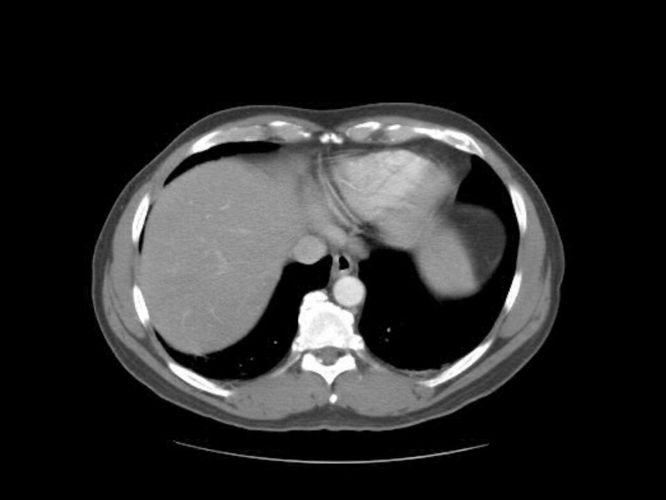
In this sequence of images, we will label the abdominal vasculature. The CT images are 5mm slices with soft tissue window settings. IV and
oral contrast have been administered which causes the vessels and GI tract to appear
hyperdense (white). Some images will contain labels to assist with tracking the vessels. Practice paging up and down to trace vessel’s origin and termination. It is best to view these images in the
movie mode
IMAGES ARE VIEWED AS LOOKING FROM THE FEETRIGHT LEFT
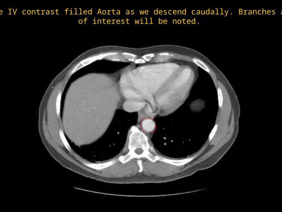
Follow the IV contrast filled Aorta as we descend caudally. Branches and pointsof interest will be noted.
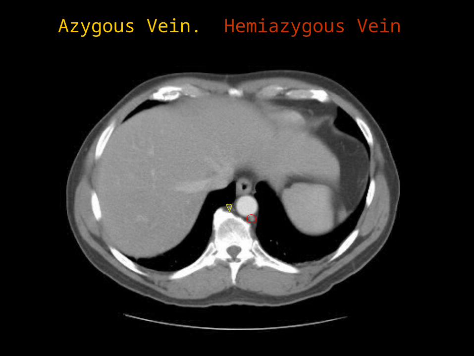
Azygous Vein. Hemiazygous Vein
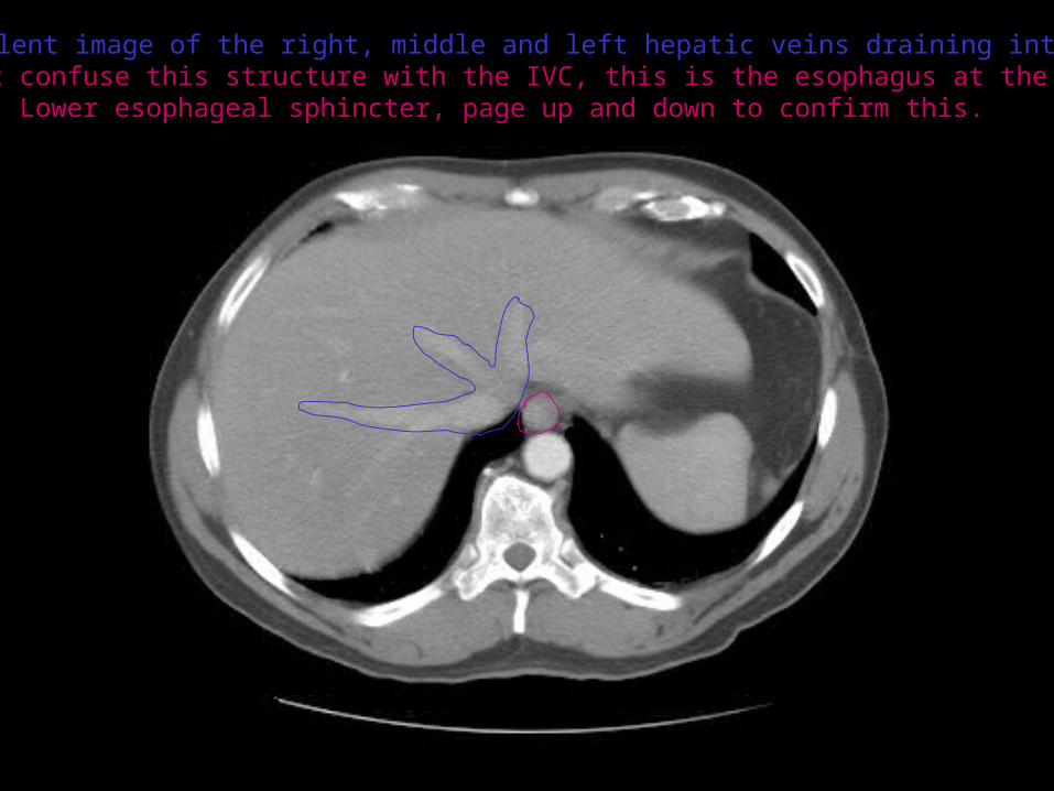
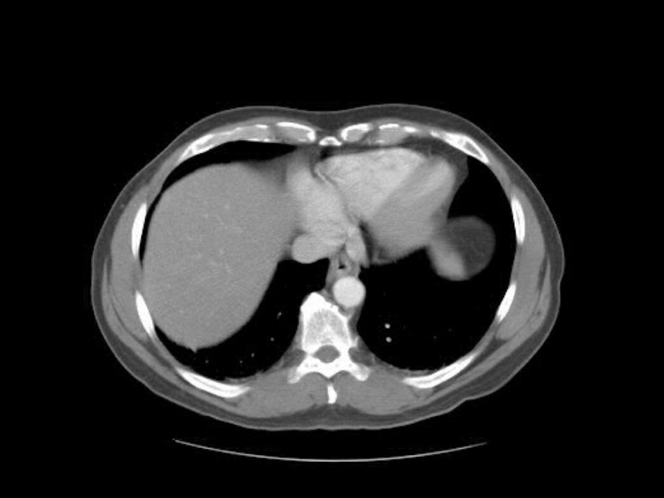
This is an excellent image of the right, middle and left hepatic veins draining into the InferiorVena Cava. Don’t confuse this structure with the IVC, this is the esophagus at the level of the
Lower esophageal sphincter, page up and down to confirm this.
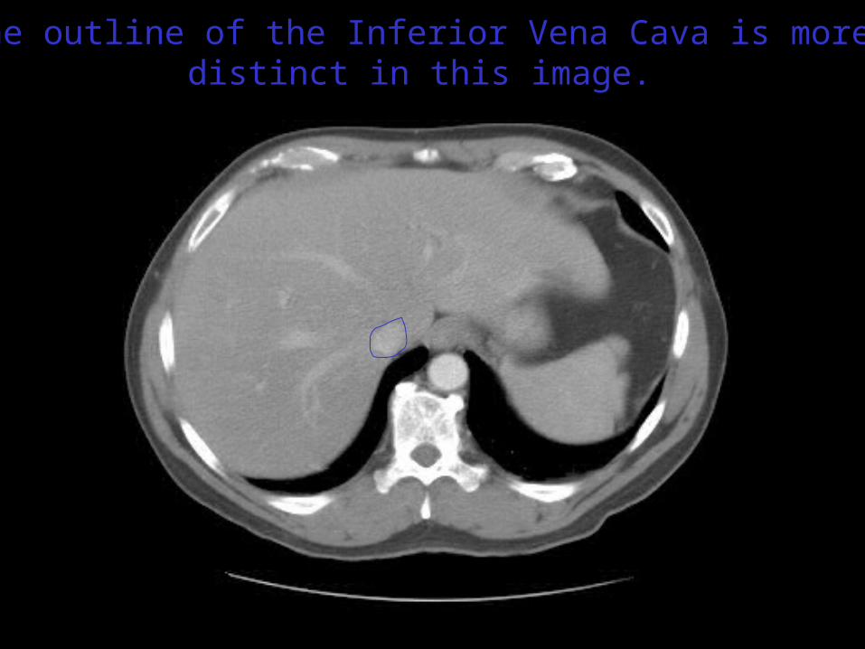
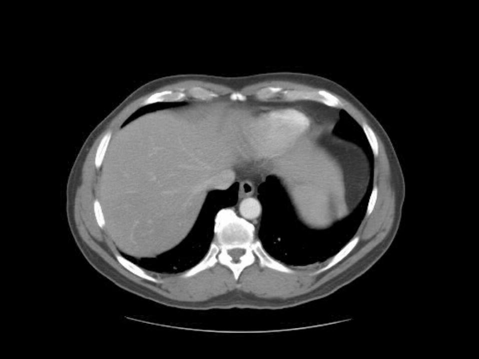
The outline of the Inferior Vena Cava is more distinct in this image.
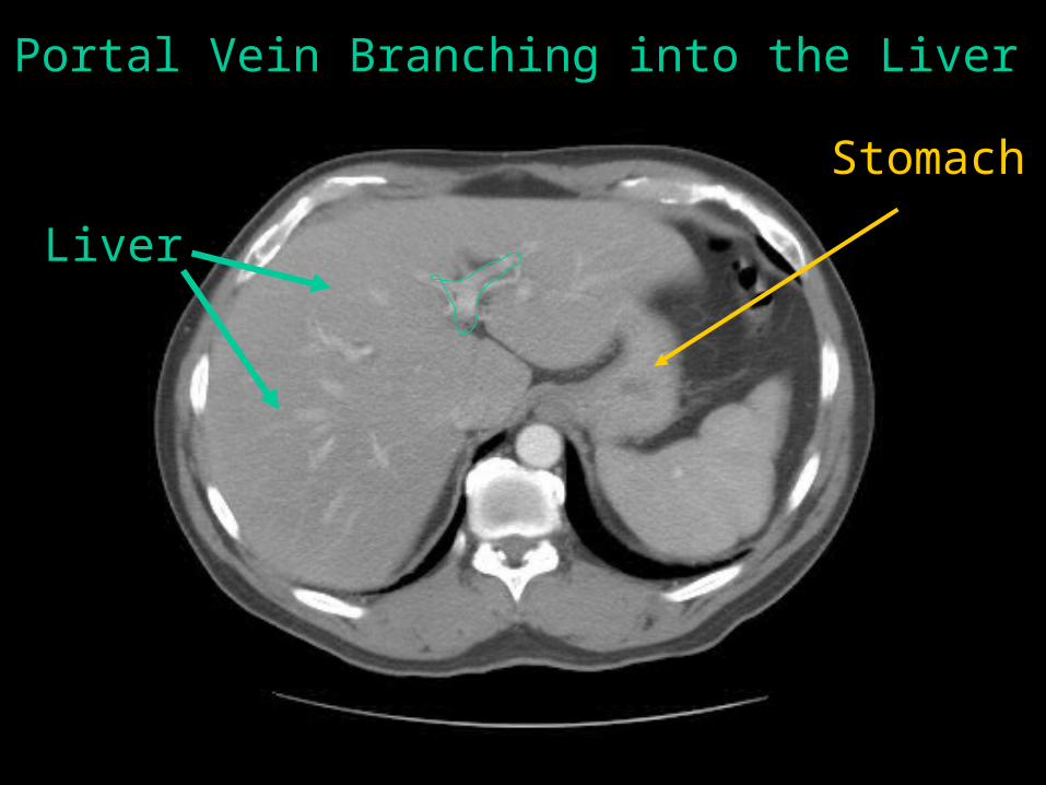
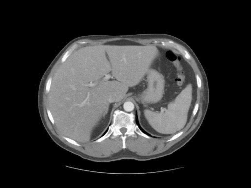
Portal Vein Branching into the Liver
Liver
Stomach
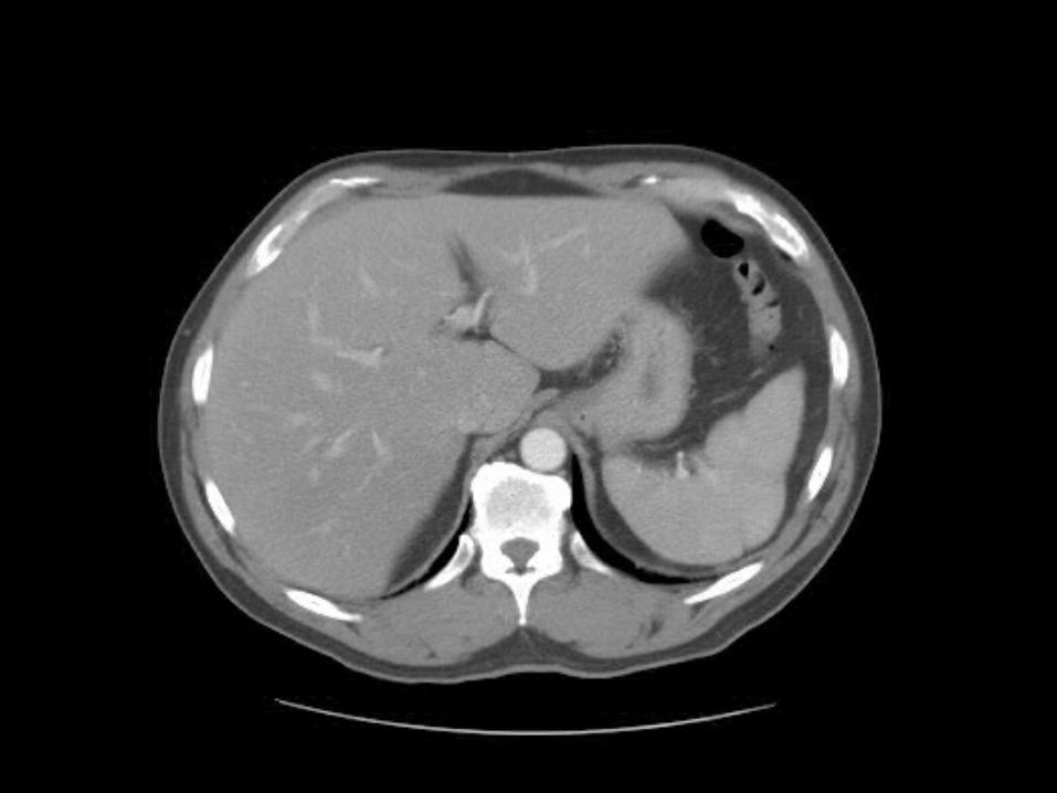
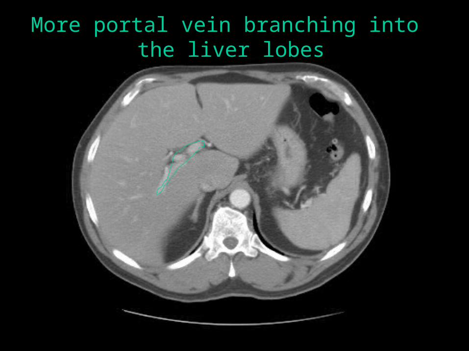
More portal vein branching into the liver lobes
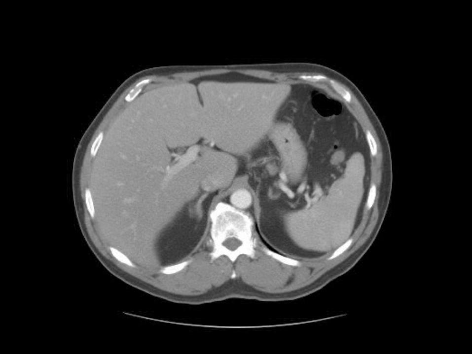
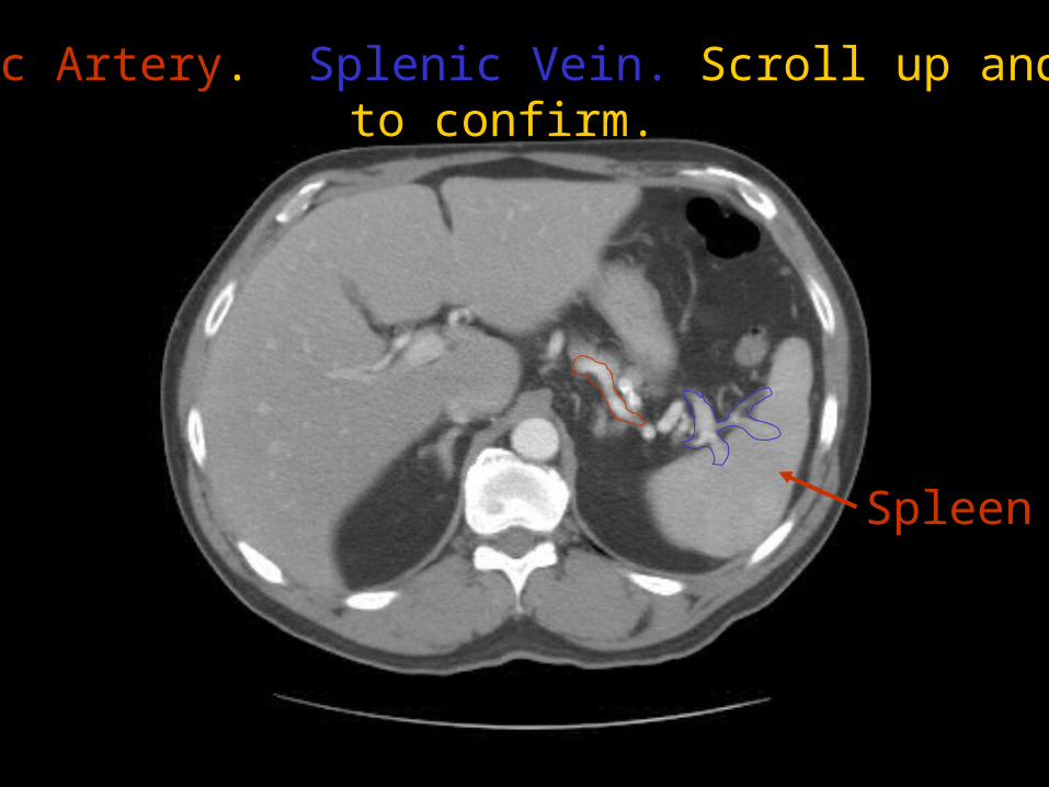
Splenic Artery. Splenic Vein. Scroll up and downto confirm.
Spleen
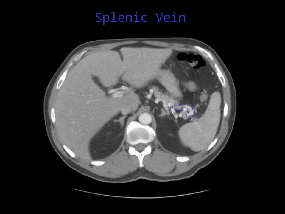
Splenic Vein
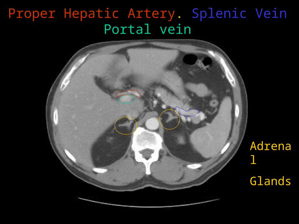
Proper Hepatic Artery. Splenic VeinPortal vein
Adrenal
Glands
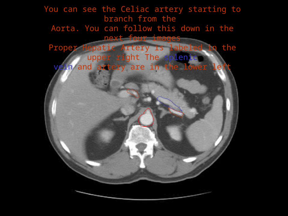
You can see the Celiac artery starting to branch from the Aorta. You can follow this down in the next four images
Proper Hepatic Artery is labeled in the upper right The splenicvein and artery are in the lower left
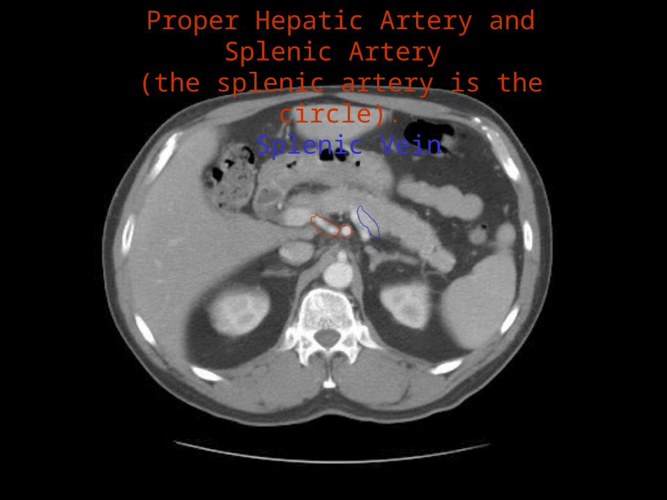
Proper Hepatic Artery and Splenic Artery (the splenic artery is the circle).
Splenic Vein
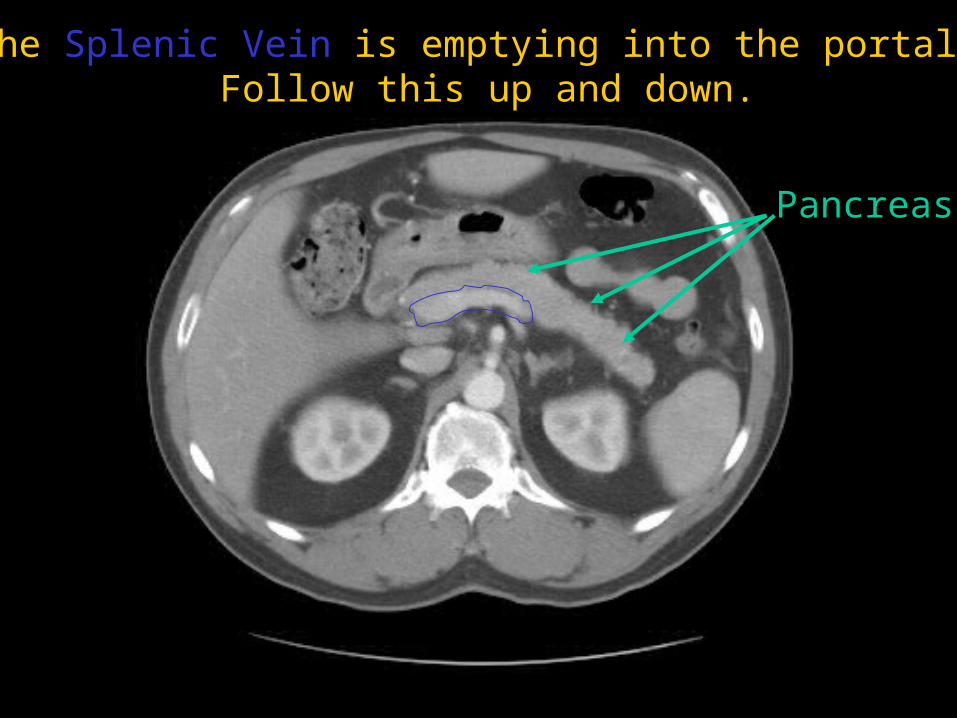
Here the Splenic Vein is emptying into the portal vein. Follow this up and down.
Pancreas
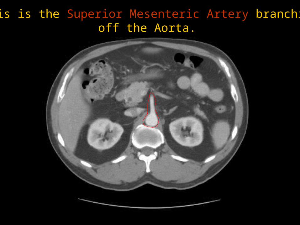
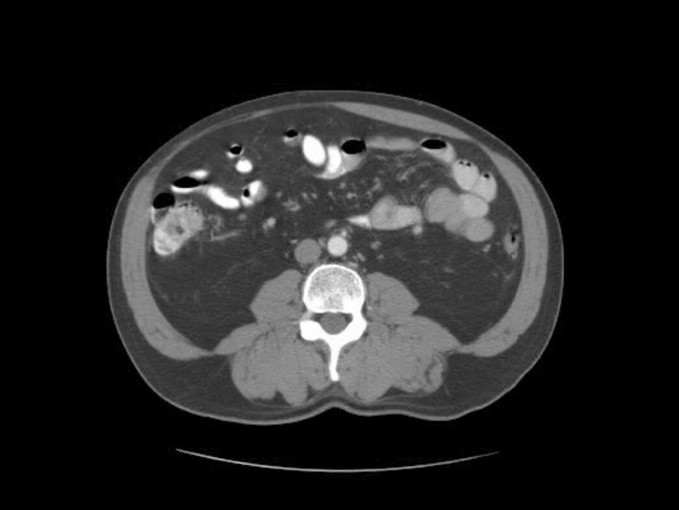
This is the Superior Mesenteric Artery branchingoff the Aorta.
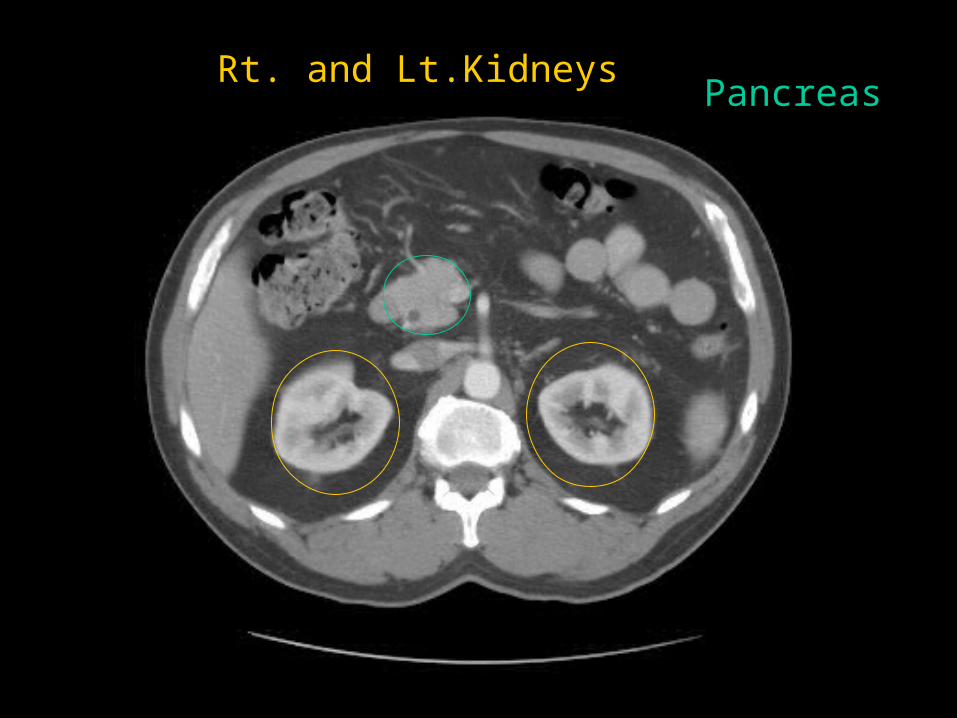
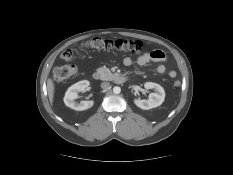
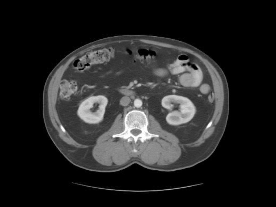
Rt. and Lt.KidneysPancreas
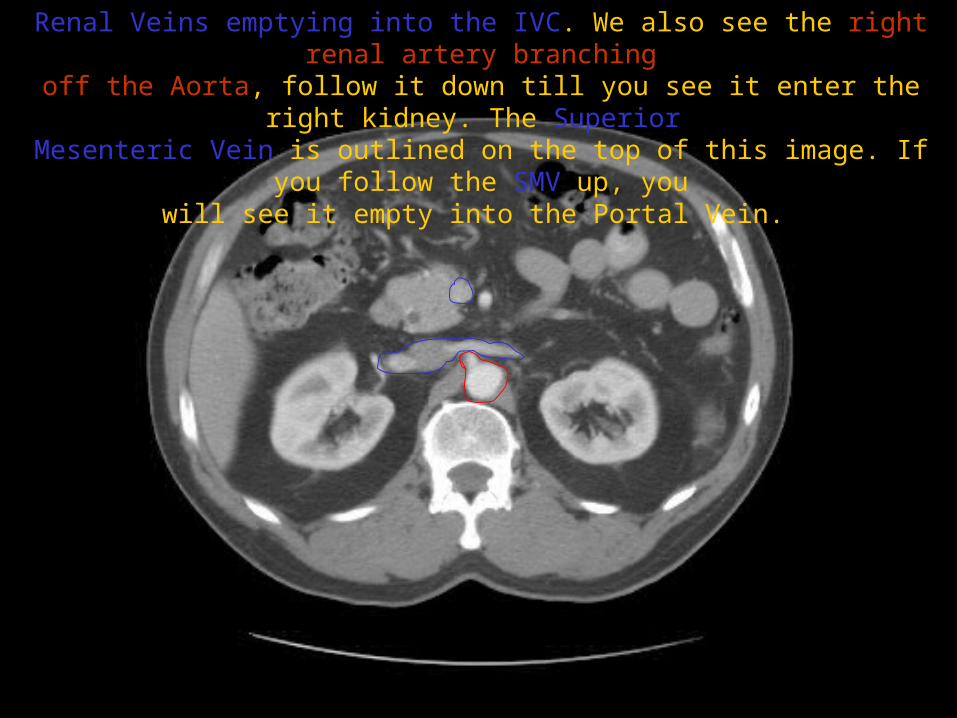
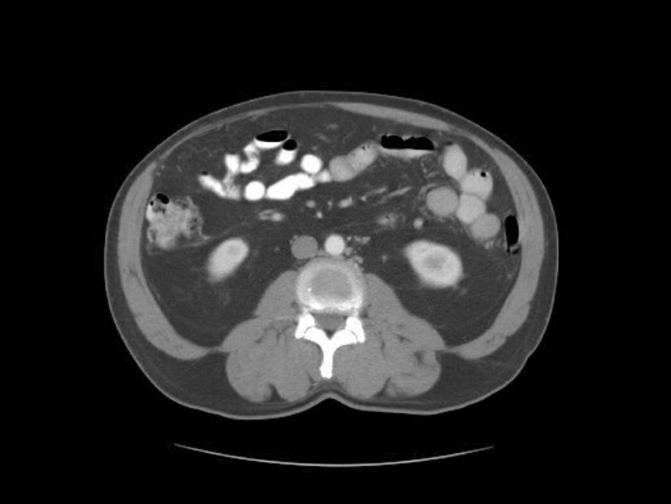
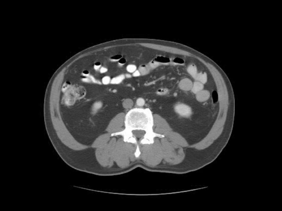
Renal Veins emptying into the IVC. We also see the right renal artery branchingoff the Aorta, follow it down till you see it enter the right kidney. The Superior Mesenteric Vein is outlined on the top of this image. If you follow the SMV up,
youwill see it empty into the Portal Vein.
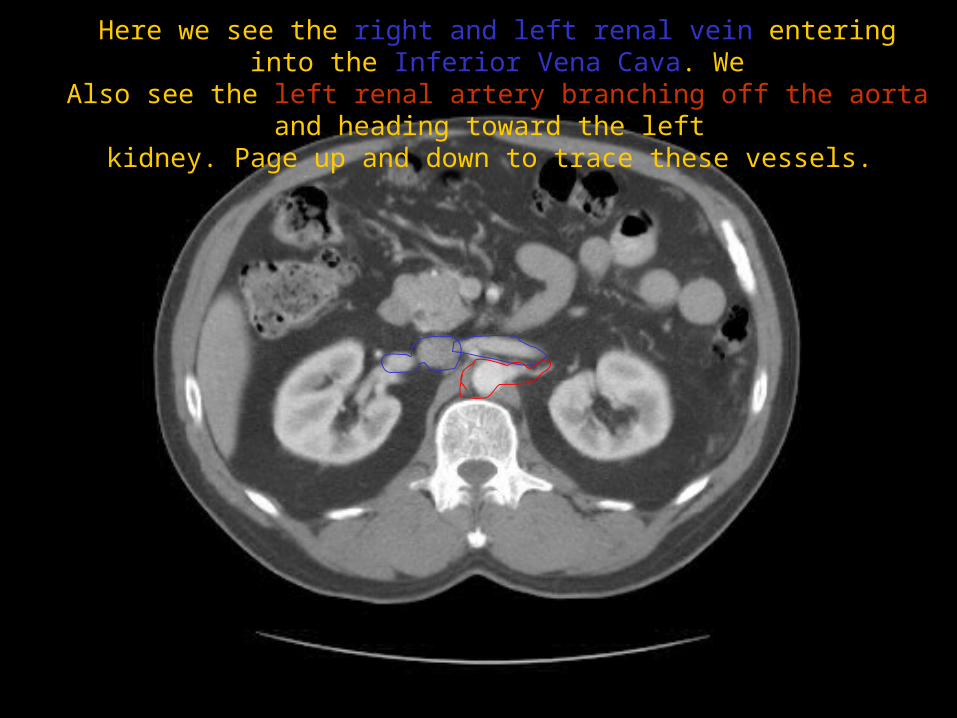
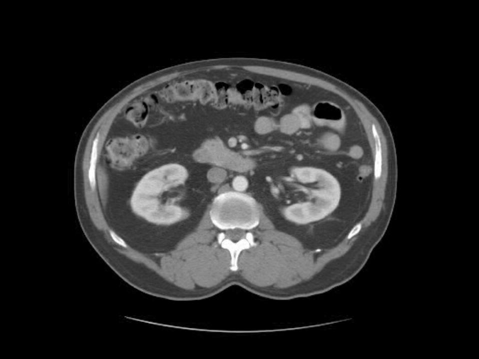
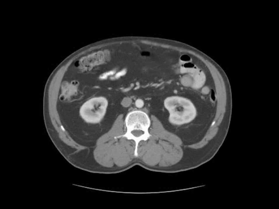
Here we see the right and left renal vein entering into the Inferior Vena Cava. We
Also see the left renal artery branching off the aorta and heading toward the left kidney. Page up and down to trace these vessels.
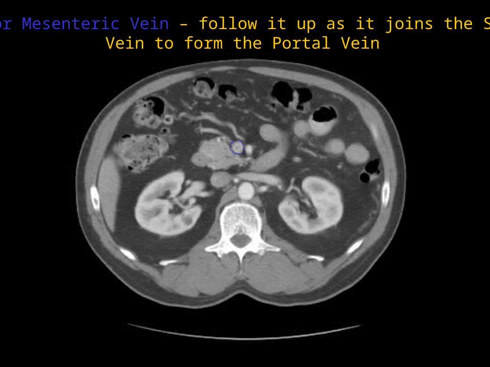
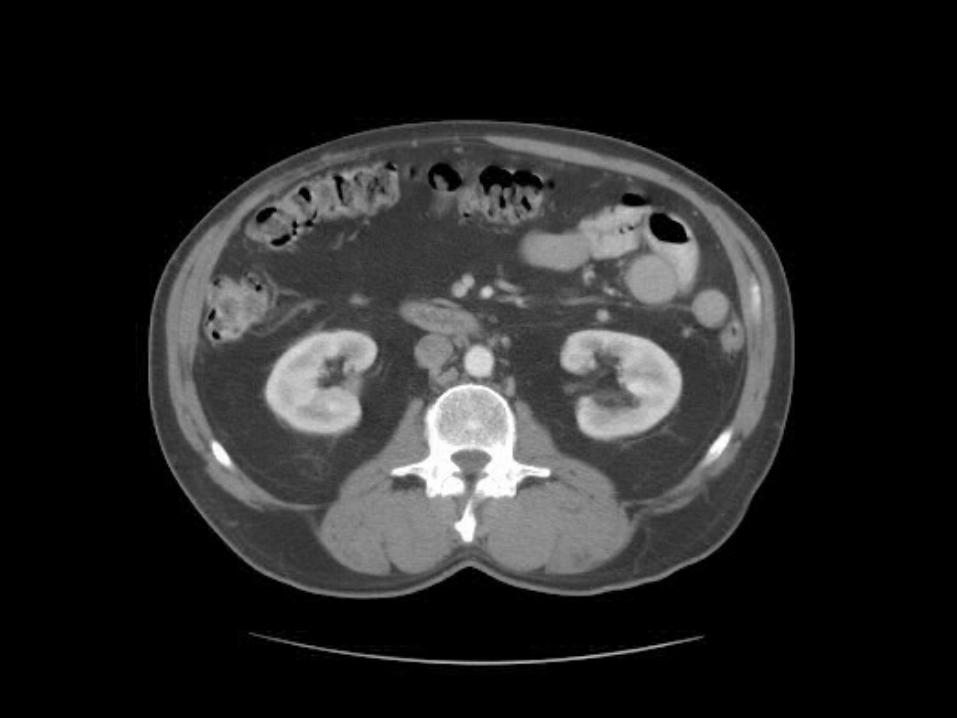
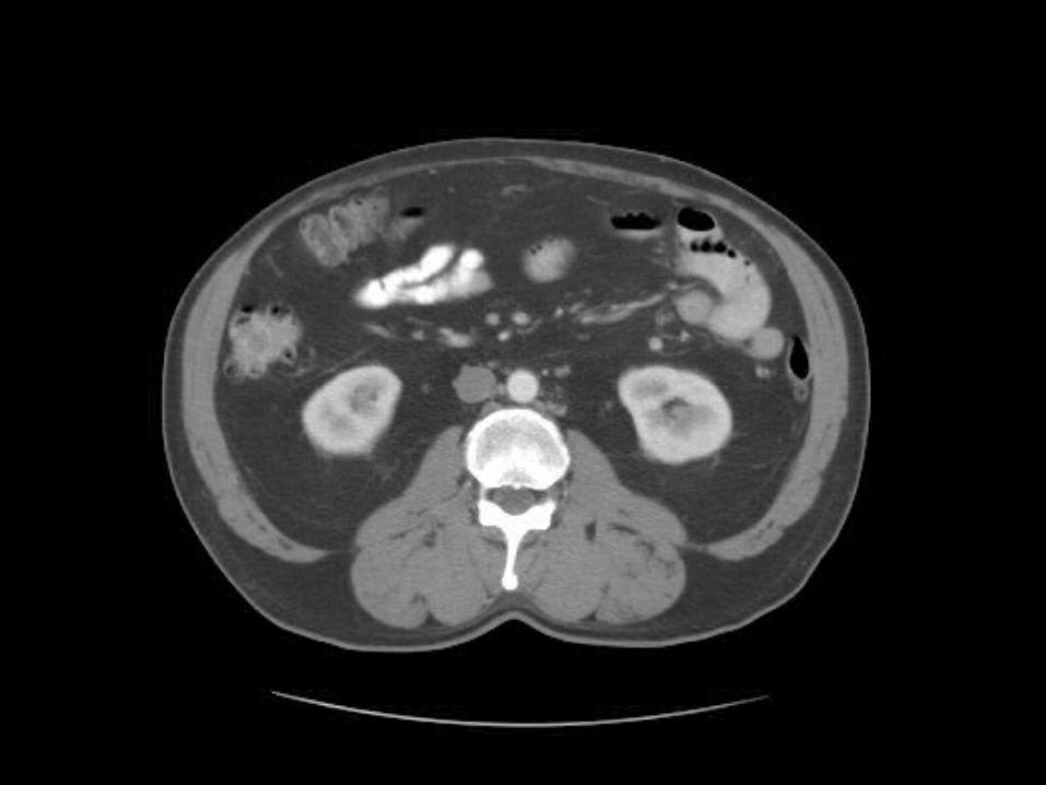
Superior Mesenteric Vein – follow it up as it joins the SplenicVein to form the Portal Vein
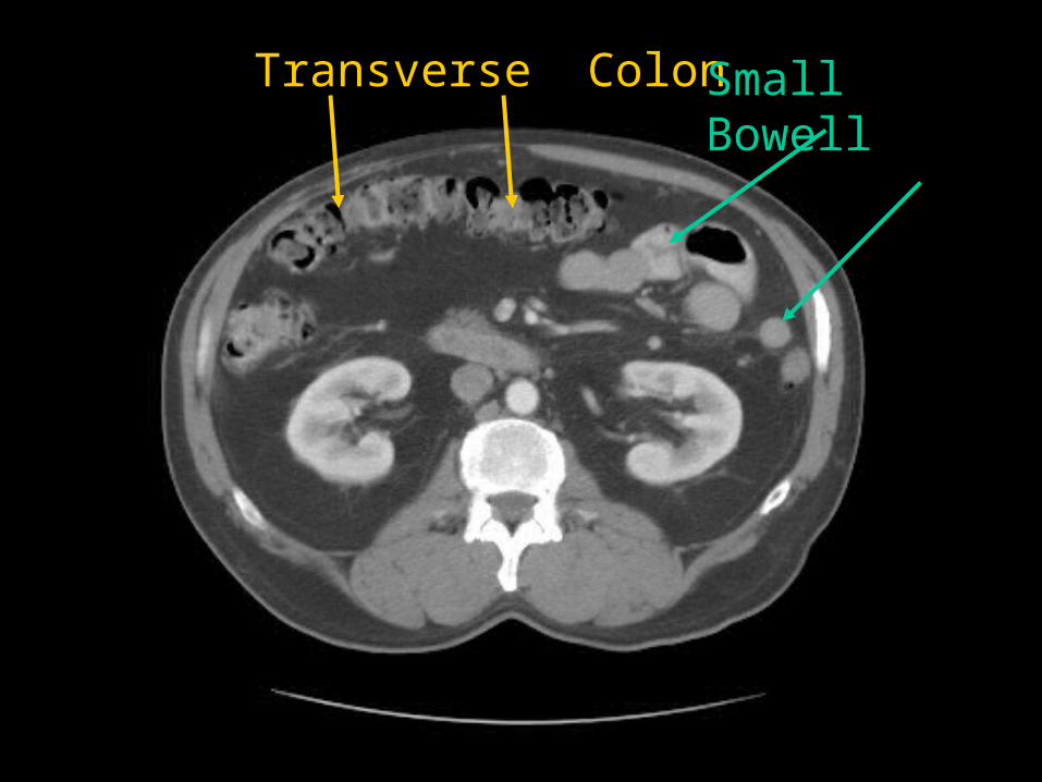
Transverse Colon Small Bowell
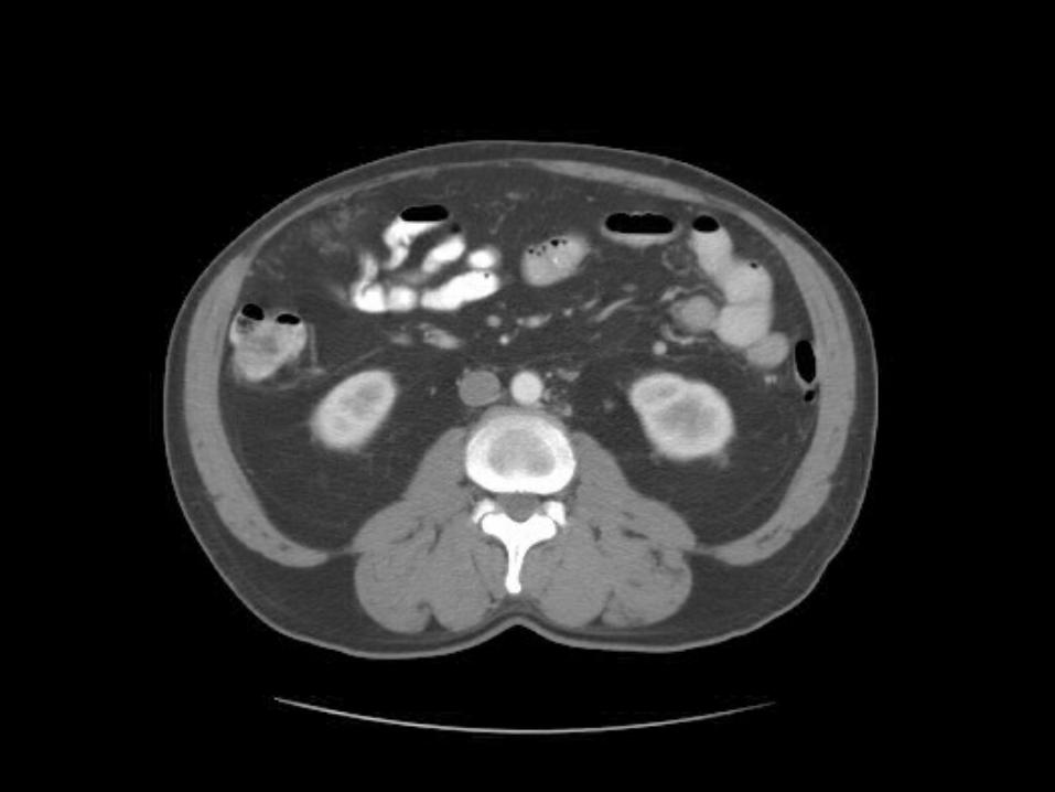
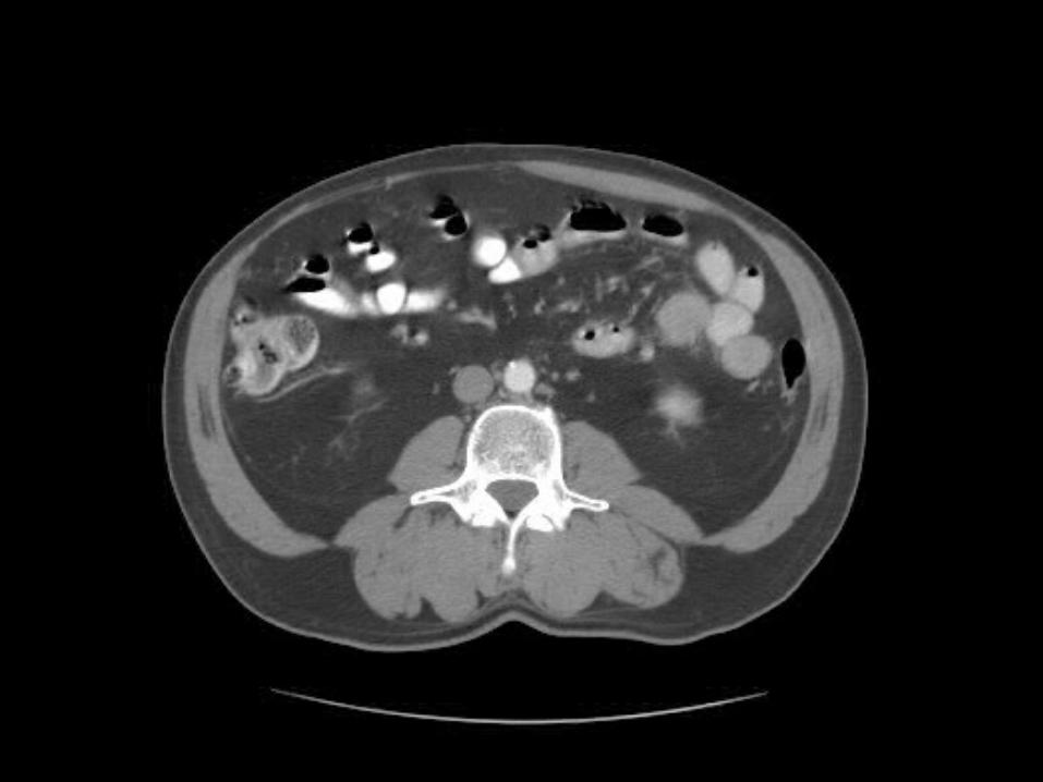
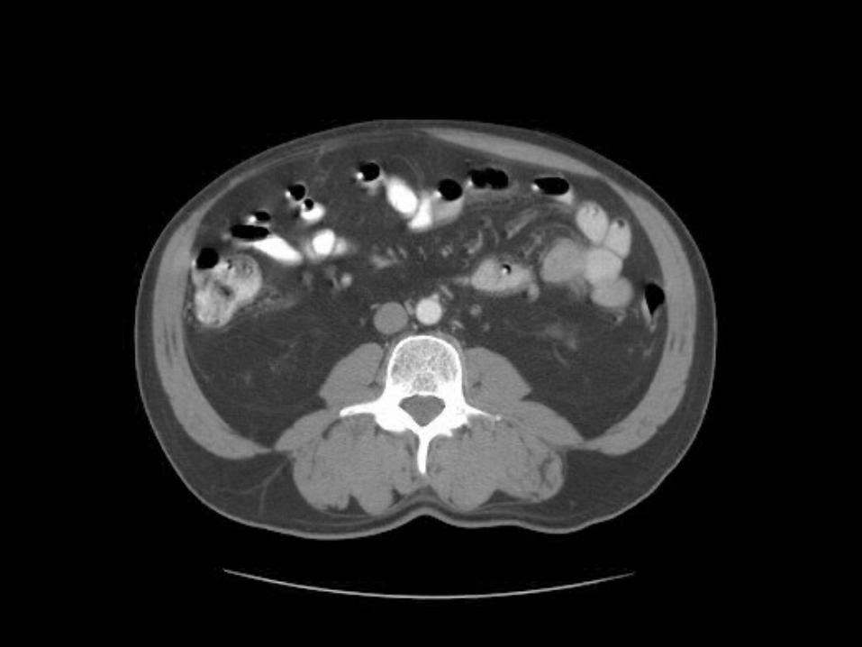
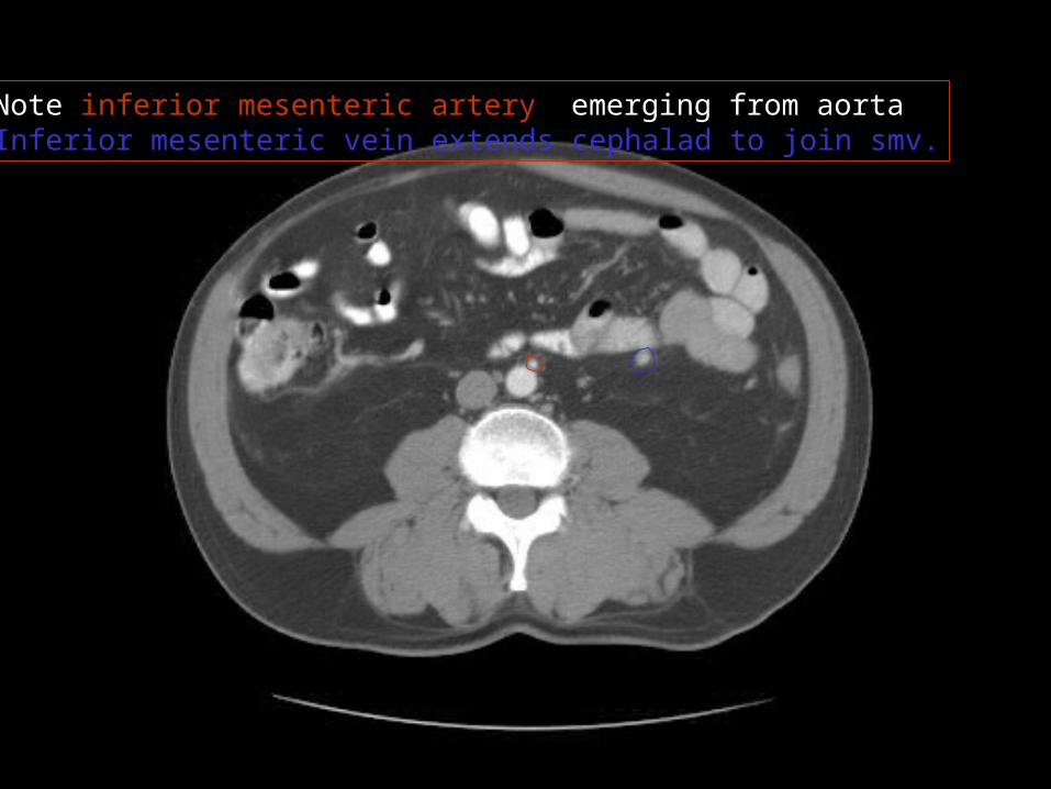
Note inferior mesenteric artery emerging from aortaInferior mesenteric vein extends cephalad to join smv.
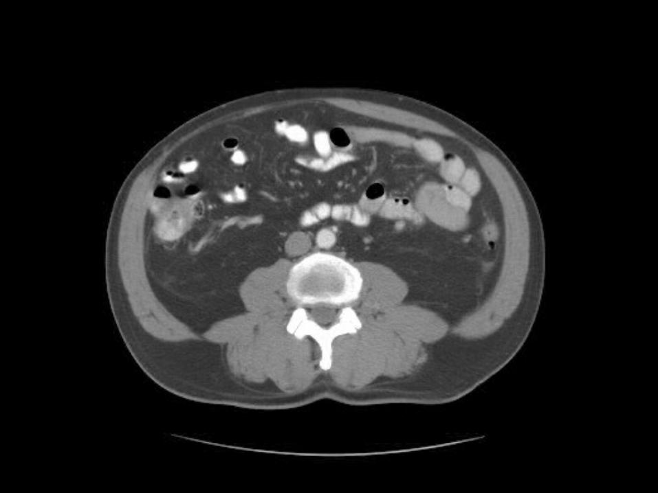
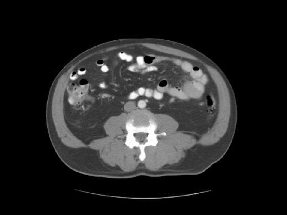
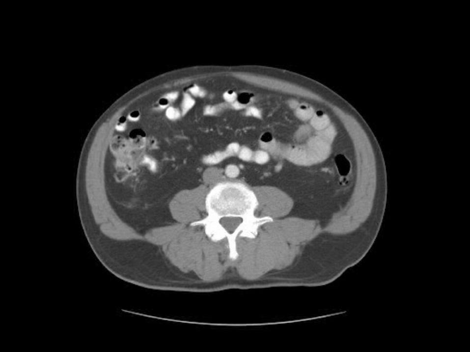
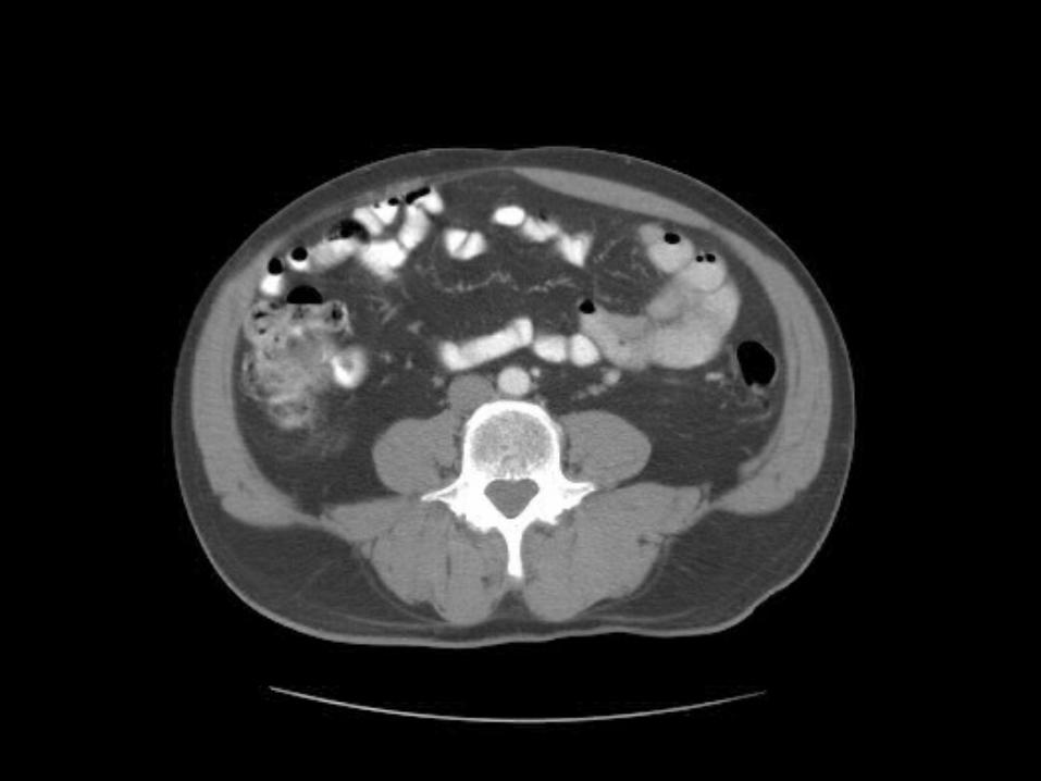
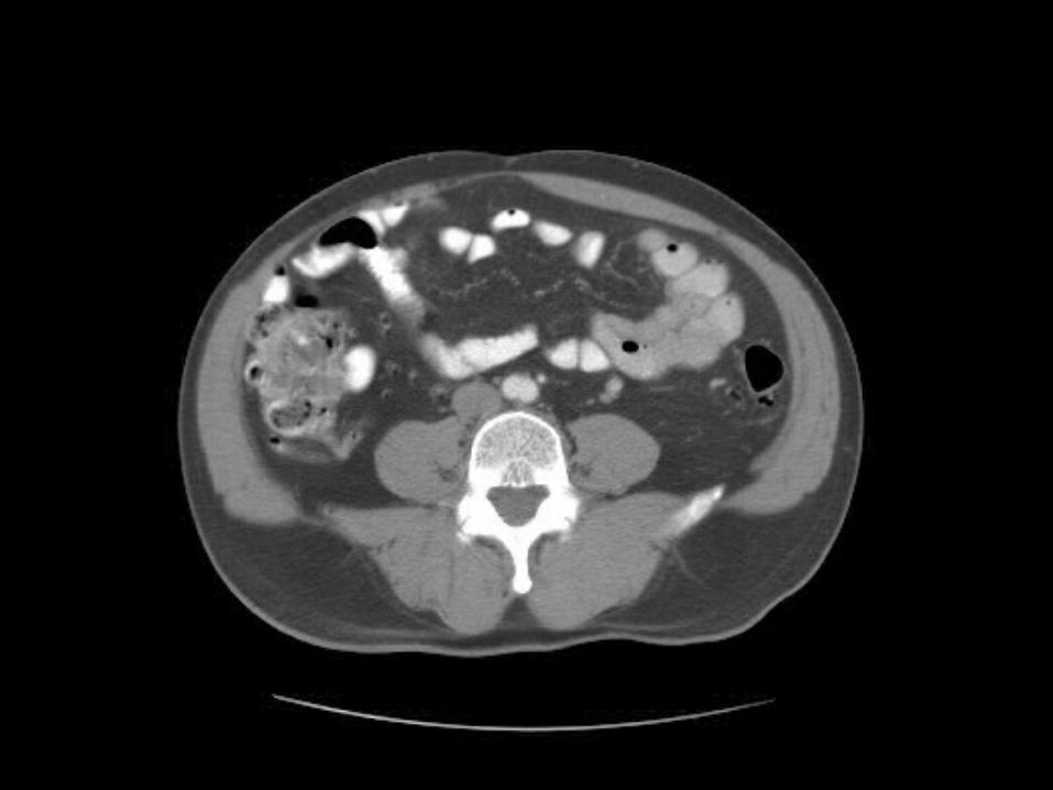
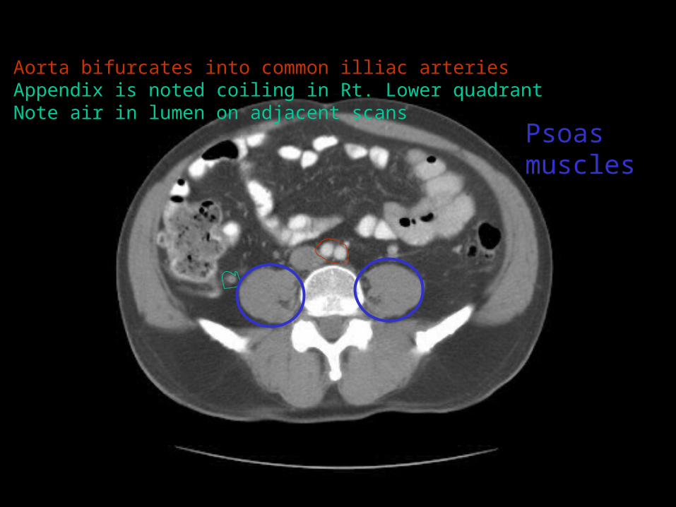
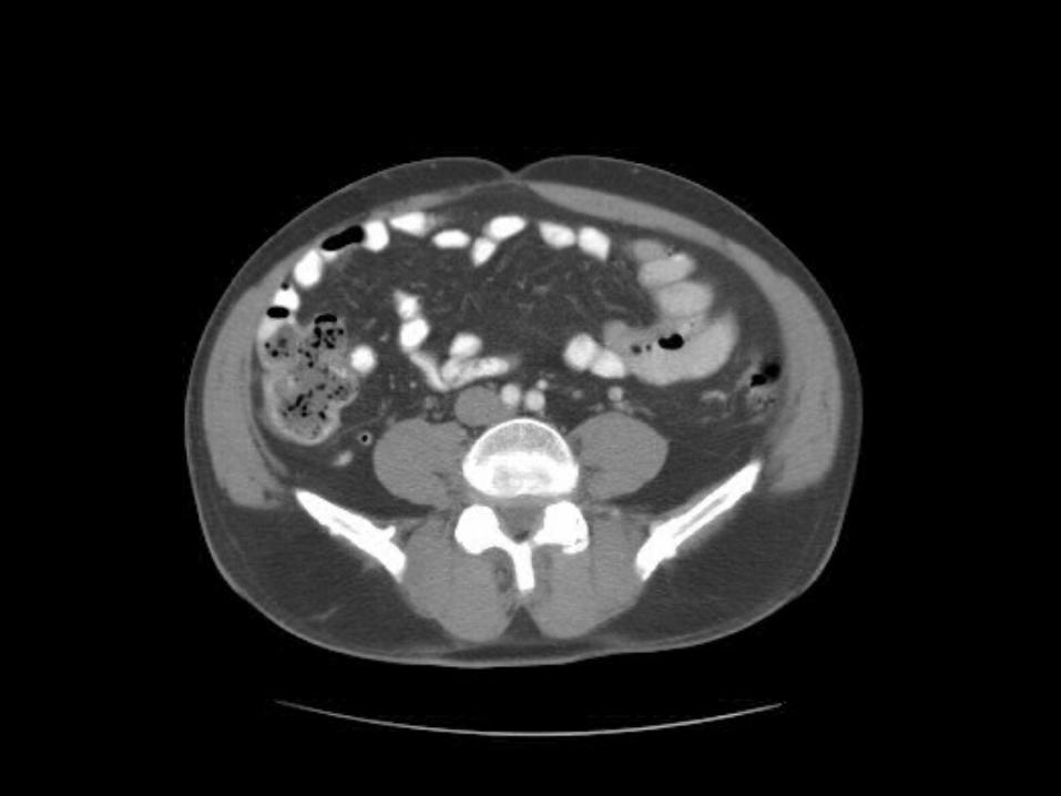
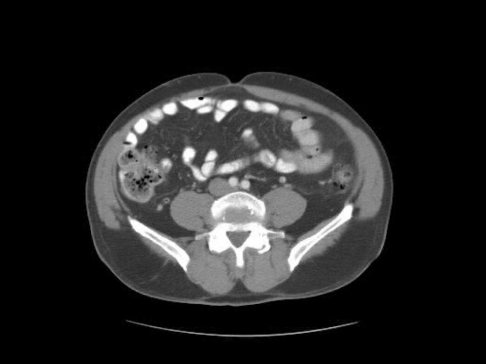
Aorta bifurcates into common illiac arteriesAppendix is noted coiling in Rt. Lower quadrantNote air in lumen on adjacent scans
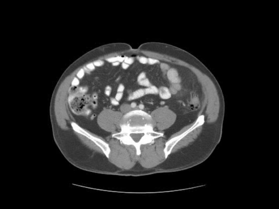
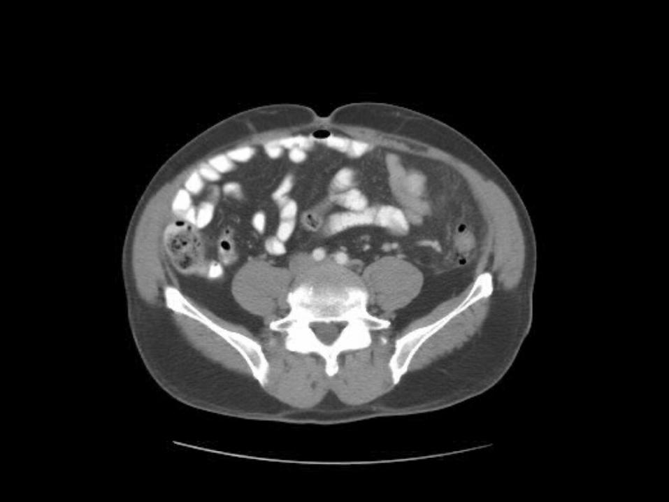
Psoas muscles