
Crystal structure of an HIV assembly and maturation switch
18
*For correspondence: my3r@ virginia.edu (MY); bpornillos@ virginia.edu (BKG-P); owp3a@ eservices.virginia.edu (OP) † These authors contributed equally to this work Competing interests: The authors declare that no competing interests exist. Funding: See page 15 Received: 19 April 2016 Accepted: 13 June 2016 Published: 14 July 2016 Reviewing editor: Volker Do ¨ tsch, Goethe University, Germany Copyright Wagner et al. This article is distributed under the terms of the Creative Commons Attribution License, which permits unrestricted use and redistribution provided that the original author and source are credited. Crystal structure of an HIV assembly and maturation switch Jonathan M Wagner 1† , Kaneil K Zadrozny 1† , Jakub Chrustowicz 1 , Michael D Purdy 1 , Mark Yeager 1,2 *, Barbie K Ganser-Pornillos 1 *, Owen Pornillos 1 * 1 Department of Molecular Physiology and Biological Physics, University of Virginia, Charlottesville, United States; 2 Department of Medicine, Division of Cardiovascular Medicine, University of Virginia Health System, Charlottesville, United States Abstract Virus assembly and maturation proceed through the programmed operation of molecular switches, which trigger both local and global structural rearrangements to produce infectious particles. HIV-1 contains an assembly and maturation switch that spans the C-terminal domain (CTD) of the capsid (CA) region and the first spacer peptide (SP1) of the precursor structural protein, Gag. The crystal structure of the CTD-SP1 Gag fragment is a goblet-shaped hexamer in which the cup comprises the CTD and an ensuing type II b-turn, and the stem comprises a 6-helix bundle. The b-turn is critical for immature virus assembly and the 6-helix bundle regulates proteolysis during maturation. This bipartite character explains why the SP1 spacer is a critical element of HIV-1 Gag but is not a universal property of retroviruses. Our results also indicate that HIV-1 maturation inhibitors suppress unfolding of the CA-SP1 junction and thereby delay access of the viral protease to its substrate. DOI: 10.7554/eLife.17063.001 Introduction Assembly and maturation of HIV-1 and other retroviruses is driven by the viral structural polyprotein, Gag, which consists of a series of independently folding domains, called MA (matrix), CA (capsid), and NC (nucleocapsid) (reviewed in Bush and Vogt, 2014; Ganser-Pornillos et al., 2012; Lingappa et al., 2014). During assembly, Gag makes a spherical, immature capsid shell that pack- ages the viral genome, directs interactions with the plasma membrane to promote envelopment, and recruits cellular machinery to release the new virus particle from the host cell. The Gag shell has hexagonal paracrystalline lattice symmetry, wherein the central CA region makes two layers of inter- actions through its two domains (NTD and CTD, for N-terminal and C-terminal domain). During mat- uration, Gag is cleaved by the viral protease at specific sites. This results in disassembly of the Gag shell and release of new structural proteins, which then rearrange into the mature, infectious virion. In the mature virion, the genome is repackaged inside a cone-shaped capsid made up of the CA protein, with the NTD forming hexameric or pentameric rings that are connected by the CTD. As with other animal viruses, the large-scale capsid rearrangements that occur during retroviral maturation are associated with the operation of structural switches that undergo dramatic conforma- tional changes. In HIV-1, two such putative switches flank the CA region of Gag. One switch spans the MA-CA junction, which is in an extended configuration in the immature virion (Kelly et al., 2006; Tang et al., 2002). Upon proteolysis, the new CA N-terminus folds into a b-hairpin stabilized by a buried salt bridge between the new N-terminal proline and a conserved aspartate residue and pro- motes assembly of the mature capsid (Gitti et al., 1996; von Schwedler et al., 1998). A second putative switch spans the junction between CA and the first spacer peptide (SP1) (Gross et al., 2000). The CA-SP1 junction is predicted to fold into an a-helix (Accola et al., 1998; Liang et al., 2002), and forms the binding site of so-called ’maturation inhibitors,’ as exemplified by 3-O-(3’,3’- Wagner et al. eLife 2016;5:e17063. DOI: 10.7554/eLife.17063 1 of 18 RESEARCH ARTICLE
Transcript of Crystal structure of an HIV assembly and maturation switch

For correspondencemy3r
virginiaedu (MY) bpornillos
virginiaedu (BKG-P) owp3a
eservicesvirginiaedu (OP)
daggerThese authors contributed
equally to this work
Competing interests The
authors declare that no
competing interests exist
Funding See page 15
Received 19 April 2016
Accepted 13 June 2016
Published 14 July 2016
Reviewing editor Volker
Dotsch Goethe University
Germany
Copyright Wagner et al This
article is distributed under the
terms of the Creative Commons
Attribution License which
permits unrestricted use and
redistribution provided that the
original author and source are
credited
Crystal structure of an HIV assembly andmaturation switchJonathan M Wagner1dagger Kaneil K Zadrozny1dagger Jakub Chrustowicz1Michael D Purdy1 Mark Yeager12 Barbie K Ganser-Pornillos1 Owen Pornillos1
1Department of Molecular Physiology and Biological Physics University of VirginiaCharlottesville United States 2Department of Medicine Division of CardiovascularMedicine University of Virginia Health System Charlottesville United States
Abstract Virus assembly and maturation proceed through the programmed operation of
molecular switches which trigger both local and global structural rearrangements to produce
infectious particles HIV-1 contains an assembly and maturation switch that spans the C-terminal
domain (CTD) of the capsid (CA) region and the first spacer peptide (SP1) of the precursor
structural protein Gag The crystal structure of the CTD-SP1 Gag fragment is a goblet-shaped
hexamer in which the cup comprises the CTD and an ensuing type II b-turn and the stem comprises
a 6-helix bundle The b-turn is critical for immature virus assembly and the 6-helix bundle regulates
proteolysis during maturation This bipartite character explains why the SP1 spacer is a critical
element of HIV-1 Gag but is not a universal property of retroviruses Our results also indicate that
HIV-1 maturation inhibitors suppress unfolding of the CA-SP1 junction and thereby delay access of
the viral protease to its substrate
DOI 107554eLife17063001
IntroductionAssembly and maturation of HIV-1 and other retroviruses is driven by the viral structural polyprotein
Gag which consists of a series of independently folding domains called MA (matrix) CA (capsid)
and NC (nucleocapsid) (reviewed in Bush and Vogt 2014 Ganser-Pornillos et al 2012
Lingappa et al 2014) During assembly Gag makes a spherical immature capsid shell that pack-
ages the viral genome directs interactions with the plasma membrane to promote envelopment
and recruits cellular machinery to release the new virus particle from the host cell The Gag shell has
hexagonal paracrystalline lattice symmetry wherein the central CA region makes two layers of inter-
actions through its two domains (NTD and CTD for N-terminal and C-terminal domain) During mat-
uration Gag is cleaved by the viral protease at specific sites This results in disassembly of the Gag
shell and release of new structural proteins which then rearrange into the mature infectious virion
In the mature virion the genome is repackaged inside a cone-shaped capsid made up of the CA
protein with the NTD forming hexameric or pentameric rings that are connected by the CTD
As with other animal viruses the large-scale capsid rearrangements that occur during retroviral
maturation are associated with the operation of structural switches that undergo dramatic conforma-
tional changes In HIV-1 two such putative switches flank the CA region of Gag One switch spans
the MA-CA junction which is in an extended configuration in the immature virion (Kelly et al 2006
Tang et al 2002) Upon proteolysis the new CA N-terminus folds into a b-hairpin stabilized by a
buried salt bridge between the new N-terminal proline and a conserved aspartate residue and pro-
motes assembly of the mature capsid (Gitti et al 1996 von Schwedler et al 1998) A second
putative switch spans the junction between CA and the first spacer peptide (SP1) (Gross et al
2000) The CA-SP1 junction is predicted to fold into an a-helix (Accola et al 1998 Liang et al
2002) and forms the binding site of so-called rsquomaturation inhibitorsrsquo as exemplified by 3-O-(3rsquo3rsquo-
Wagner et al eLife 20165e17063 DOI 107554eLife17063 1 of 18
RESEARCH ARTICLE
dimethylsuccinyl)betulinic acid (bevirimat) (reviewed in Adamson et al 2009) These inhibitors delay
proteolysis of the CA-SP1 junction and induce aberrant maturation The importance of the CA-SP1
junction as a regulator of proteolysis during maturation is well established (Krausslich et al 1995
Pettit et al 1994 Wiegers et al 1998) but the molecular basis of this function has been
unknown
Mutagenesis studies also indicate that the CA-SP1 junction is important for assembly of the
immature HIV-1 Gag shell (Accola et al 1998 Al-Mawsawi et al 2014 Datta et al 2011
Guo et al 2005 Krausslich et al 1995 Liang et al 2002 Liang et al 2003 Melamed et al
2004 Pettit et al 1994 Rihn et al 2013 von Schwedler et al 2003) Low resolution electron
microscopy studies indicate that the junction forms what appears to be a pillar of density or helical
bundle and so is likely to stabilize the Gag lattice (Schur et al 2015a Schur et al 2015b
Wright et al 2007) However neither the SP1 spacer nor the putative helical bundle is a universal
feature of retroviral Gag shells (Bharat et al 2012) This suggests that the SP1 spacer is not gener-
ally important for Gag assembly or that perhaps the CA-SP1 junction has another as yet structurally
undefined assembly determinant
Structural studies of the HIV-1 CA-SP1 junction in context of longer Gag fragments (CTD-SP1 and
CTD-SP1-NC) have shown that the junction is primarily disordered (Newman et al 2004
Worthylake et al 1999) Nevertheless the backbone NMR chemical shifts of junction residues
deviate from expected random coil values indicating a small propensity towards an a-helical confor-
mation (Newman et al 2004) Indeed the isolated SP1 peptide displays concentration dependent
secondary structure in aqueous solution and folds into an a-helix at the millimolar protein concen-
trations found in virions (Datta et al 2011) Nucleic acids (both RNA and DNA) can promote Gag
assembly in vitro presumably because of proximity-induced interactions between multiple copies of
Gag that bind to the same nucleic acid molecule (Campbell and Rein 1999 Campbell and Vogt
1995 Gross et al 2000) It therefore seems that nucleic acid-induced Gag clustering might also
promote folding of the SP1 helix
To learn how the CA-SP1 junction functions as a molecular switch during HIV-1 assembly and mat-
uration we determined the structure of its immature assembled form by X-ray crystallography at a
resolution of 327 A Our analysis elucidates how local conformational changes in the CA-SP1 switch
eLife digest Viruses like HIV must undergo a process called maturation in order to successfully
infect cells Maturation involves a dramatic rearrangement in the architecture of the virus That is to
say the virusrsquos internal protein coat ndash called the capsid ndash must change from an immature sphere into
a mature cone-shaped coat Notably this maturation process is what is disrupted by the protease
inhibitors that are a major component of anti-HIV drug cocktails
Structural changes in small portions of the capsid protein termed molecular switches commonly
trigger the viral capsids to reorganize The HIV capsid has two of these switches and Wagner
Zadrozny et al set out to understand how one of them ndash called the CA-SP1 switch ndash works
Solving the three-dimensional structure of the immature form of the CA-SP1 switch revealed that
it forms a well-structured bundle of six helices This helical bundle captures another section of the
capsid protein that would otherwise be cut by a viral protease The CA-SP1 switch therefore controls
how quickly the protein is cut and the start of the maturation process
Wagner Zadrozny et al then discovered that other small molecule inhibitors of HIV called
maturation inhibitors work by binding to and disrupting the transformation of the CA-SP1 switch
Finally further experiments showed that the formation of the CA-SP1 helical bundle controls when
the immature capsid shell forms and coordinates the process with the capsid gaining the genetic
material of the virus
The new structure means that researchers now know what the HIV capsid looks like at the start
and end of maturation The next challenge will be to figure out exactly how the capsid changes from
one form to the next as HIV matures
DOI 107554eLife17063002
Wagner et al eLife 20165e17063 DOI 107554eLife17063 2 of 18
Research article Biochemistry Biophysics and Structural Biology
promote Gag assembly drive large-scale capsid rearrangements and regulate proteolysis during
maturation We also gained new insights on the mechanism of action of maturation inhibitors
Results
HIV-1 CA CTD-SP1 recapitulates the immature Gag latticeIn the immature HIV-1 Gag shell the NTD CTD and SP1 regions form three layers of lattice-stabiliz-
ing interactions (Schur et al 2015b Wright et al 2007) The CTD-SP1 fragment of HIV-1 Gag
contains the minimal information required to assemble the immature lattice whereas the NTD is dis-
pensable (Accola et al 2000) Accordingly we found that purified CTD-SP1 protein assembled in
vitro into flat sheets with the subunits organized into a hexagonal lattice with the expected unit cell
spacing of the CTD layer of the immature Gag lattice (~74 A) (Wright et al 2007 ) (Figure 1mdash
figure supplement 1) We then performed crystallization screens using a large number of commer-
cial and in-house precipitants We obtained many crystal hits but invariably these were of the
mature-like dimer form of the CTD with disordered SP1 tails as observed previously
(Worthylake et al 1999) Using electron diffraction we identified small and scarce plate crystals
that gave a hexagonal diffraction pattern and unit cell spacing close to 70 A (not shown) Upon opti-
mization these crystals diffracted X-rays to about 327 A resolution (mean IsI 1) and we deter-
mined the crystal structure to RRfree values of 02460278 (Table 1 and Figure 1mdash
figure supplement 2) Our post hoc analysis is that these crystals were rare because the CA-SP1
junction residues had to fold and become ordered during crystallization We speculate that this rate
limiting step of crystallization reflects the behavior of the junction during assembly of HIV-1 Gag
The CTD-SP1 plate crystals were made up of stacked sheets of flat hexagonal lattices with each
sheet consisting of CTD-SP1 hexamers connected by CTD dimer linkages (Figure 1A) Our structure
therefore represents a flattened version of the immature HIV-1 Gag lattice The entire CTD-SP1 hex-
amer ndash including both the main CTD fold and ordered SP1 residues ndash gave an excellent fit to an
88 A resolution cryoEM map of the immature HIV-1 Gag lattice (Schur et al 2015b) (Figure 1mdashfig-
ure supplement 3A) The dimer also gave a good fit (Figure 1mdashfigure supplement 3B) but in this
case the SP1 helix was more laterally displaced from its corresponding density in the cryoEM map
(asterisk in Figure 1mdashfigure supplement 3B) This is likely because the cryoEM structure is of a
curved Gag lattice whereas our crystal structure is of a flat lattice To gain insight on how loss of cur-
vature in our crystal lattice is accommodated by the subunits we superimposed the crystal structure
with the model reported by Schur et al which was created by flexible fitting of the main CTD fold
(but not the CA-SP1 junction) into the cryoEM map (PDB 4USN) (Schur et al 2015b) We found
that superposition of the hexamer units resulted in an average root mean square displacement of
192 A over equivalent Ca atoms whereas superposition of the dimers resulted in a comparable dis-
placement of 193 A We therefore surmise that any changes in tertiary or quaternary structure that
were caused by flattening of the lattice in our crystals have been dispersed across the subunits
Unlike the mature capsid which contains sharp pentameric declinations and a variably curving lattice
(Ganser et al 1999 Pornillos et al 2011 Zhao et al 2013) the immature HIV-1 Gag shell is a
more smoothly curving sphere that is interrupted by large discontinuities (Briggs et al 2009
Keller et al 2011 Wright et al 2007) It therefore seems that generation of Gag curvature does
not necessarily require the kinds of conformational variations observed in the mature capsid
subunits
The CA-SP1 junction is structurally bipartiteIn our crystal structure each subunit contains the main CTD fold (amino acids 281ndash351 in the HIV-1
Gag numbering scheme) and the CA-SP1 switch region (residues 352ndash370) The CTD adopts the
canonical fold a short 310 helix followed by a strandturn element called the major homology region
(MHR) and then four a-helices (helices 8ndash11) (Gamble et al 1997 Worthylake et al 1999)
(Figure 2A) The switch region immediately follows the main CTD fold and consists of two parts a
type II b-turn (residues 352ndash355) and an a-helix that spans the CA-SP1 junction (residues 356ndash370)
This bipartite character was not anticipated but could explain the apparent structural variability in
the C-terminal switch regions of immature retroviral capsids which were discerned from subnanome-
ter resolution cryoEM maps (Bharat et al 2012 Schur et al 2015a 2015b)
Wagner et al eLife 20165e17063 DOI 107554eLife17063 3 of 18
Research article Biochemistry Biophysics and Structural Biology
Comparison of the immature CTD-SP1 subunit with the mature CTD subunit reveals a local con-
formational change (presence or absence of a kink) in the dimerization helix as noted previously
(Bartonova et al 2008) (arrows in Figure 2AB) This correlates with the different orientations of
the subunits across the immature and mature dimer interfaces (Figure 2mdashfigure supplement 1)
(Bartonova et al 2008 Gres et al 2015) Another difference is that in the mature CTD residues
downstream of helix 11 are disordered (Bartonova et al 2008 Gamble et al 1997
Table 1 Structure statistics for HIV-1 Gag CTD-SP1
Diffraction
Beamline APS 22ID
Wavelength (A) 10
Processing program HKL2000
Space group C2
Cell dimensions a = 7096 A
b = 12273 A
c = 8541 A
a = g = 90˚ b = 943˚Resolution range A 50-327 (342-327)
Rmerge Rpim 022 (074) 011 (047)
Mean IsltIgt 599 (128)
Completeness 870 (664)
Average redundancy 37 (25)
Wilson B-factor A2 8521
Refinement
Refinement program PHENIX
Resolution range 4259-327 (345-327)
No of unique reflections 9710 (908)
Reflections in free set 1009 (88)
Rwork 0246 (0369)
Rfree 0278 (0408)
No of nonhydrogen atoms
protein 3865
solvent 0
Average B-factor A2
protein 8409
solvent na
Coordinate deviations
bond lengths A 0003
bond angles ˚ 0513
Validation and Deposition
Ramachandran plot
favored 989
outliers 0
MolProbity clash score 013
PDB ID 5I4T
Values in parenthesis are for the highest resolution shell
DOI 107554eLife17063003
Wagner et al eLife 20165e17063 DOI 107554eLife17063 4 of 18
Research article Biochemistry Biophysics and Structural Biology
Worthylake et al 1999) (Figure 2BC) These observations provide further support for the pre-
sumption that the C-terminal tail of CA becomes unfolded upon maturation and has no apparent
role in assembly of the mature capsid
The CTD-SP1 hexamer looks like a goblet with the main CTD fold forming the cup and the CA-
SP1 junction forming the stem (Figure 1BC) Lateral packing between the subunits is quite loose
along the body of the cup and close contacts involving the main CTD fold occur only where the cup
meets the stem In this region the MHR loops line the bottom inner surface of the cup (Figure 1BC
colored in red) whereas the loops connecting helices 9 and 10 (910 loop orange) line the outer sur-
face of the cup and contact the tops of the junction helices in the stem (bluemagenta) Underneath
the cup the junction helices form a 6-helix bundle as predicted (Accola et al 1998 Wright et al
2007) All of the aliphatic sidechains in the junction helix make rsquoknobs-in-holesrsquo interactions similar
to classical coiled-coils (Figure 3AndashC)
The CA-SP1 junction helix regulates proteolysis by sequestering thescissile bondDuring HIV-1 maturation the CA-SP1 junction is one of ten cleavage sites in the precursor Gag and
Gag-Pol polyproteins that are cleaved by the viral protease (PR) Proteolytic processing occurs at dif-
ferent rates with the CTD-SP1 junction processed with the slowest rate and the downstream SP1-
NC junction the fastest (reviewed in Lee et al 2012) The C-terminal boundary of the junction helix
Figure 1 Crystal structure of the immature HIV-1 Gag CTD-SP1 lattice (A) Top view of the lattice with symmetry equivalent subunits in the same color
A hexameric unit is outlined in red and a dimeric unit is outlined in blue (BC) Side views of the hexamer The structural elements that make
intermolecular contacts are colored and labeled MHR (red) helix 910 loop (orange) GVGGP b-turn motif (yellow) and junction helix (bluemagenta)
DOI 107554eLife17063004
The following figure supplements are available for figure 1
Figure supplement 1 Initial characterization of CTD-SP1
DOI 107554eLife17063005
Figure supplement 2 Maps and model building
DOI 107554eLife17063006
Figure supplement 3 Comparison with the 88 A cryoEM map of the immature HIV-1 Gag hexamer
DOI 107554eLife17063007
Wagner et al eLife 20165e17063 DOI 107554eLife17063 5 of 18
Research article Biochemistry Biophysics and Structural Biology
tracks precisely with sequence conservation downstream residues that are disordered in our crystal
structure are not conserved (Figure 2C) This is consistent with the notion that the downstream SP1-
NC junction has the fastest rate of processing because it is disordered in the immature virion
In our structure the scissile bond (between Leu363 and Ala364) is located inside the helical barrel
(yellow spheres in Figure 3A) where it is sequestered within a pocket covered by a methionine side-
chain (Met367) (Figure 3D) When the CA-SP1 junction binds to PR it adopts a fully extended b-
strand configuration wherein sidechains on either side of the scissile bond occupy sub-pockets within
the enzyme active site (Prabu-Jeyabalan et al 2000) (Figure 3E right) In the CTD-SP1 hexamer
these same sidechains mediate the rsquoknobs-in-holesrsquo interactions of the 6-helix bundle (Figure 3E
left) Therefore for PR to gain access to its substrate the 6-helix bundle must unfold Since proteoly-
sis rates for the different cleavage sites are comparable when measured with peptide substrates
(Lee et al 2012) the rate limiting step for the CA-SP1 site is unfolding of the 6-helix bundle This
explains why the CA-SP1 junction displays the slowest rate of proteolysis and is the final trigger that
elicits HIV-1 maturation
Figure 2 Comparison of immature and mature forms of the CTD-SP1 switch (A) The immature CTD-SP1 subunit
Helices are shown as colored ribbons Absolutely conserved MHR residues that mediate a salt bridgehydrogen
bond network are shown as yellow sticks as are contributing residues from the helix 910 loop (B) Equivalent view
of the mature CTD (PDB 1A43) (Worthylake et al 1999) Disordered residues at the C-terminus are in dashes A
pronounced kink in the dimerization helix (H9 pink) is absent in the immature form (black arrows) (C) Sequence
conservation in the CTD-SP1 junction derived from the curated Los Alamos HIV sequence database
(Kuiken et al 2003) (6824 sequences) Secondary structure of the immature and mature forms are shown above
with dashes indicating disordered regions Proteolytic processing sites are marked by red arrowheads
DOI 107554eLife17063008
The following figure supplement is available for figure 2
Figure supplement 1 Quasi-equivalent conformations of the immature and mature dimers
DOI 107554eLife17063009
Wagner et al eLife 20165e17063 DOI 107554eLife17063 6 of 18
Research article Biochemistry Biophysics and Structural Biology
The b-turn organizes multiple hexamer-stabilizing elementsOur structure revealed a second structural component of the CA-SP1 switch which consists of five
amino acid residues that connect the main fold of the CTD and the junction helix (352GVGGP356)
(Figure 4A) This motif makes a right-handed type II b-turn that buries the Val353 sidechain within a
shallow sub-pocket at the bottom of the CTD which is made of the MHR and 910 loops In context
of the hexamer the entire b-turn is almost completely buried within a larger pocket made up of two
MHR loops two 910 loops and the junction helices from three adjacent subunits (Figure 4BC)
Thus the b-turn brings together multiple structural elements to generate the Gag hexamer This key
structural role indicates that the b-turn is a critical Gag assembly determinant
The entire CA-SP1 junction is required for immature Gag assembly invitroNumerous mutagenesis studies have established the importance of the CA-SP1 switch in assembly
of the HIV-1 Gag protein (Accola et al 1998 Datta et al 2011 Guo et al 2005
Figure 3 The junction 6-helix bundle (A) Top view of the 6-helix bundle The helical backbone is in ribbons and sidechains that mediate rsquoknobs-in-
holesrsquo type packing are shown as sticks Yellow spheres indicate the scissile peptide bond between Leu363 and Ala364 (CA residue Leu231 and SP1
residue Ala1) (B) Same top view with the subunits rendered in surface representation (C) Side view with alternating subunits rendered as ribbons or
surfaces rsquoKnobs-in-holesrsquo sidechains are shown as orange sticks and labeled (D) Close-up of the scissile bond which is sequestered at the bottom of a
pocket occupied by the Met367 sidechain Mesh shows a composite simulated annealing omit map (1s) (E) Comparison of the CA-SP1 junction in
context of the 6-helix bundle (left) and bound to the viral protease active site (right) (PDB 1 KJH) (Prabu-Jeyabalan et al 2000) Sidechains that
mediate both types of interactions are encircled with equivalent residues in the same color
DOI 107554eLife17063010
Wagner et al eLife 20165e17063 DOI 107554eLife17063 7 of 18
Research article Biochemistry Biophysics and Structural Biology
Krausslich et al 1995 Liang et al 2002 Liang et al 2003 Melamed et al 2004 Pettit et al
1994) Assembly defects in previous studies were assessed in different ways however so we per-
formed our own alanine scan to obtain a uniform dataset Mutations were made in context of the
previously described 4MA-CA-SP1-NC HIV-1 Gag construct which displays pH-dependent assembly
behavior upon dialysis with a single-stranded DNA template (Gross et al 2000) At high pH 4MA-
CA-SP1-NC assembles into thick-walled immature virus-like particles whereas at low pH it assem-
bles mature-like tubes cones and spheres The immature 4MA-CA-SP1-NC VLPs are nearly identi-
cal to the immature Gag shell of authentic virions (Briggs et al 2009 Gross et al 2000)
When we dialyzed 2 mgmL of wildtype (WT) 4MA-CA-SP1-NC with a 26-mer DNA oligonucleo-
tide at pH 8 we obtained immature VLPs as expected with little evidence of off-pathway assemblies
or aggregation (Figure 5B and Figure 5mdashfigure supplement 1) Under the same conditions we
obtained three mutant phenotypes (which are color coded in Figure 5A and summarized in Table 2
a representative image of each mutant is shown in Figure 5mdashfigure supplement 1) WT-like
mutants assembled immature VLPs (green +) (Figure 5B) rsquonon-assemblingrsquo mutants primarily pro-
duced aberrant particles or aggregates (magenta ndash) (Figure 5C) and intermediate mutants formed
Figure 4 The b-turn rsquoclasprsquo motif (A) Close-up view of the GVGGP motif in context of a single subunit An ii+3 hydrogen bond characteristic of a right-
handed b-turn is indicated as is the sub-pocket occupied by the Val353 sidechain Mesh shows a composite simulated annealing omit map (1s) (B)
Stereo view of the b-turn and its surrounding pocket which is made up of two MHR loops (red) two helix 910 loops (orange) and the N-terminal ends
of three junction helices (blue) The hexamer is rendered as a translucent surface (C) The six b-turns (yellow sticks) in context of the hexamer The
surrounding MHR 910 loops and junction helices are colored as in (B)
DOI 107554eLife17063011
The following figure supplement is available for figure 4
Figure supplement 1 Comparison of the HIV and MPMV switch regions
DOI 107554eLife17063012
Wagner et al eLife 20165e17063 DOI 107554eLife17063 8 of 18
Research article Biochemistry Biophysics and Structural Biology
both immature VLPs and aberrant particlesaggregates (yellow +ndash) (Figure 5D) We also observed
mature-like tubes for some of the mutants but these were relatively rare (indicated by asterisks in
Table 2 representative images in Figure 5mdashfigure supplement 1)
As expected from the structure mutations in the main fold of the CTD that are located within the
loosely packed cup region of the hexamer (R286A and R294A) were WT-like whereas CTD muta-
tions closer to the tightly packed junction region (M347A and Q351A) were more disruptive of
assembly (Table 2 and Figure 5mdashfigure supplement 1) In contrast virtually all of the 19 single-
point mutations in the structured region of the CA-SP1 switch (residues 352ndash370) resulted in aber-
rantly assembled particles andor protein aggregates (Figure 5A Table 2 and Figure 5mdashfigure
supplement 1) Two exceptions were G357A and K359A both of which are located near the N-
-terminal end of the junction helix The G357A phenotype was not unexpected because this is a
polymorphic position (Figure 2C) K359A on the other hand replaced a lysine residue lining the
inside of the helical barrel with a hydrophobic residue and the substitution may have eliminated
clashes between like charges inside the 6-helix bundle Thick-walled immature VLPs were also
obtained with the S368A mutation near the C-terminal end of the junction helix
Apart from residues close to the C-terminal end of the junction helix many of the switch mutants
appeared to behave in an rsquoall-or-nonersquo manner (ie they either assembled immature VLPs or aber-
rantly) indicating that both the b-turn and junction helix must be functional for assembly to occur in
Figure 5 Summary of structure-based alanine scanning mutagenesis (AndashD) Phenotypes of HIV-1 Gag 4MA-CA-SP1-NC assembled with DNA The
ribbon diagram is of two opposing subunits in the hexamer with Ca atoms shown as spheres and color-coded according to assembly phenotypes as
shown in panels B C and D (EndashH) Phenotypes of 4MA-CA-SP1-NC assembled with tartrate Scale bars = 150 nm
DOI 107554eLife17063013
The following figure supplements are available for figure 5
Figure supplement 1 Alanine scanning mutagenesis of CA-SP1
DOI 107554eLife17063014
Figure supplement 2 Alanine scanning mutagenesis of CA-SP1
DOI 107554eLife17063015
Wagner et al eLife 20165e17063 DOI 107554eLife17063 9 of 18
Research article Biochemistry Biophysics and Structural Biology
vitro Such a behavior was unexpected because in high-order assembly systems local defects in pro-
tein-protein interactions are typically buffered by cooperativity Therefore the rsquoall-or-nonersquo behavior
must be due to a defect in nucleation To test this idea more rigorously we used tartrate salts to
induce assembly of the 4MA-CA-SP1-NC VLPs (Figure 5E) The absence of nucleic acids ensured
that nucleation was triggered only by protein-protein interactions In this format most of the CA-
Table 2 In vitro assembly phenotypes of HIV-1 4MA-CA-SP1-NC Gag
Mutation Location DNA template1 Tartrate2
Wildtype + +
R286A MHR strand + +m
P289A MHR loop ndash m
K290A MHR loop ndash m
R294A MHR helix + +
P328A 910 loop ndash m
D329A 910 loop ndash m
M347A Helix 11 +ndash +
Q351A Helix 11 ndash m
G352A b-turn ndash m
V353A b-turn ndash m
G354A b-turn ndash m
G355A b-turn ndash m
P356A b-turn Junction helix ndash m
G357A Junction helix +ndash +m
H358A Junction helix ndash m
K359A Junction helix + +m
A360V Junction helix ndash m
R361A Junction helix ndash +m
V362A Junction helix ndash m
L363A Junction helix ndash m
A364V Junction helix ndash m
E365A Junction helix ndash m
A366V Junction helix ndash m
M367A Junction helix ndash m
S368A Junction helix +ndash m
Q369A Junction helix ndash m
V370A Junction helix ndash m
T371A Disordered +ndash +m
N372A Disordered +ndash +m
P373A Disordered +ndash m
A374V Disordered +ndash +m
T375A Disordered +ndash +m
I376A Disordered +ndash +m
M377A Disordered +ndash +m
1 + as in Figure 5B ndash as in Figure 5C +ndash as in Figure 5D2 + as in Figure 5F m as in Figure 5G +m as in Figure 5H
rare mature tubes
DOI 107554eLife17063016
Wagner et al eLife 20165e17063 DOI 107554eLife17063 10 of 18
Research article Biochemistry Biophysics and Structural Biology
SP1 switch mutants again failed to form immature VLPs but now instead assembled efficiently into
thin-walled mature-like particles (Figure 5EndashH Table 2 and Figure 5mdashfigure supplement 2) The
same phenotype is observed when the entire SP1 spacer is deleted (Gross et al 2000) Since junc-
tion residues downstream of the main CTD fold are not required for assembly of the mature lattice
and are almost certainly unfolded in the thin-walled particles these results further indicate that
nucleation of the immature particles requires folding of both the b-turn and junction helix
Some of the mutants formed both immature VLPs and mature particles in the presence of tar-
trate in similar proportions (R286A G357A K359A R361A) (Figure 5H and Figure 5mdash
figure supplement 2) Examination of well-separated particles indicated that each particle was
either immature-like or mature-like ie we did not observe ones that were partly thin-walled and
partly thick-walled We believe that this is an important observation because it indicates that the
identity of the particle is established at nucleation (Gross et al 2000) and is more supportive of a
disassemblyreassembly pathway of HIV-1 capsid maturation than a displacive or condensation
mechanism (Keller et al 2013) Another notable mutant was K359A which assembled into imma-
ture VLPs in the presence of DNA (Figure 5mdashfigure supplement 1) but assembled into predomi-
nantly mature-like particles by the tartrate method (Figure 5mdashfigure supplement 2) These results
indicate that although the HIV-1 Gag protein does not require nucleic acid to assemble into imma-
ture particles in vitro folding of the CA-SP1 switch can be promoted by nucleic acid likely due to
clustering
We then extended our alanine scan to include SP1 residues downstream of the junction helix (res-
idues 371ndash377) As expected all of these mutants were competent to form immature VLPs in the
presence of DNA but surprisingly were still significantly impaired in assembly efficiency (Table 2
and Figure 5mdashfigure supplement 1) By the tartrate method the mutants formed both mature and
immature VLPs with the exception of P373A which was predominantly mature (Table 2 and Fig-
ure 5mdash figure supplement 2) Our interpretation of these results is that even though the C-terminal
end of SP1 is not highly conserved in sequence (Figure 2C) the mutants were still defective in fold-
ing of the CA-SP1 junction and again nucleic acid (DNA) can partially compensate for the folding
defect Alternatively it is possible that the helical character of SP1 extends downstream all the way
to the NC domain as previously proposed (Morellet et al 2005) However our crystal structure
the cryoEM reconstruction (Figure 1 mdashfigure supplement 3) (Schur et al 2015a) and sequence
conservation (Figure 2C) appear to be in good agreement as to the boundaries of the junction helix
We therefore conclude that our structure defines the core helical bundle although an extended helix
may occur in a fraction of Gag molecules for example the ones that nucleate assembly
Finally we tested mutations at the MHR loop (P289A K290A) and the 910 loop (P328A D329A)
that surround the Val353 pocket at the bottom of the CTD These mutants behaved in a similar way
as the b-turn and junction helix mutations (Figure 5mdashfigure supplement 1 Figure 5mdash
figure supplement 2 and Table 2) These results confirm that binding of Val353 to the bottom of
the CTD ndash and by inference proper folding of the b-turn ndash is a critical step in nucleation of the imma-
ture VLPs We speculate that the valine pocket at the bottom of the CTD functions as a folding
chamber or rsquochaperonersquo for the b-turn
DiscussionOur crystal structure of CTD-SP1 represents the highest resolution structure of the immature HIV-1
Gag lattice to date and confirms prediction that the CA-SP1 junction forms a 6-helix bundle to
sequester the PR cleavage site The CA-SP1 junction elicits the slowest rate of proteolysis during
maturation because the 6-helix bundle must unfold to bind PR Binding energy might be sufficient to
induce unfolding but this may be unlikely since PR has low intrinsic affinity for its substrates (mM
range in vitro (Pettit et al 1994)) Another attractive possibility is that PR gains substrate access by
transient cooperative unfolding and refolding of the 6-helix bundle (Bush and Vogt 2014) Our
proposed mechanism is analogous to the predicted transient unfolding of the collagen triple helix
that permits cleavage by matrix metalloproteases (Lu and Stultz 2013) In non-enveloped viruses
such a mechanism allows surface exposure of otherwise internal and structured capsid protein seg-
ments which then permits access to antibodies and exogenously added proteases (Bothner et al
1998 Lin et al 2012)
Wagner et al eLife 20165e17063 DOI 107554eLife17063 11 of 18
Research article Biochemistry Biophysics and Structural Biology
Our analysis also supports a model wherein maturation inhibitors like bevirimat deny PR access
not by sequestering the scissile bond (the 6-helix bundle already does this) but rather by stabilizing
the 6-helix bundle and inhibiting its unfolding This model predicts that resistance can develop by
means of mutations that destabilize the junction Such mutations would enhance proteolysis in the
absence of the maturation inhibitor and indeed this seems to be a general property of well-charac-
terized escape mutations (Adamson et al 2006 Li et al 2006 Sakalian et al 2006 Zhou et al
2004) In fact some escape mutations that have been selected for in tissue culture appear to desta-
bilize the Gag hexamer altogether and the mutant viruses become dependent on the inhibitor to
assemble (Waki et al 2012) Conversely mutations that stabilize the 6-helix bundle would be
expected to inhibit proteolysis even in the absence of inhibitor One such mutation is T371I
(Fontana et al 2015) This residue immediately follows the junction helix and we speculate that
the isoleucine substitution stabilizes and extends the helix Extending the helix will place this side-
chain inside the barrel underneath a ring of methionine residues where it can provide additional
non-native hydrophobic contacts We have not yet been able to determine whether bevirimat binds
outside or inside the bundle but the compound has been crosslinked to both the junction helix and
the MHR (Nguyen et al 2011) which seems consistent with the second possibility
Another important finding in this study is that the HIV-1 CA-SP1 switch has a second structural
component a b-turn that connects the junction helix to the main CTD fold The b-turn is almost
completely buried within an assembly-induced pocket surrounded by three adjacent subunits Both
its location and the fact that it interacts with 7 other hexamer-stabilizing structural elements indicate
that the b-turn is a critical Gag assembly determinant and may act as a rsquoclasprsquo that locks the
interactions
Although our in vitro assembly experiments indicated that virtually the entire CA-SP1 switch
region is intolerant of mutations we did observe that assembly defects were graded ndash the most
severe mutations mapped to the b-turn and severity fell off towards the C-terminus of the junction
helix (ie the SP1 spacer) A similar trend has been observed in previous studies and indeed the
gradation in assembly defect is significantly more pronounced in cells (Accola et al 1998 Al-
Mawsawi et al 2014 Datta et al 2011 Guo et al 2005 Krausslich et al 1995 Liang et al
2002 Liang et al 2003 Melamed et al 2004 Rihn et al 2013 von Schwedler et al 2003)
For example HIV-1 Gag that completely lacks the SP1 spacer can still assemble into large mem-
brane-associated patches (Krausslich et al 1995) whereas b-turn mutants do not form plasma
membrane-associated patches and remain diffusely distributed throughout the cytosol (Liang et al
2003) These observations indicate that SP1 is not strictly required for the fundamental higher-order
self-association property of HIV-1 Gag On the other hand the b-turn is required very early in assem-
bly perhaps during nucleation of the viral particle
In context of full-length HIV-1 Gag the entire CA-SP1 switch region was suggested to be unstruc-
tured but with weak helical propensity (Newman et al 2004 Worthylake et al 1999) A fully
folded b-turn (but not junction helix) was also observed in the crystal structure of the immature-like
dimer form of the CTD (Bartonova et al 2008) We therefore speculate that in the unassembled
soluble Gag protein the b-turn fluctuates between folded and unfolded states and that the entire
CA-SP1 junction folds only upon assembly of the immature lattice Since we found that an intact b-
turn sequence cannot rescue a defective junction helix and vice versa (at least in vitro) we propose
that folding of the CA-SP1 switch and in particular the b-turn is a critical step in nucleation of the
immature Gag lattice
Assembly of retroviral particles is thought to initiate when the viral RNA genome dimerizes and
exposes high-affinity binding sites for the Gag C-terminal NC domain (DrsquoSouza and Summers
2004 Keane et al 2015) The resulting ribonucleoprotein complex containing several Gag mole-
cules is reasonably assumed to nucleate assembly of the immature viral particle RNA-mediated
Gag clustering has been proposed to nucleate assembly by promoting the CTD dimerization interac-
tion (Amarasinghe et al 2000 Bush and Vogt 2014 Ma and Vogt 2004) Since the isolated SP1
peptide folds into a helix in a concentration dependent manner (Datta et al 2011) we suggest
instead that RNA-mediated clustering directly promotes folding of the CA-SP1 junction and the 6-
helix bundle and thereby formation of Gag hexamers Compared to a dimer a hexamer is more
intuitively appealing as the nucleator of the spherical Gag shell since it is the smallest unit compris-
ing the set of protein-protein interactions that are exclusive to the immature lattice and not found in
the mature capsid or other functional states of the protein We further speculate that folding of the
Wagner et al eLife 20165e17063 DOI 107554eLife17063 12 of 18
Research article Biochemistry Biophysics and Structural Biology
junction will have the additional role of reeling in the NC domain and its bound genome increasing
the probability that the accumulating Gag molecules will bind to the same RNA It is likely that the
RNA in turn exerts some corresponding pulling force on the assembling Gag lattice helping to pro-
mote its encapsidation Our analysis therefore supports the proposal that the CA-SP1 junction allo-
sterically communicates the status of downstream NCRNA events to the upstream assembly
domains of Gag (Bush and Vogt 2014)
In summary our studies reveal the molecular details of a bipartite HIV-1 CA-SP1 switch with criti-
cal functions in both virus particle assembly and proteolytic maturation Retroviruses that do not con-
tain a spacer between their CA and NC domains might have eschewed sophisticated protease
regulation but have retained the more fundamental assembly determinant at the C-terminal end of
CA An example is Mason-Pfizer monkey virus (MPMV) whose immature capsid structure does not
contain a 6-helix bundle (Bharat et al 2012) Sequence comparison confirms that MPMV does not
have a spacer element but does appear to have a rsquoclasprsquo motif that is similar to that of HIV-1 (Fig-
ure 4mdashfigure supplement 1) Rous sarcoma virus (RSV) appears to be a contrasting example ndash it
has a well-characterized spacer (Bush et al 2014 Keller et al 2008 Schur et al 2015a) but
does not have a recognizable turn motif The RSV junction helix is longer and more stable than that
of HIV-1 (Bush et al 2014 Keller et al 2008) and we surmise that the RSV spacer confers both
the assembly and regulatory functions that are mediated by two distinct parts in HIV
Note added in proofIn a concurrent publication Schur et al report a cryoEM structure of the immature HIV-1 Gag lattice
at near-atomic resolution (Schur et al 2016) They also conclude that the CA-SP1 boundary folds
into a 6-helix bundle that is important for assembly of Gag and its proteolytic maturation
Materials and methods
Protein expression and purificationCTD-SP1 (residues 281ndash377 of HIV-1 NL4-3 Gag) was cloned with a non-cleavable His-tag (His6-Gly2)
into pET30a (NovagenEMD Millipore Germany) The construct also contained the P373T substitu-
tion which is a natural sequence variant (Kuiken et al 2003) Protein was expressed in E coli BL21
(DE3) cells by induction with 1 mM IPTG for 4 hr at 25˚C in shake cultures Bacteria were harvested
by centrifugation and resuspended in 50 mM Tris pH 83 1 M LiCl 10 mM b-mercaptoethanol
(bME) supplemented with 03 (wv) deoxycholate and protease inhibitor tablets (Roche) Cells were
lysed by incubation with lysozyme and sonication Lysates were clarified by centrifugation and then
incubated with Ni-agarose resin (Qiagen Germany) for 30 min at 4˚C Bound fractions were washed
and eluted with a step gradient of 15ndash300 mM imidazole The protein was purified to homogeneity
using anion exchange and size exclusion chromatography in 20 mM Tris pH 80 05 M NaCl
20 mM bME Pure proteins were concentrated to 15ndash20 mgmL
Two-dimensional crystallographyScreening for 2D crystals was performed as described (Yeager et al 2013) CTD-SP1 (1 mM) was
mixed with an equal volume of 04 M sodium-potassium tartrate and incubated overnight at room
temperature Samples were placed on a carbon-coated grid washed with 01 M KCl and preserved
with 2 glucose in 01 M KCl Low-dose images of vitrified samples were recorded with a Titan Krios
transmission electron microscope (PhilipsFEI Hillsboro OR) operating at 120 kV A merged projec-
tion map (Figure 1mdashfigure supplement 1) was calculated from 7 images using the program 2dx
(Gipson et al 2007) A B-factor of -500 A2 was imposed to sharpen the map
X-ray crystallographyScreening for three-dimensional crystals was performed using a large number of commercial and in-
house precipitants Plate crystals that formed in 01 M Bis-Tris propane pH 7ndash8 08ndash10 M LiSO4
were initially identified by electron diffraction as being composed of stacked hexagonal sheets Crys-
tals for X-ray diffraction experiments were optimized in sitting drops which were set up at a 12 pro-
teinprecipitant ratio We found that the best diffracting crystals formed when drops were made
with freshly purified protein Ethylene glycol (25) in mother liquor was used as cryoprotectant
Wagner et al eLife 20165e17063 DOI 107554eLife17063 13 of 18
Research article Biochemistry Biophysics and Structural Biology
Diffraction data were collected from a single crystal at beamline 22-ID at the Advanced Photon
Source and processed with HKL2000 (Otwinowski and Minor 1997) The phase problem was
solved by molecular replacement with an immature CTD hexamer model (PDB 4USN) (Schur et al
2015b) Upon rigid body refinement unbiased densities for the 6-helix bundle were readily
observed in model-phased maps (Figure 1mdashfigure supplement 2A) Multiple rounds of iterative
model building and refinement were performed with the programs PHENIX (version 19ndash1692)
(Adams et al 2010) and Coot (Emsley et al 2010) Due to the small size of the crystal (~20
microns in the longest dimension) the diffraction data were weak (mean IsltIgt = 6 and complete-
ness = 87 Table 1) Nevertheless we obtained very high quality maps for model building due to
the fortuitous existence of 6-fold non-crystallographic symmetry (NCS) and through the use of mod-
ern density modification techniques implemented in PHENIX To obtain the best unbiased map for
building the CTD-SP1 junction we first extensively refined the main CTD fold using reference model
restraints (to PDB 3DS2) (Bartonova et al 2008) A 6-fold NCS averaged map was then calculated
which clearly revealed helical densities (unbiased) for the junction (Figure 1 mdash figure supplement
2B) The junction helix was built into these densities as a polyalanine model using the rsquoPlace Helix
Herersquo command in Coot After additional rounds of building and refinement a feature-enhanced
map was calculated with PHENIX (Afonine et al 2015) which gave a unique solution to the helical
registry (Figure 1 mdashfigure supplement 2CD) At low contour levels (~05 s) residual densities that
appeared to correspond to N-terminal His-tag residues were also observed but these were left
unmodeled Secondary structure hydrogen bonding restraints riding hydrogens and local (torsion
angle) 6-fold NCS restraints were used throughout the refinement process as were structure valida-
tion tools implemented in both PHENIX and Coot The current model was also validated with a com-
posite simulated annealing omit map shown in Figure 3D Figure 4A and Figure 1mdash
figure supplement 2E Structure statistics are summarized in Table 1
Alanine-scanning mutagenesis and in vitro assembly assaysFor in vitro assembly assays we used the 4MA-CA-SP1-NC construct which is a well-validated
model system for the immature HIV-1 Gag shell (Briggs et al 2009 Gross et al 2000) WT and
mutant proteins were expressed and purified as described (Gross et al 2000) Assembly reactions
were set up as follows DNA templated assembly Purified protein (148 mL at 2 mgmL) was mixed
with 26-mer single-stranded DNA oligonucleotide (5rsquo-GGGAGTGGGGGGACTGAAGCAATGAG-3rsquo)
(2 mL at 1 mM) and dialyzed for 2 hr at 4˚C into 1 L of 50 mM Tris pH 80 01 M NaCl 1 mM EDTA
2 mM bME Particles were concentrated by centrifugation in a microcentrifuge at maximum speed
for 10 min at 4˚C and resuspended in 15 mL of dialysis buffer Tartrate-induced assembly Protein
(50 mL at 10ndash15 mgmL) was mixed with 1925 mL of 15 M tartrate and 77 mL of 1 M Tris pH 75
and incubated for 2 hr at 37˚C This second approach was more efficient at promoting assembly
and so the centrifugation step was omitted
For electron microscopy each sample (3 mL) was applied to a glow-discharged continuous car-
bon-coated grid for 2 min Excess liquid was blotted off by touching the edge of the grid with filter
paper The grid was washed with distilled water blotted stained with 2 (wv) uranyl acetate for
2 min and blotted again Images of the negatively stained samples were recorded using a Tecnai
F20 transmission electron microscope (PhilipsFEI) operating at 120 kV
The assembly experiments were performed with two independent protein preparations for each
mutant In the second set of experiments the grids were randomized so that the individual who
acquired the images was unaware of the identity of the samples
Accession codesCoordinates and structure factors are deposited in the Protein Data Bank under accession number
5I4T
AcknowledgementsWe thank Dan Shi and Tamir Gonen for help and advice with electron diffraction experiments This
study was supported by NIH grants R01-GM066087 (MY BKG-P and OP) P50-GM082545 (MY) and
P50-GM103297 (OP) JMW was supported by a postdoctoral NIH fellowship (F32-GM115007) X-ray
diffraction data were collected at beamlines 22-BM and 22-ID at the Advanced Photon Source
Wagner et al eLife 20165e17063 DOI 107554eLife17063 14 of 18
Research article Biochemistry Biophysics and Structural Biology
Argonne National Laboratory Electron microscopy data were collected at the Molecular Electron
Microscopy Core facility at University of Virginia The Titan Krios microscope was funded in part by
NIH grant S10-RR025067
Additional information
Funding
Funder Grant reference number Author
National Institutes of Health R01-GM066087 Mark YeagerBarbie K Ganser-PornillosOwen Pornillos
National Institutes of Health P50-GM082545 Mark Yeager
National Institutes of Health P50-GM103297 Owen Pornillos
National Institutes of Health F32-GM115007 Jonathan M Wagner
The funders had no role in study design data collection and interpretation or the decision tosubmit the work for publication
Author contributions
JMW KKZ JC MDP Acquisition of data Analysis and interpretation of data Drafting or revising
the article MY BKG-P OP Conception and design Acquisition of data Analysis and interpretation
of data Drafting or revising the article
Author ORCIDs
Owen Pornillos httporcidorg0000-0001-9056-5002
Additional files
Major datasets
The following dataset was generated
Author(s) Year Dataset title Dataset URL
Database licenseand accessibilityinformation
Jonathan MWagner Kaneil KZadrozny Barbie KGanser-PornillosOwen Pornillos
2016 Immature hexagonal lattice of HIV-1 Gag
httpwwwrcsborgpdbexploreexploredostructureId=5I4T
Publicly available atthe RCSB ProteinData Bank (accessionno 5I4T)
ReferencesAccola MA Hoglund S Gottlinger HG 1998 A putative a-helical structure which overlaps the capsid-p2boundary in the human immunodeficiency virus type 1 Gag precursor is crucial for viral particle assemblyJournal of Virology 722072ndash2078
Accola MA Strack B Gottlinger HG 2000 Efficient particle production by minimal Gag constructs which retainthe carboxy-terminal domain of human immunodeficiency virus type 1 capsid-p2 and a late assembly domainJournal of Virology 745395ndash5402 doi 101128JVI74125395-54022000
Adams PD Afonine PV Bunkoczi G Chen VB Davis IW Echols N Headd JJ Hung LW Kapral GJ Grosse-Kunstleve RW McCoy AJ Moriarty NW Oeffner R Read RJ Richardson DC Richardson JS Terwilliger TCZwart PH 2010 PHENIX a comprehensive Python-based system for macromolecular structure solution ActaCrystallographica Section D Biological Crystallography 66213ndash221 doi 101107S0907444909052925
Adamson CS Ablan SD Boeras I Goila-Gaur R Soheilian F Nagashima K Li F Salzwedel K Sakalian M WildCT Freed EO 2006 In vitro resistance to the human immunodeficiency virus type 1 maturation inhibitor PA-457 (Bevirimat) Journal of Virology 8010957ndash10971 doi 101128JVI01369-06
Adamson CS Salzwedel K Freed EO 2009 Virus maturation as a new HIV-1 therapeutic target Expert Opinionon Therapeutic Targets 13895ndash908 doi 10151714728220903039714
Wagner et al eLife 20165e17063 DOI 107554eLife17063 15 of 18
Research article Biochemistry Biophysics and Structural Biology
Afonine PV Moriarty NW Mustyakimov M Sobolev OV Terwilliger TC Turk D Urzhumtsev A Adams PD 2015FEM feature-enhanced map Acta Crystallographica Section D Biological Crystallography 71646ndash666 doi101107S1399004714028132
Al-Mawsawi LQ Wu NC Olson CA Shi VC Qi H Zheng X Wu TT Sun R 2014 High-throughput profiling ofpoint mutations across the HIV-1 genome Retrovirology 11 doi 101186s12977-014-0124-6
Amarasinghe GK De Guzman RN Turner RB Chancellor KJ Wu ZR Summers MF 2000 NMR structure of theHIV-1 nucleocapsid protein bound to stem-loop SL2 of the -RNA packaging signal Implications for genomerecognition Journal of Molecular Biology 301491ndash511 doi 101006jmbi20003979
Bartonova V Igonet S Sticht J Glass B Habermann A Vaney MC Sehr P Lewis J Rey FA Krausslich HG 2008Residues in the HIV-1 capsid assembly inhibitor binding site are essential for maintaining the assembly-competent quaternary structure of the capsid protein The Journal of Biological Chemistry 28332024ndash32033doi 101074jbcM804230200
Bharat TA Davey NE Ulbrich P Riches JD de Marco A Rumlova M Sachse C Ruml T Briggs JA 2012Structure of the immature retroviral capsid at 8 A resolution by cryo-electron microscopy Nature 487385ndash389doi 101038nature11169
Bothner B Dong XF Bibbs L Johnson JE Siuzdak G 1998 Evidence of viral capsid dynamics using limitedproteolysis and mass spectrometry The Journal of Biological Chemistry 273673ndash676 doi 101074jbc2732673
Briggs JA Riches JD Glass B Bartonova V Zanetti G Krausslich HG 2009 Structure and assembly of immatureHIV Proceedings of the National Academy of Sciences of the United States of America 10611090ndash11095 doi101073pnas0903535106
Bush DL Monroe EB Bedwell GJ Prevelige PE Phillips JM Vogt VM 2014 Higher-order structure of the Roussarcoma virus SP assembly domain Journal of Virology 885617ndash5629 doi 101128JVI02659-13
Bush DL Vogt VM 2014 In vitro assembly of retroviruses Annual Review of Virology 1561ndash580 doi 101146annurev-virology-031413-085427
Campbell S Rein A 1999 In vitro assembly properties of human immunodeficiency virus type 1 Gag proteinlacking the p6 domain Journal of Virology 732270ndash2279
Campbell S Vogt VM 1995 Self-assembly in vitro of purified CA-NC proteins from Rous sarcoma virus andhuman immunodeficiency virus type 1 Journal of Virology 696487ndash6497
DrsquoSouza V Summers MF 2004 Structural basis for packaging the dimeric genome of Moloney murine leukaemiavirus Nature 431586ndash590 doi 101038nature02944
Datta SA Temeselew LG Crist RM Soheilian F Kamata A Mirro J Harvin D Nagashima K Cachau RE Rein A2011 On the role of the SP1 domain in HIV-1 particle assembly a molecular switch Journal of Virology 854111ndash4121 doi 101128JVI00006-11
Emsley P Lohkamp B Scott WG Cowtan K 2010 Features and development of Coot Acta CrystallographicaSection D Biological Crystallography 66486ndash501 doi 101107S0907444910007493
Fontana J Keller PW Urano E Ablan SD Steven AC Freed EO 2016 Identification of an HIV-1 mutation inspacer peptide 1 that stabilizes the immature CA-SP1 lattice Journal of Virology 90972ndash978 doi 101128JVI02204-15
Gamble TR Yoo S Vajdos FF von Schwedler UK Worthylake DK Wang H McCutcheon JP Sundquist WI HillCP 1997 Structure of the carboxyl-terminal dimerization domain of the HIV-1 capsid protein Science 278849ndash853 doi 101126science2785339849
Ganser BK Li S Klishko VY Finch JT Sundquist WI 1999 Assembly and analysis of conical models for the HIV-1core Science 28380ndash83 doi 101126science283539880
Ganser-Pornillos BK Yeager M Pornillos O 2012 Assembly and architecture of HIV Advances in ExperimentalMedicine and Biology 726441ndash465 doi 101007978-1-4614-0980-9_20
Gipson B Zeng X Zhang ZY Stahlberg H 2007 2dxndashuser-friendly image processing for 2D crystals Journal ofStructural Biology 15764ndash72 doi 101016jjsb200607020
Gitti RK Lee BM Walker J Summers MF Yoo S Sundquist WI 1996 Structure of the amino-terminal coredomain of the HIV-1 capsid protein Science 273231ndash235 doi 101126science2735272231
Gres AT Kirby KA KewalRamani VN Tanner JJ Pornillos O Sarafianos SG 2015 X-ray crystal structures ofnative HIV-1 capsid protein reveal conformational variability Science 34999ndash103 doi 101126scienceaaa5936
Gross I Hohenberg H Wilk T Wiegers K Grattinger M Muller B Fuller S Krausslich HG 2000 A conformationalswitch controlling HIV-1 morphogenesis The EMBO Journal 19103ndash113 doi 101093emboj191103
Guo X Roldan A Hu J Wainberg MA Liang C 2005 Mutation of the SP1 sequence impairs bothmultimerization and membrane-binding activities of human immunodeficiency virus type 1 Gag Journal ofVirology 791803ndash1812 doi 101128JVI7931803-18122005
Keane SC Heng X Lu K Kharytonchyk S Ramakrishnan V Carter G Barton S Hosic A Florwick A Santos JBolden NC McCowin S Case DA Johnson BA Salemi M Telesnitsky A Summers MF 2015 Structure of theHIV-1 RNA packaging signal Science 348917ndash921 doi 101126scienceaaa9266
Keller PW Adamson CS Heymann JB Freed EO Steven AC 2011 HIV-1 maturation inhibitor bevirimatstabilizes the immature Gag lattice Journal of Virology 851420ndash1428 doi 101128JVI01926-10
Keller PW Huang RK England MR Waki K Cheng N Heymann JB Craven RC Freed EO Steven AC 2013 Atwo-pronged structural analysis of retroviral maturation indicates that core formation proceeds by adisassembly-reassembly pathway rather than a displacive transition Journal of Virology 8713655ndash13664 doi101128JVI01408-13
Wagner et al eLife 20165e17063 DOI 107554eLife17063 16 of 18
Research article Biochemistry Biophysics and Structural Biology
Keller PW Johnson MC Vogt VM 2008 Mutations in the spacer peptide and adjoining sequences in Roussarcoma virus Gag lead to tubular budding Journal of Virology 826788ndash6797 doi 101128JVI00213-08
Kelly BN Howard BR Wang H Robinson H Sundquist WI Hill CP 2006 Implications for viral capsid assemblyfrom crystal structures of HIV-1 Gag1-278 and CAN
133-278 Biochemistry 4511257ndash11266 doi 101021bi060927x
Krausslich HG Facke M Heuser AM Konvalinka J Zentgraf H 1995 The spacer peptide between humanimmunodeficiency virus capsid and nucleocapsid proteins is essential for ordered assembly and viral infectivityJournal of Virology 693407ndash3419
Kuiken C Korber B Shafer RW 2003 HIV sequence databases AIDS Reviews 552ndash61Lee SK Potempa M Swanstrom R 2012 The choreography of HIV-1 proteolytic processing and virion assemblyThe Journal of Biological Chemistry 28740867ndash40874 doi 101074jbcR112399444
Li F Zoumplis D Matallana C Kilgore NR Reddick M Yunus AS Adamson CS Salzwedel K Martin DE AllawayGP Freed EO Wild CT 2006 Determinants of activity of the HIV-1 maturation inhibitor PA-457 Virology 356217ndash224 doi 101016jvirol200607023
Liang C Hu J Russell RS Roldan A Kleiman L Wainberg MA 2002 Characterization of a putative a-helix acrossthe capsid-SP1 boundary that is critical for the multimerization of human immunodeficiency virus type 1 GagJournal of Virology 7611729ndash11737 doi 101128JVI762211729-117372002
Liang C Hu J Whitney JB Kleiman L Wainberg MA 2003 A structurally disordered region at the C terminus ofcapsid plays essential roles in multimerization and membrane binding of the gag protein of humanimmunodeficiency virus type 1 Journal of Virology 771772ndash1783 doi 101128JVI7731772-17832003
Lin J Lee LY Roivainen M Filman DJ Hogle JM Belnap DM 2012 Structure of the Fab-labeled breathingstate of native poliovirus Journal of Virology 865959ndash5962 doi 101128JVI05990-11
Lingappa JR Reed JC Tanaka M Chutiraka K Robinson BA 2014 How HIV-1 Gag assembles in cells puttingtogether pieces of the puzzle Virus Research 19389ndash107 doi 101016jvirusres201407001
Lu KG Stultz CM 2013 Insight into the degradation of type-I collagen fibrils by MMP-8 Journal of MolecularBiology 4251815ndash1825 doi 101016jjmb201302002
Ma YM Vogt VM 2004 Nucleic acid binding-induced Gag dimerization in the assembly of Rous sarcoma virusparticles in vitro Journal of Virology 7852ndash60 doi 101128JVI78152-602004
Melamed D Mark-Danieli M Kenan-Eichler M Kraus O Castiel A Laham N Pupko T Glaser F Ben-Tal NBacharach E 2004 The conserved carboxy terminus of the capsid domain of human immunodeficiency virustype 1 Gag protein is important for virion assembly and release Journal of Virology 789675ndash9688 doi 101128JVI78189675-96882004
Morellet N Druillennec S Lenoir C Bouaziz S Roques BP 2005 Helical structure determined by NMR of theHIV-1 (345-392)Gag sequence surrounding p2 implications for particle assembly and RNA packaging ProteinScience 14375ndash386 doi 101110ps041087605
Newman JL Butcher EW Patel DT Mikhaylenko Y Summers MF 2004 Flexibility in the P2 domain of the HIV-1Gag polyprotein Protein Science 132101ndash2107 doi 101110ps04614804
Nguyen AT Feasley CL Jackson KW Nitz TJ Salzwedel K Air GM Sakalian M 2011 The prototype HIV-1maturation inhibitor bevirimat binds to the CA-SP1 cleavage site in immature Gag particles Retrovirology 8doi 1011861742-4690-8-101
Otwinowski Z Minor W 1997 Processing of X-ray diffraction data collected in oscillation mode Methods inEnzymology 276307ndash326 doi 101016s0076-6879(97)76066-x
Pettit SC Moody MD Wehbie RS Kaplan AH Nantermet PV Klein CA Swanstrom R 1994 The p2 domain ofhuman immunodeficiency virus type 1 Gag regulates sequential proteolytic processing and is required toproduce fully infectious virions Journal of Virology 688017ndash8027
Pornillos O Ganser-Pornillos BK Yeager M 2011 Atomic-level modelling of the HIV capsid Nature 469424ndash427 doi 101038nature09640
Prabu-Jeyabalan M Nalivaika E Schiffer CA 2000 How does a symmetric dimer recognize an asymmetricsubstrate A substrate complex of HIV-1 protease Journal of Molecular Biology 3011207ndash1220 doi 101006jmbi20004018
Rihn SJ Wilson SJ Loman NJ Alim M Bakker SE Bhella D Gifford RJ Rixon FJ Bieniasz PD 2013 Extremegenetic fragility of the HIV-1 capsid PLoS Pathogens 9e1003461 doi 101371journalppat1003461
Sakalian M McMurtrey CP Deeg FJ Maloy CW Li F Wild CT Salzwedel K 2006 3-O-(3rsquo3rsquo-dimethysuccinyl)betulinic acid inhibits maturation of the human immunodeficiency virus type 1 Gag precursor assembled invitro Journal of Virology 805716ndash5722 doi 101128JVI02743-05
Schur FK Dick RA Hagen WJ Vogt VM Briggs JA 2015 The structure of immature virus-like Rous sarcomavirus Gag particles reveals a structural role for the p10 domain in assembly Journal of Virology 8910294ndash10302 doi 101128JVI01502-15
Schur FKM Hagen WJH Rumlova M Ruml T Muller B Krausslich H-G Briggs JAG 2015b Structure of theimmature HIV-1 capsid in intact virus particles at 88 A resolution Nature 517505ndash508 doi 101038nature13838
Schur FKM Obr M Hagen WJH Wan W Jakobi AJ Kirkpatrick JM Sachse C Krausslich H-G Briggs JAG 2016An atomic model of HIV-1 capsid-SP1 reveals structures regulating assembly and maturation Science doi 101126scienceaaf9620
Tang C Ndassa Y Summers MF 2002 Structure of the N-terminal 283-residue fragment of the immature HIV-1Gag polyprotein Nature Structural Biology 9537ndash543 doi 101038nsb806
Wagner et al eLife 20165e17063 DOI 107554eLife17063 17 of 18
Research article Biochemistry Biophysics and Structural Biology
von Schwedler UK Stemmler TL Klishko VY Li S Albertine KH Davis DR Sundquist WI 1998 Proteolyticrefolding of the HIV-1 capsid protein amino-terminus facilitates viral core assembly The EMBO Journal 171555ndash1568 doi 101093emboj1761555
von Schwedler UK Stray KM Garrus JE Sundquist WI 2003 Functional surfaces of the humanimmunodeficiency virus type 1 capsid protein Journal of Virology 775439ndash5450 doi 101128JVI7795439-54502003
Waki K Durell SR Soheilian F Nagashima K Butler SL Freed EO 2012 Structural and functional insights intothe HIV-1 maturation inhibitor binding pocket PLoS Pathogens 8e1002997 doi 101371journalppat1002997
Wiegers K Rutter G Kottler H Tessmer U Hohenberg H Krausslich HG 1998 Sequential steps in humanimmunodeficiency virus particle maturation revealed by alterations of individual Gag polyprotein cleavage sitesJournal of Virology 722846ndash2854
Worthylake DK Wang H Yoo S Sundquist WI Hill CP 1999 Structures of the HIV-1 capsid protein dimerizationdomain at 26 A resolution Acta Crystallographica Section D Biological Crystallography 5585ndash92 doi 101107S0907444998007689
Wright ER Schooler JB Ding HJ Kieffer C Fillmore C Sundquist WI Jensen GJ 2007 Electroncryotomography of immature HIV-1 virions reveals the structure of the CA and SP1 Gag shells The EMBOJournal 262218ndash2226 doi 101038sjemboj7601664
Yeager M Dryden KA Ganser-Pornillos BK 2013 Lipid monolayer and sparse matrix screening for growing two-dimensional crystals for electron crystallography methods and examples Methods in Molecular Biology 955527ndash537 doi 101007978-1-62703-176-9_28
Zhao G Perilla JR Yufenyuy EL Meng X Chen B Ning J Ahn J Gronenborn AM Schulten K Aiken C Zhang P2013 Mature HIV-1 capsid structure by cryo-electron microscopy and all-atom molecular dynamics Nature497643ndash646 doi 101038nature12162
Zhou J Chen CH Aiken C 2004 The sequence of the CA-SP1 junction accounts for the differential sensitivity ofHIV-1 and SIV to the small molecule maturation inhibitor 3-O-3rsquo3rsquo-dimethylsuccinyl-betulinic acidRetrovirology 1 doi 1011861742-4690-1-15
Wagner et al eLife 20165e17063 DOI 107554eLife17063 18 of 18
Research article Biochemistry Biophysics and Structural Biology
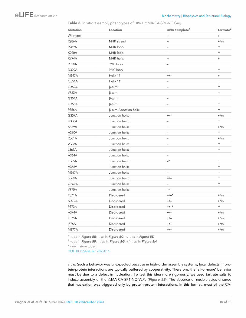


