Crystal Structure of a Polyhistidine-Tagged Recombinant Catalytic Subunit of cAMP-Dependent Protein...
11
Crystal Structure of a Polyhistidine-Tagged Recombinant Catalytic Subunit of cAMP-Dependent Protein Kinase Complexed with the Peptide Inhibitor PKI(5-24) and Adenosine ² Narendra Narayana, ‡ Sarah Cox, ‡,§ Shmuel Shaltiel, | Susan S. Taylor,* ,‡ and Nguyen-huu Xuong ⊥ Departments of Chemistry and Biochemistry, Biology, and Physics, UniVersity of California, San Diego, 9500 Gilman DriVe, La Jolla, California 92093-0654, and Department of Biological Regulation, The Weizmann Institute of Science, RehoVot 76100, Israel ReceiVed August 5, 1996; ReVised Manuscript ReceiVed December 16, 1996 X ABSTRACT: The crystal structure of the hexahistidine-tagged mouse recombinant catalytic subunit (H 6 -rC) of cAMP-dependent protein kinase (cAPK), complexed with a 20-residue peptide inhibitor from the heat- stable protein kinase inhibitor PKI(5-24) and adenosine, was determined at 2.2 Å resolution. Novel crystallization conditions were required to grow the ternary complex crystals. The structure was refined to a final crystallographic R-factor of 18.2% with good stereochemical parameters. The “active” enzyme adopts a “closed” conformation as found in rC:PKI(5-24) [Knighton et al. (1991a,b) Science 253, 407- 414, 414-420] and packs in a similar manner with the peptide providing a major contact surface. This structure clearly defines the subsites of the unique nucleotide binding site found in the protein kinase family. The adenosine occupies a mostly hydrophobic pocket at the base of the cleft between the two lobes and is completely buried. The missing triphosphate moiety of ATP is filled with a water molecule (Wtr 415) which replaces the γ-phosphate of ATP. The glycine-rich loop between 1 and 2 helps to anchor the phosphates while the ribose ring is buried beneath -strand 2. Another ordered water molecule (Wtr 375) is pentacoordinated with polar atoms from adenosine, Leu 49 in -strand 1, Glu 127 in the linker strand between the two lobes, Tyr 330, and a third water molecule, Wtr 359. The conserved nucleotide fold can be defined as a lid comprised of -strand 1, the glycine-rich loop, and -strand 2. The adenine ring is buried beneath -strand 1 and the linker strand (120-127) that joins the small and large lobes. The C-terminal tail containing Tyr 330, a segment that lies outside the conserved core, covers this fold and anchors it in a closed conformation. The main-chain atoms of the flexible glycine- rich loop (residues 50-55) in the ATP binding domain have a mean B-factor of 41.4 Å 2 . This loop is quite mobile, in striking contrast to the other conserved loops that converge at the active site cleft. The catalytic loop (residues 166-171) and the Mg 2+ positioning loop (residues 184-186) are a stable part of the large lobe and have low B-factors in all structures solved to date. The stability of the glycine-rich loop is highly dependent on the ligands that occupy the active site cleft with maximum stability achieved in the ternary complex containing Mg‚ATP and the peptide inhibitor. In this ternary complex the γ-phosphate is secured between both lobes by hydrogen bonds to the backbone amide of Ser 53 in the glycine-rich loop and the amino group of Lys 168 in the catalytic loop. In the adenosine ternary complex the water molecule replacing the γ-phosphate hydrogen bonds between Lys 168 and Asp 166 and makes no contact with the small lobe. This glycine-rich loop is thus the most mobile component of the active site cleft, with the tip of the loop being highly sensitive to what occupies the γ-subsite. The catalytic (C) subunit of cAMP-dependent protein kinase (cAPK), one of the smallest and simplest members of the protein kinase family (Hanks & Hunter, 1995), serves as a prototype for the large and very diverse enzyme family (Taylor et al., 1992). Its simplicity derives from its mech- anism of activation whereby in the absence of cAMP it is sequestered as a fully phosphorylated protein in an inactive complex with regulatory (R) subunits. Binding of cAMP to R triggers release of the active C-subunits. The biochemi- cal, kinetic, and structural analyses of the C-subunit have contributed significantly to our understanding of general protein kinase function. The C-subunit of cAPK is not only the first protein kinase structure to be solved but it is still the only structure of an active protein kinase which has been cocrystallized with both peptide and nucleotide substrates. The enzyme is bilobal, with the smaller amino-terminal lobe constituting a unique nucleotide binding domain and the larger lobe providing residues important for peptide binding and catalysis. The nucleotide is buried at the base of the cleft, with the phosphates extending to the edge of the cleft where the peptide binds. ² Supported by grants from the National Institutes of Health (GM19301) to S.S.T., the National Center for Research Resources (NIH, RR01644) to N.X., the Markey Foundation to S.S.T. and N.X., and the Minerva Foundation, Munich, Germany, to S.S. * Corresponding author. ‡ Department of Chemistry and Biochemistry, UCSD. § Present address: DuPont Merck Pharmaceutical Co., Experimental Station, E336/36A, Route 141 and Henry Clay Rd., Wilmington, DE 19880-0361. | The Weizmann Institute of Science. ⊥ Departments of Biology, Physics, and Chemistry and Biochemistry, UCSD. X Abstract published in AdVance ACS Abstracts, April 1, 1997. 4438 Biochemistry 1997, 36, 4438-4448 S0006-2960(96)01947-2 CCC: $14.00 © 1997 American Chemical Society
-
Upload
nguyen-huu -
Category
Documents
-
view
212 -
download
0
Transcript of Crystal Structure of a Polyhistidine-Tagged Recombinant Catalytic Subunit of cAMP-Dependent Protein...
Crystal Structure of a Polyhistidine-Tagged Recombinant Catalytic Subunit ofcAMP-Dependent Protein Kinase Complexed with the Peptide Inhibitor PKI(5-24)
and Adenosine†
Narendra Narayana,‡ Sarah Cox,‡,§ Shmuel Shaltiel,| Susan S. Taylor,*,‡ and Nguyen-huu Xuong⊥
Departments of Chemistry and Biochemistry, Biology, and Physics, UniVersity of California, San Diego, 9500 Gilman DriVe,La Jolla, California 92093-0654, and Department of Biological Regulation, The Weizmann Institute of Science,
RehoVot 76100, Israel
ReceiVed August 5, 1996; ReVised Manuscript ReceiVed December 16, 1996X
ABSTRACT: The crystal structure of the hexahistidine-tagged mouse recombinant catalytic subunit (H6-rC)of cAMP-dependent protein kinase (cAPK), complexed with a 20-residue peptide inhibitor from the heat-stable protein kinase inhibitor PKI(5-24) and adenosine, was determined at 2.2 Å resolution. Novelcrystallization conditions were required to grow the ternary complex crystals. The structure was refinedto a final crystallographicR-factor of 18.2% with good stereochemical parameters. The “active” enzymeadopts a “closed” conformation as found in rC:PKI(5-24) [Knighton et al. (1991a,b)Science 253, 407-414, 414-420] and packs in a similar manner with the peptide providing a major contact surface. Thisstructure clearly defines the subsites of the unique nucleotide binding site found in the protein kinasefamily. The adenosine occupies a mostly hydrophobic pocket at the base of the cleft between the twolobes and is completely buried. The missing triphosphate moiety of ATP is filled with a water molecule(Wtr 415) which replaces theγ-phosphate of ATP. The glycine-rich loop betweenâ1 andâ2 helps toanchor the phosphates while the ribose ring is buried beneathâ-strand 2. Another ordered water molecule(Wtr 375) is pentacoordinated with polar atoms from adenosine, Leu 49 inâ-strand 1, Glu 127 in thelinker strand between the two lobes, Tyr 330, and a third water molecule, Wtr 359. The conservednucleotide fold can be defined as a lid comprised ofâ-strand 1, the glycine-rich loop, andâ-strand 2.The adenine ring is buried beneathâ-strand 1 and the linker strand (120-127) that joins the small andlarge lobes. The C-terminal tail containing Tyr 330, a segment that lies outside the conserved core,covers this fold and anchors it in a closed conformation. The main-chain atoms of the flexible glycine-rich loop (residues 50-55) in the ATP binding domain have a meanB-factor of 41.4 Å2. This loop isquite mobile, in striking contrast to the other conserved loops that converge at the active site cleft. Thecatalytic loop (residues 166-171) and the Mg2+ positioning loop (residues 184-186) are a stable part ofthe large lobe and have lowB-factors in all structures solved to date. The stability of the glycine-richloop is highly dependent on the ligands that occupy the active site cleft with maximum stability achievedin the ternary complex containing Mg‚ATP and the peptide inhibitor. In this ternary complex theγ-phosphate is secured between both lobes by hydrogen bonds to the backbone amide of Ser 53 in theglycine-rich loop and the amino group of Lys 168 in the catalytic loop. In the adenosine ternary complexthe water molecule replacing theγ-phosphate hydrogen bonds between Lys 168 and Asp 166 and makesno contact with the small lobe. This glycine-rich loop is thus the most mobile component of the activesite cleft, with the tip of the loop being highly sensitive to what occupies theγ-subsite.
The catalytic (C) subunit of cAMP-dependent proteinkinase (cAPK), one of the smallest and simplest membersof the protein kinase family (Hanks & Hunter, 1995), servesas a prototype for the large and very diverse enzyme family(Taylor et al., 1992). Its simplicity derives from its mech-
anism of activation whereby in the absence of cAMP it issequestered as a fully phosphorylated protein in an inactivecomplex with regulatory (R) subunits. Binding of cAMPto R triggers release of the active C-subunits. The biochemi-cal, kinetic, and structural analyses of the C-subunit havecontributed significantly to our understanding of generalprotein kinase function.The C-subunit of cAPK is not only the first protein kinase
structure to be solved but it is still the only structure of anactive protein kinase which has been cocrystallized with bothpeptide and nucleotide substrates. The enzyme is bilobal,with the smaller amino-terminal lobe constituting a uniquenucleotide binding domain and the larger lobe providingresidues important for peptide binding and catalysis. Thenucleotide is buried at the base of the cleft, with thephosphates extending to the edge of the cleft where thepeptide binds.
† Supported by grants from the National Institutes of Health(GM19301) to S.S.T., the National Center for Research Resources (NIH,RR01644) to N.X., the Markey Foundation to S.S.T. and N.X., andthe Minerva Foundation, Munich, Germany, to S.S.* Corresponding author.‡ Department of Chemistry and Biochemistry, UCSD.§ Present address: DuPont Merck Pharmaceutical Co., Experimental
Station, E336/36A, Route 141 and Henry Clay Rd., Wilmington, DE19880-0361.
| The Weizmann Institute of Science.⊥ Departments of Biology, Physics, and Chemistry and Biochemistry,
UCSD.X Abstract published inAdVance ACS Abstracts,April 1, 1997.
4438 Biochemistry1997,36, 4438-4448
S0006-2960(96)01947-2 CCC: $14.00 © 1997 American Chemical Society
The combination of structures of binary and ternarycomplexes of the C-subunit containing both substrate andinhibitor peptides provides a foundation for understandingthe mechanism of catalysis whereby a set of highly conservedresidues converge at the active site to facilitate phosphoryltransfer (Madhusudan et al., 1994). The nucleotide base isburied in a mostly hydrophobic pocket at the base of thecleft, while the phosphates are at the edge of the cleft, andit is here where most of the conserved residues converge.These active site residues, contributed by various parts ofthe molecule, are located primarily in three loopssa glycine-rich loop in the small lobe betweenâ-strands 1 and 2, acatalytic loop in the large lobe betweenâ-strands 6 and 7,and a Mg2+ positioning loop. The various crystal structuresalso define the mobility of the two domains and reveal openand closed conformations. One of the binary complexes ofthe mammalian enzyme containing an inhibitor peptideassumes an open conformation while all of the others areclosed. These closed conformations include an ADP:PKS-(5-24) complex, an ATP:PKI(5-24) complex, a phospho-PKS(5-24) complex, and a PKI(5-24) binary complex.Opening and closing of this cleft must to some extent be anintrinsic part of the catalytic mechanism. Since the actualphosphoryl transfer step is rapid (500 s-1), the slower rate-limiting step (20 s-1) corresponds to opening of the cleftand ADP release (Grant & Adams, 1996).Kinetic evidence indicates that most of the energy for
nucleotide binding is associated with the adenine ring, sinceadenine has aKd of 30µM which is only 3-fold higher thantheKd or Km of Mg‚ATP. Thermostability is also enhancednearly equivalently by Mg‚ATP or adenosine (Herberg,manuscript in preparation). Most protein kinase inhibitorsare competitive with ATP, and it appears that occupancy ofthe adenine binding site is critical for these inhibitors. Tofurther define the nucleotide binding site of cAPK and themobility of loops at the active site cleft, a poly(His)-taggedform of the C-subunit was cocrystallized with PKI(5-24)1and adenosine, leaving the phosphate subsites unoccupied.Although novel conditions were required to obtain crystals,the structure assumed a closed conformation similar to earlierones with a water molecule occupying theγ-phosphate inthe active site cleft. The structured water molecules at theactive site are described. Analysis of the various closedconformations reveals that the glycine-rich loop is the singlemobile component of the active site cleft and assumes a fullyclosed and stable conformation only when Mg‚ATP and theinhibitor peptide are present.This structure of the H6-rC shows that the fundamental
features of the kinase structure are not disturbed by the His6
tag. In the rC-subunit which lacks the N-terminal myristicacid, the first 10-15 residues are disordered. The stretchof 20 residues containing the His6 tag is also disorderedalthough this does not interfere with crystal packing.Nevertheless, the presence of the His6 tag required different
crystallization conditions. This is extremely important forsubsequent analysis of mutants of the C-subunit that cannotbe purified by conventional methods.
EXPERIMENTAL PROCEDURES
Protein, Peptide Inhibitor, and Nucleoside.The construc-tion of the His6-tagged mouse rC will be described elsewhere(Cox et al., manuscript in preparation). To summarize, a20-residue peptide including six contiguous histidines wasfused to the N-terminus of rC. The His6-rC was expressedin Escherichia coli(Slice & Taylor, 1989) and purified usingnickel chelate affinity chromatography. Isoforms wereseparated by chromatography on a Mono-S column asdescribed by Herberg et al. (1993), except that dialysis wascarried out in a two-step procedure, first against 20 mMpotassium phosphate (pH 6.2) and 50 mM KCl and thenagainst 20 mM potassium phosphate, pH 6.2, and 25 mMKCl, before being loaded onto the Mono-S 10/10 column(Pharmacia). The H6-rC was resolved into three isoformsby eluting with a 25 mM-1 M KCl linear gradient. IsoformII, phosphorylated at three sitessSer 10, Thr 197, and Ser338 (Herberg et al., 1993)swas used for crystallization. Thepeptide inhibitor PKI(5-24), a 20-residue segment of theheat-stable protein kinase inhibitor (PKI) with an apparentKi of 2.3 nM (Cheng et al., 1986), was synthesized on aMilligen peptide synthesizer and purified to homogeneityby high-performance liquid chromatography (HPLC). Ad-enosine was purchased from Sigma Chemical Co.Crystallization. Since the conditions used previously for
crystallizing the rC-subunit with poly(ethylene glycol) 400(PEG-400) (Zheng et al., 1992) were unsuccessful for theH6-rC complex, novel crystallization conditions were devel-oped. Orthorhombic crystals of the ternary complex, com-prised of mouse H6-rC, PKI(5-24), and adenosine, weregrown by the hanging-drop vapor diffusion method using2-methyl-2,4-pentanediol (MPD) as the precipitating agent.The mother liquor (20µL) contained protein at a concentra-tion of 0.25 mM in 100 mM Bicine buffer at pH 8.0, 0.75mM PKI(5-24), 4 mM adenosine, and 4% MPD. Thereservoir solution (1 mL) was made up of 20% MPD in 100mM Bicine buffer (pH 8.0). X-ray diffraction quality crystals(0.2× 0.15× 0.8 mm3) were grown at 4°C in approximately4-6 weeks. The crystallization mixture did not containdivalent metal ions. Several trials using poly(ethyleneglycol) 400 (PEG-400) as precipitant according to theprotocol described in the crystallization of rC:PKI(5-24) andrC:PKI(5-24):Mg2‚ATP (Zheng et al., 1992) complexeswere unsuccessful.Data Collection. The crystals belong to the space group
P212121 with unit cell dimensions ofa) 73.08 Å,b) 78.44Å, andc) 80.49 Å. There is one ternary complex moleculeper asymmetric unit (Matthews coefficient,Vm ) 2.7 Å3/Da; 54% crystal solvent content). X-ray intensity data on asingle crystal were collected at 4°C on Xuong-Hamlinmultiwire area detectors (Hamlin, 1985), with a graphitemonochromated Cu KR beam from a Rigaku RU-200rotating anode X-ray generator operating at 5 kW power.Data reduction was done with the UCSD area-detector dataprocessing programs (Howard et al., 1985). The crystaldiffracted to 2.2 Å resolution with anRsym (∑|Iobs - Iav|/∑Iav) of 0.064 and 92% completeness. There was a decayof 8.0% over an X-ray exposure period of 2 days. TheWilson B-factor is 34 Å2.
1 Abbreviations: PKI(5-24) corresponds to residues 5-24 in thenaturally occurring heat-stable protein kinase inhibitor (TTYADFIAS-GRTGRRNAIHD). PKS(5-24) represents the corresponding substratepeptide, TTYADFIASGRTGRRASIHD, where the residues shown inbold were changed to NA, respectively, in the peptide inhibitor. PKS-(5-24)* represents the same peptide phosphorylated at Ser 21. Thecomplexes rC:PKI(5-24):Mn2‚ATP, rC:PKI(5-24):adenosine, rC:PKI-(5-24), and mC:PKI(5-24):ATP are abbreviated as rCI‚ATP, rCIA,rCI, and mCI‚ATP, respectively.
Ternary Complex Structure of C-Subunit of cAPK Biochemistry, Vol. 36, No. 15, 19974439

Structure Refinement. The cell dimensions of the ortho-rhombic crystal form of different complexes of mouse rC,whose structures are reported previously, are compared inTable 1. The length of theb-axis in the present structure isapproximately 3% longer than the corresponding axis in theother crystals. The variations in the other two dimensions(a- andc-axes) are less than 0.5% for all crystals obtainedso far. This elongation along theb-axis is significant andwas observed previously in crystals of a rC binary complexwith PKI(5-24) where variations range between 75.36 and77.61 Å, with one crystal whoseb-axis was even moreelongated (80.1 Å) (Knighton et al., personal communica-tion). Possible reasons for the observed variation in the celldimension along theb-axis are discussed further in thesection pertaining to crystal packing. This is the firstreported structure analysis of an rC complex grown using aprecipitant other than PEG-400, and all crystals tested forX-ray diffraction during this work consistently had anelongatedb-axis, unlike the other rC binary and ternarycomplexes (see Table 1).The coordinates of the mouse rC complexed with PKI-
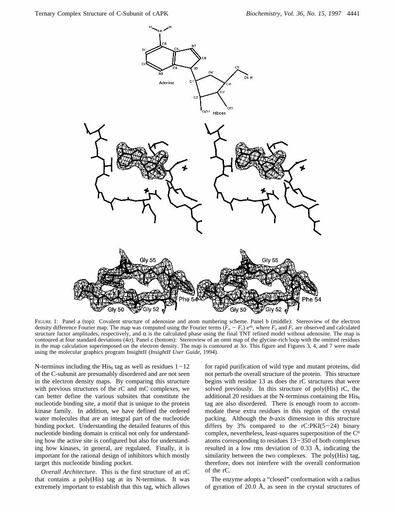
(5-24) (PDB entry 1APM) were used as the starting modelfor refinement against the X-ray data obtained from thepresent crystal of the ternary complex. The isotropicB-factors were set uniformly to 15 Å2 for all protein andpeptide inhibitor atoms. The model was initially subjectedto rigid body refinement and was then followed by severalcycles of conjugate-gradient positional and overall isotropicand individual restrainedB-factor (thermal factor) refinementusing the X-PLOR program (Brunger, 1992). The data usedin these refinement cycles were gradually increased from3.0 to 2.2 Å in three steps. Refinement of this binary modelconverged at a crystallographicR-factor of 21.6% for data(F > 2σF) between 10 and 2.2 Å. A difference Fourier mapcalculated at this stage using 20-2.2 Å data clearly showedelectron density for adenosine (see Figure 1). Furtherrefinement was continued with the inclusion of adenosine.The simulated annealing slow-cool refinement protocol wasrun fromT ) 3000 K down toT ) 277 K in steps of 25 K(time step) 0.5 fs and tolerance) 0.2 Å). This refinementwas followed by 40 cycles of Powell minimization, 20 cyclesof overallB-factor refinement, and 25 cycles of restrainedindividual B-factor refinement, which converged atR )21.0% for 10-2.2 Å resolution. A simulated annealed omitmap was computed by excluding residues within a 4 Åradiusof the adenosine. All omitted atoms were readily fitted intothe electron density map with the aid of the moleculargraphics program package TOM-FRODO (Cambillau &
Horjales, 1987). Electron densities for some side-chainatoms, particularly Lys and Arg residues on the surface suchas Lys 63, Lys 78, and Arg 256, that were not found in thestarting model were located reliably in the present structurevia several difference Fourier maps. The two C-terminalresidues, His 23 and Asp 24, of the peptide inhibitor arewell defined, and there was no indication of alternate con-formations for any of the residues, unlike those in the binarycomplex structure of rC:PKI(5-24) (Knighton et al., 1993).At the completion of X-PLOR refinement, the model con-sisted of all atoms of the rC from residue 13 to residue 350,the peptide inhibitor PKI(5-24), and adenosine. The His6
tag as well as residues 1-12 of the rC was not seen in theelectron density maps and is assumed to be thermally disor-dered. All atoms have full occupancy, and their individualB’s refined with restraints. The target standard deviationsbetweenB-factors of bonded backbone and side-chain atomswere 1.5 and 2.0, respectively. The target standard deviationsbetweenB-factors of backbone and side-chain atoms con-nected by an angle were 2.0 and 2.5, respectively.This refined model was further subjected to least-squares
refinement using the conjugate-direction algorithm in theTNT program (Tronrud et al., 1987). After the convergenceof the model to anR-factor of 20%, water molecules werebuilt into the structure in five rounds of alternate structurerefinement and identification of solvent peaks in the differ-ence Fourier maps. Waters were assigned to peaks greaterthan 3σ in the difference Fourier maps and concomitantlyat a hydrogen bonding distance with other polar atoms. Theinclusion of 90 water molecules with an additional five cyclesof refinement resulted in a finalR-value of 18.2% for databetween 10 and 2.2 Å. In the glycine-rich loop (residues50-55), the meanB’s for main-chain and side-chain atomsare 41.4 and 67.3 Å2, respectively. The unrestrained averageB excluding atoms corresponding mostly to the surface sidechains which hit the maximum allowed value of 100 Å2 was32.4 Å2. The structure has good stereochemical parametersas estimated by the program PROCHECK (Laskowski et al.,1993), and there are no non-glycine residues within disal-lowed (φ,ψ) regions in a Ramachandran plot (Ramachandran& Sasisekharan, 1968). Some results pertaining to TNTrefinement are summarized in Table 2.
RESULTS
The overall structure of this rCIA ternary complex assumesa closed conformation with the adenosine deeply buried ina solvent-inaccessible pocket at the base of the cleft betweenthe two lobes. The additional 20 amino acids at the
Table 1: Unit Cell Dimensions of Mouse rC Complexes with the Space GroupP212121a
unit cell lengths (Å)
compound a b c reference
rC+ PKI(5-24) 73.84 75.76 81.01 Knighton et al. (1993)rC+ PKI(5-24)+ Mn2‚ATP 73.58 76.28 80.58 Zheng et al. (1993b)rC+ PKS(5-24)+ ADP 73.96 76.11 81.00 Madhusudan et al. (1994)rC+ PKS(5-24)* 73.87 75.58 80.70 Madhusudan et al. (1994)rC + PKI(5-24)+ adenosine 73.08 78.44 80.49 this work
aPKI(5-24) corresponds to residues 5-24 in the naturally occurring heat-stable protein kinase inhibitor (TTYADFIASGRTGRRNAIHD). PKS(5-24) represents the corresponding substrate peptide, TTYADFIASGRTGRRASIHD, where the residues shown in bold were changed to NA, respectively,in the peptide inhibitor. PKS(5-24)* represents the same peptide phosphorylated at Ser 21. The average estimated standard deviation (esd) ofunit cell lengths is 0.05 Å. The esds for unit cell lengths were obtained by taking the average of the refined cell parameters for each orientation(total of seven runs) and then calculating the standard deviation for each cell axis. The meanB-factors for the main-chain atoms in the above listedcomplexes are 28, 33, 24, 29, and 38 Å2, respectively.
4440 Biochemistry, Vol. 36, No. 15, 1997 Narayana et al.
N-terminus including the His6 tag as well as residues 1-12of the C-subunit are presumably disordered and are not seenin the electron density maps. By comparing this structurewith previous structures of the rC and mC complexes, wecan better define the various subsites that constitute thenucleotide binding site, a motif that is unique to the proteinkinase family. In addition, we have defined the orderedwater molecules that are an integral part of the nucleotidebinding pocket. Understanding the detailed features of thisnucleotide binding domain is critical not only for understand-ing how the active site is configured but also for understand-ing how kinases, in general, are regulated. Finally, it isimportant for the rational design of inhibitors which mostlytarget this nucleotide binding pocket.OVerall Architecture. This is the first structure of an rC
that contains a poly(His) tag at its N-terminus. It wasextremely important to establish that this tag, which allows
for rapid purification of wild type and mutant proteins, didnot perturb the overall structure of the protein. This structurebegins with residue 13 as does the rC structures that weresolved previously. In this structure of poly(His) rC, theadditional 20 residues at the N-terminus containing the His6
tag are also disordered. There is enough room to accom-modate these extra residues in this region of the crystalpacking. Although theb-axis dimension in this structurediffers by 3% compared to the rC:PKI(5-24) binarycomplex, nevertheless, least-squares superposition of the CR
atoms corresponding to residues 13-350 of both complexesresulted in a low rms deviation of 0.33 Å, indicating thesimilarity between the two complexes. The poly(His) tag,therefore, does not interfere with the overall conformationof the rC.The enzyme adopts a “closed” conformation with a radius
of gyration of 20.0 Å, as seen in the crystal structures of
FIGURE 1: Panel a (top): Covalent structure of adenosine and atom numbering scheme. Panel b (middle): Stereoview of the electrondensity difference Fourier map. The map was computed using the Fourier terms (Fo - Fc) eiR, whereFo andFc are observed and calculatedstructure factor amplitudes, respectively, andR is the calculated phase using the final TNT refined model without adenosine. The map iscontoured at four standard deviations (4σ). Panel c (bottom): Stereoview of an omit map of the glycine-rich loop with the omitted residuesin the map calculation superimposed on the electron density. The map is contoured at 3σ. This figure and Figures 3, 4, and 7 were madeusing the molecular graphics program InsightII (InsightII User Guide, 1994).
Ternary Complex Structure of C-Subunit of cAPK Biochemistry, Vol. 36, No. 15, 19974441
rC:PKI(5-24) and rC:PKI(5-24):Mg2‚ATP. The secondarystructure elements in the bilobal enzyme (Figure 2) arelabeled according to Knighton et al. (1991a). The adenosineis buried between the large and small lobes. Access to theactive site cleft is blocked by the peptide inhibitor, by theglycine-rich loop betweenâ-strands 1 and 2, and by theenzyme’s C-terminal tail segment (residues 315-335),resulting in a solvent-inaccessible enclosure for the nucleo-side. The bound peptide inhibitor, PKI(5-24), contains anamphipathic helix followed by a turn and an extended region,similar to that observed in the rC:PKI(5-24) and rC:PKI-(5-24):Mg2‚ATP complexes.Geometry of the Adenosine and Its Interactions with the
Protein and PKI(5-24). (A) Adenine Binding Site. Ad-enosine is bound to the unique protein kinase nucleotide
binding fold with interactions that are essentially the sameas observed in the ternary complex of rC:PKI(5-24):Mn2‚ATP (Zheng et al., 1993b) and the porcine C-subunitcomplexed with PKI(5-24) and Mn2‚AMP-PNP (Bosse-meyer et al., 1993). The adenine base is completely buriedin a hydrophobic pocket formed at the junction of the smalland large lobes. The planar adenine ring is involved innumerous interactions, both hydrophobic and hydrogenbonding, with the enzyme. With only a few exceptions, theseinteractions involve residues that are located either in thesmall lobe or in the linker strand residues 120-127 that jointhe two lobes (Table 3). The residues involved in hydro-phobic interactions are Leu 49, Val 57, Ala 70, Val 104,Met 120, Glu 121, Tyr 122, Val 123, Leu 173, Thr 183, andPhe 327. In general, the hydrophobicity of these residuesis conserved throughout the protein kinase family. Inaddition to these interactions, the purine base forms threehydrogen bonds (see Figure 3 and Table 4). The N1 interactswith the main-chain amide of Val 123, the N6 amino groupdonates a hydrogen bond (H-bond) to the backbone carbonylof Glu 121, and N7 is bonded to OG1 of Thr 183. Theinvolvement of the N6 amino group in the hydrogen bondformation is consistent with prior analogue studies, sinceremoval of a hydrogen bond donor at this position results ina decrease in binding affinity (Hoppe et al., 1978; Taylor etal., 1990). Replacing one of the N6 hydrogens with a methylgroup has an even greater effect onKm, which confirms thefact that the adenine ring is tightly packed in this pocket.Therefore, the tetrapeptide segment Met 120-Val 123 playsa crucial role in the recognition of the adenine base both byhydrogen-bonding and hydrophobic interactions. This seg-ment, indicated in Figure 2, is part of the short strand(residues 120-127) that links the two domains in theconserved catalytic core. With the exception of Leu 173,Thr 183, and Phe 327, all of the residues that comprise theadenine binding pocket and are in close contact with thepurine base are located in either the small lobe or the linkerstrand (120-127).
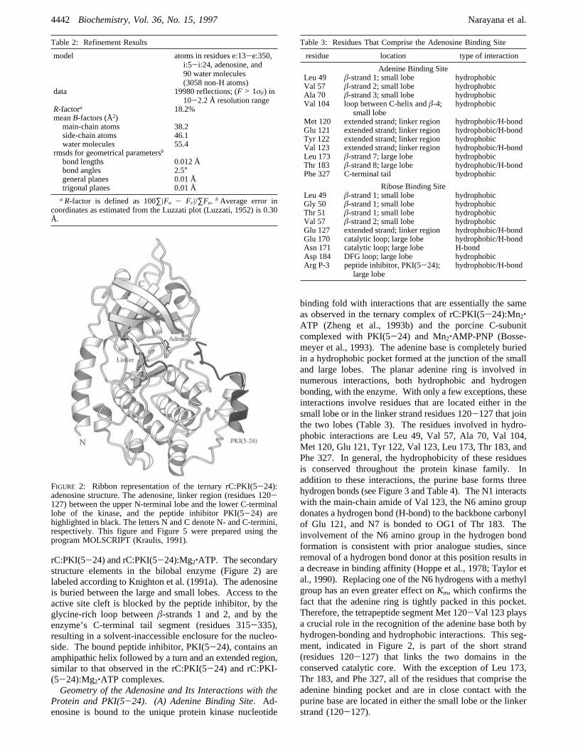
Table 2: Refinement Results
model atoms in residues e:13-e:350,i:5-i:24, adenosine, and90 water molecules(3058 non-H atoms)
data 19980 reflections; (F > 1σF) in10-2.2 Å resolution range
R-factora 18.2%meanB-factors (Å2)main-chain atoms 38.2side-chain atoms 46.1water molecules 55.4
rmsds for geometrical parametersb
bond lengths 0.012 Åbond angles 2.5°general planes 0.01 Åtrigonal planes 0.01 Åa R-factor is defined as 100∑|Fo - Fc|/∑Fo. b Average error in
coordinates as estimated from the Luzzati plot (Luzzati, 1952) is 0.30Å.
FIGURE 2: Ribbon representation of the ternary rC:PKI(5-24):adenosine structure. The adenosine, linker region (residues 120-127) between the upper N-terminal lobe and the lower C-terminallobe of the kinase, and the peptide inhibitor PKI(5-24) arehighlighted in black. The letters N and C denote N- and C-termini,respectively. This figure and Figure 5 were prepared using theprogram MOLSCRIPT (Kraulis, 1991).
Table 3: Residues That Comprise the Adenosine Binding Site
residue location type of interaction
Adenine Binding SiteLeu 49 â-strand 1; small lobe hydrophobicVal 57 â-strand 2; small lobe hydrophobicAla 70 â-strand 3; small lobe hydrophobicVal 104 loop between C-helix andâ-4;
small lobehydrophobic
Met 120 extended strand; linker region hydrophobic/H-bondGlu 121 extended strand; linker region hydrophobic/H-bondTyr 122 extended strand; linker region hydrophobicVal 123 extended strand; linker region hydrophobic/H-bondLeu 173 â-strand 7; large lobe hydrophobicThr 183 â-strand 8; large lobe hydrophobic/H-bondPhe 327 C-terminal tail hydrophobic
Ribose Binding SiteLeu 49 â-strand 1; small lobe hydrophobicGly 50 â-strand 1; small lobe hydrophobicThr 51 â-strand 1; small lobe hydrophobicVal 57 â-strand 2; small lobe hydrophobicGlu 127 extended strand; linker region hydrophobic/H-bondGlu 170 catalytic loop; large lobe hydrophobic/H-bondAsn 171 catalytic loop; large lobe H-bondAsp 184 DFG loop; large lobe hydrophobicArg P-3 peptide inhibitor, PKI(5-24);
large lobehydrophobic/H-bond
4442 Biochemistry, Vol. 36, No. 15, 1997 Narayana et al.
(B) Ribose Binding Site.The ribose adopts an unsym-metrical twist (major C3′-endo pucker) about the planeformed by atoms O4′, C1′, and C4′. The sugar assumes ananti conformation (ø ) -151.7°; Saenger, 1984) with respectto the adenine base. Unlike the purine ring, the sugarinteracts with both the enzyme and the peptide inhibitor PKI-(5-24). The exocyclic oxygens O2′, O3′, and O5′ are allinvolved in hydrogen bonding (see Figure 3 and also Table4). As seen in the ternary complex of rC:PKI(5-24):Mn2‚ATP (Zheng et al., 1993b), the O2′ interacts with the sidechain of Glu 127 while the O3′ interacts with the P-3 arginineof PKI(5-24) and the backbone carbonyl of Glu 170. Inaddition to these interactions, in both structures the O2′ formsa hydrogen bond with a water molecule (Wtr 375) and theO3′ is hydrogen-bonded to OE2 of Glu 127. However, thetorsion angle C3′-C4′-C5′-O5′ ) -38.3° is significantlydifferent from that found in the ternary complexes rC:PKI-
(5-24):Mn2‚ATP (73.7°) and mC:PKI(5-24):Mn2‚AMP-PNP (61.9°). Therefore, the O5′ atom has moved signifi-cantly away from the base and the sugar ring compared toboth the ternary complexes containing the phosphate groups.This O5′ atom has aB-factor of 76 Å2. Rotation about theexocyclic C4′-C5′ bond allows O5′ to assume differentpositions relative to the base and the furanose. In purinenucleosides, the orientation about this bond is preferentially+synclinal (∼60°), but both-synclinalandanti (∼180°)are sometimes found (Saenger, 1984).(C) Phosphate Binding Site. In contrast to the hydrophobic
adenosine binding pocket, the binding site filled by thephosphates of ATP in the ternary rCI‚ATP is very polar. Inthis structure all three phosphates are missing. This site isfilled by a water molecule, Wtr 415, that occupies theposition of theγ-phosphate of ATP. Many of the conservedresidues such as Lys 72, Glu 91, and Asp 184 that clusteraround this site of phosphoryl transfer do not move signifi-cantly in this structure. The crucial placement of this watermolecule also means that the conserved residues do not needto move. See section on Solvent Structure in the Active SiteCleft for details.Conformation of the Peptide Inhibitor PKI(5-24) and Its
Interactions with the Protein. In this structure the peptideinhibitor PKI(5-24) is bound in a similar conformation tothat described for the other rC ternary and binary complexes.The key ionic interactions between the arginines at P-2, P-3,and P-6 with glutamates 230 and 170 (P-2), 127 (P-3), and203 (P-6) are all conserved. In addition to the interactionof the P-3 Arg with Glu 127, there is also a weak interactionwith the side-chain hydroxyl of Tyr 330 (3.4 Å). Thisinteraction was seen in the previous “closed” structures butwas not emphasized. In addition, the same hydroxyl of Tyr330 interacts with the carbonyl of Leu 49 and the 2′-OH ofthe ribose of adenosine, via a water bridge. Mutationalstudies indicate that Tyr 330 is important for both peptidebinding and catalysis (Chestukhin et al., 1996). The P-3 Argdonates a hydrogen bond to the carbonyl oxygen of Thr 51in the glycine-rich loop betweenâ-strands 1 and 2. Alsoconserved in this structure are the hydrophobic interactionsof the P-11 Phe with Arg 133, Gly 234, Tyr 235, Pro 236,
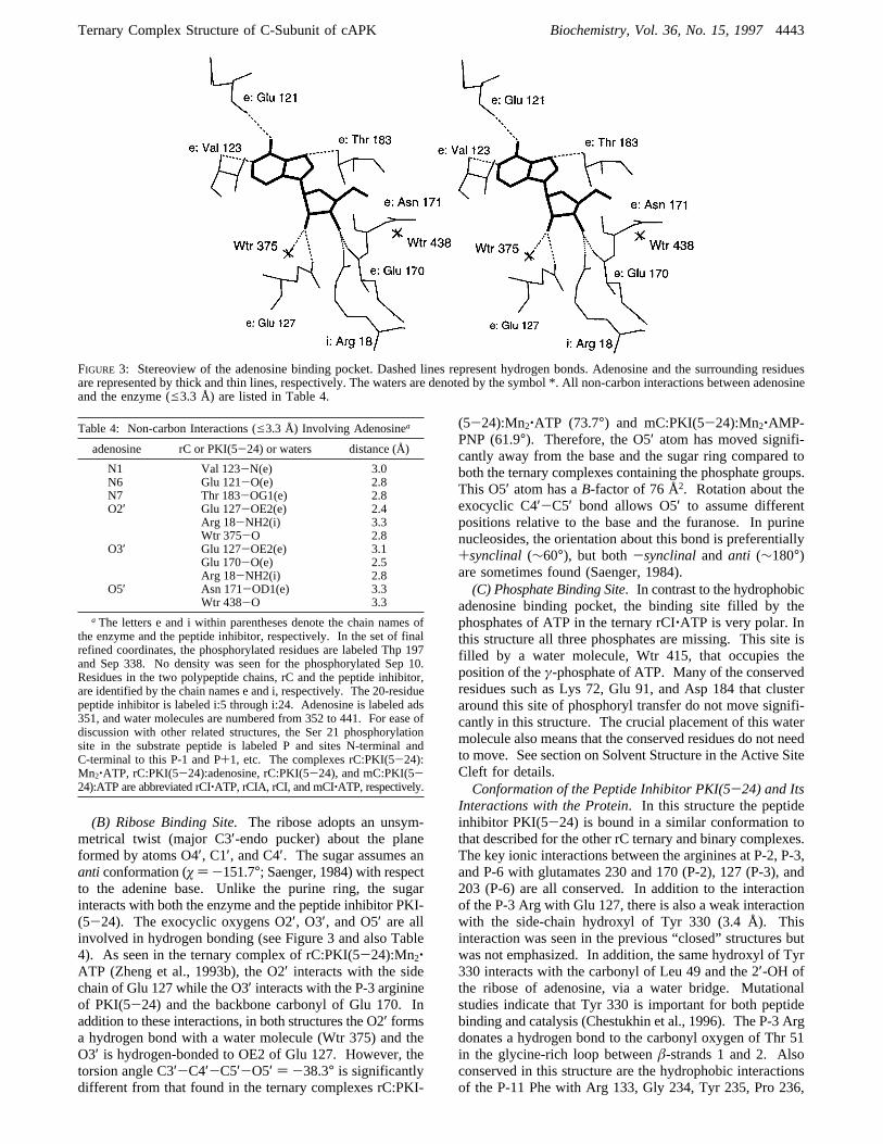
FIGURE 3: Stereoview of the adenosine binding pocket. Dashed lines represent hydrogen bonds. Adenosine and the surrounding residuesare represented by thick and thin lines, respectively. The waters are denoted by the symbol *. All non-carbon interactions between adenosineand the enzyme (e3.3 Å) are listed in Table 4.
Table 4: Non-carbon Interactions (e3.3 Å) Involving Adenosinea
adenosine rC or PKI(5-24) or waters distance (Å)
N1 Val 123-N(e) 3.0N6 Glu 121-O(e) 2.8N7 Thr 183-OG1(e) 2.8O2′ Glu 127-OE2(e) 2.4
Arg 18-NH2(i) 3.3Wtr 375-O 2.8
O3′ Glu 127-OE2(e) 3.1Glu 170-O(e) 2.5Arg 18-NH2(i) 2.8
O5′ Asn 171-OD1(e) 3.3Wtr 438-O 3.3
a The letters e and i within parentheses denote the chain names ofthe enzyme and the peptide inhibitor, respectively. In the set of finalrefined coordinates, the phosphorylated residues are labeled Thp 197and Sep 338. No density was seen for the phosphorylated Sep 10.Residues in the two polypeptide chains, rC and the peptide inhibitor,are identified by the chain names e and i, respectively. The 20-residuepeptide inhibitor is labeled i:5 through i:24. Adenosine is labeled ads351, and water molecules are numbered from 352 to 441. For ease ofdiscussion with other related structures, the Ser 21 phosphorylationsite in the substrate peptide is labeled P and sites N-terminal andC-terminal to this P-1 and P+1, etc. The complexes rC:PKI(5-24):Mn2‚ATP, rC:PKI(5-24):adenosine, rC:PKI(5-24), and mC:PKI(5-24):ATP are abbreviated rCI‚ATP, rCIA, rCI, and mCI‚ATP, respectively.
Ternary Complex Structure of C-Subunit of cAPK Biochemistry, Vol. 36, No. 15, 19974443
and Phe 239. There is a weak interaction between theC-terminal Asp of the peptide inhibitor and the side chainof Lys 83. The imidazole of the P+2 His is positionedwithin van der Waals distance from the side chain of Phe54 in the glycine-rich loop. The P-site Ala is marginallycloser to the enzyme active site. The distances between thehydroxyl of Ser 53 in the glycine-rich loop and P-site’sbackbone amide and the carbonyl oxygen are 3.2 and 2.9Å, compared to 3.5 and 2.8 Å, respectively, in the Mn2‚ATP-containing ternary complex structure and 3.7 and 3.6Å, respectively, in the binary complex rC:PKI(5-24). TheaverageB-factors (Å2) for Ser 53 in the rCI‚ATP, mCI‚-ATP, rCIA, and rCI structures are 23.95, 20.40, 64.42, and72.31, respectively.Crystal Packing. The enzyme complex is packed in an
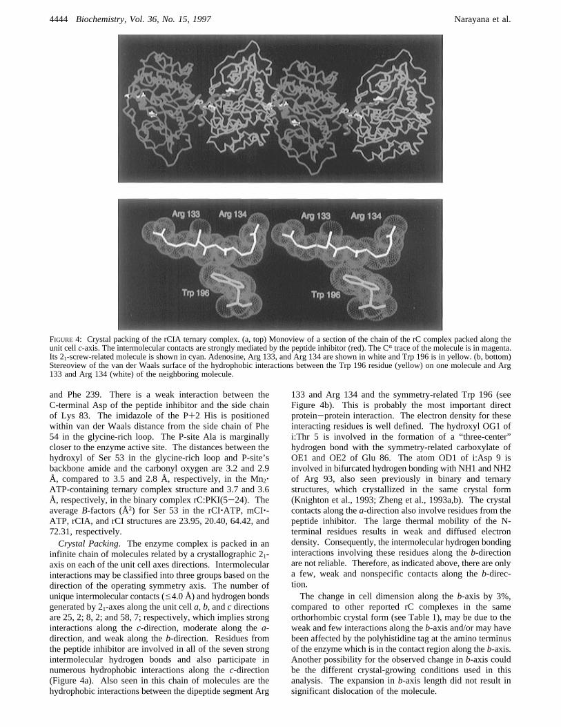
infinite chain of molecules related by a crystallographic 21-axis on each of the unit cell axes directions. Intermolecularinteractions may be classified into three groups based on thedirection of the operating symmetry axis. The number ofunique intermolecular contacts (e4.0 Å) and hydrogen bondsgenerated by 21-axes along the unit cella, b, andc directionsare 25, 2; 8, 2; and 58, 7; respectively, which implies stronginteractions along thec-direction, moderate along thea-direction, and weak along theb-direction. Residues fromthe peptide inhibitor are involved in all of the seven strongintermolecular hydrogen bonds and also participate innumerous hydrophobic interactions along thec-direction(Figure 4a). Also seen in this chain of molecules are thehydrophobic interactions between the dipeptide segment Arg
133 and Arg 134 and the symmetry-related Trp 196 (seeFigure 4b). This is probably the most important directprotein-protein interaction. The electron density for theseinteracting residues is well defined. The hydroxyl OG1 ofi:Thr 5 is involved in the formation of a “three-center”hydrogen bond with the symmetry-related carboxylate ofOE1 and OE2 of Glu 86. The atom OD1 of i:Asp 9 isinvolved in bifurcated hydrogen bonding with NH1 and NH2of Arg 93, also seen previously in binary and ternarystructures, which crystallized in the same crystal form(Knighton et al., 1993; Zheng et al., 1993a,b). The crystalcontacts along thea-direction also involve residues from thepeptide inhibitor. The large thermal mobility of the N-terminal residues results in weak and diffused electrondensity. Consequently, the intermolecular hydrogen bondinginteractions involving these residues along theb-directionare not reliable. Therefore, as indicated above, there are onlya few, weak and nonspecific contacts along theb-direc-tion.The change in cell dimension along theb-axis by 3%,
compared to other reported rC complexes in the sameorthorhombic crystal form (see Table 1), may be due to theweak and few interactions along theb-axis and/or may havebeen affected by the polyhistidine tag at the amino terminusof the enzyme which is in the contact region along theb-axis.Another possibility for the observed change inb-axis couldbe the different crystal-growing conditions used in thisanalysis. The expansion inb-axis length did not result insignificant dislocation of the molecule.
FIGURE 4: Crystal packing of the rCIA ternary complex. (a, top) Monoview of a section of the chain of the rC complex packed along theunit cellc-axis. The intermolecular contacts are strongly mediated by the peptide inhibitor (red). The CR trace of the molecule is in magenta.Its 21-screw-related molecule is shown in cyan. Adenosine, Arg 133, and Arg 134 are shown in white and Trp 196 is in yellow. (b, bottom)Stereoview of the van der Waals surface of the hydrophobic interactions between the Trp 196 residue (yellow) on one molecule and Arg133 and Arg 134 (white) of the neighboring molecule.
4444 Biochemistry, Vol. 36, No. 15, 1997 Narayana et al.

The crystal contacts are in broad agreement with theobservations made in the other orthorhombic mouse rCcomplexes (Karlsson et al., 1994). The overall structure ofthis polyhistidine-tagged rC is very similar to other rCstructures, indicating that the 20-residue N-terminal extensiondoes not significantly affect the protein fold. Also from thecrystal packing, where the lattice contacts are stronglymediated by the peptide inhibitor, we infer that the crystalsgrown without the peptide inhibitor would very likely belongto a different lattice, while still maintaining the observedkey intermolecular hydrophobic interactions between theenzyme residues only. This is, in fact, corroborated by therecently solved crystal structure of the binary rC:adenosinecomplex, where there is no peptide inhibitor. Although thiscomplex crystallizes in a new crystal form, the indole ringof Trp 196 still stacks on Arg 133, 134 of the symmetry-related molecule (Narayana et al., manuscript in preparation).Recently it was shown that Trp 196 is a major determinantfor recognition of the R-subunit of cAPK (Orellana &McKnight, 1992), while Arg 133 is an important determinantfor recognition of PKI(5-24) but not R-subunit (Wen &Taylor, 1994).
DISCUSSION
General Discussion of the Nucleotide Binding Site. Sev-eral conserved motifs in the small lobe contribute to thenucleotide binding site. Of key importance is a nucleotidepositioning motif composed of three parts:â-strand 1 whichpositions the adenine ring, the glycine-rich loop that positionsthe phosphates of ATP (Figure 1c), andâ-strand 2 whichpositions the ribose (Figure 5). The three elements serve as
a lid that covers the entire nucleotide and helps to positionthe γ-phosphate for transfer. Other elements that help toposition the phosphates are Lys 72 inâ-strand 3 and Lys168 in the catalytic loop as well as Mg and Asp 184. Thetwo strands linked by the glycine-rich loop function as a lidor a cap that locks the nucleotide into place. This lid issecured further by interactions involving residues from theC-terminal tail, a region which is not conserved in most otherprotein kinases. The strands, in general, do not move, whilethe tip of the loop is highly variable, depending on whatoccupies the active site cleft. This nucleotide positioningmotif is conserved throughout the entire protein kinase family(Hanks & Hunter, 1995).Loops at the ActiVe Site Cleft. Many of the conserved
residues that cluster around the active site are contained inthree loops. The glycine-rich loop described above linksâ-strands 1 and 2 in the small lobe and helps to anchor theâ- andγ-phosphates of ATP. The other two loops are inthe large lobe. The catalytic loop (residues 166-171) linksâ-strands 6 and 7 while the Mg2+ positioning (DFG) loop(residues 184-186) links â-strands 8 and 9. The single-letter representation for amino acid residues 184-186denotes DFG loop. Figure 6 shows the temperature factorsfor residues in the glycine-rich, catalytic, and DFG loopsaveraged over the backbone atoms in each case. Fivestructures of rC complexes are comparedsthe high-resolutionbinary complex with PKI(5-24) (Knighton et al., 1993), thePKI(5-24) and Mn2‚ATP ternary complex, the binarycomplex with the phosphorylated substrate peptide [{PKI-(5-24)N20A,A21SP}, where SP denotes the phosphorylatedSer residue], the ternary complex with ADP and unphos-phorylated substrate peptide, and the PKI(5-24) and adeno-sine ternary complex described here. It can be seen that theglycine-rich loop is less ordered or more labile than thecatalytic and DFG loops, in all the structures (Figure 6). Thisfigure shows a similar trend with respect to the relativethermal factors among these loops within each structure. Inthe adenosine structure there are no phosphates, but thecontact between the Ser 53 hydroxyl and the peptide inhibitoris maintained in a manner similar to that seen in the ternarycomplexes (rC:PKI(5-24):Mn2‚ATP and mC:PKI(5-24):Mn2‚AMP-PNP). The distance of 2.9 Å indicates a stronghydrogen bond. Thus occupying the adenine pocket canconfer stability on the glycine-rich loop; however, it is thepresence of theγ-phosphate and the Mg2+ ion which pullsthe glycine-rich loop into its most stable conformation. Theplacement of this phosphate and the filling of the adenosinepocket are both critical. As mentioned above, the stronghydrogen bonds between Ser 53 and i:Ala 21, together withthe movement of the glycine-rich loop, resulting in acomplete and tight embedding of adenosine/ATP, supportthe property of synergism observed with respect to thesimultaneous binding of the peptide inhibitor PKI(5-24) andATP (Whitehouse & Walsh, 1983; Lew et al., 1997).The thermal stability of the C-subunit, its catalytic activity,
and the stability of the holoenzyme complex of the C-subunitwith the RI subunit are all influenced by the occupation ofthe ATP binding pocket by nucleotide and metal ions(Herberg & Taylor, manuscript in preparation). There aretwo metal binding sites associated with the phosphates ofthe ATP. Catalysis is most efficient when only one metalis present; however, the thermal stability is maximized whenboth metals are present. Adenosine alone can confer almost
FIGURE5: Nucleotide binding lobe. Panel a (top) shows the bindingof adenosine in the hydrophobic pocket beneath the twistedâ-sheet.The adenine base makes important H-bonding interactions with thelinker region. Panel b (bottom) shows the binding of triphosphatesbelow the glycine-rich loop in a very polar environment. Thespheres represent the enzyme residues Ser 53, Gly 55, and Lys 72involved in phosphate anchoring (figure generated using PDB1ATP).
Ternary Complex Structure of C-Subunit of cAPK Biochemistry, Vol. 36, No. 15, 19974445
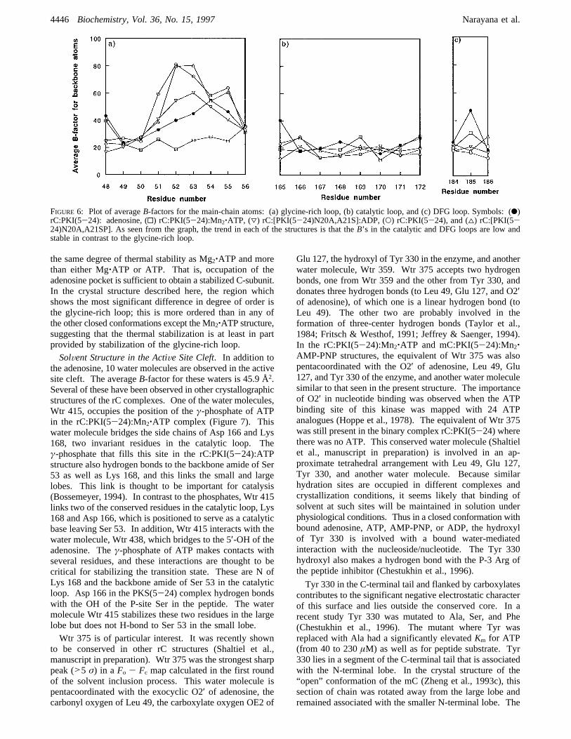
the same degree of thermal stability as Mg2‚ATP and morethan either Mg‚ATP or ATP. That is, occupation of theadenosine pocket is sufficient to obtain a stabilized C-subunit.In the crystal structure described here, the region whichshows the most significant difference in degree of order isthe glycine-rich loop; this is more ordered than in any ofthe other closed conformations except the Mn2‚ATP structure,suggesting that the thermal stabilization is at least in partprovided by stabilization of the glycine-rich loop.SolVent Structure in the ActiVe Site Cleft. In addition to
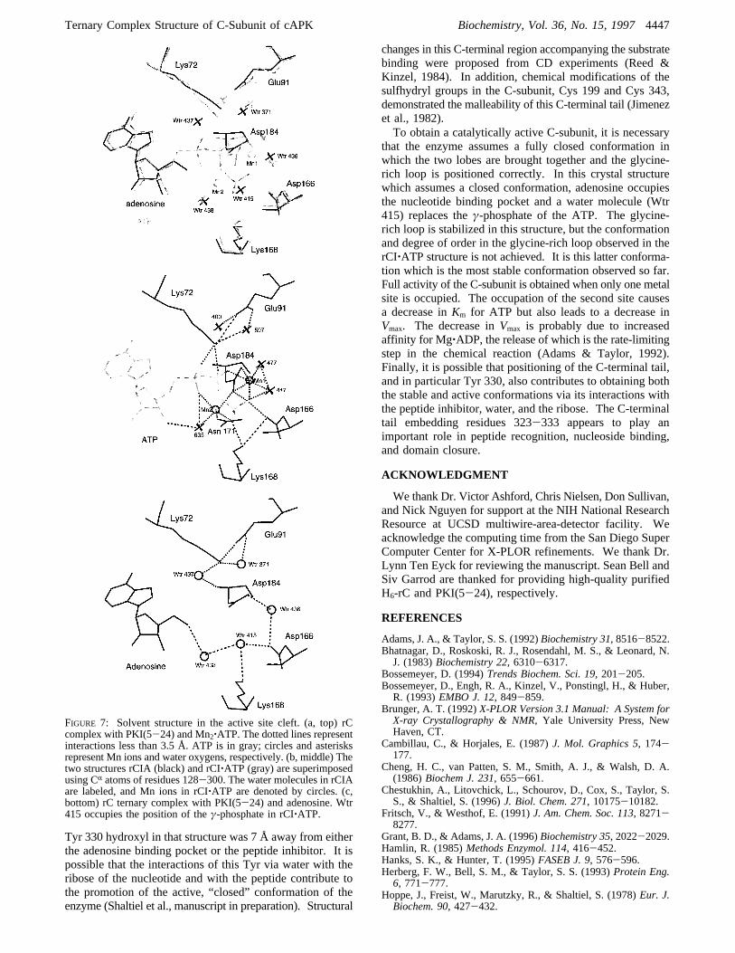
the adenosine, 10 water molecules are observed in the activesite cleft. The averageB-factor for these waters is 45.9 Å2.Several of these have been observed in other crystallographicstructures of the rC complexes. One of the water molecules,Wtr 415, occupies the position of theγ-phosphate of ATPin the rC:PKI(5-24):Mn2‚ATP complex (Figure 7). Thiswater molecule bridges the side chains of Asp 166 and Lys168, two invariant residues in the catalytic loop. Theγ-phosphate that fills this site in the rC:PKI(5-24):ATPstructure also hydrogen bonds to the backbone amide of Ser53 as well as Lys 168, and this links the small and largelobes. This link is thought to be important for catalysis(Bossemeyer, 1994). In contrast to the phosphates, Wtr 415links two of the conserved residues in the catalytic loop, Lys168 and Asp 166, which is positioned to serve as a catalyticbase leaving Ser 53. In addition, Wtr 415 interacts with thewater molecule, Wtr 438, which bridges to the 5′-OH of theadenosine. Theγ-phosphate of ATP makes contacts withseveral residues, and these interactions are thought to becritical for stabilizing the transition state. These are N ofLys 168 and the backbone amide of Ser 53 in the catalyticloop. Asp 166 in the PKS(5-24) complex hydrogen bondswith the OH of the P-site Ser in the peptide. The watermolecule Wtr 415 stabilizes these two residues in the largelobe but does not H-bond to Ser 53 in the small lobe.Wtr 375 is of particular interest. It was recently shown
to be conserved in other rC structures (Shaltiel et al.,manuscript in preparation). Wtr 375 was the strongest sharppeak (>5 σ) in a Fo - Fc map calculated in the first roundof the solvent inclusion process. This water molecule ispentacoordinated with the exocyclic O2′ of adenosine, thecarbonyl oxygen of Leu 49, the carboxylate oxygen OE2 of
Glu 127, the hydroxyl of Tyr 330 in the enzyme, and anotherwater molecule, Wtr 359. Wtr 375 accepts two hydrogenbonds, one from Wtr 359 and the other from Tyr 330, anddonates three hydrogen bonds (to Leu 49, Glu 127, and O2′of adenosine), of which one is a linear hydrogen bond (toLeu 49). The other two are probably involved in theformation of three-center hydrogen bonds (Taylor et al.,1984; Fritsch & Westhof, 1991; Jeffrey & Saenger, 1994).In the rC:PKI(5-24):Mn2‚ATP and mC:PKI(5-24):Mn2‚AMP-PNP structures, the equivalent of Wtr 375 was alsopentacoordinated with the O2′ of adenosine, Leu 49, Glu127, and Tyr 330 of the enzyme, and another water moleculesimilar to that seen in the present structure. The importanceof O2′ in nucleotide binding was observed when the ATPbinding site of this kinase was mapped with 24 ATPanalogues (Hoppe et al., 1978). The equivalent of Wtr 375was still present in the binary complex rC:PKI(5-24) wherethere was no ATP. This conserved water molecule (Shaltielet al., manuscript in preparation) is involved in an ap-proximate tetrahedral arrangement with Leu 49, Glu 127,Tyr 330, and another water molecule. Because similarhydration sites are occupied in different complexes andcrystallization conditions, it seems likely that binding ofsolvent at such sites will be maintained in solution underphysiological conditions. Thus in a closed conformation withbound adenosine, ATP, AMP-PNP, or ADP, the hydroxylof Tyr 330 is involved with a bound water-mediatedinteraction with the nucleoside/nucleotide. The Tyr 330hydroxyl also makes a hydrogen bond with the P-3 Arg ofthe peptide inhibitor (Chestukhin et al., 1996).
Tyr 330 in the C-terminal tail and flanked by carboxylatescontributes to the significant negative electrostatic characterof this surface and lies outside the conserved core. In arecent study Tyr 330 was mutated to Ala, Ser, and Phe(Chestukhin et al., 1996). The mutant where Tyr wasreplaced with Ala had a significantly elevatedKm for ATP(from 40 to 230µM) as well as for peptide substrate. Tyr330 lies in a segment of the C-terminal tail that is associatedwith the N-terminal lobe. In the crystal structure of the“open” conformation of the mC (Zheng et al., 1993c), thissection of chain was rotated away from the large lobe andremained associated with the smaller N-terminal lobe. The
FIGURE 6: Plot of averageB-factors for the main-chain atoms: (a) glycine-rich loop, (b) catalytic loop, and (c) DFG loop. Symbols: (b)rC:PKI(5-24): adenosine, (0) rC:PKI(5-24):Mn2‚ATP, (3) rC:[PKI(5-24)N20A,A21S]:ADP, (O) rC:PKI(5-24), and (4) rC:[PKI(5-24)N20A,A21SP]. As seen from the graph, the trend in each of the structures is that theB’s in the catalytic and DFG loops are low andstable in contrast to the glycine-rich loop.
4446 Biochemistry, Vol. 36, No. 15, 1997 Narayana et al.
Tyr 330 hydroxyl in that structure was 7 Å away from eitherthe adenosine binding pocket or the peptide inhibitor. It ispossible that the interactions of this Tyr via water with theribose of the nucleotide and with the peptide contribute tothe promotion of the active, “closed” conformation of theenzyme (Shaltiel et al., manuscript in preparation). Structural
changes in this C-terminal region accompanying the substratebinding were proposed from CD experiments (Reed &Kinzel, 1984). In addition, chemical modifications of thesulfhydryl groups in the C-subunit, Cys 199 and Cys 343,demonstrated the malleability of this C-terminal tail (Jimenezet al., 1982).To obtain a catalytically active C-subunit, it is necessary
that the enzyme assumes a fully closed conformation inwhich the two lobes are brought together and the glycine-rich loop is positioned correctly. In this crystal structurewhich assumes a closed conformation, adenosine occupiesthe nucleotide binding pocket and a water molecule (Wtr415) replaces theγ-phosphate of the ATP. The glycine-rich loop is stabilized in this structure, but the conformationand degree of order in the glycine-rich loop observed in therCI‚ATP structure is not achieved. It is this latter conforma-tion which is the most stable conformation observed so far.Full activity of the C-subunit is obtained when only one metalsite is occupied. The occupation of the second site causesa decrease inKm for ATP but also leads to a decrease inVmax. The decrease inVmax is probably due to increasedaffinity for Mg‚ADP, the release of which is the rate-limitingstep in the chemical reaction (Adams & Taylor, 1992).Finally, it is possible that positioning of the C-terminal tail,and in particular Tyr 330, also contributes to obtaining boththe stable and active conformations via its interactions withthe peptide inhibitor, water, and the ribose. The C-terminaltail embedding residues 323-333 appears to play animportant role in peptide recognition, nucleoside binding,and domain closure.
ACKNOWLEDGMENT
We thank Dr. Victor Ashford, Chris Nielsen, Don Sullivan,and Nick Nguyen for support at the NIH National ResearchResource at UCSD multiwire-area-detector facility. Weacknowledge the computing time from the San Diego SuperComputer Center for X-PLOR refinements. We thank Dr.Lynn Ten Eyck for reviewing the manuscript. Sean Bell andSiv Garrod are thanked for providing high-quality purifiedH6-rC and PKI(5-24), respectively.
REFERENCES
Adams, J. A., & Taylor, S. S. (1992)Biochemistry 31, 8516-8522.Bhatnagar, D., Roskoski, R. J., Rosendahl, M. S., & Leonard, N.J. (1983)Biochemistry 22, 6310-6317.
Bossemeyer, D. (1994)Trends Biochem. Sci. 19, 201-205.Bossemeyer, D., Engh, R. A., Kinzel, V., Ponstingl, H., & Huber,R. (1993)EMBO J. 12, 849-859.
Brunger, A. T. (1992)X-PLOR Version 3.1 Manual: A System forX-ray Crystallography & NMR, Yale University Press, NewHaven, CT.
Cambillau, C., & Horjales, E. (1987)J. Mol. Graphics 5, 174-177.
Cheng, H. C., van Patten, S. M., Smith, A. J., & Walsh, D. A.(1986)Biochem J. 231, 655-661.
Chestukhin, A., Litovchick, L., Schourov, D., Cox, S., Taylor, S.S., & Shaltiel, S. (1996)J. Biol. Chem. 271, 10175-10182.
Fritsch, V., & Westhof, E. (1991)J. Am. Chem. Soc. 113, 8271-8277.
Grant, B. D., & Adams, J. A. (1996)Biochemistry 35, 2022-2029.Hamlin, R. (1985)Methods Enzymol. 114, 416-452.Hanks, S. K., & Hunter, T. (1995)FASEB J. 9, 576-596.Herberg, F. W., Bell, S. M., & Taylor, S. S. (1993)Protein Eng.6, 771-777.
Hoppe, J., Freist, W., Marutzky, R., & Shaltiel, S. (1978)Eur. J.Biochem. 90, 427-432.
FIGURE 7: Solvent structure in the active site cleft. (a, top) rCcomplex with PKI(5-24) and Mn2‚ATP. The dotted lines representinteractions less than 3.5 Å. ATP is in gray; circles and asterisksrepresent Mn ions and water oxygens, respectively. (b, middle) Thetwo structures rCIA (black) and rCI‚ATP (gray) are superimposedusing CR atoms of residues 128-300. The water molecules in rCIAare labeled, and Mn ions in rCI‚ATP are denoted by circles. (c,bottom) rC ternary complex with PKI(5-24) and adenosine. Wtr415 occupies the position of theγ-phosphate in rCI‚ATP.
Ternary Complex Structure of C-Subunit of cAPK Biochemistry, Vol. 36, No. 15, 19974447
Howard, A. J., Nielsen, C., & Xuong, N.-h. (1985)MethodsEnzymol. 114A, 452-472.
Insight II User Guide, Version 2.3.9(1994) Biosym Technologies,Inc., San Diego, CA.
Jeffrey, G. A., & Saenger, W. (1994)Hydrogen bonding inbiological structures, Springer-Verlag, New York.
Jimenez, J. S., Kupfer, A., Gani, V., & Shaltiel, S. (1982)Biochemistry 21, 1623-1630.
Karlsson, R., Madhusudan, Taylor, S. S., & Sowadski, J. M. (1994)Acta Crystallogr. D50, 657-662.
Knighton, D. R., Zheng, J., Ten Eyck, L. F., Ashford, V. A., Xuong,N.-h., Taylor, S. S., & Sowadski, J. M. (1991a)Science 253,407-414.
Knighton, D. R., Zheng, J., Ten Eyck, L. F., Xuong, N.-h., Taylor,S. S., & Sowadski, J. M. (1991b)Science 253, 414-420.
Knighton, D. R., Bell, S. M., Zheng, J., Ten Eyck, L. F., Xuong,N.-h., Taylor, S. S., & Sowadski, J. M. (1993)Acta Crystallogr.D49, 357-361.
Kraulis, P. J. (1991)J. Appl. Crystallogr. 24, 946-950.Laskowski, R. A., MacArthur, M. W., Moss, D. S., & Thornton, J.M. (1993)J. Appl. Crystallogr. 26, 283-291.
Lew, J., Coruh, N., Tsigelny, I., Garrod, S., & Taylor, S. S. (1997)J. Biol. Chem. 272, 1507-1513.
Luzzati, P. V. (1952)Acta Crystallogr. 5, 802-810.Madhusudan, Trafny, E. A., Xuong, N.-h., Adams, J. A., Ten Eyck,L. F., Taylor, S. S., & Sowadski, J. M. (1994)Protein Sci. 3,176-187.
Orellana, S. A., & McKnight, G. S. (1992)Proc. Natl. Acad. Sci.U.S.A. 89, 4726-4730.
Ramachandran, G. N., & Sasisekharan, V. (1968)AdV. ProteinChem. 23, 283-438.
Reed, J., & Kinzel, V. (1984)Biochemistry 23, 968-973.Saenger, W. (1984)Principles of Nucleic Acid Structure, Springer-Verlag, New York.
Slice, L. W., & Taylor, S. S. (1989)J. Biol. Chem. 264, 20940-20946.
Taylor, R., Kennard, O., & Versichel, W. (1984)J. Am. Chem.Soc. 106, 244-248.
Taylor, S. S., Buechler, J. A., & Knighton, D. R. (1990) inPeptidesand Protein Phosphorylation(Kemp, B. E., Ed.) pp 1-42, CRCPress, Inc., Boca Raton, FL.
Taylor, S. S., Knighton, D. R., Zheng, J., Ten Eyck, L. F., &Sowadski, J. M. (1992)Annu. ReV. Cell Biol. 8, 429-462.
Taylor, S. S., Knighton, D. R., Zheng, J., Sowadski, J. M., Gibbs,C. S., & Zoller, M. J. (1993)Trends Biochem. Sci. 18, 84-89.
Tronrud, D. E., Ten Eyck, L. F., & Matthews, B. W. (1987)ActaCrystallogr. A43, 489-501.
Wen, W., & Taylor, S. S. (1994)J. Biol. Chem. 269,8423-8430.Whitehouse, S., & Walsh, D. A. (1983)J. Biol. Chem. 258, 3682-3692.
Zheng, J., Knighton, D. R., Xuong, N.-h., Parello, J., Taylor, S. S.,& Sowadski, J. M. (1992)Acta Crystallogr. B48, 241-244.
Zheng, J., Knighton, D. R., Ten Eyck, L. F., Karlsson, R., Xuong,N.-h., Taylor, S. S., & Sowadski, J. M. (1993a)Biochemistry32, 2154-2161.
Zheng, J., Trafny, E. A., Knighton, D. R., Xuong, N.-h., Taylor, S.S., Ten Eyck, L. F., & Sowadski, J. M. (1993b)Acta Crystallogr.D49, 362-365.
Zheng, J., Knighton, D. R., Xuong, N.-h., Taylor, S. S., Sowadski,J. M., & Ten Eyck, L. F. (1993c)Protein Sci. 2, 1559-1573.
BI961947+
4448 Biochemistry, Vol. 36, No. 15, 1997 Narayana et al.