Crystal fingerprinting: elucidating the crystals of ... · Crystal fingerprinting: elucidating the...
14
ORIGINAL PAPER Crystal fingerprinting: elucidating the crystals of Cheddar, Parmigiano-Reggiano, Gouda, and soft washed-rind cheeses using powder x-ray diffractometry G. F. Tansman 1 & P. S. Kindstedt 1 & J. M. Hughes 2 Received: 15 January 2015 /Revised: 12 March 2015 /Accepted: 20 March 2015 / Published online: 8 May 2015 # The Author(s) 2015. This article is published with open access at Springerlink.com Abstract Crystals in cheese may be considered defects or positive features, depending on the variety and mode of production (industrial, artisanal). Powder x-ray diffractometry (PXRD) offers a simple means to identify and resolve complex combinations of crystals that contribute to cheese characteristics. The objective of the present research was to demonstrate the application of PXRD to study crystals from a range of different cheese types, specifically Cheddar, Parmigiano-Reggiano, Gouda, and soft washed-rind (smear ripened) cheeses. In studies of Parmigiano-Reggiano and long-aged Gouda, PXRD has confirmed that hard (crunchy) crystals that form abundantly within these cheeses consist of tyrosine. Furthermore, PXRD has tentatively identified the presence of an unusual form of crystalline leucine in large (up to 6 mm in diameter) spherical entities, or “pearls”, that occur abundantly in 2-year-old Parmigiano Reggiano and long-aged Gouda cheeses, and on the surface of rindless hard Italian-type cheese. Ongoing investigations into the nature of these “pearls” are providing new insight into the roles that crystals play in the visual appearance and texture of long-aged cheeses. Crystals also sometimes develop profusely in the eyes of long-aged Gouda, which have been shown by PXRD to consist of tyrosine and the aforementioned presumptive form of crystalline leucine. Finally, crystals have been shown by PXRD to form in the smears of soft washed-rind cheeses. These crystals may be associated in some cheeses with gritty mouth feel and with zonal body softening that occurs during ripening. Heightened interest in artisanal cheeses highlights the need to better understand crystals and their contributions to cheese characteristics. Keywords X-ray diffraction . Crystals . Cheese . Tyrosine . Leucine Dairy Sci. & Technol. (2015) 95:651–664 DOI 10.1007/s13594-015-0225-6 This paper is part of the Special issue dedicated to the 9th International Cheese Symposium held in Cork, Ireland and organized by Teagasc in collaboration with University College Cork and INRA, 12th & 13th November 2014. * P. S. Kindstedt [email protected] 1 Department of Nutrition and Food Sciences, University of Vermont, Burlington, VT, USA 2 Department of Geology, University of Vermont, Burlington, VT, USA
Transcript of Crystal fingerprinting: elucidating the crystals of ... · Crystal fingerprinting: elucidating the...
ORIGINAL PAPER
Crystal fingerprinting: elucidating the crystals of Cheddar,Parmigiano-Reggiano, Gouda, and soft washed-rindcheeses using powder x-ray diffractometry
G. F. Tansman1& P. S. Kindstedt1 & J. M. Hughes2
Received: 15 January 2015 /Revised: 12 March 2015 /Accepted: 20 March 2015 /Published online: 8 May 2015# The Author(s) 2015. This article is published with open access at Springerlink.com
Abstract Crystals in cheesemay be considered defects or positive features, depending onthe variety and mode of production (industrial, artisanal). Powder x-ray diffractometry(PXRD) offers a simple means to identify and resolve complex combinations of crystalsthat contribute to cheese characteristics. The objective of the present research was todemonstrate the application of PXRD to study crystals from a range of different cheesetypes, specifically Cheddar, Parmigiano-Reggiano, Gouda, and soft washed-rind (smearripened) cheeses. In studies of Parmigiano-Reggiano and long-aged Gouda, PXRD hasconfirmed that hard (crunchy) crystals that form abundantly within these cheeses consistof tyrosine. Furthermore, PXRD has tentatively identified the presence of an unusual formof crystalline leucine in large (up to 6 mm in diameter) spherical entities, or “pearls”, thatoccur abundantly in 2-year-old Parmigiano Reggiano and long-aged Gouda cheeses, andon the surface of rindless hard Italian-type cheese. Ongoing investigations into the natureof these “pearls” are providing new insight into the roles that crystals play in the visualappearance and texture of long-aged cheeses. Crystals also sometimes develop profuselyin the eyes of long-aged Gouda, which have been shown by PXRD to consist of tyrosineand the aforementioned presumptive form of crystalline leucine. Finally, crystals havebeen shown by PXRD to form in the smears of soft washed-rind cheeses. These crystalsmay be associated in some cheeses with gritty mouth feel and with zonal body softeningthat occurs during ripening. Heightened interest in artisanal cheeses highlights the need tobetter understand crystals and their contributions to cheese characteristics.
Keywords X-ray diffraction . Crystals . Cheese . Tyrosine . Leucine
Dairy Sci. & Technol. (2015) 95:651–664DOI 10.1007/s13594-015-0225-6
This paper is part of the Special issue dedicated to the 9th International Cheese Symposium held in Cork, Irelandand organized by Teagasc in collaboration with University College Cork and INRA, 12th& 13th November 2014.
* P. S. [email protected]
1 Department of Nutrition and Food Sciences, University of Vermont, Burlington, VT, USA2 Department of Geology, University of Vermont, Burlington, VT, USA
1 Introduction
Crystals that form in cheese have been the subject of scientific inquiry for over acentury, and x-ray diffraction (XRD) has been marshaled as a direct analytical approachto identify cheese crystals for almost as long, the earliest studies dating back to the1930s (Tuckey and Ruehe 1938; Tuckey et al. 1938). Most XRD investigations ofcheese crystals have been restricted to Cheddar cheese and have focused primarily oncalcium lactate pentahydrate (CLP) crystals (Agarwal et al. 2006; Chou et al. 2003;Conchie et al. 1960; Dybing et al. 1988; Harper et al. 1953; Tuckey et al. 1938;Washam et al. 1985). A few studies of Cheddar also identified the occurrence ofcrystals consisting of tyrosine (Conchie et al. 1960; Harper et al. 1953; Shock et al.1948), cysteine (Harper et al. 1953; Shock et al. 1948), and calcium phosphate(Conchie and Sutherland 1965). In the United States, visible crystals are often viewedby the Cheddar industry as defects and sources of confusion for consumers (Johnson2014). Therefore, there has been considerable interest to understand the factors thatpromote crystal formation and develop measures to prevent their occurrence. Incontrast, in some traditional cheeses such as Parmigiano-Reggiano and hand-craftedartisanal cheeses, crystals are viewed as important contributors to cheese character andquality (Noël et al. 1996; Zannoni et al. 1994). Growing consumer appreciation oftraditional and artisanal cheeses is also fueling interest in understanding the nature andorigins of crystals and their contributions to cheese quality and character.
Studies of crystals in cheeses other than Cheddar, such as tyrosine crystals in Roquefort(Dox 1911), CLP, calcium phosphate and tyrosine crystals in Grana cheeses (Bottazzi et al.1982, 1994), calcium phosphate crystals in soft white mold surface-ripened (bloomy rind)cheeses (Boutrou et al. 1999; Brooker 1987; Gaucheron et al. 1999; Karahadrian andLindsay 1987), and CLP and calcium phosphate crystals in Serra cheese (Parker et al.1998), have generally employed indirect methods such as chemical analyses or micros-copy coupled with differential staining and x-ray microanalysis to identify presumedcrystalline species. Although indirect approaches may provide useful presumptive iden-tification of crystalline species, only XRD can furnish direct and definitive results becausethe XRD pattern of a specific crystal is determined by its three-dimensional atomicarrangement, which is unique to that crystal and thus analogous to a fingerprint.Consequently, the XRD pattern of an unknown species can be compared to those ofknown crystals; a perfect match with one of the known patterns provides definitiveidentification. Today, a growing database of over 250,000 known XRD patterns isaccessible through the International Centre for Diffraction Data (ICDD).
Powder x-ray diffractometry (PXRD), which utilizes crystalline specimens thatideally have been pulverized to a fine powder, is a versatile and user-friendly applica-tion of XRD. Major enhancements in the computing power of PXRD instrumentation,and advances in the software algorithms for data analysis, have opened up newopportunities for the study of cheese crystals. For example, the authors recentlydemonstrated that the two different enantiomeric forms of CLP that were presumedto occur in Cheddar cheese (i.e., the L(+) and D(−)/L(+) forms) can be identified anddifferentiated using PXRD, based on subtle differences in the XRD patterns of the twoenantiomeric configurations (Tansman et al. 2014). The application of PXRD to studycrystals in Cheddar and other cheese varieties could offer new insights into the factorsthat govern crystallization phenomena in cheese and the roles that crystals play in
652 G.F. Tansman et al.
determining cheese character and quality. Therefore, the objective of the presentresearch was to demonstrate the application of PXRD to study crystals from a rangeof different cheese types, specifically Cheddar, Parmigiano-Reggiano, Gouda, and softwashed-rind (smear ripened) cheeses.
2 Materials and methods
2.1 Cheese samples and crystal collection
All cheese samples were commercially produced and purchased from local retailoutlets, and crystals were harvested for analysis as described below, except forCheddar cheese and a hard Italian-style cheese. In the case of Cheddar, the manufac-turer harvested large (up to ca. 5 mm) internal crystals from the body of a Cheddarcheese sample (aged 2 years before sale) and delivered the crystals to the University ofVermont for identification after the manufacturer received a consumer complaint aboutunknown objects in their cheese. Crystals from a hard Italian-style cheese (obtainedcourtesy of Mark Johnson, Center for Dairy Research, University of Wisconsin) werescraped from the cheese surface using a spatula. Small (ca. 1–2 mm) dense crystalsembedded in the body of Parmigiano-Reggiano cheese samples (aged 2 years) andGouda cheese samples (aged 2 years) were physically excised from the surroundingcheese matrix using a dissecting needle and tweezers. The isolated crystals werebrushed free of adhering cheese matrix. Parmigiano-Reggiano samples also containedwhite spherical entities that ranged from barely visible to around 6 mm in diameter,which will be referred to as “pearls” in this publication. Pearls were separated from thesurrounding cheese matrix by slicing the sample with a wire cutter into 1-cm sectionsand gently applying pressure to the cheese surrounding the pearl using thumb and indexfinger. Gentle pressure caused the surrounding matrix to fracture and separate from thepearls, enabling the pearls to be isolated and brushed free of adhering cheese matrix.Similar pearls, though smaller in size distribution, also were harvested from the Goudasamples. Gouda samples also displayed extensive crystal formation along the surfacesof internal eyes. Crystals were scraped free from the surfaces of eyes using a spatula.The six soft washed-rind (smear ripened) cheeses that were examined in this study wereproduced by different companies, four located in the United States and two in Italy.
2.2 Analytical methods
Pearls collected from Parmigiano-Reggiano samples were evaluated for size distribu-tion by placing them on a vibrating stack of four sieves of successively smaller meshsize (5.6, 4.00, 3.35, and 2.00 mm) and counting the number of pearls retained by eachsieve. The pearls were then pooled to form a composite sample, grated to fine particlesin a blender, and analyzed in duplicate for total solids content by drying in a forced-draft oven at 100 °C for 24 h, fat content by a modified Babcock method (Atherton andNewlander 1977), and for soluble leucine (i.e., free amino acid) content by a modifiedgas chromatography–mass spectrometry method as follows. Thirteen milligrams ofpearls was homogenized in 100 mL of deionized water adjusted to a pH of 1 with HCl.The suspension was vortexed for 10 min and centrifuged at 5000 rpm for an additional
X-ray diffraction of cheese crystals 653
5 min to remove particulate material from the solution. A 1-mL aliquot of the mixturewas removed for analysis and to this was added 0.1 mL of 8.37 mM 555 d3 leucine(Isotec 486825 Lot TPI633-SP). The sample was mixed with 1 mL of 50% acetic acidand passed through cation exchange resin (Biorad AG50W-x8 cation exchange resin100–200 mesh). The resin was then rinsed three times with 2 mL of deionized water.Amino acids were eluted off the resin by the addition of 2 mL of 4 N ammoniumhydroxide. The eluate was collected and evaporated under nitrogen gas. To the dry vialwas added 200 µL of a 50:50 mixture of N-methyl-N-(t-butyldimethylsilyl) trifluoro-acetamide+1% t-butyldimethylchlorosilane (MtBSTFA+1% t-BDMCS)/acetonitrile.The solution was heated for 30 min in a heating block at 80 °C and then transferredto an autosampler vial. A 1-µL aliquot was injected into a ZB1 gas chromatographer(GC) column (30 m in length; 0.25 mm inner diameter with 0.25 μm film). The GCwas run isothermally at 165 °C with a column flow rate of 1 mL helium per minute.The mass spectrometer (MS) was run in selected ion monitoring mode and monitoredfor a mass to charge of 200m/z for leucine and 203m/z for labeled leucine.
Pooled samples of the cheese matrix surrounding the pearls were similarly grated andanalyzed for total solids and fat contents. Gouda samples that displayed extensive crystalformation along the surfaces of internal eyes were examined by stereoscopic microscopybefore the crystals were harvested for analysis. Smear material was scraped from thesurface of soft washed-rind (smear ripened) cheeses employing a spatula, using care notto remove the underlying cheese matrix along with the smear. Smear material wasanalyzed for selected mineral content by inductively coupled atomic emission spec-trometry as follows. A scraping of surface smear from one Vermont washed-rind cheesewas dried and defatted in acetone and analyzed for Ca, Mg, K, and P by ICP-OES(Optima 3000DV; Perkin Elmer Corp, Norwalk, CT, USA). Calibration standards wereprepared according to the instrument manufacturer’s suggested guidelines, to cover therange of concentrations in the sample set. Two-point calibrations (plus a CalibrationBlank) were used for ICP analysis. Continuing Calibration Verification samples, pre-pared from an independent source, were used to check the calibration periodically.
In preparation for PXRD, dense internal crystals obtained from Cheddar,Parmigiano-Reggiano, and Gouda cheeses, as well as crystals scraped from the surfaceof the hard Italian-style cheese and from the eyes of Gouda, were dried and defatted inacetone, finely ground using a mortar and pestle, and transferred to a glass diffractionslide with a well. The powder in the slide well was leveled with the surface of thediffraction slide using a microscope slide. Diffraction patterns were generated fromcheese crystals using a MiniFlex II powder x-ray diffractometer (Rigaku, TheWoodlands, TX). Diffractograms were generated at a speed of 2° 2θ.min−1 between 5and 50° 2θ. Diffraction patterns were compared to existing entries archived in theICDD database to determine matches using a proprietary calculation called a “figure ofmerit”. The figure of merit was calculated automatically using PDXL, thediffractometer software package (Rigaku, The Woodlands, TX). An acceptancethreshold of 1.0 was set for the figure of merit. Diffraction patterns were alsogenerated from finely grated samples of pearls, and samples of the surroundingcheese matrix, from Parmigiano-Reggiano and Gouda cheeses as described. Samplesof smears scraped from the surfaces of soft washed-rind (smear ripened) cheeses wereloaded directly onto the slide well and immediately analyzed without any additionalsample preparation.
654 G.F. Tansman et al.
3 Results and discussion
3.1 Cheddar
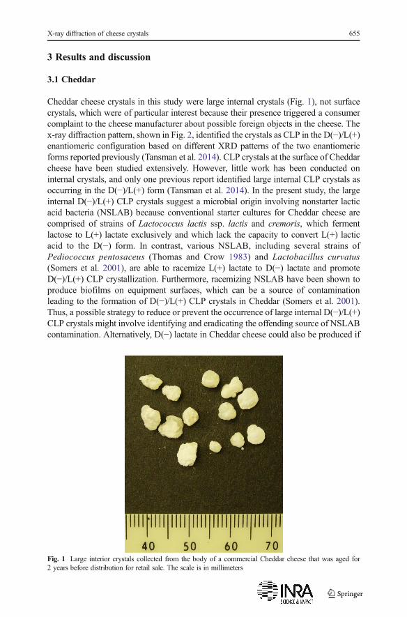
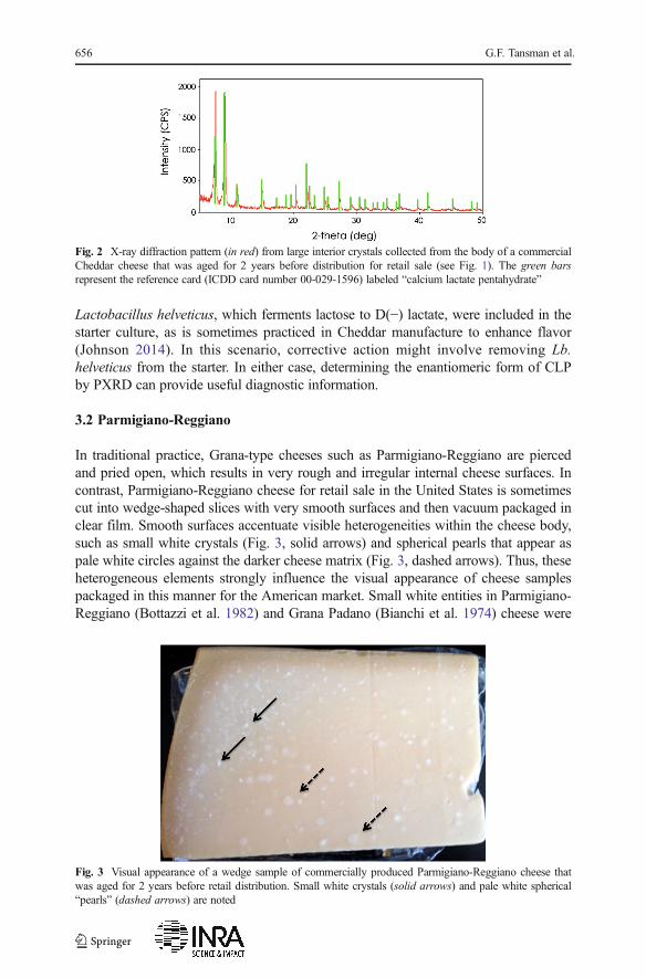
Cheddar cheese crystals in this study were large internal crystals (Fig. 1), not surfacecrystals, which were of particular interest because their presence triggered a consumercomplaint to the cheese manufacturer about possible foreign objects in the cheese. Thex-ray diffraction pattern, shown in Fig. 2, identified the crystals as CLP in the D(−)/L(+)enantiomeric configuration based on different XRD patterns of the two enantiomericforms reported previously (Tansman et al. 2014). CLP crystals at the surface of Cheddarcheese have been studied extensively. However, little work has been conducted oninternal crystals, and only one previous report identified large internal CLP crystals asoccurring in the D(−)/L(+) form (Tansman et al. 2014). In the present study, the largeinternal D(−)/L(+) CLP crystals suggest a microbial origin involving nonstarter lacticacid bacteria (NSLAB) because conventional starter cultures for Cheddar cheese arecomprised of strains of Lactococcus lactis ssp. lactis and cremoris, which fermentlactose to L(+) lactate exclusively and which lack the capacity to convert L(+) lacticacid to the D(−) form. In contrast, various NSLAB, including several strains ofPediococcus pentosaceus (Thomas and Crow 1983) and Lactobacillus curvatus(Somers et al. 2001), are able to racemize L(+) lactate to D(−) lactate and promoteD(−)/L(+) CLP crystallization. Furthermore, racemizing NSLAB have been shown toproduce biofilms on equipment surfaces, which can be a source of contaminationleading to the formation of D(−)/L(+) CLP crystals in Cheddar (Somers et al. 2001).Thus, a possible strategy to reduce or prevent the occurrence of large internal D(−)/L(+)CLP crystals might involve identifying and eradicating the offending source of NSLABcontamination. Alternatively, D(−) lactate in Cheddar cheese could also be produced if
Fig. 1 Large interior crystals collected from the body of a commercial Cheddar cheese that was aged for2 years before distribution for retail sale. The scale is in millimeters
X-ray diffraction of cheese crystals 655
Lactobacillus helveticus, which ferments lactose to D(−) lactate, were included in thestarter culture, as is sometimes practiced in Cheddar manufacture to enhance flavor(Johnson 2014). In this scenario, corrective action might involve removing Lb.helveticus from the starter. In either case, determining the enantiomeric form of CLPby PXRD can provide useful diagnostic information.
3.2 Parmigiano-Reggiano
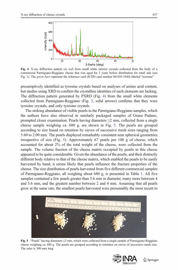
In traditional practice, Grana-type cheeses such as Parmigiano-Reggiano are piercedand pried open, which results in very rough and irregular internal cheese surfaces. Incontrast, Parmigiano-Reggiano cheese for retail sale in the United States is sometimescut into wedge-shaped slices with very smooth surfaces and then vacuum packaged inclear film. Smooth surfaces accentuate visible heterogeneities within the cheese body,such as small white crystals (Fig. 3, solid arrows) and spherical pearls that appear aspale white circles against the darker cheese matrix (Fig. 3, dashed arrows). Thus, theseheterogeneous elements strongly influence the visual appearance of cheese samplespackaged in this manner for the American market. Small white entities in Parmigiano-Reggiano (Bottazzi et al. 1982) and Grana Padano (Bianchi et al. 1974) cheese were
Fig. 2 X-ray diffraction pattern (in red) from large interior crystals collected from the body of a commercialCheddar cheese that was aged for 2 years before distribution for retail sale (see Fig. 1). The green barsrepresent the reference card (ICDD card number 00-029-1596) labeled “calcium lactate pentahydrate”
Fig. 3 Visual appearance of a wedge sample of commercially produced Parmigiano-Reggiano cheese thatwas aged for 2 years before retail distribution. Small white crystals (solid arrows) and pale white spherical“pearls” (dashed arrows) are noted
656 G.F. Tansman et al.
presumptively identified as tyrosine crystals based on analyses of amino acid content,but studies using XRD to confirm the crystalline identities of such elements are lacking.The diffraction pattern generated by PXRD (Fig. 4) from the small white elementscollected from Parmigiano-Reggiano (Fig. 3, solid arrows) confirms that they weretyrosine crystals, and only tyrosine crystals.
The striking abundance of visible pearls in the Parmigiano-Reggiano samples, whichthe authors have also observed in similarly packaged samples of Grana Padano,prompted closer examination. Pearls having diameters ≥2 mm, collected from a singlecheese sample weighing ca. 680 g, are shown in Fig. 5. The pearls are groupedaccording to size based on retention by sieves of successive mesh sizes ranging from5.60 to 2.00 mm. The pearls displayed remarkably consistent near spherical geometriesirrespective of size (Fig. 5). Approximately 67 pearls per 100 g of cheese, whichaccounted for about 2% of the total weight of the cheese, were collected from thesample. The volume fraction of the cheese matrix occupied by pearls in this cheeseappeared to be quite considerable. Given the abundance of the pearls, and their distinctlydifferent body relative to that of the cheese matrix, which enabled the pearls to be easilyharvested by hand, it seems likely that pearls influence the fracture properties of thecheese. The size distribution of pearls harvested from five different commercial samplesof Parmigiano-Reggiano, all weighing about 680 g, is presented in Table 1. All fivesamples contained a few pearls greater than 5.6 mm in diameter, many more between 4and 5.6 mm, and the greatest number between 2 and 4 mm. Assuming that all pearlsgrow at the same rate, the smallest pearls harvested were presumably the most recent to
Fig. 4 X-ray diffraction pattern (in red) from small white interior crystals collected from the body of acommercial Parmigiano-Reggiano cheese that was aged for 2 years before distribution for retail sale (seeFig. 3). The green bars represent the reference card (ICDD card number 00-039-1840) labeled “tyrosine”
Fig. 5 “Pearls” having diameters ≥2 mm, which were collected from a single sample of Parmigiano-Reggianocheese weighing ca. 680 g. The pearls are grouped according to retention on sieves of successive mesh size.The ruler is 300 mm long
X-ray diffraction of cheese crystals 657
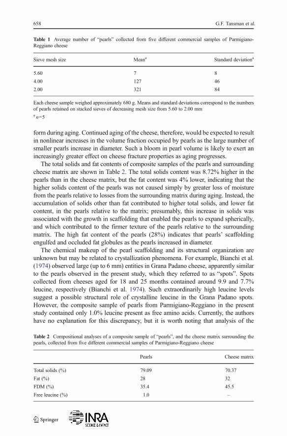
form during aging. Continued aging of the cheese, therefore, would be expected to resultin nonlinear increases in the volume fraction occupied by pearls as the large number ofsmaller pearls increase in diameter. Such a bloom in pearl volume is likely to exert anincreasingly greater effect on cheese fracture properties as aging progresses.
The total solids and fat contents of composite samples of the pearls and surroundingcheese matrix are shown in Table 2. The total solids content was 8.72% higher in thepearls than in the cheese matrix, but the fat content was 4% lower, indicating that thehigher solids content of the pearls was not caused simply by greater loss of moisturefrom the pearls relative to losses from the surrounding matrix during aging. Instead, theaccumulation of solids other than fat contributed to higher total solids, and lower fatcontent, in the pearls relative to the matrix; presumably, this increase in solids wasassociated with the growth in scaffolding that enabled the pearls to expand spherically,and which contributed to the firmer texture of the pearls relative to the surroundingmatrix. The high fat content of the pearls (28%) indicates that pearls’ scaffoldingengulfed and occluded fat globules as the pearls increased in diameter.
The chemical makeup of the pearl scaffolding and its structural organization areunknown but may be related to crystallization phenomena. For example, Bianchi et al.(1974) observed large (up to 6 mm) entities in Grana Padano cheese, apparently similarto the pearls observed in the present study, which they referred to as “spots”. Spotscollected from cheeses aged for 18 and 25 months contained around 9.9 and 7.7%leucine, respectively (Bianchi et al. 1974). Such extraordinarily high leucine levelssuggest a possible structural role of crystalline leucine in the Grana Padano spots.However, the composite sample of pearls from Parmigiano-Reggiano in the presentstudy contained only 1.0% leucine present as free amino acids. Currently, the authorshave no explanation for this discrepancy, but it is worth noting that analysis of the
Table 1 Average number of “pearls” collected from five different commercial samples of Parmigiano-Reggiano cheese
Sieve mesh size Meana Standard deviationa
5.60 7 8
4.00 127 46
2.00 321 84
Each cheese sample weighed approximately 680 g. Means and standard deviations correspond to the numbersof pearls retained on stacked sieves of decreasing mesh size from 5.60 to 2.00 mma n=5
Table 2 Compositional analyses of a composite sample of “pearls”, and the cheese matrix surrounding thepearls, collected from five different commercial samples of Parmigiano-Reggiano cheese
Pearls Cheese matrix
Total solids (%) 79.09 70.37
Fat (%) 28 32
FDM (%) 35.4 45.5
Free leucine (%) 1.0 –
658 G.F. Tansman et al.
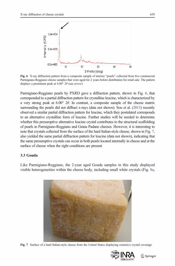
Parmigiano-Reggiano pearls by PXRD gave a diffraction pattern, shown in Fig. 6, thatcorresponded to a partial diffraction pattern for crystalline leucine, which is characterized bya very strong peak at 6.00° 2θ. In contrast, a composite sample of the cheese matrixsurrounding the pearls did not diffract x-rays (data not shown). Sou et al. (2013) recentlyobserved a similar partial diffraction pattern for leucine, which they postulated correspondsto an alternative crystalline form of leucine. Further studies will be needed to determinewhether this presumptive alternative leucine crystal contributes to the structural scaffoldingof pearls in Parmigiano-Reggiano and Grana Padano cheeses. However, it is interesting tonote that crystals collected from the surface of the hard Italian-style cheese, shown in Fig. 7,also yielded the same partial diffraction pattern for leucine (data not shown), indicating thatthe same presumptive crystals can occur in both pearls located internally in cheese and at thesurface of cheese when the right conditions are present.
3.3 Gouda
Like Parmigiano-Reggiano, the 2-year aged Gouda samples in this study displayedvisible heterogeneities within the cheese body, including small white crystals (Fig. 8a,
Fig. 6 X-ray diffraction pattern from a composite sample of interior “pearls” collected from five commercialParmigiano-Reggiano cheese samples that were aged for 2 years before distribution for retail sale. The patterndisplays a prominent peak at 6.00° 2θ (see arrow)
Fig. 7 Surface of a hard Italian-style cheese from the United States displaying extensive crystal coverage
X-ray diffraction of cheese crystals 659
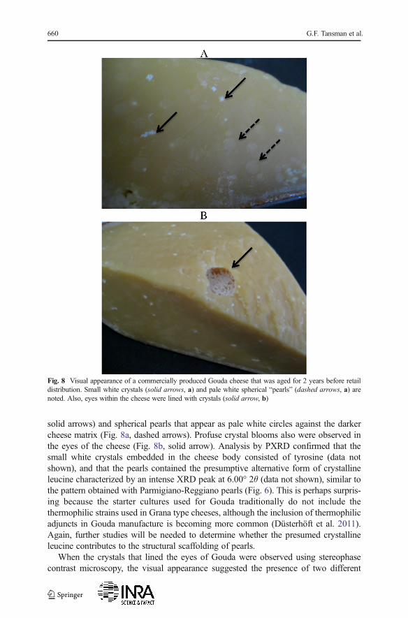
solid arrows) and spherical pearls that appear as pale white circles against the darkercheese matrix (Fig. 8a, dashed arrows). Profuse crystal blooms also were observed inthe eyes of the cheese (Fig. 8b, solid arrow). Analysis by PXRD confirmed that thesmall white crystals embedded in the cheese body consisted of tyrosine (data notshown), and that the pearls contained the presumptive alternative form of crystallineleucine characterized by an intense XRD peak at 6.00° 2θ (data not shown), similar tothe pattern obtained with Parmigiano-Reggiano pearls (Fig. 6). This is perhaps surpris-ing because the starter cultures used for Gouda traditionally do not include thethermophilic strains used in Grana type cheeses, although the inclusion of thermophilicadjuncts in Gouda manufacture is becoming more common (Düsterhöft et al. 2011).Again, further studies will be needed to determine whether the presumed crystallineleucine contributes to the structural scaffolding of pearls.
When the crystals that lined the eyes of Gouda were observed using stereophasecontrast microscopy, the visual appearance suggested the presence of two different
Fig. 8 Visual appearance of a commercially produced Gouda cheese that was aged for 2 years before retaildistribution. Small white crystals (solid arrows, a) and pale white spherical “pearls” (dashed arrows, a) arenoted. Also, eyes within the cheese were lined with crystals (solid arrow, b)
660 G.F. Tansman et al.
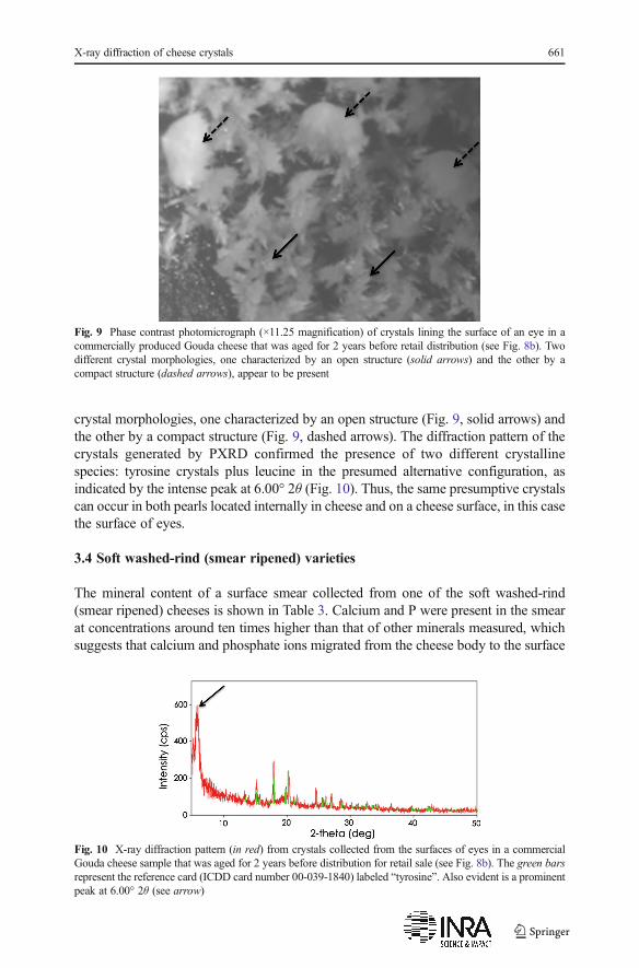
crystal morphologies, one characterized by an open structure (Fig. 9, solid arrows) andthe other by a compact structure (Fig. 9, dashed arrows). The diffraction pattern of thecrystals generated by PXRD confirmed the presence of two different crystallinespecies: tyrosine crystals plus leucine in the presumed alternative configuration, asindicated by the intense peak at 6.00° 2θ (Fig. 10). Thus, the same presumptive crystalscan occur in both pearls located internally in cheese and on a cheese surface, in this casethe surface of eyes.
3.4 Soft washed-rind (smear ripened) varieties
The mineral content of a surface smear collected from one of the soft washed-rind(smear ripened) cheeses is shown in Table 3. Calcium and P were present in the smearat concentrations around ten times higher than that of other minerals measured, whichsuggests that calcium and phosphate ions migrated from the cheese body to the surface
Fig. 9 Phase contrast photomicrograph (×11.25 magnification) of crystals lining the surface of an eye in acommercially produced Gouda cheese that was aged for 2 years before retail distribution (see Fig. 8b). Twodifferent crystal morphologies, one characterized by an open structure (solid arrows) and the other by acompact structure (dashed arrows), appear to be present
Fig. 10 X-ray diffraction pattern (in red) from crystals collected from the surfaces of eyes in a commercialGouda cheese sample that was aged for 2 years before distribution for retail sale (see Fig. 8b). The green barsrepresent the reference card (ICDD card number 00-039-1840) labeled “tyrosine”. Also evident is a prominentpeak at 6.00° 2θ (see arrow)
X-ray diffraction of cheese crystals 661
in a manner similar to what is believed to occur in bloomy rind cheeses, i.e., as a resultof high surface pH that develops during ripening, which triggers surface crystallizationof calcium phosphate (Boutrou et al. 1999; Brooker 1987; Gaucheron et al. 1999;Karahadrian and Lindsay 1987). Therefore, the authors expected to find one or moreforms of crystalline calcium phosphate in the smears of the six soft washed-rindcheeses that were analyzed by PXRD. However, smears from all six cheeses eachproduced a unique XRD pattern that could not be matched to any pattern in the ICDDdatabase. Diffraction of x-rays indicates the presence of crystals in a sample, thus theXRD patterns confirmed that crystalline species were present in the smears of all sixcheeses but none could be identified. A possible explanation for the unknown XRDpatterns became evident during the course of the analyses when the authors noted thesamples dried out extensively during the diffraction procedure, dehydrating the extantphases. This strongly suggested that the PXRD results contained diffraction artifacts ofthe experimental conditions. Consequently, the preparation of smear samples for PXRDwas modified to include the addition of a thin layer mineral oil over the sample well toprevent sample desiccation. This modification produced reproducible data, therebypreempting artifacts obtained previously. When the modified sample preparation pro-cedure was applied to smear material from two of the washed-rind cheeses analyzedpreviously, the XRD pattern for ikaite, which consists of calcium carbonate in anunusual hexahydrate form, was identified in the smears (Fig. 11). Additional analysesof other washed-rind cheeses are underway and will be presented in a subsequentreport.
The formation of surface crystals, whether of calcium phosphate or other species, isof great interest because of the possible implications for zonal softening, which hasbeen studied extensively in bloomy rind cheeses but not well characterized in soft
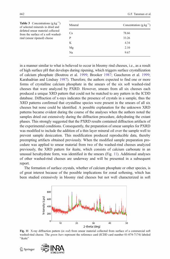
Table 3 Concentrations (g.kg−1)of selected minerals in dried anddefatted smear material collectedfrom the surface of a soft washed-rind (smear ripened) cheese
Mineral Concentration (g.kg−1)
Ca 78.66
P 33.26
K 4.34
Mg 2.10
Na 9.67
Fig. 11 X-ray diffraction pattern (in red) from smear material collected from surface of a commercial softwashed-rind cheese. The green bars represent the reference card (ICDD card number 01-074-7174) labeled“ikaite”
662 G.F. Tansman et al.
washed-rind cheeses. Surface crystal formation also has implications for the develop-ment of sandiness or grittiness, which the authors have heard anecdotally fromcheesemakers and cheesemongers may be considered desirable or undesirable depend-ing on the market.
4 Conclusion
Powder x-ray diffractometry is a versatile and powerful analytical approach to identifyand differentiate crystalline species in cheese. Crystals influence the quality andcharacter of many cheeses, ranging from extra hard, long-aged types, such as granacheeses, to soft, rapidly ripened cheeses such as surface ripened washed-rind andbloomy-rind types. This study confirmed the presence of tyrosine crystals and acrystalline species presumptively identified as leucine in both Parmigiano-Reggianoand Gouda cheeses, as well as large interior crystals of D(−)/L(+) calcium lactatepentahydrate in Cheddar cheese and ikaite crystals at the surface of a soft washed-rindcheese. Advancements in PXRD technology have facilitated new avenues of researchrelating to structure-function implications of crystallization phenomena, such as the roleof leucine crystals in the formation and growth of pearls in Parmigiano-Reggiano andGouda cheeses, and the possible role of surface crystals in the zonal softening of softsurface ripened cheeses.
Acknowledgments This study was funded by the United States Department of Agriculture Hatch ProjectVT-H01905. Purchase of the x-ray diffractometer was funded byNational Science FoundationGrant EAR-0922961.
Conflict of interest Author Tansman, author Kindstedt, and author Hughes declare that they have noconflict of interest.
Statement of human and animal rights This article does not contain any studies with human or animalsubjects performed by any of the authors.
Open Access This article is distributed under the terms of the Creative Commons Attribution 4.0 InternationalLicense (http://creativecommons.org/licenses/by/4.0/), which permits unrestricted use, distribution, and repro-duction in any medium, provided you give appropriate credit to the original author(s) and the source, provide alink to the Creative Commons license, and indicate if changes were made.
References
Agarwal S, Sharma K, Swanson BG, Yüksel GÜ, Clark S (2006) Nonstarter lactic acid bacteria biofilms andcalcium lactate crystals in Cheddar cheese. J Dairy Sci 89:1452–1466
Atherton HV, Newlander JA (1977) Chemistry and testing of dairy products, 4th ed. Avi Pub Co,Westport, CTBianchi A, Beretta G, Caserio G, Giolitti G (1974) Amino acid composition of granules and spots in Grana
Padano cheeses. J Dairy Sci 57:1504–1508Bottazzi V, Battistotto B, Bianchi F (1982) The microscopic crystalline inclusions in Grana cheese and their
x-ray microanalysis. Milchwissenschaft Milk Sci Internat 37(10):577–580Bottazzi V, Lucchini F, Rebecchi A, Scolari GL (1994) I cristalli del formaggio grana (crystals present in
Grana cheese). Scienza e Tecnica Lattiero-Casearia 45(1):7–14Boutrou R, Gaucheron F, Piot M, Michel F, Maubois J-L, Léonil J (1999) Changes in the composition of juice
expressed from Camembert cheese during ripening. Lait 79:503–513
X-ray diffraction of cheese crystals 663
Brooker BE (1987) The crystallization of calcium phosphate at the surface of mould-ripened cheeses. FoodMicrostruct 6:25–33
Chou Y-E, Edwards CG, Luedecke LO, Bates MP, Clark S (2003) Nonstarter lactic acid bacteria and agingtemperature affect calcium lactate crystallization in Cheddar cheese. J Dairy Sci 86:2516–2524
Conchie J, Sutherland BJ (1965) The nature of steaminess in Cheddar cheese. J Dairy Res 32:35–44Conchie A, Lawrence J, Czulak J, Cole WF (1960) Aust J Dairy Technol 15:120Dox AW (1911) The occurrence of tyrosine crystals in Roquefort cheese. J Amer Chem Soc 33:423–425Düsterhöft EM, Engels W, van den Berg G (2011) Dutch-type cheeses. In Encylopedia of dairy sciences, 2nd
edn. Academic Press/Elsevier, LondonDybing ST, Wiegand JA, Brudvig SA, Huang EA, Chandan RC (1988) Effect on processing variables on the
formation of calcium lactate crystals on Cheddar cheese. J Dairy Sci 71:1701–1710Gaucheron F, Le Graët Y, Michel F, Briard V, Piot M (1999) Evolution of various salt concentrations in the
moisture and in the outer layer and centre of a model cheese during its brining and storage in anammoniacal atmosphere. Lait 79:553–566
Harper WJ, Swanson AM, Sommer HH (1953) Observations on the chemical composition of white particlesin several lots of Cheddar cheese. J Dairy Sci 36:368–372
Johnson M (2014) Crystallization in cheese. Dairy Pipeline, Wisconsin Center for Dairy Research, 26(3): 1–5Karahadrian C, Lindsay RC (1987) Integrated roles of lactate, ammonia, and calcium in texture development
of mod surface-ripened cheese. J Dairy Sci 70:909–918Noël Y, Zannoni M, Hunter EA (1996) Texture of Parmigiano Reggiano cheese: statistical relationship
between rheological and sensory variates. Lait 76:243–254Parker ML, Gunning PA, Macedo AC, Malcata FX, Brocklehurst TF (1998) The microstructure and
distribution of micro-organisms within mature Serra cheese. J Appl Microbiol 84:523–530Shock AA, Harper WJ, Swanson AM, Sommer HH (1948) What’s in those “white specks” on Cheddar.
Wisconsin Agric Experiment Station Bull 474:31–32Somers EB, Johnson ME, Wong ACL (2001) Biofilm formation and contamination of cheese by nonstarter
lactic acid bacteria in the dairy environment. J Dairy Sci 84:1926–1936Sou T, Kaminskas LM, Nguyen T-H, Calberg R, McIntosh MP, Morton DAV (2013) The effect of amino acid
excipients on morphology and solid-state properties of multi-component spray dried formulations forpulmonary delivery of biomacromolecules. Eur J Pharm Biopharm 83:234–243
Tansman G, Kindstedt PS, Hughes JM (2014) Powder x-ray diffraction can differentiate between enantiomericvariants of calcium lactate pentahydrate crystal in cheese. J Dairy Sci 97:7354–7362
Thomas TD, Crow VL (1983) Mechanism of D(−)-lactic acid formation in Cheddar cheese. N Z J Dairy SciTechnol 18:131–141
Tuckey SL, Ruehe HA (1938) An x-ray diffraction analysis of Cheddar cheese. J Dairy Sci 21:777–789Tuckey SL, Ruehe HA, Clark GL (1938) X-ray diffraction analysis of white specks in Cheddar cheese. J Dairy
Sci 21:161Washam CJ, Kerr TJ, Hurst VJ, Rigsby WE (1985) A scanning electron microscopy study of crystalline
structures in commercial cheese. Dev Ind Microbiol 26:749–761Zannoni M, Bertozzi L, Hunter EA (1994) Comparison of Parmigiano-Reggiano and American Parmesan
cheeses by sensory analysis of texture. Scienza E Technica Lattiero-Casearia 45(6):505–518
664 G.F. Tansman et al.


![arXiv:1903.08390v1 [cond-mat.mtrl-sci] 20 Mar 2019propagation has been studied in b.c.c. crystals using MD/FEM [28] and in aragonite using MD/PF [29, 30]. In this article, elucidating](https://static.fdocuments.in/doc/165x107/600bd5cad75ce15fd149ece1/arxiv190308390v1-cond-matmtrl-sci-20-mar-2019-propagation-has-been-studied.jpg)