Clinical Presentation of Obesity Hypoventilation adn Right Heart ...

Crushing Chest Injury
Time is short, the odds great, the margin
small, and the stakes infinite.” Churchill
Daria C. Ruffolo

THINK ENERGY 85% of all chest trauma is fixed with a CT

Background
Chest trauma responsible for 25% of trauma deaths and contributes to another 50%
Mechanism
• Blunt
• Penetrating

The Fragile Cavity
Vital Structures •Heart, Great Vessels, Esophagus, Tracheobronchial Tree, & Lungs
25% of MVC deaths are due to
thoracic trauma •12,000 annually in US
Abdominal injuries are common with chest trauma.

Mechanics of Breathing
Two pleural membranes – known space
Opposing forces – “lung recoil vs. ribs”

Negative Intrathoracic Pressure
This pressure facilitates air movement into alveoli
Augments venous return
Loss of negative intrathoracic pressure results in loss of pressure gradient needed for ventilation
Venous return is hampered
The pleural space is site of collection of fluid and/or air
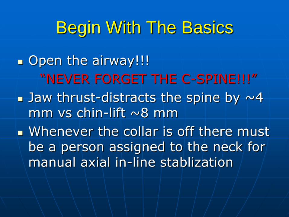
Begin With The Basics
Open the airway!!!
“NEVER FORGET THE C-SPINE!!!”
Jaw thrust-distracts the spine by ~4 mm vs chin-lift ~8 mm
Whenever the collar is off there must be a person assigned to the neck for manual axial in-line stablization

Airway Obstruction
Clinical Presentation
• “Distress” cyanosis – remember need adequate hemoglobin
• Stridor / hoarse
• Let them assume a desired position

Are You In???
The verification is more important then the intubation itself.
Direct visualiztion-- “I saw the cords”
Auscultation-- ~75% accuracy
CXR-- time-limiting
Pulse oximetry—some limitations
End-tidal CO2

End Tidal CO2 Monitoring
Place on ETT immediately after intubation
Deliver ~6 breaths to analyze to eliminate false positives that are seen after long-term BVM or carbonated beverage ingestion
100% sensitive with a spontaneous pulse
~30% false negative in cardiac arrest

Pulse Oximetry
Looks at the ratio of saturated hemoglobin molecules and though UV rays depicts them to an analog monitor.
They require perfusion, normothermia, protection from ambient light, cannot depict the structures of saturation
Have ~75% prediction of Sa02 Rodicks 2010
Decreased accuracy with saturations<85%

Airway Obstruction
*WHEN IN DOUBT INTUBATE*
• Cricothyroidotomy

Blunt Trauma
• Results from kinetic energy forces Blast
•Pressure wave causes tissue disruption
•Tear blood vessels & disrupt alveolar tissue
•Disruption of tracheobronchial tree •Traumatic diaphragm rupture
Crush (Compression) •Body is compressed between an object and a hard surface
•Direct injury of chest wall and internal structures

Deceleration • Body in motion strikes a fixed object • Blunt trauma to chest wall • Internal structures continue in motion
Ligamentum Arteriosum shears aorta
• Age Factors Pediatric Thorax: More cartilage = Absorbs
forces Geriatric Thorax: Calcification &
osteoporosis = More fractures

Simple Pneumothorax
• Occurs when lung tissue is disrupted and air leaks into the pleural space
•Progressive Pathology Air accumulates in pleural space
Lung collapses
Alveoli collapse (atelectasis)
Reduced oxygen and carbon dioxide exchange
Ventilation/Perfusion Mismatch • Increased ventilation but no alveolar perfusion
• Reduced respiratory efficiency results in HYPOXIA
• Typical MOI: “Paper Bag Syndrome”

Tension Pneumothorax
“A Fatal Sequelae”
Defined
• A pulmonary collapse that results in hemodynamic embarrassment and shock
Etiology
• Penetrating chest injury
• Blunt chest injury
• Iatrogenic barotrauma

Tension Pneumothorax
Clinical Presentation
• Severe respiratory distress hypoxemia
• Hemodynamic embarrassment
• Unilateral absence of breath sounds or chest expansion
• Tracheal (?) and PMI deviation

Tension Pneumothorax
Management
• Support – ABC
• Needle decompression
• Tube thoracostomy

Massive Hemothorax
Defined • Blood in pleural space
• Mortality rate of 75%
• Each side of thorax may hold up to 3,000 mL
• Blood loss in thorax causes a decrease in tidal volume
• Ventilation/Perfusion Mismatch & Shock
• Typically accompanies pneumothorax Hemopneumothorax

Massive Hemothorax
Management
• Support ABC – good venous access -> autotransfusion
• Tube thoracostomy – BIG TUBE
• Indications for thoracotomy
Ongoing shock
1,500 cc initial evacuation
200 cc for 3 hours
Failure to drain blood

Massive Hemothorax
Clinical Presentation
• Mechanism
• Rib fractures
• Signs of hemorrhagic shock
• Dullness to percussion / absence of breath sounds
• Signs of hypoxemia

Chest Tubes
Indications: loss of negative inspiratory force!!!!
--air/fluid/blood
--big tube-- blood/little tube-- air
--Initial return of 1500cc blood or > 200cc for > 3 hours to OR
--Call if > 100cc/hr or > 3-5cc/kg for children

Keep to suction at all times unless there is an order
Maintain at 20cm H2O unless otherwise noted
Closely monitor output
--color
--volume
Monitor for airleak
NEVER CLAMP
Chest tube ties

Indications for removal
--<200cc/24 hours
--lung fully inflated=no air leak
--full inspiration and occlusive dressing
when pulled
After removal
--Occlusive dressing for 48-72 hours DATE the dressing
--CXR as ordered
--Assess the patient

Autotransfusion
Indication with chest trauma
Special collection reservoir
THINK
May stay extracorporeal for ~4
hours
Filter, no addtives
Complete paperwork

Heimlich Valves
May be utilized on airlifted patients
May be utilized on “pigtails”
Only used when there is air in the pleural cavity

Chest Catheters
Utilized for simple pneumothorax
Usually placed in anterior chest or midaxillary line
Can be connected to a pleurovac or Heimlich valve
Less morbidity

Rib Fractures • >50% of significant chest trauma cases
due to blunt trauma • Compressional forces flex and fracture
ribs at weakest points • Ribs 1-3 requires great force to fracture
Possible underlying lung injury • Ribs 4-9 are most commonly fractured • Ribs 9-12 less likely to be fractured
Transmit energy of trauma to internal organs
If fractured, suspect liver and spleen injury
• Hypoventilation is COMMON due to PAIN

Facts That Frighten
There is a 27% increase in complications for EVERY rib fractured in the elderly

Flail Chest
Defined
• Multiple rib fractures occurring in two or more places along the same rib
• Paradoxical movement
• VC, WOB, VO2
• Venous return, hypoperfusion

Flail Chest
Clinical Presentation
• Diagnosis is clinical NOT radiologic
• Chest bruising, deformity, asymmetry, concavity
• Hypoxemia
• Associated sequelae
Pulmonary contusion
Hemopneumothorax
Abdominal injuries (15%)
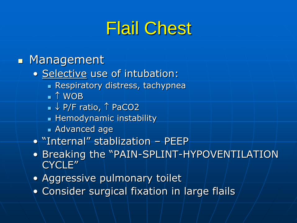
Flail Chest
Management • Selective use of intubation:
Respiratory distress, tachypnea
WOB
P/F ratio, PaCO2
Hemodynamic instability
Advanced age
• “Internal” stablization – PEEP
• Breaking the “PAIN-SPLINT-HYPOVENTILATION CYCLE”
• Aggressive pulmonary toilet
• Consider surgical fixation in large flails

Sternal Fracture & Dislocation • Associated with severe blunt anterior trauma • Typical MOI
Direct Blow (i.e. Steering wheel) • Incidence: 5-8% • Mortality: 25-45%
Myocardial contusion Pericardial tamponade Cardiac rupture Pulmonary contusion
• Dislocation uncommon but same MOI as fracture Tracheal depression if posterior

Pulmonary Contusion
Defined
• Parenchymal destruction, interstitial hemorrhage, edema, and capillary leak
• “Blossoming” – inflammatory cascade
• Intrapulmonary shunting hypoxemia
• Children – “Plastic”

Pulmonary Contusion
Clinical Presentation
• Dypnea
• Hypoxemia / hypercarbia
• Decreased compliance
• Diminished breath sounds
• Hemoptysis
• Microhemorrhage may account for 1- 1 ½ L of blood loss in alveolar tissue
• CXR – findings

Pulmonary Contusion
Management
• “SUPPORT”
• Intensive pulmonary toilet – rotational beds
• Judicious use of fluids – pulmonary artery catheters
• No prophylactic ABX

Pulmonary Contusion
MILD • Monitor SpO2
• Aggressive toilet / mobility
• Analgesia
Moderate Severe • Intubate with positive pressure ventilation
(IDLV)
Catastrophic • Non-conventional therapy PC-IRV, HFJV, ECMO
• Full body support

Pericardial Tamponade
Defined
• Perforation of the pericardial sac / chamber / vessel with rapid accumulation of blood
• Blunt injury
• Non-elastic parietal sac results in compression of structures – hypoperfused tissue beds
• Primary mechanism – stab wound

Pericardial Tamponade • Restriction to cardiac filling caused by
blood or other fluid within the pericardium
• Occurs in <2% of all serious chest trauma However, very high mortality
• Results from tear in the coronary artery or penetration of myocardium Blood seeps into pericardium and is unable
to escape
200-300 ml of blood can restrict effectiveness of cardiac contractions
• Removing as little as 20 ml can provide relief

Pericardial Tamponade
Clinical Presentation • “Suspect” – low BP, acidosis, BD
• Beck’s Triad
hypotension (↓SV), JVD, muffled heart tones
• Pulses paradoxus
• ECG Electrical Alterans
P, QRS, & T amplitude changes in every other cardiac cycle
PEA
• Echo
• Hemodynamics

Pericardial Tamponade
Hemodynamically stable
• PERICARDIOCENTESIS
“Primarily diagnostic”
Left costal margin
Utilize alligator clamp
Assessment of fluid / status
“Problems”

Pericardial Tamponade
Hemodynamically unstable
• Subxiphoid window
Small surgical window
Open visualization
“Problems”

Pericardial Tamponade
Hemodynamically Unstable • ED THORACOTOMY
• Indications Penetrating
Recently lost signs
• Procedure
• Management of wound
• Survivability
CPR 5 minutes with no airway
CPR 15 minutes with airway

Diaphragmatic Tear
Defined
• MOI – deceleration injury
• Left hemidiaphragm injured > right hemidiaphragm

Diaphragm
• Muscular, dome-like structure
• Separates abdomen from the thoracic cavity
• Affixed to the lower border of the rib cage
• Major muscle of respiration
Draws downward during inspiration
Moves upward during exhalation

Diaphragmatic Tear
Clinical presentation • CXR – 25-50% - diagnostic
• Stomach, colon, bowel in chest
• NGT in chest
• Hemidiaphragmatic elevation
• DPL – 35% false negative
• DPL FLUID EXITING CT
• Bowel sounds in chest
• Dyspnea

Diaphragmatic Tear
Management
• Surgical repair
• High mortality due to co-morbidities

Tracheobronchial Injury
Defined • Tear of bronchus
• MOI – deceleration, compression
• >80% occur with 2.5 cm of the carina
• 50% of patients with injury die within 1 hr of injury
Clinical Presentation
• Subcutaneous air
• Dyspnea, pain, dysphagia, cough, hemoptysis
• Radiologic findings
Subcutaneous – posterior soft tissue – mediastinal air

Tracheobronchial Injury
Management
• ABC’s
• Tube thoracostomy
• Bronchoscopy
• OR repair

Traumatic Asphyxia
•Results from severe compressive forces applied to the thorax
•Causes backwards flow of blood from right side of heart into superior vena cava and the upper extremities
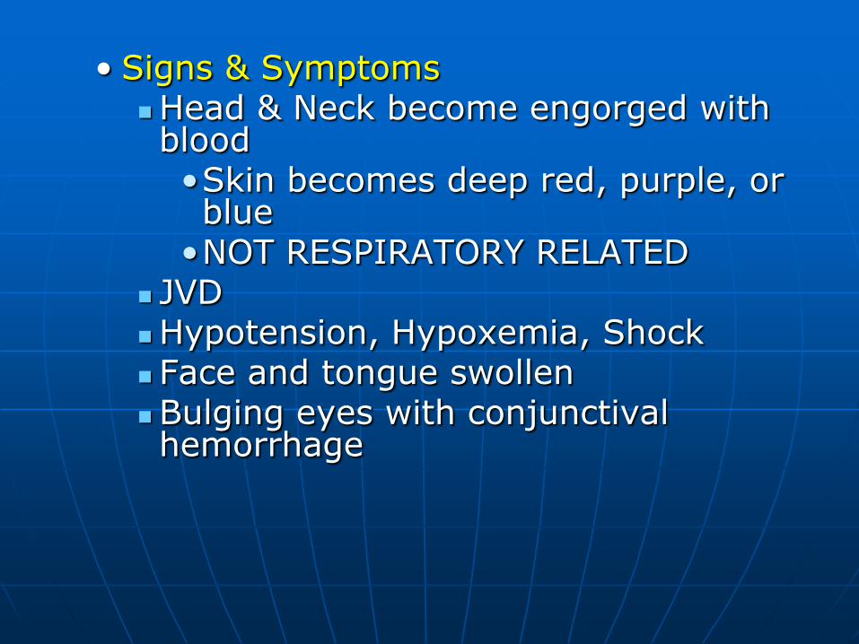
• Signs & Symptoms Head & Neck become engorged with blood •Skin becomes deep red, purple, or blue
•NOT RESPIRATORY RELATED JVD Hypotension, Hypoxemia, Shock Face and tongue swollen Bulging eyes with conjunctival hemorrhage

Blunt Cardiac Injury
Defined
• Blunt MOI with subendocardial and interstial hemorrhage, edema, and inflammatory cascade
• 15% occurance rate all comers
• Occurs in 76% of patients with severe blunt
chest trauma
• Right ventricle primarily injured
• Impairment of conduction and/or contractility 20%

Blunt Cardiac Injury
Clinical presentation (cont‘d)
• ECG
ST * (PVC’s)
Afib
RBBB
ST, T changes
• ECHO
Verify wall motility and valvular function – RVEF

• Enzymes
– CKMB – index >5% - significant
– Troponin I
– Enzymes weak indicator for BMI
• Seat belt sign
• BP, pain, N/V

Blunt Cardiac Injury
Management
• Monitor for 24 hours for dysrhythmias
• Ventricular hypokinesis ICU management
Fluids
Inotropes
Optimize DO2/VO2
NO VASODIALATORS
• Supplemental O2
• Monitor conduction disturbances
• Pain relief NSAIDS

Esophageal Injuries
Defined
• Penetrating in nature and involve the thoracic esophagus
Clinical Presentations
• Subcutaneous air, mediastinal air
• Fever 24 hours after injury
• Diagnostics
Esophagoscopy – 60%
Esophagram – 90%

• Rare complication of blunt thoracic trauma
• 30% mortality
• Contents in esophagus/stomach may move into mediastinum
Serious Infection occurs
Chemical irritation
Damage to mediastinal structures
Air enters mediastinum

Esophageal Injuries
Management
• Injury < 6 hours out primary repair
• Injury > 12 hours out laceration repair
• With diversion of proximal surgical feeding tube
• Injuries 6-12 hours old controversial management

DUTIES OF THE FLOOR
NURSE-1887 In addition to caring for your 50
patients each nurse will follow these regulations:
1. Daily sweep and mop the floors of the wards, dust patient’s furniture and window sills.
2. Maintain an even temperature in your ward by bringing in a scuttle of coal each day.

• Light is important to observe
your patient’s condition,
therefore, each day fill the
kerosene lamps, clean chimneys,
and trim wicks.
Nurses notes are important to
add the physician. Make your
pens carefully with crisp whittle
your nibs.

Each nurse should lay aside from each pay a goodly sum of her earnings for her declining years so that she will not be a burden.
Any nurse who smokes, uses liquor, gets her hair done at a beauty salon, or frequents dance halls give the director good reason to suspect her worth, intentions and integrity.

Each nurse will report everyday at 7
AM and leave at 8 PM except on the
Sabbath on which you may be off
from 12 noon until 2 PM.
Graduate nurses in good standing
with the director of nurses will be
given an evening off each week if you
go regularly to church.

The nurse who performs her labors, serves her patients and doctors faithfully for 5 years will be given an increase in pay by 5 cents per day providing no hospital debts are outstanding.