ADAMTS-5: THE STORY SO FAR - eCM - eCells & Materials Journal
Case ReportTreatment of Concurrent Thrombotic ThrombocytopenicPurpura and Graves’ Disease: A Report on Two Cases
Karl Lhotta ,1 Emanuel Zitt,1 Hannelore Sprenger-Mähr,1
Lorin Loacker,2 and Alexander Becherer3
1Department of Internal Medicine 3, Academic Teaching Hospital Feldkirch, Feldkirch, Austria2Central Institute for Medical and Chemical Laboratory Diagnostics, Medical University Innsbruck, Innsbruck, Austria3Department of Nuclear Medicine, Academic Teaching Hospital Feldkirch, Feldkirch, Austria
Correspondence should be addressed to Karl Lhotta; [email protected]
Received 20 April 2018; Accepted 1 August 2018; Published 9 August 2018
Academic Editor: Thomas Gruning
Copyright © 2018 Karl Lhotta et al. This is an open access article distributed under the Creative Commons Attribution License,which permits unrestricted use, distribution, and reproduction in any medium, provided the original work is properly cited.
Graves’ disease (GD) and thrombotic thrombocytopenic purpura (TTP) are autoimmunediseases caused by autoantibodies againstthe TSH receptor (TRAb) and the enzymeADAMTS13.We here report on two patients with concurrentGD andTTP, who achievedsustained remission of both conditions with the TTP treatment regimen and thiamazole. Both patients suffered from relapsing TTPand were diagnosed with GD concomitantly at the time of relapse. They were treated with steroids, plasma exchange, rituximab,and thiamazole. This therapy induced complete remission of TTP. TRAb levels also decreased rapidly and both patients developedsubclinical hypothyroidism three and five weeks later. Our observations suggest that TTP and GD may be concomitant and thatGD possibly triggers a relapse of TTP. The combination of thyrostatic treatment and immunosuppression with PE, rituximab, andsteroids is able to induce rapid and prolonged remission of GD.
1. Introduction
Graves’ disease (GD) is the most common cause of hyper-thyroidism with a lifetime risk of 3% for women and 0.5%for men [1]. The disease is caused by activating autoan-tibodies directed against the alpha subunit of the TSHreceptor (TRAb) on thyroid follicular cells [2]. Graves’ diseaseis currently treated with either thyrostatic drugs such asthiamazole or propylthiouracil, which block thyroid hor-mone synthesis, radioactive iodine, or surgical thyroidec-tomy [3]. Remission after medical thyrostatic therapy isachieved in about 50% of patients. Although a theoreticaloption, targeting autoimmunity with immunosuppressionis currently not pursued in patients with the disorder. Wehere report on two patients with concurrent thromboticthrombocytopenic purpura (TTP), an autoimmune diseasecaused by autoantibodies against the von Willebrand factor-cleaving protease ADAMTS13, and GD. Treatment consistingof steroids, plasma exchange, and rituximab induced rapidand sustained remission of both conditions.
2. Patient 1
A 40-year-old woman sought medical treatment becauseof petechia, hematuria, and headache. Laboratory analy-sis revealed severe hemolytic anemia with schistocytosisand thrombopenia. ADAMTS13 activity was absent (<24ng/mL, reference range 530-800 ng/mL), but no inhibitorcould be detected. A diagnosis of thrombotic thrombo-cytopenic purpura was made despite a negative test foranti-ADAMTS13 antibodies [4]. She made a quick recov-ery with steroids and daily plasma exchange (PE) usingfresh frozen plasma as a substitution fluid. After one weekshe experienced a severe relapse with microangiopathicinvolvement of the brain, heart, lung, kidneys, liver, spleen,stomach, and gut. PE was performed twice daily. Altogether,the patient had 41 exchanges over a six-week period. Inaddition, she received two 1g infusions of rituximab. Thy-roid function was normal. The patient made a completerecovery and ADAMTS13 activity remained in the normalrange.
HindawiCase Reports in EndocrinologyVolume 2018, Article ID 5747969, 4 pageshttps://doi.org/10.1155/2018/5747969
2 Case Reports in Endocrinology
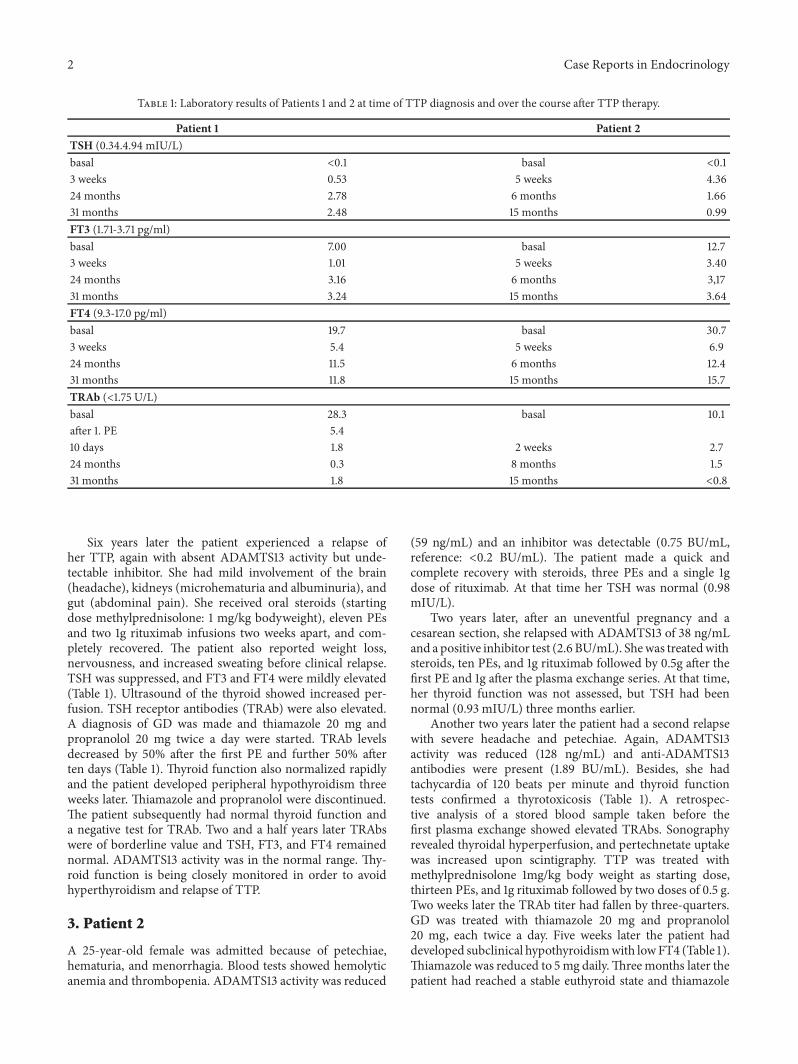
Table 1: Laboratory results of Patients 1 and 2 at time of TTP diagnosis and over the course after TTP therapy.
Patient 1 Patient 2TSH (0.34.4.94 mIU/L)basal <0.1 basal <0.13 weeks 0.53 5 weeks 4.3624 months 2.78 6 months 1.6631 months 2.48 15 months 0.99FT3 (1.71-3.71 pg/ml)basal 7.00 basal 12.73 weeks 1.01 5 weeks 3.4024 months 3.16 6 months 3,1731 months 3.24 15 months 3.64FT4 (9.3-17.0 pg/ml)basal 19.7 basal 30.73 weeks 5.4 5 weeks 6.924 months 11.5 6 months 12.431 months 11.8 15 months 15.7TRAb (<1.75 U/L)basal 28.3 basal 10.1after 1. PE 5.410 days 1.8 2 weeks 2.724 months 0.3 8 months 1.531 months 1.8 15 months <0.8
Six years later the patient experienced a relapse ofher TTP, again with absent ADAMTS13 activity but unde-tectable inhibitor. She had mild involvement of the brain(headache), kidneys (microhematuria and albuminuria), andgut (abdominal pain). She received oral steroids (startingdose methylprednisolone: 1 mg/kg bodyweight), eleven PEsand two 1g rituximab infusions two weeks apart, and com-pletely recovered. The patient also reported weight loss,nervousness, and increased sweating before clinical relapse.TSH was suppressed, and FT3 and FT4 were mildly elevated(Table 1). Ultrasound of the thyroid showed increased per-fusion. TSH receptor antibodies (TRAb) were also elevated.A diagnosis of GD was made and thiamazole 20 mg andpropranolol 20 mg twice a day were started. TRAb levelsdecreased by 50% after the first PE and further 50% afterten days (Table 1). Thyroid function also normalized rapidlyand the patient developed peripheral hypothyroidism threeweeks later. Thiamazole and propranolol were discontinued.The patient subsequently had normal thyroid function anda negative test for TRAb. Two and a half years later TRAbswere of borderline value and TSH, FT3, and FT4 remainednormal. ADAMTS13 activity was in the normal range. Thy-roid function is being closely monitored in order to avoidhyperthyroidism and relapse of TTP.
3. Patient 2
A 25-year-old female was admitted because of petechiae,hematuria, and menorrhagia. Blood tests showed hemolyticanemia and thrombopenia. ADAMTS13 activity was reduced
(59 ng/mL) and an inhibitor was detectable (0.75 BU/mL,reference: <0.2 BU/mL). The patient made a quick andcomplete recovery with steroids, three PEs and a single 1gdose of rituximab. At that time her TSH was normal (0.98mIU/L).
Two years later, after an uneventful pregnancy and acesarean section, she relapsed with ADAMTS13 of 38 ng/mLand a positive inhibitor test (2.6 BU/mL). Shewas treatedwithsteroids, ten PEs, and 1g rituximab followed by 0.5g after thefirst PE and 1g after the plasma exchange series. At that time,her thyroid function was not assessed, but TSH had beennormal (0.93 mIU/L) three months earlier.
Another two years later the patient had a second relapsewith severe headache and petechiae. Again, ADAMTS13activity was reduced (128 ng/mL) and anti-ADAMTS13antibodies were present (1.89 BU/mL). Besides, she hadtachycardia of 120 beats per minute and thyroid functiontests confirmed a thyrotoxicosis (Table 1). A retrospec-tive analysis of a stored blood sample taken before thefirst plasma exchange showed elevated TRAbs. Sonographyrevealed thyroidal hyperperfusion, and pertechnetate uptakewas increased upon scintigraphy. TTP was treated withmethylprednisolone 1mg/kg body weight as starting dose,thirteen PEs, and 1g rituximab followed by two doses of 0.5 g.Two weeks later the TRAb titer had fallen by three-quarters.GD was treated with thiamazole 20 mg and propranolol20 mg, each twice a day. Five weeks later the patient haddeveloped subclinical hypothyroidismwith lowFT4 (Table 1).Thiamazole was reduced to 5mg daily.Threemonths later thepatient had reached a stable euthyroid state and thiamazole
Case Reports in Endocrinology 3
was further reduced to 5 mg on alternate days. Eight andtwelve months after diagnosis of GD, TRAb levels were in thenormal range below 1.75 U/L.
4. Discussion
We here report on two patients with concomitant TTP andGD.This combination has been described before in four casereports [5–8]. An association between both TTP [9] and GD[10] and other autoimmune diseases is well described, butconcurrence of TTP and GD is not mentioned in those publi-cations, probably because of the rarity of TTP. TTP has a lowincidence of 3 cases permillion persons per year [11], whereasGD is rather common with 20 to 50 cases per 100.000 [1]. Inour patients TTPpresented beforeGDandGDwas associatedwith a relapse of TTP. Whether this is a coincidence orwhether GD is indeed the cause of the TTP relapse remainsunknown, but we assume a causal relationship is very likely.The nature of this causal link remains to be determined, butcould be by augmented stimulation of synthesis of (possiblypreformed) ADAMTS13 autoantibodies or some endothelialactivation, thus triggering a TTP relapse. A causal link is alsosupported by one of the case reports describing remissionof TTP without PE after successful treatment of GD withradioactive iodine [5].
The four previous case reports all describe good responseof TTP to PE and of GD to PE and either antithyroid drugs [6,8], radioiodine [7], or radioiodine and surgery [5]. The cur-rent therapy for relapsing TTP is a combination of steroids,PEwith FFP as substitutionfluid and rituximab [12, 13]. In ourtwo patients, this therapy caused complete remission of TTP,but also rapid recovery fromhyperthyroidism.One reason forthat may be that PE is able to remove TRAbs, as shown in ourpatients, in whomTRAb levels fell rapidly (Table 1) comparedto the expected normal half-life of 21 days of IgG TRAbs[14]. Number and frequency of PE sessions (11 in Patient 1and 13 in Patient 2) were determined by TTP response totreatment. PE is also considered to be a therapeutic optionin thyroid storm, not only because it removes TRAbs, butalso because it lowers the pool of T3 and T4 bound toplasma proteins [15]. Rituximab is an anti-CD20 monoclonalantibody that eliminates B lymphocytes. It is used in B-celllymphomas and autoimmune diseases such as rheumatoidarthritis or anti-neutrophil cytoplasmic antibody-associatedvasculitis. In TTP, rituximab and steroids are applied to blockADAMTS13 autoantibody synthesis. It can be expected thatthis therapy also affects TRAb formation. Indeed, our patientsquickly became TRAb-negative and remained in remissionthereafter. In Patient 1, a borderline TRAb level was detectedtwo and a half years after the attack, when recovery fromrituximab-induced B-cell depletion is likely.
In addition to removal and suppression of TRAb syn-thesis, our patients also received standard therapy withthiamazole to block thyroid hormone synthesis. This com-bination therapy actually led to subclinical hypothyroidismwith low FT4 levels after three and five weeks, respectively,and to normalization of TSH levels after four and six weeks,when thiamazole was able to be discontinued or the dosedramatically reduced.
Rituximab has been used in the treatment of GD andespecially Graves’ orbitopathy (GO) with mixed results. Onesmall study described sustained remission in four out of tenGD patients after rituximab compared to none of the tenpatients not on rituximab. Rituximab seemed to be effectivein patients with low TRAb levels but had no effect onTRAb titers in addition to thiamazole [16]. Another study inrelapsing patients found a persistent remission in nine outof 13 patients [17]. In addition, rituximab led to a decreasein TRAb levels. Two studies in GO compared rituximaband iv methylprednisolone. In the first study including 32patients, rituximab was superior to iv methylprednisolone[18], whereas in the second study comprising 21 patients, itwas not [19]. In both studies TRAb titers fell with rituximab.The decrease in TRAb levels induced by rituximab occursslowly over several months [17, 20], which is comparable tothe effect of antithyroid drugs on TRAb levels [21]. Ritux-imab, however, has been shown to specifically reduce theproduction of thyroid-stimulating autoantibodies, whereasmethimazole has no such effect [22]. In conclusion, we reporton two patients, in whom GD possibly caused a relapse ofpreexisting TTP. GD should be considered as a risk factor forTTP recurrence and we suggest routine assessment of thyroidfunction in such patients. Initiation of TTP treatment withsteroids, PE, and rituximab with the addition of thiamazolecaused rapid and prolonged remission of GD.This treatmentregimen could also be considered, for example, in GDpatients with thyroid storm. Further studies are required toprove its effectiveness and to determine the optimal dose ofsteroids, PE, and rituximab.
Conflicts of Interest
The authors declare that there are no conflicts of interestregarding the publication of this article.
References
[1] M. B. Zimmermann and K. Boelaert, “Iodine deficiency andthyroid disorders,”The Lancet Diabetes & Endocrinology, vol. 3,no. 4, pp. 286–295, 2015.
[2] T. J. Smith and L. Hegedus, “Graves’ disease,”The New EnglandJournal of Medicine, vol. 375, no. 16, pp. 1552–1565, 2016.
[3] H. B. Burch and D. S. Cooper, “Management of Graves Disease:AReview,” Journal of the AmericanMedical Association, vol. 314,no. 23, pp. 2544–2554, 2015.
[4] H. M. Tsai, “Measurement of ADAMTS13,” Int RevThromb, vol.1, pp. 272–280, 2006.
[5] F. Bellante, P. Redondo Saez, C. Springael, and S. Dethy, “Strokein thrombotic thrombocytopenic purpura induced by thyro-toxicosis: A case report,” Journal of Stroke and CerebrovascularDiseases, vol. 23, no. 6, pp. 1744–1746, 2014.
[6] B. T. Chaar, G. C. Kudva, T. J. Olsen, A. B. Silverberg, and B. J.Grossman, “Thrombotic thrombocytopenic purpura andgravesdisease,”The American Journal of the Medical Sciences, vol. 334,no. 2, pp. 133–135, 2007.
[7] S. Chhabra and G. Tenorio, “Thrombotic thrombocytopenicpurpura precipitated by thyrotoxicosis,” Journal of ClinicalApheresis, vol. 27, no. 5, pp. 265-266, 2012.
4 Case Reports in Endocrinology
[8] W.-L. Zheng, G.-S. Zhang, and M.-Y. Deng, “Thromboticthrombocytopenic purpura complicating Graves disease: Dra-matic response to plasma exchange and infusion,” TransfusionMedicine, vol. 21, no. 5, pp. 354-355, 2011.
[9] P. Coppo, D. Bengoufa, A. Veyradier et al., “Severe ADAMTS13deficiency in adult idiopathic thrombotic microangiopathiesdefines a subset of patients characterized by various autoim-mune manifestations, lower platelet count, and mild renalinvolvement,”Medicine, vol. 83, no. 4, pp. 233–244, 2004.
[10] K. Boelaert, P. R. Newby, M. J. Simmonds et al., “Prevalenceand relative risk of other autoimmune diseases in subjects withautoimmune thyroid disease,” American Journal of Medicine,vol. 123, no. 2, pp. 183.e1–183.e9, 2010.
[11] J. A. Reese, D. S. Muthurajah, J. A. K. Hovinga, S. K. Vesely,D. R.Terrell, and J. N. George, “Children and adults with thromboticthrombocytopenic purpura associated with severe, acquiredAdamts13 deficiency: comparison of incidence, demographicand clinical features,” Pediatric Blood & Cancer, vol. 60, no. 10,pp. 1676–1682, 2013.
[12] Y. Benhamou, G. Paintaud, E. Azoulay et al., “Efficacy of arituximab regimen based on B cell depletion in thromboticthrombocytopenic purpura with suboptimal response to stan-dard treatment: Results of a phase II, multicenter noncompara-tive study,” American Journal of Hematology, vol. 91, no. 12, pp.1246–1251, 2016.
[13] W. F. Clark, G. Rock, D. Barth et al., “A phase-II sequential case-series study of all patients presenting to four plasma exchangecentres with presumed relapsed/refractory thrombotic throm-bocytopenic purpura treated with rituximab,” British Journal ofHaematology, vol. 170, no. 2, pp. 208–217, 2015.
[14] L. S. Zuckier, L. D. Rodriguez, andM.D. Scharff, “Immunologicand pharmacologic concepts of monoclonal antibodies,” Semi-nars in Nuclear Medicine, vol. 19, no. 3, pp. 166–186, 1989.
[15] J. Schwartz, A. Padmanabhan, N. Aqui et al., “Guidelines onthe Use ofTherapeutic Apheresis in Clinical Practice-Evidence-Based Approach from the Writing Committee of the AmericanSociety for Apheresis: The Seventh Special Issue,” Journal ofClinical Apheresis, vol. 31, no. 3, pp. 149–162, 2016.
[16] D. El Fassi, C. H. Nielsen, S. J. Bonnema, H. C. Hasselbalch,and L. Hegedus, “B lymphocyte depletion with the monoclonalantibody rituximab in graves’ disease: A controlled pilot study,”The Journal of Clinical Endocrinology &Metabolism, vol. 92, no.5, pp. 1769–1772, 2007.
[17] K. A. Heemstra, R. E. Toes, J. Sepers et al., “Rituximab inrelapsing Graves’ disease, a phase II study,” European Journal ofEndocrinology, vol. 159, no. 5, pp. 609–615, 2008.
[18] M. Salvi, G. Vannucchi, N. Curro et al., “Efficacy of B-cell tar-geted therapywith rituximab in patientswith activemoderate tosevere graves’ orbitopathy: a randomized controlled study,”TheJournal of Clinical Endocrinology & Metabolism, vol. 100, no. 2,pp. 422–431, 2015.
[19] M. N. Stan, J. A. Garrity, B. G. C. Leon, T. Prabin, E. A. Bradley,and R. S. Bahn, “Randomized controlled trial of rituximabin patients with graves’ orbitopathy,” The Journal of ClinicalEndocrinology & Metabolism, vol. 100, no. 2, pp. 432–441, 2015.
[20] A. L. Mitchell, E. H. Gan, M. Morris et al., “The effect of B celldepletion therapy on anti-TSH receptor antibodies and clini-cal outcome in glucocorticoid-refractory Graves’ orbitopathy,”Clinical Endocrinology, vol. 79, no. 3, pp. 437–442, 2013.
[21] P. Laurberg, G. Wallin, L. Tallstedt, M. Abraham-Nordling,G. Lundell, and O. Torring, “TSH-receptor autoimmunity inGraves’ disease after therapywith anti-thyroid drugs, surgery, or
radioiodine: A 5-year prospective randomized study,” EuropeanJournal of Endocrinology, vol. 158, no. 1, pp. 69–75, 2008.
[22] D. El Fassi, J. P. Banga, J. A. Gilbert, C. Padoa, L. Hegedus,and C. H. Nielsen, “Treatment of Graves’ disease with ritux-imab specifically reduces the production of thyroid stimulatingautoantibodies,” Clinical Immunology, vol. 130, no. 3, pp. 252–258, 2009.

Stem Cells International
Hindawiwww.hindawi.com Volume 2018
Hindawiwww.hindawi.com Volume 2018
MEDIATORSINFLAMMATION
of
EndocrinologyInternational Journal of
Hindawiwww.hindawi.com Volume 2018
Hindawiwww.hindawi.com Volume 2018
Disease Markers
Hindawiwww.hindawi.com Volume 2018
BioMed Research International
OncologyJournal of
Hindawiwww.hindawi.com Volume 2013
Hindawiwww.hindawi.com Volume 2018
Oxidative Medicine and Cellular Longevity
Hindawiwww.hindawi.com Volume 2018
PPAR Research
Hindawi Publishing Corporation http://www.hindawi.com Volume 2013Hindawiwww.hindawi.com
The Scientific World Journal
Volume 2018
Immunology ResearchHindawiwww.hindawi.com Volume 2018
Journal of
ObesityJournal of
Hindawiwww.hindawi.com Volume 2018
Hindawiwww.hindawi.com Volume 2018
Computational and Mathematical Methods in Medicine
Hindawiwww.hindawi.com Volume 2018
Behavioural Neurology
OphthalmologyJournal of
Hindawiwww.hindawi.com Volume 2018
Diabetes ResearchJournal of
Hindawiwww.hindawi.com Volume 2018
Hindawiwww.hindawi.com Volume 2018
Research and TreatmentAIDS
Hindawiwww.hindawi.com Volume 2018
Gastroenterology Research and Practice
Hindawiwww.hindawi.com Volume 2018
Parkinson’s Disease
Evidence-Based Complementary andAlternative Medicine
Volume 2018Hindawiwww.hindawi.com
Submit your manuscripts atwww.hindawi.com