Crotacetin, a novel snake venom C-type lectin homolog of ...
12
© Copyright 2006 by Humana Press Inc. All rights of any nature whatsoever reserved. 1085-9195/(Online)1559-0283/06/44:412–423/$30.00 Cell Biochemistry and Biophysics 412 Volume 44, 2006 INTRODUCTION By molecular cloning and polymerase chain reaction (PCR) homology screening, several snake venom C- type lectin homologs can be isolated that share nucleotide (nt) and predicted amino acid sequences with one another. The conformational topologies of these toxin homologs must present common features, but not necessarily common biological or pharmacolog- ical functions. Based on this premise, we used reverse biology to isolate and characterize a novel convulxin (CVX)-like toxin with the ability to aggregate platelets and to inhibit bacterial growth. Snake venoms are complex mixtures of polypeptides and nonprotein components able to disturb the home- ostasis of prey organisms. The venom composition is diverse, differing among species (interspecific venom variations) and individuals (intraspecific venom varia- tions) (1). Hereditary and epigenetic factors (e.g., geo- graphical distribution, diet, and snake maturity) contribute to the venom variability (2–6). Based on a vic- tim’s symptoms of snakebite, snake venom can basically be classified as neurotoxic or hemorrhagic. Neurotoxins act on ion channels and neural receptors (7). Hemorrhagic toxins interfere with blood stasis, and major classes of Crotacetin, a Novel Snake Venom C-Type Lectin Homolog of Convulxin, Exhibits an Unpredictable Antimicrobial Activity Gandhi Rádis-Baptista, 1, * ,# Frederico Bruno Mendes Batista Moreno, 1,2 Lucas de Lima Nogueira, 1 Alice M. C. Martins, 3 Daniela de Oliveira Toyama, 4 Marcos Hikari Toyama, 5 Benildo Sousa Cavada, 1 Walter Filgueira de Azevedo, Jr., 2 and Tetsuo Yamane 6 Departments of 1 Biochemistry and Molecular Biology and 3 Clinical and Toxicological Analysis, Federal University of Ceará, Ceará, Brazil; 2 Department of Physics–IBILCE/UNESP, São Paulo, Brazil; 4 Deptartment of Biochemistry, UNICAMP, São Paulo, Brazil; 5 Department of Fundamental Chemistry, UNESP, São Paulo, Brazil; and 6 Laboratory of Molecular Biology, IPEN-CNEN, São Paulo, Brazil and Center of Biothecnology of Amazon, CBA, Manaus, Brazil Abstract Snake venom (sv) C-type lectins encompass a group of hemorrhagic toxins that are capable of interfering with blood stasis. A very well-studied svC-type lectin is the heterodimeric toxin, convulxin (CVX), from the venom of South American rattlesnake Crotalus durissus terrificus. CVX is able to activate platelets and induce their aggrega- tion by acting via p62/GPVI collagen receptor. By using polymerase chain reaction homology screening, we have cloned several cDNA precursors of CVX subunit homologs. One of them, named crotacetin (CTC) β-subunit, pre- dicts a polypeptide with a topology very similar to the tridimensional conformations of other subunits of CVX- like snake toxins, as determined by computational analysis. Using gel permeation and reverse-phase high-performance liquid chromatography, CTC was purified from C. durissus venoms. CTC can be isolated from the venom of several C. durissus subspecies, but its quantitative predominance is in the venom of C. durissus cas- cavella. Functional analysis indicates that CTC induces platelet aggregation, and, importantly, exhibits an antimi- crobial activity against Gram-positive and -negative bacteria, comparable with CVX. Index Entries: Crotalus durisssus venom; snake venom C-type lectin; crotacetin; convulxin; platelet aggrega- tion; antimicrobial activity. * Author to whom all correspondence and reprint requests should be addressed. E-mail: [email protected] # Present Address: Department of Biochemistry and Laboratory of Immunopathology Keizo Asami, Federal University of Pernambuco, Brazil. ORIGINAL ARTICLE
Transcript of Crotacetin, a novel snake venom C-type lectin homolog of ...
© Copyright 2006 by Humana Press Inc.All rights of any nature whatsoever reserved.1085-9195/(Online)1559-0283/06/44:412–423/$30.00
Cell Biochemistry and Biophysics 412 Volume 44, 2006
INTRODUCTION
By molecular cloning and polymerase chain reaction(PCR) homology screening, several snake venom C-type lectin homologs can be isolated that sharenucleotide (nt) and predicted amino acid sequenceswith one another. The conformational topologies ofthese toxin homologs must present common features,but not necessarily common biological or pharmacolog-ical functions. Based on this premise, we used reverse
biology to isolate and characterize a novel convulxin(CVX)-like toxin with the ability to aggregate plateletsand to inhibit bacterial growth.
Snake venoms are complex mixtures of polypeptidesand nonprotein components able to disturb the home-ostasis of prey organisms. The venom composition isdiverse, differing among species (interspecific venomvariations) and individuals (intraspecific venom varia-tions) (1). Hereditary and epigenetic factors (e.g., geo-graphical distribution, diet, and snake maturity)contribute to the venom variability (2–6). Based on a vic-tim’s symptoms of snakebite, snake venom can basicallybe classified as neurotoxic or hemorrhagic. Neurotoxinsact on ion channels and neural receptors (7). Hemorrhagictoxins interfere with blood stasis, and major classes of
Crotacetin, a Novel Snake Venom C-Type Lectin Homolog of Convulxin, Exhibits an Unpredictable Antimicrobial Activity
Gandhi Rádis-Baptista,1,* ,# Frederico Bruno Mendes Batista Moreno,1,2
Lucas de Lima Nogueira,1 Alice M. C. Martins,3 Daniela de Oliveira Toyama,4
Marcos Hikari Toyama,5 Benildo Sousa Cavada,1
Walter Filgueira de Azevedo, Jr.,2 and Tetsuo Yamane6
Departments of 1Biochemistry and Molecular Biology and 3Clinical and Toxicological Analysis, Federal University of Ceará,Ceará, Brazil; 2Department of Physics–IBILCE/UNESP, São Paulo, Brazil; 4Deptartment of Biochemistry, UNICAMP,
São Paulo, Brazil; 5Department of Fundamental Chemistry, UNESP, São Paulo, Brazil; and 6Laboratory of Molecular Biology,IPEN-CNEN, São Paulo, Brazil and Center of Biothecnology of Amazon, CBA, Manaus, Brazil
Abstract
Snake venom (sv) C-type lectins encompass a group of hemorrhagic toxins that are capable of interfering withblood stasis. A very well-studied svC-type lectin is the heterodimeric toxin, convulxin (CVX), from the venom ofSouth American rattlesnake Crotalus durissus terrificus. CVX is able to activate platelets and induce their aggrega-tion by acting via p62/GPVI collagen receptor. By using polymerase chain reaction homology screening, we havecloned several cDNA precursors of CVX subunit homologs. One of them, named crotacetin (CTC) β-subunit, pre-dicts a polypeptide with a topology very similar to the tridimensional conformations of other subunits of CVX-like snake toxins, as determined by computational analysis. Using gel permeation and reverse-phasehigh-performance liquid chromatography, CTC was purified from C. durissus venoms. CTC can be isolated fromthe venom of several C. durissus subspecies, but its quantitative predominance is in the venom of C. durissus cas-cavella. Functional analysis indicates that CTC induces platelet aggregation, and, importantly, exhibits an antimi-crobial activity against Gram-positive and -negative bacteria, comparable with CVX.
Index Entries: Crotalus durisssus venom; snake venom C-type lectin; crotacetin; convulxin; platelet aggrega-tion; antimicrobial activity.
* Author to whom all correspondence and reprint requestsshould be addressed. E-mail: [email protected]
# Present Address: Department of Biochemistry and Laboratoryof Immunopathology Keizo Asami, Federal University ofPernambuco, Brazil.
ORIGINAL ARTICLE
these toxins encompass metalloproteases, phospholipases,disintegrins, and C-type lectins (reviewed in ref. 8). C-type lectins are animal proteins of approx 130 aminoacids, containing at least a carbohydrate recognitiondomain (CRD) capable of mediating sugar and calciumbinding. The carbohydrate recognition is directly relatedto some biological activities, such as cell–cell adhesion,serum glycoprotein turnover, and innate immune res-ponses against potential pathogens (9). Snake venom C-type lectins also contain the conserved CRD and sharesignificant primary structure similarities with the animalC-type lectins, but they neither necessarily bind carbohy-drate molecules nor require calcium ions for their activ-ity. More than 80 C-type lectin-like proteins that affectthrombosis and hemostasis by inhibiting or activatingspecific platelet receptors or blood coagulation factorshave been characterized from the venom of various snakespecies (10).
A very well-studied snake venom C-type lectin is theheterodimeric toxin CVX, from the venom of SouthAmerican rattlesnake Crotalus durissus terrificus (11). Thisprotein consists of two subunits, α (CVXα, 13.9 kDa) andβ (CVXβ, 12.6 kDa), joined by inter- and intrachain disul-fide bounds, arranged in a tetrameric α4β4 conformation(12,13). CVX activates platelets and induces their aggre-gation via p62/GPVI collagen receptor (14). Some othersnake toxins with similar properties of activating andaggregating platelets are also known. They are, forexample, bitiscetin, from the venoms of Bitis arietans (15);botrocetin, from Bothrops jararaca (16); flavocetin A (17);ophioluxin, a protein from Ophiophagus hannah (18), andmucrocetin, a platelet-agglutinin from Trimerusurusmucrosquamatus (19).
Recombinant convulxin (rCVX) has been cloned andexpressed in Drosophila cells. Purified rCVX from cell cul-ture supernatants binds strongly to human platelet GPVIin Western blot assay when using whole platelet proteinsor recombinant human GPVI as target. Importantly, rCVXinduces the aggregation of platelets in platelet-rich-plasma, indicating that the recombinant CVX subunitscan assemble into a functionally competent complex (20).
By PCR homology screening, we have cloned severalcomplementary DNA (cDNA) precursors that arehomologous of CVX subunits. In this work, we havecharacterized one of them, named crotacetin (CTC), anew member of snake venom C-type lectin family, withthe property of aggregating platelets and with an unex-pected antimicrobial activity.
MATERIALS AND METHODS
Snake and Snake VenomFor the construction of venom gland cDNA library, a
pair of glands was excised from an adult specimen of C.durissus terrificus, captured in São Paulo state and pro-
vided by the Laboratory of Herpetology, InstitutoButantan (São Paulo, Brazil). The snake was milked tocollect the venom and to induce the maximum level ofRNA synthesis in venom gland (21). The crude venomwas vacuum-dried and kept at –20°C until proteinpurification. The venom of C. durissus cascavella was agift from the Regional Snake Laboratory of Fortaleza(LAROF), Ceará, Brazil. The venom of C. durissuscollilineatus was purchased from the Bio-AgentsSerpentarium (Batatais, São Paulo, Brazil).
C. durissus terrificus Venom Gland cDNA Library Construction
The cDNA library was constructed as described pre-viously (22). Briefly, poly(A+) RNAs were purified froma pair of venom glands excised from a single specimenof South American rattlesnake, C. durissus terrificus (Cdt9706). The cDNAs were synthesized, selected by size,and cloned into a phagemide vector—a λ phage deriva-tive (λ ZapII; Strategene, La Jolla, CA). Recombinantphagemids were packed into viable phage particles andused to infect Escherichia coli cells XL1 Blue MRF′(Stratagene). The venom gland cDNA library wastitrated, amplified, and stored at –80°C, in 7% dimethylsulfoxide, for posterior utilization.
PCR Homology CloningBased on nt sequences of subunit α (or A) and β (or B)
of CVX from C. durissus terrificus (GenBank accessionno. AF541882 and Y16348, CVX subunit α, AF541881 andY16349, CVX subunit β) one oligonucleotide primerforward CVXA/B-FW1 (5′-TCTCTCTGCAGGGAAG-GAAG-3′), and two reverse primers, CVXA-RV1A (5′-TCCTTGCTTCTCCAGACTTCA-3′) and CVXB-RV2B(5′-ACTTCACACAGCCGGATCTT- 3′) were synthesized(Invitrogen, São Paulo, Brazil), which correspond to the5′-untranslated region (UTR) of CVX subunit α and βgenes (forward primer), and to 3′-UTRs of CVX subunitα (reverse primer RV1A) and of CVX subunit β (reverseprimer RV2B), respectively. The gene of one subunit ofCTC (β-subunit) was amplified with the same primerpair used to amplify CVX subunit α, in the followingway: phage particles (107–108 plaque-forming units) of C.durissus terrificus venom gland cDNA library, 10 pmol ofeach primer and 25 µL of ExLONGase enzyme mix(Invitrogen, Carlsbad, CA), were mixed and the PCR wasperformed according to the manufacturer’s instructions.The long-distance–PCR products were purified from thegel slice and cloned into pCR2.1-TOPO (Invitrogen).
The gene was sequenced with ABI Prism Big DyeTerminator (Applied Biosystems, Foster City, CA) in anautomated sequencer (ABI Prism 373 or 377; AppliedBiosystems) by using synthetic oligonucleotidesdesigned for CVXA/B sequences (this work). The clonedCTC mRNA (accession no. AF541884) was compared
Crotacetin Exhibits an Unpredictable Antimicrobial Activity 413
Cell Biochemistry and Biophysics Volume 44, 2006
against nt sequences in the GenBank at National Centerfor Biotechnology Information (http://www.ncbi.nlm.nih.gov), using the BLAST algorithm and the Bio-computing software Lasergene (DNAStar, Madison, WI).
Similarity Search and Homology ModelingThe predicted amino acid sequence from the respec-
tive C. durissus terrificus precursor cDNA was comparedagainst the Protein Data Bank (PDB) (http://www.rcsb.org/pdb/) by using BLASTP, a tool for proteinsearch and alignment (23). From PDB, several sequenceswere chosen as the best score after alignment andused to identify conserved residues in homologoussequences. Snake venom C-type lectin sequences fromthe venoms of Trimeresurus flavoviridis (PDB entry code1C3A; chain B) (17), T. mucrosquamatus (1V4L; chain B)(19), and C. durissus terrificus (1UOS, chain B; and1UMR, chain B) (12,13) were aligned with ClustalW(24), by using default parameters.
To build a tridimensional structure of the CTC β-subunit C-type lectin, 1000 models were generatedwith PARMODEL, a Web server at http://www.biocristalografia.df.ibilce.unesp.br/tools/parmodel(25), which paralyzes the MODELLER (26) software ina Beowulf cluster (16 nodes). The best model wasselected according to the Modeller objective functionand evaluated by PROCHECK (27), WHATCHECK(28), and 3DANALYSIS (29). The permissible angles ofamino acid residues in the spatial structure of CTC β-subunit were also evaluated by RAMACHANDRANplot (30).
CTC PurificationWhole venom (35 mg) was dissolved in a 0.2 M
ammonium bicarbonate (pH 8.0) buffer and then clari-fied by high-speed centrifugation (4500g for 1 min). Thesupernatant was injected onto a molecular exclusionhigh-performance liquid chromatography (HPLC) col-umn (Superdex 75; 1 × 60 cm; GE Healthcare, LittleChalfont, Buckinghamshire, UK), previously equili-brated with the same buffer that was used for solubi-lization of the whole venom. Chromatography processwas performed with a flow rate of 0.2 mL/min andmonitored at 280 nm. Fractions corresponding to CTCwere pooled and lyophilized. After this first step ofpurification, CTC was repurified by reverse-phaseHPLC. Approximately 1 mg of the purified protein wasdissolved in the same buffer (buffer A, 0.1% trifluo-roacetic acid [TFA] in aqueous solution) for equilibra-tion of the analytical µ-Bondapack C18 column (0.39 ×30 cm). The elution of highly purified CTC was carriedout using a linear and discontinuous buffer B gradient(66% acetonitrile in buffer A). The chromatographic run
was conducted at constant flow rate of 1.0 mL/min andmonitored at 214 nm.
Reduction, S-Carboxymethylation and Determination of N-Terminal Sequence
Two milligrams of purified CTC was dissolved in 200mL of a 6.0 M guanidine chloride solution (Merck,Darmstadt, Germany), containing 0.4 M of Tris-HCl and2 mM EDTA, pH 8.15. Nitrogen was blown over the topof the protein solution for 15 min, followed by molecu-lar reduction with 200 mL of a 6.0 M dithiothreitol andfurther carboxymethylated with [14 C]iodoacetic acidand cold iodoacetic acid. Nitrogen was again blownover the surface of the solution and the reaction tubewas sealed. This solution was incubated in the dark at37°C for 1 h and desalted in a Sephadex G25 column (0.7× 12 cm), equilibrated with 1.0 M acetic acid buffer. Theeluted reduced CTC was then applied on the reverse-phase HPLC µ-Bondapack C18 column (0.39 × 30 cm),previously equilibrated with buffer A (0.1% TFA inaqueous solution). The subunits of CTC were elutedwith a nonlinear gradient concentration of buffer B (66%acetonitrile in buffer A), and the fraction correspondingto each CTC subunit was recovered, lyophilized, andstored at –80°C. CTC subunits were then sequenced byautomatic Edman degradation, using a gas–liquid pro-tein sequencer (model Precise; Applied Biosystems).The amino acid phenylthiohydantoin (PTH) derivativeswere identified using a PTH-analyser (model 450 micro-gradient; Applied Biosystems).
Platelet AggregationPlatelet-rich plasma (PRP) was prepared as described
by Chudzinski-Tavassi et al. (31), and the evaluation ofplatelet aggregation was performed with a Chrono-LogLumi aggregometer (Chrono-Log, Havertown, PA).PRP samples were incubated at 37°C for 2 min with dif-ferent concentrations of purified CTC. Platelet aggluti-nation was monitored by turbidimetry and expressed asan increase of light transmittance. Collagen was used ascontrol of platelet aggregation.
Antimicrobial Activity and Electron MicroscopyXanthomonas axonopodis pv. passiflorae (Gram-nega-
tive) or Clavibacter michiganensis michigansis cells wereharvested from fresh agar plates and suspended in ster-ile distilled water (A 650nm = 0.3/cc 103 CFU/mL).Aliquots of bacterial suspension were diluted to a 10–5
colony-forming units [CFU]/mL and incubated, during20 min at 37°C, with the same concentration (150(g/mL) of purified CTC or CVX. Then, the capacity ofmicrobial cell survival was assayed on nutrient medium(Difco, Detroit, MI) plates (n = 5). In these antibacterialassays, morphological alterations were visualized by
414 Rádis-Baptista et al.
Cell Biochemistry and Biophysics Volume 44, 2006
Crotacetin Exhibits an Unpredictable Antimicrobial Activity 415
Cell Biochemistry and Biophysics Volume 44, 2006
electron microscopy, in absence (control) or presence ofCTC or CVX (A 650nm = 0.3/cc 103 CFU/mL). For thispurpose, bacterial samples were fixed with 1% osmiumtetroxide (Agar Scientific), for 2 h, at 25°C. Sections werewashed three times, dehydrated in increasing concen-trations of ethanol and propylene oxide, and embeddedin Epon resin (Agar Scientific, Stansted, UK).Polymerization was performed at 60°C for 48 h, andultra-thin sections were prepared with a Sorvall MT2ultramicrotome. Sections were placed on 5% collo-dion-coated 100-mesh grids and stained with 4% uranylacetate (Agar Scientific) for 15 min, followed by 2.6%lead citrate (Agar Scientific) for 15 min. Samples wereobserved with a Hitachi 1100 transmission electronmicroscope (Hitachi Scientific Instruments, Tokyo,Japan), operating at 100 kV.
RESULTS
PCR Homology Cloning of CTC β-SubunitThe gene coding for CTCβ (GenBank accession no.
AF541884) was isolated from C. durissus terrificusvenom gland cDNA library by PCR homology screen-ing with specific primers for the 5′-UTR and for 3′-UTRof both CVXα and CVXβ, as described in Materials andMethods.
The cloned CTCβ gene is 513-bp long with a 5′-UTRstrictly conserved in all CVX-like and antithrombin-likeprecursors (ATLs) isolated so far (Fig. 1). In CTCβ gene,the start codon (ATG) is located 28-bp downstream, andthe stop codon (TGA) is located 496-bp downstream. Asequence corresponding to the signal peptide liesbetween nt 28 and 96 (Fig. 1, underlined).
At the nt level, the similarity between CTCβ andCVXβ is higher than between CTCβ and CVXα or CTCβor ATLs. In contrast, the similarity of CVXα and ATLs isproportionally high. The CTCβ gene precursor predictsa polypeptide of 148 residues of amino acids, 23 corre-sponding to the leader sequence, and a molecular massof mature subunit of 14.3 kDa.
Sequence Alignment and TridimensionalModeling
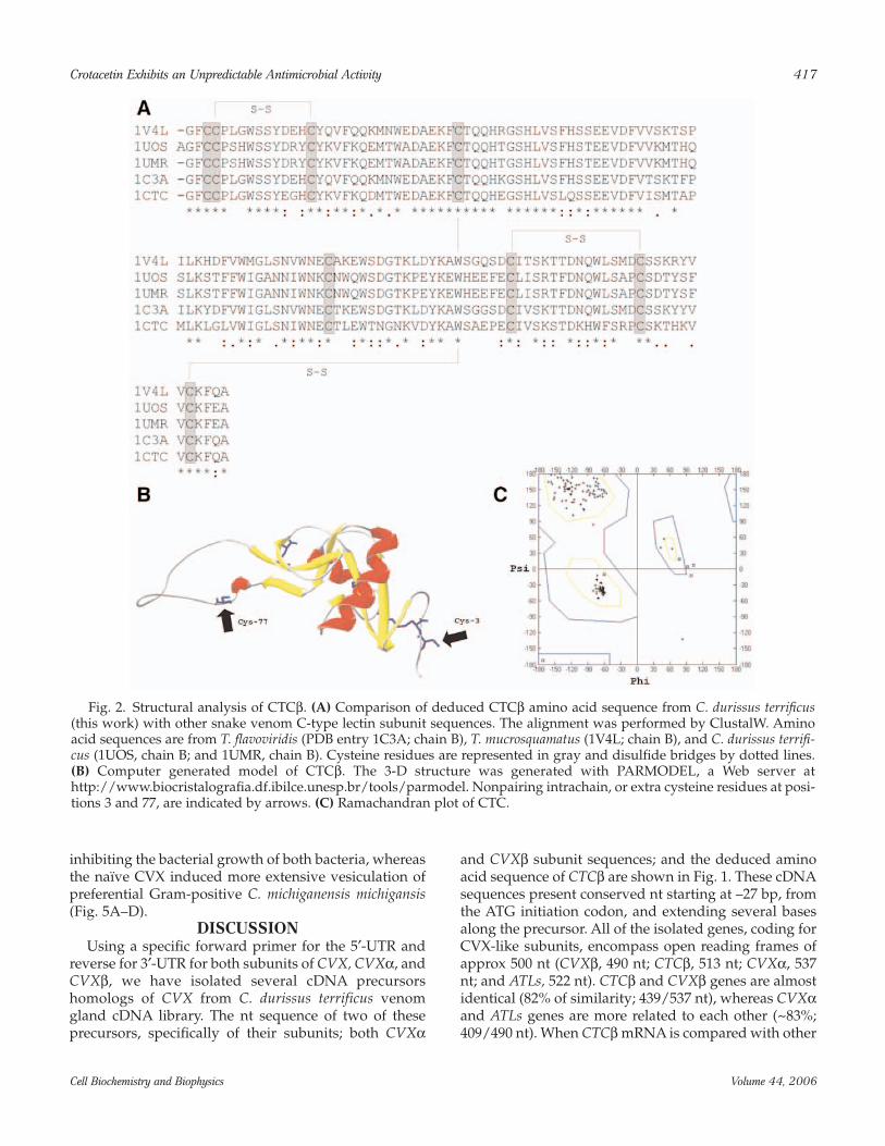
Comparison of the predicted amino acid sequence ofCTCβ gene against protein data bank reveals high simi-larity of CTCβ protein with C-type lectin subunits fromthe venom of T. flavoviridis (PDB entry code 1C3A; chainB), T. mucrosquamatus (1V4L; chain B), and C. durissusterrificus (1UOS, chain B; and 1UMR, chain B) (Fig. 2A).The similarity of CTCβ and these other snake venom-aggregating toxins is approx 50%. If conserved substitu-tions are considered, then the similarity exceeds 70%. Inprimary structure of CTCβ, the eight residues of cys-
teine are located in the same position of all alignedsequences. These positions include six residues that areinvolved in the intrachain disulfide bridges and twoother “extra” cysteines in the carboxy (Cys3) and amino(Cys77) terminals. The computer-generated model ofCTCβ shows an overall topology of five principal β-strands and two α-helices, forming a globular structurewith a lateral loop similar to the other CVX-like C-typelectins (Fig. 2B). The lateral loop contributes to the for-mation of heterodimers that are stabilized by the extracysteine residue. Ramachandran diagram of CTCβmodel indicates that psi (ψ) and phi (ϕ) angles of themajority of amino acid residues are located in thermod-inamically favorable region (Fig. 2C).
Isolation, Reduction, and N-Terminal Sequence of CTC From C. durissus
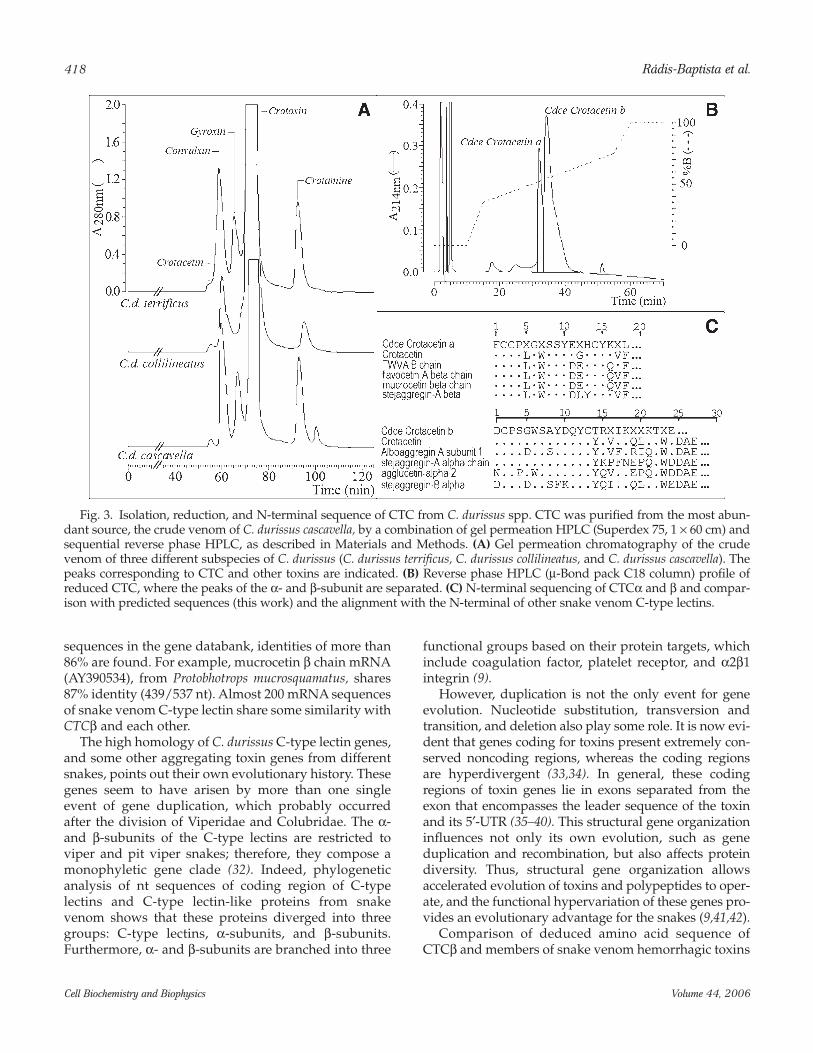
By exclusion chromatography, CTC heterodimerseems to be a minor fraction peak in the venom of atleast three C. durissus subspecies, namely, C. durissuscascavella, C. durissus terrificus, and C. durissus collilinea-tus (Fig. 3A). In the venom of C. durissus cascavella, theamount of CTC is significantly abundant (representing~0.8% of the proteins in the crude venom). Thus, CTCwas isolated and characterized from this venom.Purified CTC from C. durissus cascavella shows an appar-ent molecular mass of 70 kDa, by gel permeation chro-matography, and it appears as two subunits of differentsize by reverse phase-HPLC (Fig. 3B). Furthermore, bymolecular exclusion chromatography, it seems to have ahigh molecular weight, indicating the presence ofoligomeric forms. The partial N-terminal amino acidsequence of purified CTCβ corresponds to that pre-dicted from the gene sequence (Fig. 3C).
Platelet Aggregation AssayCTC is capable of aggregating human platelets in
PRP in a dose-dependent manner: 22% at concentrationof 32.8 µg/mL (47 µM), 36% at 49.3 µg/mL (70 µM), and84% at 65.5 µg/mL (94 µM) (data not shown).
Antimicrobial Activity of Naive CTC, CVX, and Their Isolated α- and β-Subunits
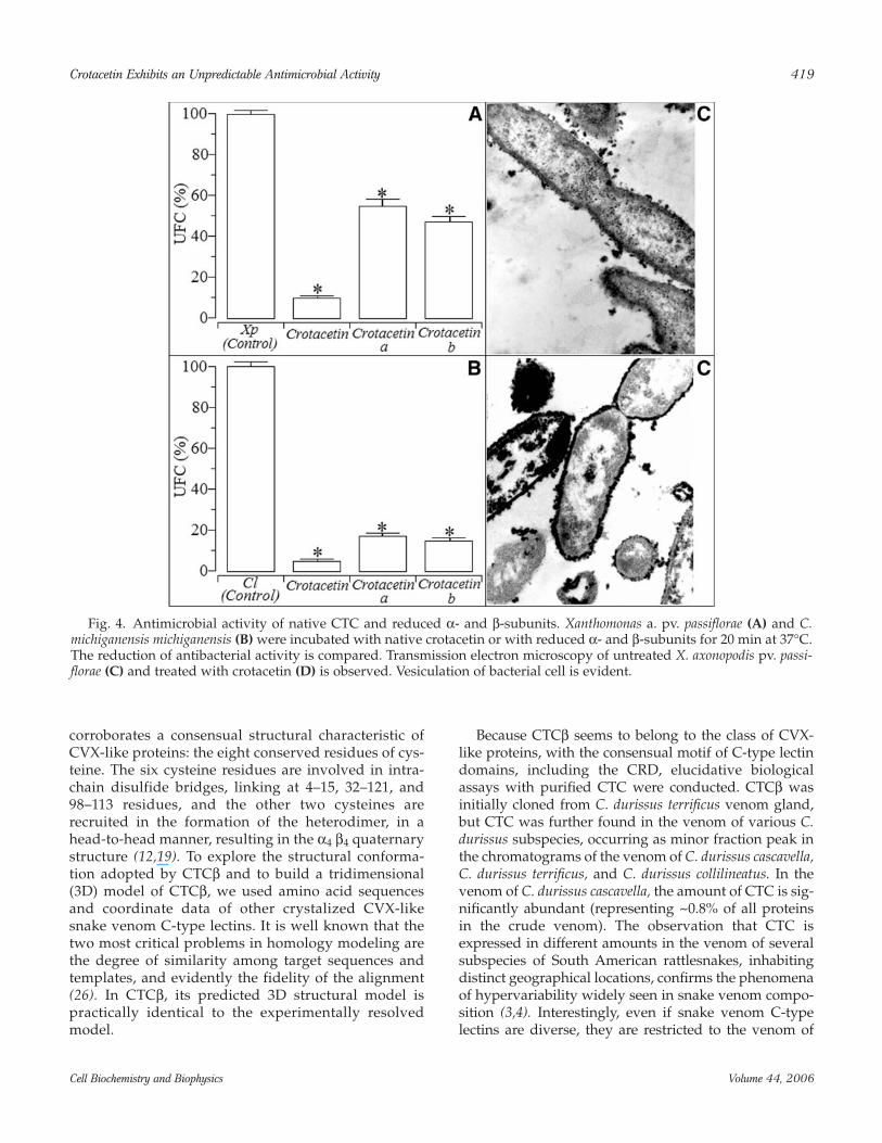
The oligomeric CTC protein decreases the bacterialgrowth of two plant pathogens: X. axonopodis pv. passi-florae, a Gram-negative bacterium and C. michiganensismichigansis , a Gram-positive bacterium. The inhibitionrates were in the range of 87.8 and 96.4%, respectively.The isolated reduced chains did not show any signifi-cant antimicrobial activity (Fig. 4A,B) against bothspecies of tested bacteria. In Xanthomonas axonopodis pv.passiflorae, CTC induced vacuolation of cell cytoplasm,and, in some cases, ruptured the cell membrane (Fig.4C,D). Similarly to CTC, reduced CVX subunits failed in

416 Rádis-Baptista et al.
Cell Biochemistry and Biophysics Volume 44, 2006
Fig. 1. Comparison of nucleotide sequence of CTCβ, CVXα and β subunits, and antithrombin-like subunit cDNA pre-cursors. All genes were isolated from C. durissus terrificus venom gland cDNA phage library by PCR homology screening.Sequences were aligned using ClustalW accessible at http://www.ebi.ac.uk/clustalw/index.html with parameters indefault. The numbers in the left side of each precursor correspond to the GenBank accession nos. AF541881, CVXβ mRNA;AF541882, CVXα subunit mRNA; AF541883, antithrombin-like (ATL) mRNA; and AF541884, CTCβ mRNA, all from C.durissus terrificus. Conserved 5′-UTRs, where the primer CVXA/B FW exactly anneals, are underlined. Dots represent con-served nucleotides in homologous sequences.
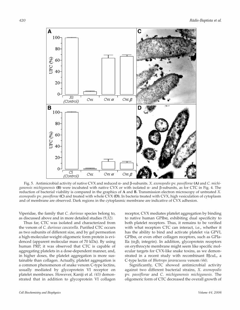
inhibiting the bacterial growth of both bacteria, whereasthe naïve CVX induced more extensive vesiculation ofpreferential Gram-positive C. michiganensis michigansis(Fig. 5A–D).
DISCUSSIONUsing a specific forward primer for the 5′-UTR and
reverse for 3′-UTR for both subunits of CVX, CVXα, andCVXβ, we have isolated several cDNA precursorshomologs of CVX from C. durissus terrificus venomgland cDNA library. The nt sequence of two of theseprecursors, specifically of their subunits; both CVXα
and CVXβ subunit sequences; and the deduced aminoacid sequence of CTCβ are shown in Fig. 1. These cDNAsequences present conserved nt starting at –27 bp, fromthe ATG initiation codon, and extending several basesalong the precursor. All of the isolated genes, coding forCVX-like subunits, encompass open reading frames ofapprox 500 nt (CVXβ, 490 nt; CTCβ, 513 nt; CVXα, 537nt; and ATLs, 522 nt). CTCβ and CVXβ genes are almostidentical (82% of similarity; 439/537 nt), whereas CVXαand ATLs genes are more related to each other (~83%;409/490 nt). When CTCβ mRNA is compared with other
Crotacetin Exhibits an Unpredictable Antimicrobial Activity 417
Cell Biochemistry and Biophysics Volume 44, 2006
Fig. 2. Structural analysis of CTCβ. (A) Comparison of deduced CTCβ amino acid sequence from C. durissus terrificus(this work) with other snake venom C-type lectin subunit sequences. The alignment was performed by ClustalW. Aminoacid sequences are from T. flavoviridis (PDB entry 1C3A; chain B), T. mucrosquamatus (1V4L; chain B), and C. durissus terrifi-cus (1UOS, chain B; and 1UMR, chain B). Cysteine residues are represented in gray and disulfide bridges by dotted lines.(B) Computer generated model of CTCβ. The 3-D structure was generated with PARMODEL, a Web server athttp://www.biocristalografia.df.ibilce.unesp.br/tools/parmodel. Nonpairing intrachain, or extra cysteine residues at posi-tions 3 and 77, are indicated by arrows. (C) Ramachandran plot of CTC.
418 Rádis-Baptista et al.
Cell Biochemistry and Biophysics Volume 44, 2006
sequences in the gene databank, identities of more than86% are found. For example, mucrocetin β chain mRNA(AY390534), from Protobhotrops mucrosquamatus, shares87% identity (439/537 nt). Almost 200 mRNA sequencesof snake venom C-type lectin share some similarity withCTCβ and each other.
The high homology of C. durissus C-type lectin genes,and some other aggregating toxin genes from differentsnakes, points out their own evolutionary history. Thesegenes seem to have arisen by more than one singleevent of gene duplication, which probably occurredafter the division of Viperidae and Colubridae. The α-and β-subunits of the C-type lectins are restricted toviper and pit viper snakes; therefore, they compose amonophyletic gene clade (32). Indeed, phylogeneticanalysis of nt sequences of coding region of C-typelectins and C-type lectin-like proteins from snakevenom shows that these proteins diverged into threegroups: C-type lectins, α-subunits, and β-subunits.Furthermore, α- and β-subunits are branched into three
functional groups based on their protein targets, whichinclude coagulation factor, platelet receptor, and α2β1integrin (9).
However, duplication is not the only event for geneevolution. Nucleotide substitution, transversion andtransition, and deletion also play some role. It is now evi-dent that genes coding for toxins present extremely con-served noncoding regions, whereas the coding regionsare hyperdivergent (33,34). In general, these codingregions of toxin genes lie in exons separated from theexon that encompasses the leader sequence of the toxinand its 5′-UTR (35–40). This structural gene organizationinfluences not only its own evolution, such as geneduplication and recombination, but also affects proteindiversity. Thus, structural gene organization allowsaccelerated evolution of toxins and polypeptides to oper-ate, and the functional hypervariation of these genes pro-vides an evolutionary advantage for the snakes (9,41,42).
Comparison of deduced amino acid sequence ofCTCβ and members of snake venom hemorrhagic toxins
Fig. 3. Isolation, reduction, and N-terminal sequence of CTC from C. durissus spp. CTC was purified from the most abun-dant source, the crude venom of C. durissus cascavella, by a combination of gel permeation HPLC (Superdex 75, 1 × 60 cm) andsequential reverse phase HPLC, as described in Materials and Methods. (A) Gel permeation chromatography of the crudevenom of three different subspecies of C. durissus (C. durissus terrificus, C. durissus collilineatus, and C. durissus cascavella). Thepeaks corresponding to CTC and other toxins are indicated. (B) Reverse phase HPLC (µ-Bond pack C18 column) profile ofreduced CTC, where the peaks of the α- and β-subunit are separated. (C) N-terminal sequencing of CTCα and β and compar-ison with predicted sequences (this work) and the alignment with the N-terminal of other snake venom C-type lectins.
corroborates a consensual structural characteristic ofCVX-like proteins: the eight conserved residues of cys-teine. The six cysteine residues are involved in intra-chain disulfide bridges, linking at 4–15, 32–121, and98–113 residues, and the other two cysteines arerecruited in the formation of the heterodimer, in ahead-to-head manner, resulting in the α4 β4 quaternarystructure (12,19). To explore the structural conforma-tion adopted by CTCβ and to build a tridimensional(3D) model of CTCβ, we used amino acid sequencesand coordinate data of other crystalized CVX-likesnake venom C-type lectins. It is well known that thetwo most critical problems in homology modeling arethe degree of similarity among target sequences andtemplates, and evidently the fidelity of the alignment(26). In CTCβ, its predicted 3D structural model ispractically identical to the experimentally resolvedmodel.
Because CTCβ seems to belong to the class of CVX-like proteins, with the consensual motif of C-type lectindomains, including the CRD, elucidative biologicalassays with purified CTC were conducted. CTCβ wasinitially cloned from C. durissus terrificus venom gland,but CTC was further found in the venom of various C.durissus subspecies, occurring as minor fraction peak inthe chromatograms of the venom of C. durissus cascavella,C. durissus terrificus, and C. durissus collilineatus. In thevenom of C. durissus cascavella, the amount of CTC is sig-nificantly abundant (representing ~0.8% of all proteinsin the crude venom). The observation that CTC isexpressed in different amounts in the venom of severalsubspecies of South American rattlesnakes, inhabitingdistinct geographical locations, confirms the phenomenaof hypervariability widely seen in snake venom compo-sition (3,4). Interestingly, even if snake venom C-typelectins are diverse, they are restricted to the venom of
Crotacetin Exhibits an Unpredictable Antimicrobial Activity 419
Cell Biochemistry and Biophysics Volume 44, 2006
Fig. 4. Antimicrobial activity of native CTC and reduced α- and β-subunits. Xanthomonas a. pv. passiflorae (A) and C.michiganensis michiganensis (B) were incubated with native crotacetin or with reduced α- and β-subunits for 20 min at 37°C.The reduction of antibacterial activity is compared. Transmission electron microscopy of untreated X. axonopodis pv. passi-florae (C) and treated with crotacetin (D) is observed. Vesiculation of bacterial cell is evident.
Viperidae, the family that C. durissus species belong to,as discussed above and in more detailed studies (9,32).
Thus far, CTC was isolated and characterized fromthe venom of C. durissus cascavella. Purified CTC occursas two subunits of different size, and by gel permeationa high-molecular-weight oligomeric form protein is evi-denced (apparent molecular mass of 70 kDa). By usinghuman PRP, it was observed that CTC is capable ofaggregating platelets in a dose-dependent manner, and,in higher doses, the platelet aggregation is more sus-tainable than collagen. Actually, platelet aggregation isa common phenomenon of snake venom C-type lectins,usually mediated by glycoprotein VI receptor onplatelet membranes. However, Kaniji et al. (43) demon-strated that in addition to glycoprotein VI collagen
receptor, CVX mediates platelet aggregation by bindingto native human GPIbα, exhibiting dual specificity toboth platelet receptors. Thus, it remains to be verifiedwith what receptors CTC can interact, i.e., whether ithas the ability to bind and activate platelet via GPVI,GPIbα, or even other collagen receptors, such as GPIa-IIa (α2β1 integrin). In addition, glycoprotein receptorson erythrocyte membrane might seem like specific mol-ecular targets for CVX-like snake toxins, as we demon-strated in a recent study with recombinant BJcuL, aC-type lectin of Bhotrops jararacussu venom (44).
Significantly, CTC showed antimicrobial activityagainst two different bacterial strains, X. axonopodispv. passiflorae and C. michiganensis michiganesis. Theoligomeric form of CTC decreased the overall growth of
420 Rádis-Baptista et al.
Cell Biochemistry and Biophysics Volume 44, 2006
Fig. 5. Antimicrobial activity of native CVX and reduced α- and β-subunits. X. axonopodis pv. passiflorae (A) and C. michi-ganensis michiganensis (B) were incubated with native CVX or with isolated α- and β-subunits, as for CTC in Fig. 4. Thereduction of bacterial viability is compared in the graphics of A and B. Transmission electron microscopy of untreated X.axonopodis pv. passiflorae (C) and treated with whole CVX (D). In bacteria treated with CVX, high vesiculation of cytoplasmand of membrane are observed. Dark regions in the cytoplasmic membrane are indicative of CVX adhesion.
both bacteria. However, the separation of intactoligomeric protein into isolated and reduced subunitssignificantly abolishes the antimicrobial activity ofCTC. As mentioned, tetrameric conformation is a typi-cal structure adopted by several snake venom CVX-likeC-type lectins, and this quaternary arrangement alsoseems essential for antimicrobial activity. The micro-scopic effect of CTC on X. axonopodis pv. passifloraeinvolved the induction of cytoplasmic vacuolation andmembrane rupture. As surprising as the antimicrobialactivity of CTC is, the fact is that CVX, another snakevenom C-type lectin and potent agonist of plateletaggregation from the venom of C. durissus terrificus,was able to abrogate the bacterial viability of bothstrains. In this respect, CVX was more effective indecreasing the growth of C. michiganensis michiganesis,a Gram-positive bacterium. Similarly to CTC, and incontrast to the corresponding natural oligomericpolypeptide, the reduced CVX subunits failed torestrain the bacterial growth.
From our point of view, these latter results are of par-ticular relevance, because they concern the descriptionof antimicrobial activities of snake venom C-typelectins. The most studied C-type lectin domain-contain-ing proteins with the property of binding to microor-ganisms are the mannose binding lectins (MBLs) andthe lectins receptors (CLRs) located on antigen-present-ing cell membranes. MBLs are involved in vertebratefirst-line defense by binding to bacteria, viruses, proto-zoa, and helminths and then initiating a range of hostresponse (45). Also, C-type lectin receptors, CLRs, ondendritic cells are type II transmembrane proteinsimplicated in the pattern recognition of pathogens andin the distinction of self- and no-self antigen recognitionin mammals (46). Thus, the antimicrobial property of C-type lectins should be further investigated.
Together, it is evident that snake venom C-typelectins have their structural domains derived from acommon ancestral precursor. Snake venom C-typelectins comprise not only a multigene family but alsohomologous polypeptides with diverse biological func-tions, including the ability to interact with glycopro-teins on blood cell membranes, and possibly onmicrobial cell.
In summary, we report (1) the cloning of severalgenes for C-type lectin subunits from C. durissus terrifi-cus, (2) the isolation and characterization of the geneproduct of CTC from the venom of three C. durissus sub-species, (3) an indication of its predominant presence inthe venom of C. durissus cascavela, (4) prediction of its3D structure, and (5) comparable, unexpected antimi-crobial activity of CTC and CVX. To our knowledge, thiswork reports for the first time the antimicrobial activi-ties of snake venom C-type lectin homologs.
ACKNOWLEDGMENTS
We thank Dr. A. M. Chudzinski-Tavassi (Laboratoryof Biochemistry and Biophysics, Insituto Butantan, SãoPaulo, Brazil) for the evaluation of platelet aggregationassays. We are also grateful to Coordenação deAperfeiçoamento de Pessoal de Nível Superior(CAPES), Conselho Nacional de DesenvolvimentoCientífico e Tecnológico (CNPq), Fundação de Amparoà Pesquisa do Estado de São Paulo (FAPESP), andFundação Cearense de Apoio ao DesenvolvimentoCientífico e Tecnológico (FUNCAP Brazil), for financialsupport. B.S.C. and T.Y. are senior investigators ofCNPq (Brazil).
REFERENCES
1. Mukherjee, A. K. and Maity, C. R. (2002) Biochemical com-position, lethality and pathophysiology of venom fromtwo cobras – Naja naja and N. kaouthia. Comp. Biochem.Physiol. B. Biochem. Mol. Biol. 131, 125–132.
2. Daltry, J. C., Wüster, W., and Thorpe, R. S. (1996) Diet andsnake venom evolution. Nature 379, 537–540.
3. Saravia, P., Rojas, E., Arce, V., et al. (2002) Geographic andontogenic variability in the venom of the Neotropical rat-tlesnake Crotalus durissus: pathophysiological and thera-peutic implications. Rev. Biol. Trop. 50, 337–346.
4. Shashidharamurthy, R., Jagadeesha, D. K., Girish, K. S.,and Kemparaju, K. (2002) Variations in biochemical andpharmacological properties of Indian cobra (Naja najanaja) venom due to geographical distribution. Mol. Cell.Biochem. 229, 93–101.
5. Lopez-Lozano, J. L., De Sousa, M. V., Ricart, C. A., et al.(2002) Ontogenetic variation of metalloproteinases andplasma coagulant activity in venoms of wild Bothrops atroxspecimens from Amazonian rain forest. Toxicon 40,997–1006.
6. Tsai, I. H., Wang, Y. M., Chen, Y. H., and Tu, A. T. (2003)Geographic variations, cloning, and functional analyses ofthe venom acidic phospholipases A2 of Crotalus viridisviridis. Arch. Biochem. Biophys. 411, 289–296.
7. Harvey, A. (1993) Natural and Synthetic Neurotoxins,Academic, New York.
8. Markland, F. S. (1998) Snake venom and the hemostaticsystem. Toxicon 36, 1749–1800.
9. Drickamer, K. (1999) C-type lectin-like domains. Curr.Opin. Struct. Biol. 9, 585–590.
10. Ogawa, T., Chijiwa, T., Oda-Ueda, N., and Ohno, M.(2005) Molecular diversity and accelerated evolution ofC-type lectin-like proteins from snake venom. Toxicon 45,1–14.
11. Prado-Franceschi, J. and Brazil, O. V. (1981) Convulxin, anew toxin from the venom of the South American rat-tlesnake Crotalus durissus terrificus. Toxicon 19, 875–887.
12. Murakami, M. T., Zela, S. P., Gava, L. M., Michelan-Duarte, S., Cintra, A.C., and Arni, R. K. (2003) Crystalstructure of the platelet activator convulxin, a disulfide-
Crotacetin Exhibits an Unpredictable Antimicrobial Activity 421
Cell Biochemistry and Biophysics Volume 44, 2006
linked alpha4beta4 cyclic tetramer from the venom ofCrotalus durissus terrificus. Biochem. Biophys. Res. Commun.310, 478–482.
13. Batuwangala, T., Leduc, M., Gibbins, J. M., Bon, C., andJones, E. Y. (2004) Structure of the snake-venom toxin con-vulxin. Acta Crystallogr. 60, 46.
14. Polgar, J., Clemetson, J. M., Kehrel, B. E., et al. (1997)Platelet activation and signal transduction by convulxin, aC-type lectin from Crotalus durissus terrificus (tropical rat-tlesnake) venom via the p62/GPVI collagen receptor. J.Biol. Chem. 272, 13,576–13,583.
15. Hamako, J., Matsui, T., Suzuki, M., et al. (1996) Purificationand characterization of bitiscetin, a novel von Willebrandfactor modulator protein from Bitis arietans snake venom.Biochem. Biophys. Res. Commun. 226, 273–279.
16. Fujimura, Y., Titani, K., Usami, Y., et al. (1991) Isolationand chemical characterization of two structurally andfunctionally distinct forms of botrocetin, the platelet coag-glutinin isolated from the venom of Bothrops jararaca.Biochemistry 30, 1957–1964.
17. Fukuda, K., Mizuno, H., Atoda, H., and Morita, T. (2000)Crystal structure of flavocetin-A, a platelet glycoproteinIb-binding protein, reveals a novel cyclic tetramer of C-type lectin-like heterodimers. Biochemistry 39, 1915.
18. Du, X. Y., Clemetson, J. M., Navdaev, A., Magnenat, E. M.,Wells, T. N., and Clemetson, K. J. (2002) Ophioluxin, a con-vulxin-like C-type lectin from Ophiophagus hannah (Kingcobra) is a powerful platelet activator via glycoprotein VI.J. Biol. Chem. 277, 35,124–35,132.
19. Huang, K. F., Ko, T. P., Hung, C. C., Chu, J., Wang, A. H.,and Chiou, S. H. (2004) Crystal structure of a platelet-agglu-tinating factor isolated from the venom of Taiwan habu(Trimeresurus mucrosquamatus). Biochem. J. 378, 399–407.
20. Furihata, K., Kato, K., Rádis-Baptista, G., and Kunicki, T. J.(2003) Recombinant convulxin induces platelet aggrega-tion. Blood 102, 2891.
21. Rottenberg, D., Bamberger, E. S., and Kochva, E. (1971)Studies on ribonucleic acid synthesis in the venom glandsof Vipera palaestinae (Ophidiae, Reptilia). Biochem. J. 121,609–612.
22. Rádis-Baptista, G., Oguiura, N., Hayashi, M. A. F., et al.(1999) Nucleotide sequence of crotamine isoform precur-sors from a single South American rattlesnake (Crotalusdurissus terrificus). Toxicon 37, 973–984.
23. Altschul, S. F., Madden, T. L., Schäffer, A. A., et al. (1997)Gapped BLAST and PSI-BLAST: a new generation of pro-tein database search programs. Nucleic Acids Res. 25,3389–3402.
24. Higgins, D., Thompson, J., Gibson, T., Thompson, J. D.,Higgins, D. G., and Gibson, T. J. (1994) CLUSTAL W:improving the sensitivity of progressive multiple sequencealignment through sequence weighting, position-specificgap penalties and weight matrix choice. Nucleic Acids Res.22, 4673–4680.
25. Uchoa, H. B., Jorge, G. E., da Silveira, N. J. F., Câmera, J.C., Jr., Canduri, F., and de Azevedo, W. F. J. (2004)Parmodel: a web server for automated comparative mod-eling of proteins. Biochem. Biophys. Res. Commun. 325,1481–1486.
26. Sali, A. and Blundell, T. L. (1993) Comparative proteinmodeling by satisfaction of spatial restraints. J. Mol. Biol.234, 779–815.
27. Laskowski, R. A., MacArthur, M. W., Moss, D. S., andThornton, J. M. (1993) PROCHECK: a program to checkthe stereochemical quality of protein structures. J. Appl.Crystallogr. 26, 283–291.
28. Hooft, R. W., Vriend, G., Sander, C., and Abola, E. E. (1996)Errors in protein structures. Nature 381, 83–85.
29. Bowie, J. U., Luthy, R., and Eisenberg, D. (1991) A methodto identify protein sequences that fold into a known three-dimensional structure. Science 253, 164–170.
30. Ramakrishnan, C. and Ramachandran, G. N. (1965)Stereochemical criteria for polypeptide and protein chainconformations. II. Allowed conformations for a pair ofpeptide units. Biophys. J. 5, 909–933.
31. Chudzinski-Tavassi, A. M., Bermej, E., Rosenstein, R. E., etal. (2003) Nitridergic platelet pathway activation byhementerin, a metalloprotease from the leech Haementeriadepressa. Biol. Chem. 384, 1333–1339.
32. Fry, B. G. and Wauuster, W. (2004) Assembling an arsenal:origin and evolution of the snake venom proteomeinferred from phylogenetic analysis of toxin sequences.Mol. Biol. Evol. 21, 871–885.
33. Nakashima, K., Nobuhisa, I., Deshimaru, M., et al. (1995)Accelerated evolution in the protein-coding regions is uni-versal in crotalinae snake venom gland phospholipase A2isozyme genes. Proc. Natl. Acad. Sci. U.S.A. 92, 5605–5609.
34. Ohno, M., Magenez, R., Ogawa, T., et al. (1998) Molecularevolution of snake toxins: Is the functional diversity ofsnake toxins associated with a mechanism of acceleratedevolution? Prog. Nucleic Acids Res. Mol. Biol. 59, 309–364.
35. Afifiyan, F., Armugam, A., Tan, C. H., Gopalakrishnakone,P., and Jeyaseelan, K. (1999) Postsynaptic alpha-neuro-toxin gene of the spitting cobra, Naja naja sputatrix: struc-ture, organization, and phylogenetic analysis. Genome Res.9, 259–266.
36. Tani, A., Ogawa, T., Nose, T., et al. (2002) Characterization,primary structure and molecular evolution of anticoagu-lant protein from Agkistrodon actus venom. Toxicon 40,803–813.
37. Olivera, B. M., Walker, C., Cartier, G. E., et al. (1999)Specification of cone snails and interspecific hyperdiver-gency of their venom peptides, potential evolutionary sig-nificance of introns. Ann. N.Y. Acad. Sci. 870, 223–237.
38. Chang, L., Lin, S., Huang, H., and Hsiao, M. (1999)Genetic organization of alpha-bungarotoxins fromBungarus multicinctus (Taiwan banded krait): evidenceshowing that the production of alpha-bungarotoxin iso-toxins is not derived from edited mRNAs. Nucleic AcidsRes. 27, 3970–3975.
39. Rádis-Baptista, G., Kubo, T., Oguiura, N., et al. (2004)Identification of crotasin, a crotamine-related gene ofCrotalus durissus terrificus. Toxicon 43, 751–759.
40. Morrison, G. M., Semple, C. A., Kilanowski, F. M., Hill, R.E., and Dorin, J. R. (2003) Signal sequence conservationand mature peptide divergence within subgroups of themurine beta-defensin gene family. Mol. Biol. Evol. 20,460–470.
422 Rádis-Baptista et al.
Cell Biochemistry and Biophysics Volume 44, 2006
Crotacetin Exhibits an Unpredictable Antimicrobial Activity 423
Cell Biochemistry and Biophysics Volume 44, 2006
41. Sumiyama, K., Saitou, N., and Ueda, S. (2002) Adaptiveevolution of the IgA hinge region in primates. Mol. Biol.Evol. 19, 1093–1009.
42. Ohno, M., Chijiwa, T., Oda-Ueda, N., Ogawa, T., andHattori, S. (2003) Molecular evolution of myotoxic phos-pholipases A2 from snake venom. Toxicon 42, 841–854.
43. Kanaji, S., Kanaji, T., Furihata, K., Kato, K., Ware, J. L., andKunicki, T. J. (2003) Convulxin binds to native, humanglycoprotein Ib alpha. J. Biol. Chem. 278, 39,452–39,460.
44. Kassab, B. H., De Carvalho, D. D., Oliveira, M. A., et al.(2004) Cloning, expression, and structural analysis of
recombinant BJcuL, a c-type lectin from the Bothropsjararacussu snake venom. Protein Expr. Purif. 35, 344–352.
45. Jack, D. L. and Turner, M. W. (2003) Anti-microbial activi-ties of mannose-binding lectin. Biochem. Soc. Trans. 31,753–757.
46. Geijtenbeek, T. B., Van Vliet, S. J., Engering, A. T., Hart, B.A., and van Kooyk, Y. (2004) Self- and nonself-recognitionby C-type lectins on dendritic cells. Annu. Rev. Immunol.22, 33–54.
All in-text references underlined in blue are linked to publications on ResearchGate, letting you access and read them immediately.