Cross-talk between macrophages and atrial myocytes in ... · ORIGINAL CONTRIBUTION Cross-talk...
19
ORIGINAL CONTRIBUTION Cross-talk between macrophages and atrial myocytes in atrial fibrillation Zewei Sun 1 • Dongchen Zhou 1 • Xudong Xie 1 • Shuai Wang 1 • Zhen Wang 1 • Wenting Zhao 1 • Hongfei Xu 2 • Liangrong Zheng 1 Received: 29 March 2016 / Accepted: 13 September 2016 / Published online: 22 September 2016 Ó The Author(s) 2016. This article is published with open access at Springerlink.com Abstract Increased macrophage accumulation occurs in the atria of patients with atrial fibrillation (AF). However, the phenotype and functions of the macrophages in AF remain unclear. We investigated the macrophage-atrial myocyte interaction in AF patients and found that the increased macrophages were mainly pro-inflammatory macrophages (iNOS ? , Arg1 - ). Tachypacing of HL-1 atrial myocytes also led to pro-inflammatory macrophage polar- ization. In addition, lipopolysaccharide (LPS)-stimulated pro-inflammatory macrophages-induced atrial electrical remodeling, evidenced by increased AF incidence and decreased atrial effective refractory period and L-type calcium currents (I Ca-L ) in both canine and mouse AF models. Depletion of macrophages relieved LPS-induced atrial electrical remodeling, confirming the role of pro-in- flammatory macrophages in the pathogenesis of AF. We also found that the effect of LPS-stimulated macrophages on atrial myocytes was mediated by secretion of interleukin 1 beta (IL-1b), which inhibited atrial myocyte quaking protein (QKI) expression. IL-1b knockout in macrophages restored the LPS-stimulated macrophage-induced inhibition of QKI and CACNA1C (a1C subunit of L-type calcium channel) in atrial myocytes. Meanwhile, QKI overexpression in atrial myocytes restored the LPS-stimu- lated macrophage-induced electrical remodeling through enhanced binding of QKI to CACNA1C mRNA, which upregulated the expression of CACNA1C as well as I Ca-L . In contrast, QKI knockout inhibited CACNA1C expres- sion. Finally, using transcription factor activation profiling plate array and chromatin immunoprecipitation, we revealed that special AT-rich sequence binding protein 1 activated QKI transcription. Taken together, our study uncovered the functional interaction between macrophages and atrial myocytes in AF. AF induced pro-inflammatory macrophage polarization while pro-inflammatory macro- phages exacerbated atrial electrical remodeling by secret- ing IL-1b, further inhibiting QKI expression in atrial myocytes, which contributed to I Ca-L downregulation. Our study demonstrates a novel molecular mechanism under- lying the pathogenesis and progression of AF and suggests that QKI is a potential therapeutic target. Keywords Atrial fibrillation Macrophage-atrial myocyte interaction Interleukin 1 beta Quaking protein Introduction Atrial fibrillation (AF) is the most common cardiac arrhythmia affecting over 300 million individuals world- wide [8, 46]. Apart from compromising quality of life, AF induces stroke and heart failure, consequently increasing mortality. Currently, the major treatments for AF include rhythm control, rate control, and anti-thrombosis. How- ever, the underlying mechanisms of AF initiation and progression are not fully understood, and, therefore, the Z. Sun and D. Zhou contributed equally to this work. Electronic supplementary material The online version of this article (doi:10.1007/s00395-016-0584-z) contains supplementary material, which is available to authorized users. & Liangrong Zheng [email protected] 1 Department of Cardiology, The First Affiliated Hospital, College of Medicine, Zhejiang University, No. 79 Qingchun Road, Hangzhou 310003, China 2 Department of Cardiothoracic Surgery, The First Affiliated Hospital, College of Medicine, Zhejiang University, No.79 Qingchun Road, Hangzhou 310003, China 123 Basic Res Cardiol (2016) 111:63 DOI 10.1007/s00395-016-0584-z
Transcript of Cross-talk between macrophages and atrial myocytes in ... · ORIGINAL CONTRIBUTION Cross-talk...

ORIGINAL CONTRIBUTION
Cross-talk between macrophages and atrial myocytes in atrialfibrillation
Zewei Sun1 • Dongchen Zhou1 • Xudong Xie1 • Shuai Wang1 • Zhen Wang1 •
Wenting Zhao1 • Hongfei Xu2 • Liangrong Zheng1
Received: 29 March 2016 / Accepted: 13 September 2016 / Published online: 22 September 2016
� The Author(s) 2016. This article is published with open access at Springerlink.com
Abstract Increased macrophage accumulation occurs in
the atria of patients with atrial fibrillation (AF). However,
the phenotype and functions of the macrophages in AF
remain unclear. We investigated the macrophage-atrial
myocyte interaction in AF patients and found that the
increased macrophages were mainly pro-inflammatory
macrophages (iNOS?, Arg1-). Tachypacing of HL-1 atrial
myocytes also led to pro-inflammatory macrophage polar-
ization. In addition, lipopolysaccharide (LPS)-stimulated
pro-inflammatory macrophages-induced atrial electrical
remodeling, evidenced by increased AF incidence and
decreased atrial effective refractory period and L-type
calcium currents (ICa-L) in both canine and mouse AF
models. Depletion of macrophages relieved LPS-induced
atrial electrical remodeling, confirming the role of pro-in-
flammatory macrophages in the pathogenesis of AF. We
also found that the effect of LPS-stimulated macrophages
on atrial myocytes was mediated by secretion of interleukin
1 beta (IL-1b), which inhibited atrial myocyte quaking
protein (QKI) expression. IL-1b knockout in macrophages
restored the LPS-stimulated macrophage-induced
inhibition of QKI and CACNA1C (a1C subunit of L-type
calcium channel) in atrial myocytes. Meanwhile, QKI
overexpression in atrial myocytes restored the LPS-stimu-
lated macrophage-induced electrical remodeling through
enhanced binding of QKI to CACNA1C mRNA, which
upregulated the expression of CACNA1C as well as ICa-L.
In contrast, QKI knockout inhibited CACNA1C expres-
sion. Finally, using transcription factor activation profiling
plate array and chromatin immunoprecipitation, we
revealed that special AT-rich sequence binding protein 1
activated QKI transcription. Taken together, our study
uncovered the functional interaction between macrophages
and atrial myocytes in AF. AF induced pro-inflammatory
macrophage polarization while pro-inflammatory macro-
phages exacerbated atrial electrical remodeling by secret-
ing IL-1b, further inhibiting QKI expression in atrial
myocytes, which contributed to ICa-L downregulation. Our
study demonstrates a novel molecular mechanism under-
lying the pathogenesis and progression of AF and suggests
that QKI is a potential therapeutic target.
Keywords Atrial fibrillation � Macrophage-atrial myocyte
interaction � Interleukin 1 beta � Quaking protein
Introduction
Atrial fibrillation (AF) is the most common cardiac
arrhythmia affecting over 300 million individuals world-
wide [8, 46]. Apart from compromising quality of life, AF
induces stroke and heart failure, consequently increasing
mortality. Currently, the major treatments for AF include
rhythm control, rate control, and anti-thrombosis. How-
ever, the underlying mechanisms of AF initiation and
progression are not fully understood, and, therefore, the
Z. Sun and D. Zhou contributed equally to this work.
Electronic supplementary material The online version of thisarticle (doi:10.1007/s00395-016-0584-z) contains supplementarymaterial, which is available to authorized users.
& Liangrong Zheng
1 Department of Cardiology, The First Affiliated Hospital,
College of Medicine, Zhejiang University, No. 79 Qingchun
Road, Hangzhou 310003, China
2 Department of Cardiothoracic Surgery, The First Affiliated
Hospital, College of Medicine, Zhejiang University, No.79
Qingchun Road, Hangzhou 310003, China
123
Basic Res Cardiol (2016) 111:63
DOI 10.1007/s00395-016-0584-z

incidence of recurrence and stroke remains high even after
treatment [4].
A number of studies have uncovered a close relationship
between inflammation and AF. For instance, several case–
control studies showed that levels of C-reactive protein
(CRP), interleukin-6 (IL-6), IL-8, and tumor necrosis fac-
tor-a (TNF-a) were significantly elevated in the atria of AF
patients [3, 24, 50]. Increased infiltration of immune cells
including monocytes, macrophages, and neutrophils were
found in the atrial myocardium in both AF patients and in
an angiotensin II-induced AF mouse model
[18, 19, 22, 60]. In addition, cardiac-specific overexpres-
sion of TNF-a or tumor growth factor-b1 (TGF-b1)aggravated atrial remodeling and increased the risk of AF
[7, 53, 55, 59]. In contrast, inhibition of TGF-b prevented
atrial remodeling and the development of AF in a canine
model [47]. Furthermore, a clinical, randomized control
study found that glucocorticoids, an anti-inflammatory
drug, decreased the recurrence of AF [14]. The above
findings support the premise that inflammation is involved
in development of AF and that anti-inflammatory therapy
could be a promising AF treatment. However, there are
several limitations that have restricted the clinical appli-
cation of anti-inflammatory therapy for AF patients. First,
the most commonly used anti-inflammatory drug, gluco-
corticoid, increases the risk of infection, bleeding, and
hyperglycemia [26]. Second, the treatments that inhibit
secretion of cytokines interfere with myocardial function
[17, 33]. Therefore, it is necessary to further understand the
underlying mechanisms of AF and develop more effective
and specific therapeutics with little or no adverse outcomes.
Recent studies suggested that macrophages are involved in
the pathogenesis of AF. For instance, macrophages were
increased in the atrial myocardium in patients with AF
[61], and stretch of atrial myocytes, which mimics atrium
enlargement, in turn also increased the number of macro-
phages [49]. Additionally, macrophages-induced prolifer-
ation of atrial fibroblasts in culture [6], indicating close
contact of macrophages with atrial fibroblasts. Despite the
progress in revealing the potential role of macrophages in
AF, several questions still need to be addressed. First,
which macrophage phenotype is increased in the atria?
Second, do different macrophage phenotypes have differ-
ent roles in AF? Third, do therapies aimed at repressing
macrophage function or downstream signaling efficiently
treat AF?
Quaking protein (QKI) is a RNA-binding protein that
was first cloned by Ebersole et al. [15]. QKI regulates RNA
splicing, export of target RNAs from the nucleus, protein
translation, and maintenance of RNA stability [34]. Recent
studies have found that QKI is closely associated with
inflammation. For instance, Tili et al. found that QKI
expression was inhibited in lipopolysaccharide (LPS)-
induced activation of pro-inflammatory macrophages [58].
Fu et al. found that QKI inhibited monocyte to macrophage
differentiation, an integral part of the inflammatory
response [20]. Recently, bioinformatics predicted that QKI
could bind to CACNA1C mRNA, the a1C subunit of L-type
calcium channel [21], indicating a potential role for QKI in
AF. However, the exact role QKI plays in the pathogenesis
of AF is not known. We hypothesized that QKI regulates
both macrophage function and atrial myocyte
electrophysiology.
In the present study, we investigated the functional
interaction between macrophages and atrial myocytes in
AF. We first determined the phenotype of macrophages in
the atrial myocardium in patients with sinus rhythm (SR) or
AF. We then explored whether and how QKI was mediated
by macrophage-atrial myocyte interaction and its involve-
ment in the pathogenesis of AF. Our findings lend novel
insight into the molecular basis underlying AF and indicate
that QKI is a potential therapeutic target for treating AF in
the clinic.
Results
AF promoted pro-inflammatory macrophage
polarization
To determine which macrophage phenotype was activated
in AF, we performed immunofluorescence on RAA sec-
tions prepared from 8 AF and 11 SR patients. Using a
macrophage specific marker (CD68), we found increased
macrophages in RAA sections from AF patients compared
to SR patients (Fig. 1a–c, white arrows). These macro-
phages in AF patients were iNOS-positve but Arg1-nega-
tive (Fig. 1a–c), suggesting that the cells were pro-
inflammatory macrophages. These results were further
confirmed by IL-1b staining, which showed increased IL-
1b expression in macrophages in AF patients (Fig. 1d, e).
To further confirm these findings, we used a model of atrial
myocyte and macrophage co-culture. Tachypaced HL-1
atrial myocytes promoted pro-inflammatory macrophage
Fig. 1 Increased atrial macrophages in AF patients were mainly pro-
inflammatory. a Increased iNOS expression was observed in
macrophages in the atria of patients with AF. b Arg-1 expression in
macrophages was low in both SR and AF patients. c The statistical
results of a, b. d Increased IL-1b expression in macrophages in the
atria of patients with AF. Immunofluorescence was performed on
RAA sections obtained from 11 patients with SR and 8 patients with
AF. The general macrophage marker CD68, the pro-inflammatory
marker iNOS, anti-inflammatory marker Arg-1, as well as IL-1b were
used for staining. DAPI was used for nuclear staining. Arrows show
macrophages. CD68, green; iNOS, Arg1 or IL-1b, red; DAPI, blue.*p\ 0.05 SR vs. AF; **p\ 0.01 SR vs. AF
c
63 Page 2 of 19 Basic Res Cardiol (2016) 111:63
123

polarization and multi-synapse formation in Raw264.7
macrophages. However, the morphology of macrophages
co-cultured with control HL-1 cells remained almost round
(Fig. 2a). These morphological phenotypes were further
confirmed by iNOS and Arg-1 expression in Western blot.
Tachypaced HL-1 cells had significantly increased iNOS
Basic Res Cardiol (2016) 111:63 Page 3 of 19 63
123

but not Arg-1 expression (Fig. 2d, e). The migration assay
showed that co-culture with tachypaced HL-1 cells
enhanced macrophage migration (Fig. 2b, c). Taken toge-
ther, these findings collectively demonstrate that AF pro-
motes pro-inflammatory macrophage polarization.
LPS-stimulated macrophages increased
the incidence of AF through IL-1b secretion
To investigate the role of pro-inflammatory macrophages
in AF pathology and its associated mechanisms, HL-1 cells
Fig. 2 Pro-inflammatory macrophage polarization was induced by
tachypaced atrial myocytes and suppressed CACNA1C expression in
atrial myocytes via IL-1b secretion. a Raw264.7 macrophages
remained round when co-cultured with normal HL-1 medium, and
formed multi-synapses when co-cultured with tachypaced HL-1
medium. b Co-culture with tachypaced HL-1 medium increased
migration of Raw264.7 cells. c The statistical result of b. d Co-culture
with tachypaced HL-1 medium significantly elevated the expression
of iNOS but not Arg1. Western blot was performed on protein lysates
purified from Raw264.7 macrophages co-cultured with control
medium or the medium from tachypaced HL-1 cells. GAPDH was
used as a loading control. e The statistical result of d. f Co-culturewith LPS-stimulated macrophage medium inhibited CACNA1C
expression in HL-1 cells. g The statistical result of f. h IL-1bknockout was achieved by CRISPR/cas9 system. IL-1b expression
was measured by western blot. i The statistical result of h. j IL-1bknockout restored CACNA1C expression repressed by LPS-stimu-
lated macrophages. k The statistical result of j. Data were compiled
from three independent experiments. **p\ 0.01 vs. control group
63 Page 4 of 19 Basic Res Cardiol (2016) 111:63
123

were co-cultured with control medium or the medium
obtained from LPS-stimulated Raw264.7 cells, which were
differentiated into pro-inflammatory macrophages. Co-
culture with LPS-stimulated macrophage medium inhibited
CACNA1C expression in atrial myocytes (Fig. 2f, g),
which was restored by IL-1b knockout in macrophages
using the CRISPR/Cas9 system, suggesting that repression
of CACNA1C expression by LPS-stimulated macrophage
medium was mediated by IL-1b (Fig. 2h–k). We further
confirmed this finding in a chronic inflammation canine
model [37]. Canines received a low dose of LPS (0.1 lg/kgin 0.9 % NaCl, i.p.) once a day for 2 weeks to stimulate
pro-inflammatory macrophages (Fig. 3a, b). As shown in
Fig. 3c–f, LPS stimulation increased the incidence of AF
and decreased atrial ERP, indicating that LPS-stimulated
macrophages aggravated atrial electrical remodeling. To
further confirm this finding, we generated a chronic LPS
stimulation mouse model in which LPS-stimulated mac-
rophages were depleted by CL injection (Fig. 4a). Mice
were injected i.p. with LPS once a week for 8 weeks to
stimulate pro-inflammatory macrophage in the atrium, and
half of the LPS-treated mice were injected intravenously
with CL twice a week for 2 weeks to deplete macrophages
[2]. The macrophage depletion group showed a significant
decrease in LPS-induced activation of pro-inflammatory
macropages and electrical remodeling compared to the
LPS-treated mice (Fig. 4b–d), suggesting that depletion of
pro-inflammatory macrophages protected atrial myocytes
from LPS-triggered electrical disorder. Mechanistically,
LPS treatment downregulated the level of ICa-L, while
macrophage depletion partly restored it, as revealed by
patch-clamp (Fig. 4e, f). These findings, therefore, support
the notion that LPS-stimulated macrophages promote
electrical remodeling of atrial cardiomyocytes in part via
altering ICa-L.
LPS-stimulated macrophage polarization induced
electrical remodeling of atrial myocytes
via regulating QKI-CACNA1C signaling
Galarneau et al. found that QKI, a RNA-binding protein
that regulates RNA splicing, transportation, stability, and
protein translation, bound to CACNA1C mRNA [21, 34]
(binding sites are shown in red in Fig. 5a). In addition, Tili
et al. discovered that LPS treatment downregulated QKI
expression, while QKI ablation increased IL-1b expression
in macrophages [58]. These findings prompted us to ask
whether LPS-stimulated macrophages induce atrial myo-
cyte electrical remodeling through regulating QKI expres-
sion. Consistent with the above findings, we found that LPS
stimulation inhibited atrial QKI expression in a mouse
model (Fig. 5b, c), while atrial myocytes co-cultured with
IL-1b knockout macrophages increased QKI expression
(Fig. 5d, e). Moreover, tachypaced HL-1 cells had
decreased QKI expression (Fig. 5f, g). We also found that
overexpression of QKI elevated CACNA1C mRNA and
protein expression (Fig. 5h–j), while QKI knockdown
mediated by the CRISPR/Cas9 system inhibited CAC-
NA1C expression in both HL-1 (Fig. 5k, l) and primary
cultured cardiomyocytes (Fig. 6a, b). RNA immunopre-
cipitation (RIP) assay showed that QKI bound to CAC-
NA1C mRNA, which was inhibited by tachypacing
(Fig. 6c, d). To further confirm the effect of QKI on
CACNA1C expression, we overexpressed QKI in a mouse
model via adenovirus transduction and found that infected
atrial myocytes (white arrows) had higher CACNA1C
expression compared to non-infected cells (yellow arrows)
(Fig. 6g, h). We also investigated the effect of QKI on ICa-
L, and found that QKI overexpression increased ICa-L(Fig. 6e, f). We further confirmed the relationship between
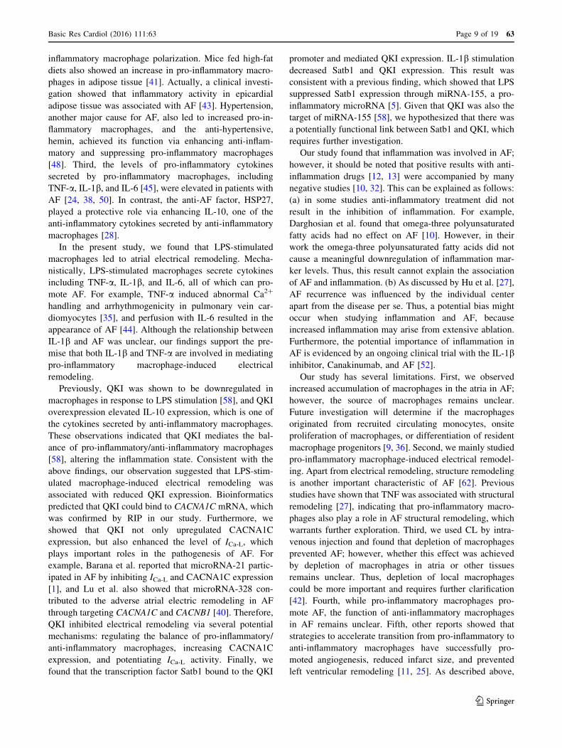
QKI and CACNA1C by QKI knockout in HEK293T cells.
Compared with the reference sequence from NCBI data-
bases, transfection with Cas9-QKI led to a frameshift
mutation (22 bp deletion, Fig. 7a, b) which further induced
the decrease of CACNA1C expression (Fig. 7c, d). Taken
together, we conclude that QKI-CACNA1C signaling is
involved in mediating LPS-stimulated macrophage-in-
duced electrical remodeling of atrial cardiomyocytes.
Satb1 mediated QKI expression
Next, we investigated the upstream regulation of QKI
expression by performing transcription factor screening. As
shown in Fig. 8a, relative light units (RLU) were signifi-
cantly decreased in the Satb1 probe ? nuclear pro-
tein ? QKI promoter group compared to the Satb1
probe ? nuclear protein group. This suggests that Satb1
binds to the QK1 promoter. Indeed, ChIP assay showed
that anti-Satb1antibody but not IgG precipitated a signifi-
cant amount of the targeted chromatin fragment of the QK1
promoter (Fig. 8b, c). Functionally, overexpression of
Satb1 increased QKI mRNA and protein expression in
cultured cardiomyocytes (Fig. 8d–f). We further confirmed
the role of Satb1 in macrophage–myocyte interaction by
IL-1b stimulation (30 pg/ml for 48 h) and found that IL-1bdecreased Satb1 and QKI expression (Fig. 8g, h). Taken
together, we argue that QK1 expression is mediated, in
part, by Satb1. Moreover, we measured the expression of
Satb1 and QKI in human samples and found that both were
decreased in patients with AF (Supplementary Fig. 2).
TNF-a was involved in electrical remodeling
induced by LPS-stimulated macrophages
Apart from IL-1b, LPS-stimulated macrophages can
secrete several other cytokines including TNF-a [45]. We
Basic Res Cardiol (2016) 111:63 Page 5 of 19 63
123
then investigated whether TNF-a was involved in elec-
trical remodeling. As shown in Supplementary Fig. 1A,
LPS stimulation induced TNF-a secretion. Since TNF-acan inhibit connexin 40 expression [39, 55], TNF-a may
be another candidate that is involved in macrophage-in-
duced electrical remodeling given that connexin 40
expression was downregulated in AF and connexin 40
transfer using adenovirus inhibited AF inducibility [29].
Consistent with the above observations, LPS stimulation
inhibited connexin 40 expression while TNFa antibody
partly abolished this inhibitory effect, indicating the
involvement of TNF-a in electrical remodeling induced
by LPS-stimulated macrophages (Supplementary
Fig. 1B, C).
Fig. 3 LPS-stimulated macrophages promoted atrial electrical
remodeling. a Chronic LPS injection into canines induced pro-
inflammatory macrophages. Immunostaining was performed on
sections of atrial myocardium prepared from control and LPS-treated
canine hearts using mac-2 antibody. Arrow indicates a mac-2-positive
macrophage. b The statistical result of a. c Representative results of
AF incidence in canines injected with PBS (control) or with LPS are
shown. d Representative results of AERP in canines injected with
PBS (control) or with LPS are shown. e, f Statistical analysis of AFincidence (c) and AERP (d). n = 7 and 6 for control and LPS-
stimulated groups, respectively. *p\ 0.05 vs. control group
63 Page 6 of 19 Basic Res Cardiol (2016) 111:63
123

Discussion
The main findings of the present study are: (1) increased
pro-inflammatory macrophages were found in the atria of
AF patients, and HL-1 cell tachypacing induced pro-in-
flammatory macrophage polarization; (2) pro-inflammatory
macrophage polarization played a major role in atrial
electrical remodeling as evidenced by the increased inci-
dence of AF in chronic LPS treatment in mice and canines,
which was inhibited by the depletion of LPS-stimulated
macrophages; (3) the effect of LPS-stimulated macro-
phages on electrical remodeling was mediated by IL-1bsecretion, which inhibited QKI expression in atrial myo-
cytes; (4) QKI bound to CACNA1C mRNA and regulated
the level of ICa-L; and (5) the transcription factor Satb1
mediated QKI expression. Our findings are summarized in
the working model shown in Fig. 9.
Although previous studies have reported increased
macrophage accumulation in AF, the macrophage pheno-
type and functions were unknown. Our study revealed that
the increased macrophages in AF patients were mainly pro-
inflammatory macrophages. This phenomenon can be
explained by a number of possibilities. First, since the
macrophages are pro-inflammatory [23], increased
inflammation in AF may be attributed, at least in part, to
increased pro-inflammatory macrophages. Second, several
systemic diseases, including obesity and hypertension, are
associated with low-grade inflammation and pro-
Fig. 4 Pro-inflammatory macrophage depletion ameliorated atrial
electrical remodeling in a mouse AF model. a Generation of chronic
LPS-stimulated animal model followed by depletion of pro-inflam-
matory macrophages by CL. b LPS-induced macrophages were
depleted by CL injection. (CD68, green; DAPI, blue). Arrows
indicated CD68-positive macrophages. c The statistical result of
b. d CL injection reduced AF incidence triggered by LPS. e LPS
inhibited L-type calcium currents while CL partly reversed its
inhibition. f The peak current at ?20 mV of each group. N = 15 for
control and LPS group, and n = 16 for LPS ? CL group. *p\ 0.05,
**p\ 0.01 vs. control group. #p\ 0.05, ##p\ 0.01 vs. LPS group
Basic Res Cardiol (2016) 111:63 Page 7 of 19 63
123

Fig. 5 Pro-inflammatory macrophages promoted electrical remodel-
ing through regulating QKI expression in atrial myocytes. a The
predicted binding sequence (red) of QKI on CACNA1C mRNA.
b LPS injection inhibited atrial QKI expression in a mouse model.
Immunostaining was performed on atrial myocardium sections using
a QKI antibody. Arrow indicates a QKI-positive atrial cardiomyocyte.
c The statistical result of b. d Co-culture with IL-1b knockout LPS-
stimulated macrophages reversed QKI expression inhibited by LPS-
stimulated macrophages. Western blot was used to measure QKI
expression in HL-1 cells that were co-cultured with wild type (WT) or
IL-1b knockout LPS-stimulated macrophages. GAPDH was used as a
loading control. e The statistical result of d. f HL-1 tachypacing
inhibited QKI expression. Western blot was used as in d to measure
QK1 expression in control or tachypaced HL-1 cells. g The statistical
result of f. h, i QKI overexpression increased CACNA1C mRNA and
protein expression. RT-qPCR (h) or western blot (i) was performed
on RNA or protein lysates obtained from cultured cardiomyocytes
with adenoviral-mediated expression of control or QKI. j The
statistical result of i. k QKI knockdown decreased CACNA1C
expression. l The statistical result of k. Cellular experiments were
repeated three times and the sample number of mice in each group
was 15. *p\ 0.05, **p\ 0.01 vs. control group
63 Page 8 of 19 Basic Res Cardiol (2016) 111:63
123
inflammatory macrophage polarization. Mice fed high-fat
diets also showed an increase in pro-inflammatory macro-
phages in adipose tissue [41]. Actually, a clinical investi-
gation showed that inflammatory activity in epicardial
adipose tissue was associated with AF [43]. Hypertension,
another major cause for AF, also led to increased pro-in-
flammatory macrophages, and the anti-hypertensive,
hemin, achieved its function via enhancing anti-inflam-
matory and suppressing pro-inflammatory macrophages
[48]. Third, the levels of pro-inflammatory cytokines
secreted by pro-inflammatory macrophages, including
TNF-a, IL-1b, and IL-6 [45], were elevated in patients withAF [24, 38, 50]. In contrast, the anti-AF factor, HSP27,
played a protective role via enhancing IL-10, one of the
anti-inflammatory cytokines secreted by anti-inflammatory
macrophages [28].
In the present study, we found that LPS-stimulated
macrophages led to atrial electrical remodeling. Mecha-
nistically, LPS-stimulated macrophages secrete cytokines
including TNF-a, IL-1b, and IL-6, all of which can pro-
mote AF. For example, TNF-a induced abnormal Ca2?
handling and arrhythmogenicity in pulmonary vein car-
diomyocytes [35], and perfusion with IL-6 resulted in the
appearance of AF [44]. Although the relationship between
IL-1b and AF was unclear, our findings support the pre-
mise that both IL-1b and TNF-a are involved in mediating
pro-inflammatory macrophage-induced electrical
remodeling.
Previously, QKI was shown to be downregulated in
macrophages in response to LPS stimulation [58], and QKI
overexpression elevated IL-10 expression, which is one of
the cytokines secreted by anti-inflammatory macrophages.
These observations indicated that QKI mediates the bal-
ance of pro-inflammatory/anti-inflammatory macrophages
[58], altering the inflammation state. Consistent with the
above findings, our observation suggested that LPS-stim-
ulated macrophage-induced electrical remodeling was
associated with reduced QKI expression. Bioinformatics
predicted that QKI could bind to CACNA1C mRNA, which
was confirmed by RIP in our study. Furthermore, we
showed that QKI not only upregulated CACNA1C
expression, but also enhanced the level of ICa-L, which
plays important roles in the pathogenesis of AF. For
example, Barana et al. reported that microRNA-21 partic-
ipated in AF by inhibiting ICa-L and CACNA1C expression
[1], and Lu et al. also showed that microRNA-328 con-
tributed to the adverse atrial electric remodeling in AF
through targeting CACNA1C and CACNB1 [40]. Therefore,
QKI inhibited electrical remodeling via several potential
mechanisms: regulating the balance of pro-inflammatory/
anti-inflammatory macrophages, increasing CACNA1C
expression, and potentiating ICa-L activity. Finally, we
found that the transcription factor Satb1 bound to the QKI
promoter and mediated QKI expression. IL-1b stimulation
decreased Satb1 and QKI expression. This result was
consistent with a previous finding, which showed that LPS
suppressed Satb1 expression through miRNA-155, a pro-
inflammatory microRNA [5]. Given that QKI was also the
target of miRNA-155 [58], we hypothesized that there was
a potentially functional link between Satb1 and QKI, which
requires further investigation.
Our study found that inflammation was involved in AF;
however, it should be noted that positive results with anti-
inflammation drugs [12, 13] were accompanied by many
negative studies [10, 32]. This can be explained as follows:
(a) in some studies anti-inflammatory treatment did not
result in the inhibition of inflammation. For example,
Darghosian et al. found that omega-three polyunsaturated
fatty acids had no effect on AF [10]. However, in their
work the omega-three polyunsaturated fatty acids did not
cause a meaningful downregulation of inflammation mar-
ker levels. Thus, this result cannot explain the association
of AF and inflammation. (b) As discussed by Hu et al. [27],
AF recurrence was influenced by the individual center
apart from the disease per se. Thus, a potential bias might
occur when studying inflammation and AF, because
increased inflammation may arise from extensive ablation.
Furthermore, the potential importance of inflammation in
AF is evidenced by an ongoing clinical trial with the IL-1binhibitor, Canakinumab, and AF [52].
Our study has several limitations. First, we observed
increased accumulation of macrophages in the atria in AF;
however, the source of macrophages remains unclear.
Future investigation will determine if the macrophages
originated from recruited circulating monocytes, onsite
proliferation of macrophages, or differentiation of resident
macrophage progenitors [9, 36]. Second, we mainly studied
pro-inflammatory macrophage-induced electrical remodel-
ing. Apart from electrical remodeling, structure remodeling
is another important characteristic of AF [62]. Previous
studies have shown that TNF was associated with structural
remodeling [27], indicating that pro-inflammatory macro-
phages also play a role in AF structural remodeling, which
warrants further exploration. Third, we used CL by intra-
venous injection and found that depletion of macrophages
prevented AF; however, whether this effect was achieved
by depletion of macrophages in atria or other tissues
remains unclear. Thus, depletion of local macrophages
could be more important and requires further clarification
[42]. Fourth, while pro-inflammatory macrophages pro-
mote AF, the function of anti-inflammatory macrophages
in AF remains unclear. Fifth, other reports showed that
strategies to accelerate transition from pro-inflammatory to
anti-inflammatory macrophages have successfully pro-
moted angiogenesis, reduced infarct size, and prevented
left ventricular remodeling [11, 25]. As described above,
Basic Res Cardiol (2016) 111:63 Page 9 of 19 63
123

63 Page 10 of 19 Basic Res Cardiol (2016) 111:63
123

QKI was downregulated in pro-inflammatory macrophages
while QKI overexpression increased IL-10 secretion [58],
indicating that QKI may serve as a switch molecule. Thus,
understanding the macrophage switch from pro-inflamma-
tory to anti-inflammatory in AF and whether QKI plays a
role in this phenotypic switch merits further investigation.
Furthermore, recent studies indicated that inhibition of
CD40 induced macrophages polarization from pro-inflam-
matory towards anti-inflammatory. Hence, CD40 could be
another target to explore the effects of different macro-
phage phenotypes on AF [30]. Finally, since the main
functions of QKI are regulating RNA splicing, transporta-
tion, stability, and protein translation, the exact mecha-
nisms involving QKI-mediated CACNA1C expression
need to be further investigated.
In conclusion, our study demonstrates that AF promotes
pro-inflammatory macrophage polarization, and that pro-
inflammatory macrophages further induce atrial electrical
remodeling through secreting IL-1b. Increased release of
IL-1b suppresses QKI expression in atrial myocytes,
leading to decreased L-type calcium currents. Our study
uncovers a novel molecular mechanism for AF and points
to QKI as a potential therapeutic target.
Materials and methods
Reagents
LPS, Claycomb Medium, fetal bovine serum (FBS), nore-
pinephrine, and L-glutamine were purchased from Sigma-
Aldrich (USA). Dulbecco’s modified Eagle medium
(DMEM) was from Thermo Fisher Scientific (USA). QKI
encoding vector and adenoviruses were purchased from
Vigene Biosciences (China). JetPRIME and JetPEI-Mac-
rophage transfection reagents were from Polyplus
Transfection (France). Antibodies against CD68, inducible
nitric oxide (iNOS), arginase-1 (Arg-1), QKI, and IL-1bwere obtained from Abcam (USA). Antibody against
CACNA1C was purchased from Santa Cruz (USA). RNA
immunoprecipitation kit was from Millipore (USA).
Chromatin immunoprecipitation (ChIP) kit was obtained
from CST (USA). Clodronate Liposomes (CL) was from
http://clodronateliposomes.com (The Netherlands). Pro-
moter-Binding TF Profiling Plate Array kit was purchased
from Signosis (USA).
Human samples
Right atrial appendages (RAA) were obtained as surgical
specimens from 11 patients with SR and 8 patients with
persistent AF undergoing cardiac valve replacement. The
detailed clinical characteristics of these patients are
described in Table 1. No patients involved in this study had
a history of myocardial infarction, febrile disorders, sys-
temic inflammatory diseases, malignancy or chronic renal
failure.
Experimental animals
C57BL/6 mice were obtained from the Shanghai Labora-
tory Animal Center of the Chinese Academy of Sciences
(China). All mice were 8 weeks old with an average weight
of 25–30 g. Beagle dogs were obtained from Huishan
Laboratory Animal Center (China) with an average weight
of 8–10 kg. All mice and dogs were housed in the animal
facility, which was maintained at 20–25 �C, 55 % relative
humidity, with an automatic 12 h light/dark cycle. All
animals received a standard laboratory diet and tap water
ad libitum, and were acclimated for 1 week before
experimentation.
Generation of animal model with chronic
inflammation
The chronic inflammation animal model was generated by
recurrent exposure to subclinical LPS as described [37].
Briefly, mice were injected intraperitoneally (i.p.) with
saline (control) or LPS (10 mg/kg) once a week for
2 months. To deplete macrophages, CL was injected
through the tail vein at a dose of 10 ll/g twice a week for
2 weeks. Dogs received a low dose of LPS (0.1 lg/kg in
0.9 % NaCl, i.p.) once a day for 2 weeks.
In vivo electrophysiology and programmed
stimulation
In mice, in vivo electrophysiology and programmed stim-
ulation experiments were conducted as previously
bFig. 6 QKI mediated CACNA1C expression in atrial cardiomy-
ocytes. a Primary cultured cardiomyocytes transfected with Cas9-
QKI-GFP plasmid (GFP positive white arrows) had lower CACNA1C
expression compared to GFP negative cells (yellow arrows). b The
statistical result of a. ***p\ 0.001 vs. GFP negative group. c QKI
bound to CACNA1C mRNA. This binding was attenuated by
tachypacing. RIP was performed. d The statistical result of
c. **p\ 0.01 control HL-1 cells anti-QKI vs. control HL-1 cells
anti-IgG; ##p\ 0.01 tachypaced HL-1 cells anti-QKI vs. control HL-
1 cells anti-QKI. e Adenoviral-mediated QKI overexpression in
primary cultured cardiomyocytes increased the L-type calcium
currents. f The peak current at 20 mV of each group. *p\ 0.05 vs.
control. g Atrial cells infected with adenovirus expressing flag-tagged
QKI exhibited higher CACNA1C expression in a mouse model. h The
statistical result of g. *p\ 0.05 vs. Flag negative group.White arrows
show cells transfected with plasmid or adenovirus, while yellow
arrows show cells that were not transfected with plasmid or
adenovirus. Cellular experiments were repeated three times and the
sample number of mice in each group was 10
Basic Res Cardiol (2016) 111:63 Page 11 of 19 63
123
described [16]. Briefly, mice were anesthetized under
isoflurane anesthesia (1.5 % vol/vol) and a subcutaneous
administration of 0.03 mg/kg buprenorphine hydrochlo-
ride. The following signs were monitored to establish the
adequacy of anesthesia: (1) no limb and palpebral with-
drawal reflexes; (2) stable respiration and heart rates. Then
the subdermal needle electrodes were placed in all four legs
to make a lead II conformation. A 1.1F electrophysiology
catheter containing eight electrodes (Scisense Inc., Canada)
was inserted through the jugular vein. The correct position
was confirmed by obtaining a sole ventricular signal in the
distal lead and a predominant atrial signal in the proximal
Fig. 7 QKI mediated CACNA1C expression in HEK293T cells. a,b Transfection with Cas9-QKI led to a frameshift mutation (22 bp
deletion). c QKI knockout induced the decrease of CACNA1C
expression in HEK293T cells. d The statistical result of c. Arrows
show a 22 bp deletion compared with reference sequences from NCBI
databases. Experiments were repeated three times, **p\ 0.01,
***p\ 0.001 vs. control group
63 Page 12 of 19 Basic Res Cardiol (2016) 111:63
123

Fig. 8 Satb1 mediated QKI
expression. a Transcription
factor binding screening assay
showed the binding of Satb1 to
the QKI promoter (arrow).
b ChIP assay confirmed the
binding of Satb1 to the QKI
promoter. c The statistical resultof b. d, e Satb1 overexpression
increased QKI mRNA and
protein expression. f Thestatistical result of e. g IL-1bstimulation (30 pg/ml, 48 h)
decreased Satb1 and QKI
expression. h The statistical
result of g. **p\ 0.01,
***p\ 0.001 vs. control.
Experiments were repeated
three times
Basic Res Cardiol (2016) 111:63 Page 13 of 19 63
123

lead. AF inducibility was determined using burst pacing in
the right atrium. In detail, three trains of 2 s burst pacing
were given as follows: the first 2 s burst was applied at a
cycle length of 40 ms with a pulse duration of 5 ms. After
3 min of stabilization, the second 2 s burst was set at a
cycle length of 20 ms with a pulse duration of 5 ms. After
another 3 min of stabilization, the final 2 s burst was given
at a cycle length of 20 ms with a pulse duration of 10 ms.
AF was defined as a rapid and irregular atrial rhythm with
irregular R–R intervals for at least 1 s on the surface
electrocardiogram (ECG). All ECG data were acquired
using a cardiac electrophysiology stimulator and multi-
channel electrophysiological recording system (Scisense
Inc., Canada). After measurement, the mice were imme-
diately sacrificed by cervical dislocation.
Canines were anesthetized with sodium pentobarbital
(initial bolus 30 mg/kg i.v., 50–100 mg as needed for
maintenance). The following signs were monitored to
establish the adequacy of anesthesia: (1) no limb and
palpebral withdrawal reflexes; (2) stable respiration and
heart rates. Atrial effective refractory period (AERP) was
determined by programmed stimulation at RAA, which
consisted of eight consecutive stimuli (S1S1 = 250 ms)
followed by a premature stimulus (S1S2). The S1S2
intervals were decreased from 200 ms to refractoriness
initially by a 2 ms decrement. The atrial ERP was defined
as the longest S1–S2 interval that failed to induce atrial
depolarization. AF inducibility was also measured by burst
pacing. Two trains of 120 s burst pacing were given as
follows: first, a 120 s burst was applied at a cycle length of
100 ms. After 5 min of stabilization, the second 120 s burst
pacing was given at a cycle length of 50 ms. AF was
defined as irregular atrial rates faster than 500 beats/min
associated with irregular atrio-ventricular conduction last-
ing longer than 5 s. After measurement, canines were
euthanized by removal of the heart.
Cell culture
Murine atrial myocytes, HL-1, were provided by Dr. Chen
and Dr. Wang’s lab (Nanjing medical university, China)
with the permission of Dr. Claycomb (LSU Health Sci-
ences Center, USA). The cells were cultured with Clay-
comb Medium supplemented with 10 % FBS, 100 U/ml
penicillin/streptomycin, 0.1 mM norepinephrine, and
2 mM L-glutamine. HL-1 cell tachypacing was performed
using a cell pacing system (Ionoptix, USA). HL-1 cells
were subjected to rapid stimulation for 24 h at 5 Hz (18 V,
4 ms) [56].
Raw264.7 macrophages and HEK293T cells were pur-
chased from American Type Culture Collection (ATCC,
USA) and cultured with DMEM supplemented with 10 %
FBS. To develop pro-inflammatory macrophages, 100 ng/
ml LPS was added for 16 h as previously described [54].
Primary neonatal rat cardiomyocytes were isolated from
1-day-old rats as described [57]. Briefly, hearts were
minced and digested with trypsin and collagenase, and the
isolated cells were pre-plated twice for 60 min to eliminate
fibroblasts. The non-adherent myocytes were then plated in
plating medium containing 199 medium supplemented
with HEPES, MEM non-essential amino acids, glucose,
glutamine, 10 % FBS, vitamin B12, penicillin, and strep-
tomycin on fibronectin-coated plates.
Macrophages and atrial myocytes were co-cultured in
exchanging medium. In brief, RAW264.7 cells were trea-
ted with 100 ng/ml LPS for 16 h, then washed with PBS,
and cultured in fresh medium for another 24 h. Thereafter,
the medium was collected and used for culturing HL-1
cells. Similarly, HL-1 cells were tachypaced for 24 h.
Thereafter, the medium was collected and used for cul-
turing RAW264.7 cells.
Transient transfection and in vivo gene transfer
DNA transfection was performed using jetPRIME or jet-
PEI-Macrophage for cardiomyocytes or macrophages,
respectively. For cardiomyocytes, 2 lg DNA was added
into 200 ll jetPRIME buffer, followed by addition of 4 lljetPRIME. After a 10 min incubation, the transfection mix
Fig. 9 Working model of cellular signaling underlying the functional
interaction between atrial cells and macrophages in AF. AF promotes
pro-inflammatory macrophage polarization by secreting cytokines.
Pro-inflammatory macrophages induce atrial electrical remodeling by
secreting IL-1b, which subsequently inhibits QKI expression in atrial
myocytes and decreases QKI-CACNA1C mRNA binding, conse-
quently suppressing L-type calcium currents, which promotes atrial
electrical remodeling and exacerbates AF
63 Page 14 of 19 Basic Res Cardiol (2016) 111:63
123
was added into each well. For macrophages, 6 lg DNA
was added into 50 ll of 150 mM NaCl, followed by
addition of 50 ll jetPEI-Macrophage solution. After a
30 min incubation, the transfection mix was added into
each well.
In vivo gene transfer was conducted by injecting aden-
oviruses at a dose of 109 plaque forming units (pfu) in
50 ll PBS through the tail vein [31].
Genome editing with CRISPR/Cas9 system
Genome engineering was performed using CRISPR/Cas9
systems according to published protocols [51]. Briefly,
guide RNA (sgRNA) was designed and inserted into
pSpCas9 (BB)-2AGFP or pSpCas9 (BB)-2A-Puro vector.
The sgRNA sequences targeting QKI were: sgRNA top,
CACCGGATCTTCAACCACCTCGAG; sgRNA bottom,
AAACCTCGAGGTGGTTGAAGATCC. The sgRNA
sequences targeting IL-1b were: sgRNA top, CACCGAG
CACCTAAGTCCCTAGGTT; sgRNA bottom, AAACAA
CCTAGGGACTTAGGTGCTC. The plasmids were
amplified in E. coli strain, purified, sequenced, and used for
transfection into macrophages, HEK293T cells or car-
diomyocytes as described above. For macrophages and
HEK293T cells, cells transfected with Cas9-IL1b-puro or
Cas9-QKI-puro vector were further treated with puromycin
(10 lg/ml) for 72 h. Surviving cells were used for mono-
clonal formation. QKI and IL-1b knockout were deter-
mined by sequencing and Western blot. Since single HL-1
cell was not able to form monoclones, we used a mixture
pool for QKI knockdown. In detail, HL-1 cells transfected
with Cas9-QKI-puro vector were treated with puromycin
and surviving cells were used for analysis as a mixture
pool. QKI knockdown was measured by Western blot.
Since primary cultured cardiomyocytes have no prolifera-
tive capability, cells were transfected with Cas9-QKI-GFP
vector, and the GFP-positive and GFP-negative cells were
used for analysis.
Immunohistochemistry and immunofluorescence
Immunohistochemistry and immunofluorescence were
performed as described [57, 63]. The primary antibodies
used were: CD68 (1:100, Abcam), iNOS (1:50, Abcam),
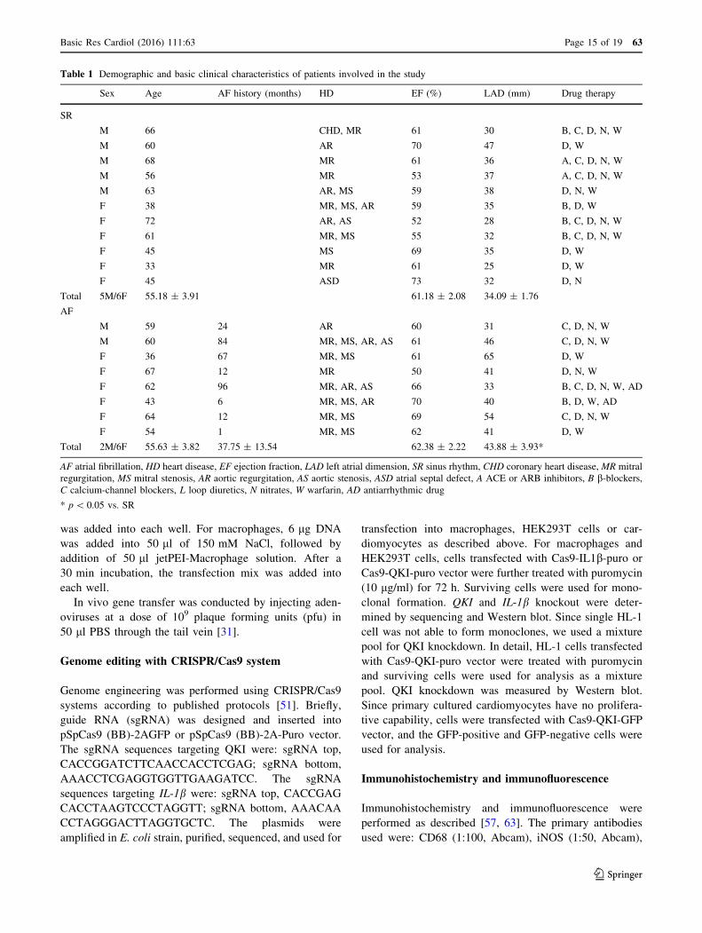
Table 1 Demographic and basic clinical characteristics of patients involved in the study
Sex Age AF history (months) HD EF (%) LAD (mm) Drug therapy
SR
M 66 CHD, MR 61 30 B, C, D, N, W
M 60 AR 70 47 D, W
M 68 MR 61 36 A, C, D, N, W
M 56 MR 53 37 A, C, D, N, W
M 63 AR, MS 59 38 D, N, W
F 38 MR, MS, AR 59 35 B, D, W
F 72 AR, AS 52 28 B, C, D, N, W
F 61 MR, MS 55 32 B, C, D, N, W
F 45 MS 69 35 D, W
F 33 MR 61 25 D, W
F 45 ASD 73 32 D, N
Total 5M/6F 55.18 ± 3.91 61.18 ± 2.08 34.09 ± 1.76
AF
M 59 24 AR 60 31 C, D, N, W
M 60 84 MR, MS, AR, AS 61 46 C, D, N, W
F 36 67 MR, MS 61 65 D, W
F 67 12 MR 50 41 D, N, W
F 62 96 MR, AR, AS 66 33 B, C, D, N, W, AD
F 43 6 MR, MS, AR 70 40 B, D, W, AD
F 64 12 MR, MS 69 54 C, D, N, W
F 54 1 MR, MS 62 41 D, W
Total 2M/6F 55.63 ± 3.82 37.75 ± 13.54 62.38 ± 2.22 43.88 ± 3.93*
AF atrial fibrillation, HD heart disease, EF ejection fraction, LAD left atrial dimension, SR sinus rhythm, CHD coronary heart disease, MR mitral
regurgitation, MS mitral stenosis, AR aortic regurgitation, AS aortic stenosis, ASD atrial septal defect, A ACE or ARB inhibitors, B b-blockers,C calcium-channel blockers, L loop diuretics, N nitrates, W warfarin, AD antiarrhythmic drug
* p\ 0.05 vs. SR
Basic Res Cardiol (2016) 111:63 Page 15 of 19 63
123

Arg-1 (1:100, Abcam), QKI (1:200, Abcam), and CAC-
NA1C (1:50, Santa Cruz).
RNA immunoprecipitation
RNA immunoprecipitation was performed according to the
manufacturer’s protocol. Briefly, 2 9 107 control or
tachypaced HL-1 cells were collected and lysed, and the
lysates were incubated with magnetic bead-QKI or IgG
antibody with rotation at 4 �C overnight. Samples were
then digested by proteinase K and RNA was purified using
phenol–chloroform–isoamyl alcohol. RT-PCR was per-
formed on purified RNA. The primers used for identifying
QKI binding sequence were as follows: forward-
50GCTCATTGCCTTCAAACC30; reverse-50GATGGAGATGCGGGAGTT30.
Real-time PCR
Real-time PCR was conducted as described previously
[57]. The primers used were as follows: QKI: forward-
50AGGCAAAGGCTCAATGAGGG30, reverse-50CCTGGGCAGTTGGTGATTT30; CACNA1C: forward-50TCCCGAGCACATCCCTACTC30, reverse-50ACTGACGGTAGAGATGGTTGC30; b-actin: forward-50GGCTGTATTCCCCTCCATCG30, reverse-50CCAGTTGGTAACAATGCCATGT30.
ChIP
ChIP was performed according to the manufacturer’s
instructions. Briefly, HL-1 cells were fixed with 1 %
formaldehyde for 10 min at 37 �C. Chromatin was digested
with 5 ll nuclease at 37 �C for 20 min, yielding DNA
fragments of 200–800 bps. After preclearing with protein
A/G agarose at 4 �C for 1 h, samples were incubated with
10 lg of Satb1 antibody or IgG (as a control) with rotation
at 4 �C overnight. The reverse crosslink was reversed by
incubation of the sample in a 5 M NaCl and proteinase K
solution at 65 �C for 2 h. The precipitated DNA was
purified using spin columns, and the purified DNA was
subjected to PCR analysis. The primers used for PCR to
identify the Satb1 binding sequence were as follows: for-
ward-50CCCAGTGAAGCAACAGGA30; reverse-50GGAATCCCGGCGGGACTC30.
Whole-cell patch-clamp
Whole-cell patch-clamp was performed using a HEKA
EPC10 amplifier (HEKA, Germany). The bath solution
contained 140 mM TEA-Cl, 2 mM MgCl2, 10 mM CaCl2,
10 mM HEPES, and 5 mM glucose at pH 7.4 (TEA-OH).
The internal solution contained 120 mM CsCl 120, 1 mM
MgCl2,10 mM HEPES, 4 mM Mg-ATP,10 mM EGTA,
and 0.3 mM Na2-GTP at pH 7.2 (CsOH). The pipettes
were created from capillary tubing (Sutter Instruments,
USA) and had resistances of 4–6 MX under these solution
conditions. All recordings were carried out at room tem-
perature. Cav IV-curve was generated using the following
protocol: a single cell was clamped at a holding potential of
-60 mV and Cav current was measured using a stimulus
voltage pattern consisting of a 500 ms test pulse from -60
to 60 mV, separated by a 1 s test interval at the holding
potential -60 mV. For steady-state channel inactivation,
the cell was clamped at a holding potential of -80 mV and
stepped to voltage between -60 and 80 mV for 2000 ms to
inactivate the Cav current. The cell was then clamped to
10 mV for 250 ms to elicit the Cav current.
Promoter-binding transcription factor profiling
plate array
Promoter-binding transcription factor profiling plate array
was performed according to the manufacturer’s instruction.
In brief, biotin-labeled transcription factor probes were
mixed with nuclear extract with or without the QKI pro-
moter. The transcription factor-DNA complex was sepa-
rated from free probes using an isolation column, and the
bound probes were eluted using elution buffer.
Hybridization of eluted probes was performed with
hybridization plate. Finally, the bound probe was detected
using a streptavidin-HRP conjugate. The QKI promoter
sequence used in this assay is shown in the Supplementary
materials.
Western blot
Western blot was performed as previously described [57].
The primary antibodies used were: iNOS (1:1000, Abcam),
Arg-1 (1:1000, Abcam), QKI (1:1000, Abcam), CAC-
NA1C (1:200, Santa Cruz), and IL-1b (1:1000, Abcam).
Statistical analysis
Data are expressed as mean ± SD. An unpaired Student’s
t test was used for statistical comparison between two
groups after the demonstration of homogeneity of variance
with an F test, and one-way ANOVA was used for com-
parison of more than two groups. Fisher exact test was used
for evaluating the incidence of AF. A p value less than 0.05
was considered statistically significant.
Acknowledgments We thank Dr. Chen, Dr. Wang and Dr. Claycomb
for providing HL-1 cells. This work was supported by the National
Natural Science Foundation of China (Nos. 81170167 and 81270002)
and the Natural Science Foundation of Zhejiang Province (No.
LZ16H020001).
63 Page 16 of 19 Basic Res Cardiol (2016) 111:63
123

Compliance with ethical standards
Ethical statement All animal studies were approved by the Animal
Care and Use Committee of Zhejiang University and conformed to
the Guide for the Care and Use of Laboratory Animals published by
the US National Institutes of Health (NIH Publication No. 85-23,
revised 1996).
Informed consent was obtained from all participants in accordance
with the guidelines of the Human Subjects Committee of the Medical
Ethical Commission of the First Affiliated Hospital of Zhejiang
University (China) and the declaration of Helsinki.
Conflict of interest The authors declared that they have no potential
conflicts of interest.
Open Access This article is distributed under the terms of the Creative
Commons Attribution 4.0 International License (http://creative
commons.org/licenses/by/4.0/), which permits unrestricted use, dis-
tribution, and reproduction in any medium, provided you give
appropriate credit to the original author(s) and the source, provide a link
to the Creative Commons license, and indicate if changes were made.
References
1. Barana A, Matamoros M, Dolz-Gaiton P, Perez-Hernandez M,
Amoros I, Nunez M, Sacristan S, Pedraz A, Pinto A, Fernandez-
Aviles F, Tamargo J, Delpon E, Caballero R (2014) Chronic atrial
fibrillation increases microRNA-21 in human atrial myocytes
decreasing L-type calcium current. Circ Arrhythm Electrophysiol
7:861–868. doi:10.1161/circep.114.001709
2. Ben-Mordechai T, Holbova R, Landa-Rouben N, Harel-Adar T,
Feinberg MS, Abd EI, Blum G, Epstein FH, Silman Z, Cohen S,
Leor J (2013) Macrophage subpopulations are essential for infarct
repair with and without stem cell therapy. J Am Coll Cardiol
62:1890–1901. doi:10.1016/j.jacc.2013.07.057
3. Boos CJ, Anderson RA, Lip GY (2006) Is atrial fibrillation an
inflammatory disorder? Eur Heart J 27:136–149. doi:10.1093/
eurheartj/ehi645
4. Camm AJ, Lip GY, De Caterina R, Savelieva I, Atar D, Hohn-
loser SH, Hindricks G, Kirchhof P (2012) 2012 focused update of
the ESC Guidelines for the management of atrial fibrillation: an
update of the 2010 ESC Guidelines for the management of atrial
fibrillation-developed with the special contribution of the Euro-
pean Heart Rhythm Association. EUROPACE 14:1385–1413.
doi:10.1093/europace/eus305
5. Chen S, Smith BA, Iype J, Prestipino A, Pfeifer D, Grundmann S,
Schmitt-Graeff A, Idzko M, Beck Y, Prinz G, Finke J, Duyster J,
Zeiser R (2015) MicroRNA-155-deficient dendritic cells cause
less severe GVHD through reduced migration and defective
inflammasome activation. Blood 126:103–112. doi:10.1182/
blood-2014-12-617258
6. Chen XQ, Zhang DL, Zhang MJ, Guo M, Zhan YY, Liu F, Jiang
WF, Zhou L, Zhao L, Wang QX, Liu X (2015) TRIF promotes
angiotensin II-induced cross-talk between fibroblasts and mac-
rophages in atrial fibrosis. Biochem Biophys Res Commun
464:100–105. doi:10.1016/j.bbrc.2015.05.131
7. Choi EK, Chang PC, Lee YS, Lin SF, Zhu W, Maruyama M,
Fishbein MC, Chen Z, Rubart-von DLM, Field LJ, Chen PS
(2012) Triggered firing and atrial fibrillation in transgenic mice
with selective atrial fibrosis induced by overexpression of TGF-
beta1. Circ J 76:1354–1362. doi:10.1253/circj.cj-11-1301
8. Chugh SS, Havmoeller R, Narayanan K, Singh D, Rienstra M,
Benjamin EJ, Gillum RF, Kim YH, McAnulty JJ, Zheng ZJ,
Forouzanfar MH, Naghavi M, Mensah GA, Ezzati M, Murray CJ
(2014) Worldwide epidemiology of atrial fibrillation: a Global
Burden of Disease 2010 Study. Circulation 129:837–847. doi:10.
1161/circulationaha.113.005119
9. Cochain C, Zernecke A (2015) Macrophages and immune cells in
atherosclerosis: recent advances and novel concepts. Basic Res
Cardiol 110:34. doi:10.1007/s00395-015-0491-8
10. Darghosian L, Free M, Li J, Gebretsadik T, Bian A, Shintani A,
McBride BF, Solus J, Milne G, Crossley GH, Thompson D,
Vidaillet H, Okafor H, Darbar D, Murray KT, Stein CM (2015)
Effect of omega-three polyunsaturated fatty acids on inflamma-
tion, oxidative stress, and recurrence of atrial fibrillation. Am J
Cardiol 115:196–201. doi:10.1016/j.amjcard.2014.10.022
11. Dayan V, Yannarelli G, Billia F, Filomeno P, Wang XH, Davies
JE, Keating A (2011) Mesenchymal stromal cells mediate a
switch to alternatively activated monocytes/macrophages after
acute myocardial infarction. Basic Res Cardiol 106:1299–1310.
doi:10.1007/s00395-011-0221-9
12. Deftereos S, Giannopoulos G, Efremidis M, Kossyvakis C, Kat-
sivas A, Panagopoulou V, Papadimitriou C, Karageorgiou S,
Doudoumis K, Raisakis K, Kaoukis A, Alexopoulos D, Manolis
AS, Stefanadis C, Cleman MW (2014) Colchicine for prevention
of atrial fibrillation recurrence after pulmonary vein isolation:
mid-term efficacy and effect on quality of life. Heart Rhythm
11:620–628. doi:10.1016/j.hrthm.2014.02.002
13. Deftereos S, Giannopoulos G, Kossyvakis C, Efremidis M,
Panagopoulou V, Kaoukis A, Raisakis K, Bouras G, Angelidis C,
Theodorakis A, Driva M, Doudoumis K, Pyrgakis V, Stefanadis
C (2012) Colchicine for prevention of early atrial fibrillation
recurrence after pulmonary vein isolation: a randomized con-
trolled study. J Am Coll Cardiol 60:1790–1796. doi:10.1016/j.
jacc.2012.07.031
14. Dernellis J, Panaretou M (2004) Relationship between C-reactive
protein concentrations during glucocorticoid therapy and recur-
rent atrial fibrillation. Eur Heart J 25:1100–1107. doi:10.1016/j.
ehj.2004.04.025
15. Ebersole TA, Chen Q, Justice MJ, Artzt K (1996) The quaking
gene product necessary in embryogenesis and myelination com-
bines features of RNA binding and signal transduction proteins.
Nat Genet 12:260–265. doi:10.1038/ng0396-260
16. Egom EE, Vella K, Hua R, Jansen HJ, Moghtadaei M, Polina I,
Bogachev O, Hurnik R, Mackasey M, Rafferty S, Ray G, Rose
RA (2015) Impaired sinoatrial node function and increased sus-
ceptibility to atrial fibrillation in mice lacking natriuretic peptide
receptor C. J Physiol 593:1127–1146. doi:10.1113/jphysiol.2014.
283135
17. Frangogiannis NG (2014) The inflammatory response in
myocardial injury, repair, and remodelling. Nat Rev Cardiol
11:255–265. doi:10.1038/nrcardio.2014.28
18. Friedrichs K, Adam M, Remane L, Mollenhauer M, Rudolph V,
Rudolph TK, Andrie RP, Stockigt F, Schrickel JW, Ravekes T,
Deuschl F, Nickenig G, Willems S, Baldus S, Klinke A (2014)
Induction of atrial fibrillation by neutrophils critically depends on
CD11b/CD18 integrins. PLoS One 9:e89307. doi:10.1371/jour
nal.pone.0089307
19. Frustaci A, Chimenti C, Bellocci F, Morgante E, Russo MA,
Maseri A (1997) Histological substrate of atrial biopsies in
patients with lone atrial fibrillation. Circulation 96:1180–1184.
doi:10.1161/01.cir.96.4.1180
20. Fu H, Yang G, Wei M, Liu L, Jin L, Lu X, Wang L, Shen L,
Zhang J, Lu H, Yao L, Lu Z (2012) The RNA-binding protein
QKI5 is a direct target of C/EBPalpha and delays macrophage
Basic Res Cardiol (2016) 111:63 Page 17 of 19 63
123

differentiation. Mol Biol Cell 23:1628–1635. doi:10.1091/mbc.
e11-05-0412
21. Galarneau A, Richard S (2005) Target RNA motif and target
mRNAs of the Quaking STAR protein. Nat Struct Mol Biol
12:691–698. doi:10.1038/nsmb963
22. Goette A, Bukowska A, Lendeckel U, Erxleben M, Hamm-
wohner M, Strugala D, Pfeiffenberger J, Rohl FW, Huth C,
Ebert MP, Klein HU, Rocken C (2008) Angiotensin II receptor
blockade reduces tachycardia-induced atrial adhesion molecule
expression. Circulation 117:732–742. doi:10.1161/circulatio
naha.107.730101
23. Gordon S, Martinez FO (2010) Alternative activation of macro-
phages: mechanism and functions. Immunity 32:593–604. doi:10.
1016/j.immuni.2010.05.007
24. Guo Y, Lip GY, Apostolakis S (2012) Inflammation in atrial
fibrillation. J Am Coll Cardiol 60:2263–2270. doi:10.1016/j.jacc.
2012.04.063
25. Harel-Adar T, Ben MT, Amsalem Y, Feinberg MS, Leor J, Cohen
S (2011) Modulation of cardiac macrophages by phos-
phatidylserine-presenting liposomes improves infarct repair. Proc
Natl Acad Sci USA 108:1827–1832. doi:10.1073/pnas.
1015623108
26. Ho KM, Tan JA (2009) Benefits and risks of corticosteroid pro-
phylaxis in adult cardiac surgery: a dose-response meta-analysis.
Circulation 119:1853–1866. doi:10.1161/circulationaha.108.
848218
27. Hu YF, Chen YJ, Lin YJ, Chen SA (2015) Inflammation and the
pathogenesis of atrial fibrillation. Nat Rev Cardiol 12:230–243.
doi:10.1038/nrcardio.2015.2
28. Hu YF, Yeh HI, Tsao HM, Tai CT, Lin YJ, Chang SL, Lo LW,
Tuan TC, Suenari K, Li CH, Chao TF, Chen SA (2012) Elec-
trophysiological correlation and prognostic impact of heat shock
protein 27 in atrial fibrillation. Circ Arrhythm Electrophysiol
5:334–340. doi:10.1161/circep.111.965996
29. Igarashi T, Finet JE, Takeuchi A, Fujino Y, Strom M, Greener ID,
Rosenbaum DS, Donahue JK (2012) Connexin gene transfer
preserves conduction velocity and prevents atrial fibrillation.
Circulation 125:216–225. doi:10.1161/CIRCULATIONAHA.
111.053272
30. Jansen MF, Hollander MR, van Royen N, Horrevoets AJ, Lutgens
E (2016) CD40 in coronary artery disease: a matter of macro-
phages? Basic Res Cardiol 111:38. doi:10.1007/s00395-016-
0554-5
31. Juan SH, Lee TS, Tseng KW, Liou JY, Shyue SK, Wu KK, Chau
LY (2001) Adenovirus-mediated heme oxygenase-1 gene transfer
inhibits the development of atherosclerosis in apolipoprotein
E-deficient mice. Circulation 104:1519–1525. doi:10.1161/
hc3801.095663
32. Kazemi B, Akbarzadeh F, Safaei N, Yaghoubi A, Shadvar K,
Ghasemi K (2013) Prophylactic high-dose oral-N-acetylcysteine
does not prevent atrial fibrillation after heart surgery: a
prospective double blind placebo-controlled randomized clinical
trial. Pacing Clin Electrophysiol 36:1211–1219. doi:10.1111/
pace.12190
33. Kleinbongard P, Schulz R, Heusch G (2011) TNFalpha in
myocardial ischemia/reperfusion, remodeling and heart failure.
Heart Fail Rev 16:49–69. doi:10.1007/s10741-010-9180-8
34. Lauriat TL, Shiue L, Haroutunian V, Verbitsky M, Ares MJ,
Ospina L, McInnes LA (2008) Developmental expression profile
of quaking, a candidate gene for schizophrenia, and its target
genes in human prefrontal cortex and hippocampus shows
regional specificity. J Neurosci Res 86:785–796. doi:10.1002/jnr.
21534
35. Lee SH, Chen YC, Chen YJ, Chang SL, Tai CT, Wongcharoen
W, Yeh HI, Lin CI, Chen SA (2007) Tumor necrosis factor-alpha
alters calcium handling and increases arrhythmogenesis of
pulmonary vein cardiomyocytes. Life Sci 80:1806–1815. doi:10.
1016/j.lfs.2007.02.029
36. Leinonen JV, Korkus-Emanuelov A, Wolf Y, Milgrom-Hoffman
M, Lichtstein D, Hoss S, Lotan C, Tzahor E, Jung S, Beeri R
(2016) Macrophage precursor cells from the left atrial appendage
of the heart spontaneously reprogram into a C-kit?/CD45- stem
cell-like phenotype. Int J Cardiol 209:296–306. doi:10.1016/j.
ijcard.2016.02.040
37. Lew WY, Bayna E, Molle ED, Dalton ND, Lai NC, Bhargava V,
Mendiola V, Clopton P, Tang T (2013) Recurrent exposure to
subclinical lipopolysaccharide increases mortality and induces
cardiac fibrosis in mice. PLoS One 8:e61057. doi:10.1371/jour
nal.pone.0061057
38. Li J, Solus J, Chen Q, Rho YH, Milne G, Stein CM, Darbar D
(2010) Role of inflammation and oxidative stress in atrial fibril-
lation. Heart Rhythm 7:438–444. doi:10.1016/j.hrthm.2009.12.
009
39. Liew R, Khairunnisa K, Gu Y, Tee N, Yin NO, Naylynn TM,
Moe KT (2013) Role of tumor necrosis factor-alpha in the
pathogenesis of atrial fibrosis and development of an arrhyth-
mogenic substrate. Circ J 77:1171–1179. doi:10.1253/circj.CJ-
12-1155
40. Lu Y, Zhang Y, Wang N, Pan Z, Gao X, Zhang F, Zhang Y, Shan
H, Luo X, Bai Y, Sun L, Song W, Xu C, Wang Z, Yang B (2010)
MicroRNA-328 contributes to adverse electrical remodeling in
atrial fibrillation. Circulation 122:2378–2387. doi:10.1161/circu
lationaha.110.958967
41. Lumeng CN, Bodzin JL, Saltiel AR (2007) Obesity induces a
phenotypic switch in adipose tissue macrophage polarization.
J Clin Invest 117:175–184. doi:10.1172/jci29881
42. Marchini T, Wolf D, Michel NA, Mauler M, Dufner B, Hoppe N,
Beckert J, Jackel M, Magnani N, Duerschmied D, Tasat D,
Alvarez S, Reinohl J, von Zur MC, Idzko M, Bode C, Hilgendorf
I, Evelson P, Zirlik A (2016) Acute exposure to air pollution
particulate matter aggravates experimental myocardial infarction
in mice by potentiating cytokine secretion from lung macro-
phages. Basic Res Cardiol 111:44. doi:10.1007/s00395-016-
0562-5
43. Mazurek T, Kiliszek M, Kobylecka M, Skubisz-Gluchowska J,
Kochman J, Filipiak K, Krolicki L, Opolski G (2014) Relation of
proinflammatory activity of epicardial adipose tissue to the
occurrence of atrial fibrillation. Am J Cardiol 113:1505–1508.
doi:10.1016/j.amjcard.2014.02.005
44. Mitrokhin VM, Mladenov MI, Kamkin AG (2015) Effects of
interleukin-6 on the bio-electric activity of rat atrial tissue under
normal conditions and during gradual stretching. Immunobiology
220:1107–1112. doi:10.1016/j.imbio.2015.05.003
45. Murray PJ, Allen JE, Biswas SK, Fisher EA, Gilroy DW, Goerdt
S, Gordon S, Hamilton JA, Ivashkiv LB, Lawrence T, Locati M,
Mantovani A, Martinez FO, Mege JL, Mosser DM, Natoli G,
Saeij JP, Schultze JL, Shirey KA, Sica A, Suttles J, Udalova I,
van Ginderachter JA, Vogel SN, Wynn TA (2014) Macrophage
activation and polarization: nomenclature and experimental
guidelines. Immunity 41:14–20. doi:10.1016/j.immuni.2014.06.
008
46. Naccarelli GV, Varker H, Lin J, Schulman KL (2009) Increasing
prevalence of atrial fibrillation and flutter in the US. Am J Cardiol
104:1534–1539. doi:10.1016/j.amjcard.2009.07.022
47. Nakatani Y, Nishida K, Sakabe M, Kataoka N, Sakamoto T,
Yamaguchi Y, Iwamoto J, Mizumaki K, Fujiki A, Inoue H (2013)
Tranilast prevents atrial remodeling and development of atrial
fibrillation in a canine model of atrial tachycardia and left ven-
tricular dysfunction. J Am Coll Cardiol 61:582–588. doi:10.1016/
j.jacc.2012.11.014
48. Ndisang JF, Mishra M (2013) The heme oxygenase system
selectively suppresses the proinflammatory macrophage m1
63 Page 18 of 19 Basic Res Cardiol (2016) 111:63
123

phenotype and potentiates insulin signaling in spontaneously
hypertensive rats. Am J Hypertens 26:1123–1131. doi:10.1093/
ajh/hpt082
49. Oishi S, Sasano T, Tateishi Y, Tamura N, Isobe M, Furukawa T
(2012) Stretch of atrial myocytes stimulates recruitment of
macrophages via ATP released through gap-junction channels.
J Pharmacol Sci 120:296–304. doi:10.1254/jphs.12202fp
50. Patel P, Dokainish H, Tsai P, Lakkis N (2010) Update on the
association of inflammation and atrial fibrillation. J Cardiovasc
Electrophysiol 21:1064–1070. doi:10.1111/j.1540-8167.2010.
01774.x
51. Ran FA, Hsu PD, Wright J, Agarwala V, Scott DA, Zhang F
(2013) Genome engineering using the CRISPR-Cas9 system. Nat
Protoc 8:2281–2308. doi:10.1038/nprot.2013.143
52. Ridker PM, Thuren T, Zalewski A, Libby P (2011) Interleukin-
1beta inhibition and the prevention of recurrent cardiovascular
events: rationale and design of the Canakinumab Anti-inflam-
matory Thrombosis Outcomes Study (CANTOS). Am Heart J
162:597–605. doi:10.1016/j.ahj.2011.06.012
53. Saba S, Janczewski AM, Baker LC, Shusterman V, Gursoy EC,
Feldman AM, Salama G, McTiernan CF, London B (2005) Atrial
contractile dysfunction, fibrosis, and arrhythmias in a mouse
model of cardiomyopathy secondary to cardiac-specific overex-
pression of tumor necrosis factor-{alpha}. Am J Physiol Heart
Circ Physiol 289:H1456–H1467. doi:10.1152/ajpheart.00733.
2004
54. Saha B, Bala S, Hosseini N, Kodys K, Szabo G (2015) Kruppel-
like factor 4 is a transcriptional regulator of M1/M2 macrophage
polarization in alcoholic liver disease. J Leukoc Biol. doi:10.
1189/jlb.4a1014-485r
55. Sawaya SE, Rajawat YS, Rami TG, Szalai G, Price RL, Siva-
subramanian N, Mann DL, Khoury DS (2007) Downregulation of
connexin40 and increased prevalence of atrial arrhythmias in
transgenic mice with cardiac-restricted overexpression of tumor
necrosis factor. Am J Physiol Heart Circ Physiol 292:H1561–
H1567. doi:10.1152/ajpheart.00285.2006
56. Sidorova TN, Yermalitskaya LV, Mace LC, Wells KS, Boutaud
O, Prinsen JK, Davies SS, Roberts LN, Dikalov SI, Glabe CG,
Amarnath V, Barnett JV, Murray KT (2015) Reactive gamma-
ketoaldehydes promote protein misfolding and preamyloid oli-
gomer formation in rapidly-activated atrial cells. J Mol Cell
Cardiol 79:295–302. doi:10.1016/j.yjmcc.2014.11.013
57. Sun Z, Han J, Zhao W, Zhang Y, Wang S, Ye L, Liu T, Zheng L
(2014) TRPV1 activation exacerbates hypoxia/reoxygenation-
induced apoptosis in H9C2 cells via calcium overload and
mitochondrial dysfunction. Int J Mol Sci 15:18362–18380.
doi:10.3390/ijms151018362
58. Tili E, Chiabai M, Palmieri D, Brown M, Cui R, Fernandes C,
Richmond T, Kim T, Sheetz T, Sun HL, Lagana A, Veneziano D,
Volinia S, Rassenti L, Kipps T, Awad H, Michaille JJ, Croce CM
(2015) Quaking and miR-155 interactions in inflammation and
leukemogenesis. Oncotarget 6:24599–24610. doi:10.18632/onco
target.5248
59. Verheule S, Sato T, Everett TT, Engle SK, Otten D, Rubart-von
DLM, Nakajima HO, Nakajima H, Field LJ, Olgin JE (2004)
Increased vulnerability to atrial fibrillation in transgenic mice
with selective atrial fibrosis caused by overexpression of TGF-
beta1. Circ Res 94:1458–1465. doi:10.1161/01.res.0000129579.
59664.9d
60. Yamashita T, Sekiguchi A, Iwasaki YK, Date T, Sagara K,
Tanabe H, Suma H, Sawada H, Aizawa T (2010) Recruitment of
immune cells across atrial endocardium in human atrial fibrilla-
tion. Circ J 74:262–270. doi:10.1253/circj.cj-09-0644
61. Yamashita T, Sekiguchi A, Suzuki S, Ohtsuka T, Sagara K,
Tanabe H, Kunihara T, Sawada H, Aizawa T (2015) Enlargement
of the left atrium is associated with increased infiltration of
immune cells in patients with atrial fibrillation who had under-
gone surgery. J Arrhythm 31:78–82. doi:10.1016/j.joa.2014.07.
003
62. Yeh YH, Hsu LA, Chen YH, Kuo CT, Chang GJ, Chen WJ
(2016) Protective role of heme oxygenase-1 in atrial remodeling.
Basic Res Cardiol 111:58. doi:10.1007/s00395-016-0577-y
63. Zhao C, Guo H, Li J, Myint T, Pittman W, Yang L, Zhong W,
Schwartz RJ, Schwarz JJ, Singer HA, Tallquist MD, Wu M
(2014) Numb family proteins are essential for cardiac morpho-
genesis and progenitor differentiation. Development
141:281–295. doi:10.1242/dev.093690
Basic Res Cardiol (2016) 111:63 Page 19 of 19 63
123