Cronicon OPEN ACCESS EC GYNAECOLOGY Case …Cronicon OPEN ACCESS EC GYNAECOLOGY Case Report...
5
Cronicon OPEN ACCESS EC GYNAECOLOGY Case Report Tubo-Ovarian Abscess Case Study Ahmed Saied Sabry 1 *, Nehal MI Saloum 1 , Sadia Sajid 2 , Reda RH Yousef 1 and Salah AA AlMughalles 2 1 Department of Clinical Imaging, Women’s Wellness and Research Center, Hamad Medical Corporation, Doha, Qatar 2 Department of Clinical Imaging, Hamad General Hospital, Hamad Medical Corporation, Doha, Qatar Citation: Ahmed Saied Sabry., et al. “Tubo-Ovarian Abscess Case Study”. EC Gynaecology 8.7 (2019): 575-579. *Corresponding Author: Ahmed Saied Sabry, Department of Clinical Imaging, Women’s and Wellness Research Center, Hamad Medical Corporation, Doha, Qatar. Received: June 05, 2019; Published: June 21, 2019 Abstract We present a case of diagnosis and successful treatment of young female with tubo-ovarian abscess, our goal is to generate more interest in and use of our online materials. Keywords: Tubo-ovarian Abscess; Pelvic Inflammatory Disease; Ultrasound; CT; MRI Background Tubo-ovarian abscess (TOA) is usually seen as a complication of pelvic inflammatory disease in sexually active females. Because it typically presents with non-specific symptoms of fever and lower abdominal pain, high index of suspicion together with imaging findings is required for its prompt diagnosis [1,2]. A pathologically proven case of left TOA in sexually active female presenting with non-specific symptoms with imaging findings and subsequent MRI showing diagnostic features of left TOA together with literature view is discussed. Case Report A 32 years old female P1 with secondary infertility presented in Emergency Department with lower abdominal pain and fever with chills. Laboratory findings showed leukocytosis. Ultrasound abdomen showed, markedly enlarged left ovary (volume 123 cc) with mul- tiple variable size cysts, some are hemorrhagic cysts showing fluid-debris level. Color and Pulse Doppler scan show preserved vascularity with normal ovarian benign Doppler arterial flow indices with RI: 0.56. The uterus is Retroflexed with minimal turbid fluid collection seen within the endometrial cavity (Figure 1). Patient was started on intravenous antibiotics. Patient improved clinically however, transvaginal ultrasound repeated after 3 days and showed, persistent left adnexal complex lesion, however the lesion appears more organizes.
Transcript of Cronicon OPEN ACCESS EC GYNAECOLOGY Case …Cronicon OPEN ACCESS EC GYNAECOLOGY Case Report...

CroniconO P E N A C C E S S EC GYNAECOLOGY
Case Report
Tubo-Ovarian Abscess Case Study
Ahmed Saied Sabry1*, Nehal MI Saloum1, Sadia Sajid2, Reda RH Yousef1 and Salah AA AlMughalles2
1Department of Clinical Imaging, Women’s Wellness and Research Center, Hamad Medical Corporation, Doha, Qatar 2Department of Clinical Imaging, Hamad General Hospital, Hamad Medical Corporation, Doha, Qatar
Citation: Ahmed Saied Sabry., et al. “Tubo-Ovarian Abscess Case Study”. EC Gynaecology 8.7 (2019): 575-579.
*Corresponding Author: Ahmed Saied Sabry, Department of Clinical Imaging, Women’s and Wellness Research Center, Hamad Medical Corporation, Doha, Qatar.
Received: June 05, 2019; Published: June 21, 2019
AbstractWe present a case of diagnosis and successful treatment of young female with tubo-ovarian abscess, our goal is to generate more
interest in and use of our online materials.
Keywords: Tubo-ovarian Abscess; Pelvic Inflammatory Disease; Ultrasound; CT; MRI
Background
Tubo-ovarian abscess (TOA) is usually seen as a complication of pelvic inflammatory disease in sexually active females. Because it typically presents with non-specific symptoms of fever and lower abdominal pain, high index of suspicion together with imaging findings is required for its prompt diagnosis [1,2]. A pathologically proven case of left TOA in sexually active female presenting with non-specific symptoms with imaging findings and subsequent MRI showing diagnostic features of left TOA together with literature view is discussed.
Case Report
A 32 years old female P1 with secondary infertility presented in Emergency Department with lower abdominal pain and fever with chills. Laboratory findings showed leukocytosis. Ultrasound abdomen showed, markedly enlarged left ovary (volume 123 cc) with mul-tiple variable size cysts, some are hemorrhagic cysts showing fluid-debris level. Color and Pulse Doppler scan show preserved vascularity with normal ovarian benign Doppler arterial flow indices with RI: 0.56. The uterus is Retroflexed with minimal turbid fluid collection seen within the endometrial cavity (Figure 1). Patient was started on intravenous antibiotics. Patient improved clinically however, transvaginal ultrasound repeated after 3 days and showed, persistent left adnexal complex lesion, however the lesion appears more organizes.

Citation: Ahmed Saied Sabry., et al. “Tubo-Ovarian Abscess Case Study”. EC Gynaecology 8.7 (2019): 575-579.
Tubo-Ovarian Abscess Case Study
576
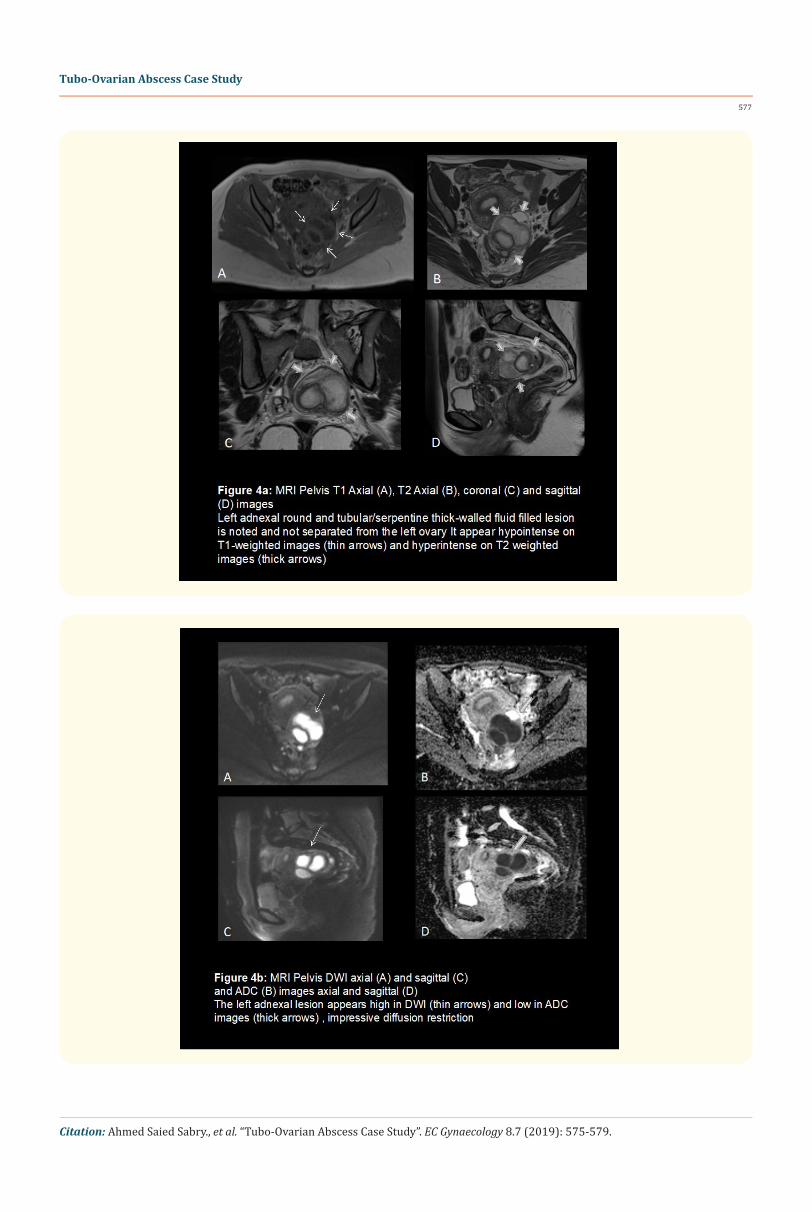
Color Doppler scan show preserved vascularity (Figure 2). Subsequent CT Scan without and with IV contrast done and demonstrates bulky left ovary with ill-defined margin in the pre contrast images. Post-contrast images show multi-loculatd left adnexal cystic lesion with enhancing wall and surrounding pelvic inflammation, fluid within the endometrial cavity also noted (Figure 3). MRI done and showed left adnexal round and tubular/serpentine thick-walled fluid filled lesion which is not separated from the left ovary. It appears hypo-intense on T1-weighted images, hyper-intense on T2 weighted images, high in DWI and low in ADC images, impressive diffusion restriction. The left adnexal lesion in post contrast images demonstrates dense rim enhancement, contrast diffuses in the surrounding stromal part of the lesion and sparing the central fluid content, the findings which are typical for tubo- ovarian abscess. Surrounding inflammatory changes along the meso-salpinx/meso-ovarium, with tenting of part of the overlying adjacent small bowel loop denoting element of adhesion process (Figure 4a-4c). Ovarian abscess drainage and pelvic wash was done by exploratory laparotomy. Patient improved postoperatively and was discharged home.
Citation: Ahmed Saied Sabry., et al. “Tubo-Ovarian Abscess Case Study”. EC Gynaecology 8.7 (2019): 575-579.
Tubo-Ovarian Abscess Case Study
577

Citation: Ahmed Saied Sabry., et al. “Tubo-Ovarian Abscess Case Study”. EC Gynaecology 8.7 (2019): 575-579.
Tubo-Ovarian Abscess Case Study
578
Discussion
TOA is a well-known entity commonly resulting as a late complication of pelvic inflammatory disease. It usually occurs in young females though rarely seen in post-menopausal women as well [4]. The disease can occurs secondary to ascending cervical or vaginal infection or through fallopian tubes into peritoneal cavity leading to secondary inflammation and finally abscess formation [2]. Mostly it is poly microbial with predominance of anaerobes. Neisseria Gonorrhoeae and Chlamydia Trachomatis are the commonly responsible for ascending infection [4,5]. Actinomycosis, tuberculosis and xanthogranulomatous inflammation are among the rare causes [3]. The other mechanism could be the secondarily involvement of adnexa due to infection or inflammation in surrounding pelvic organs e.g. appendix, colon, bladder [2].
Patients usually have vague symptoms however; the most common symptom is pelvic or lower abdominal pain of variable duration which is usually constant and worsening [1]. Other symptoms could include foul smelling vaginal discharge, dysuria, dyspareunia, vaginal bleeding and in few patients’ nausea and vomiting as well. Fever with chills along with leukocytosis is an important finding as in our patient however could be only seen in 80% of patients [5,7]. On physical examination, the patients may have lower abdominal tenderness and guarding with cervical motion tenderness [4,5].
Prompt diagnosis and treatment of pelvic inflammatory disease and tubo-ovarian abscess is of prime importance, keeping in view the severity of short and long term sequel of untreated condition [2]. These include increased risk of subsequent episodes of pelvic inflammatory disease and ectopic pregnancy, chronic pelvic pain, increased risk of and of utmost importance is the infertility [7]. The other serious complications due to abscess rupture could be sepsis, peritonitis, ovarian vein thrombosis and Fitz Hugh Curtis syndrome [5,6]. The patient’s usually require admission in hospital. The treatment options include IV antibiotics, image guided or surgical drainage of abscess or combination of all. Success rate of percutaneous drainage of tubo-ovarian abscess by simple aspiration and by catheter drainage is reported to be 94% and 77% respectively [8].
The gold standard for diagnosis of PID is laparoscopy where direct visualization is possible [7]. Ultrasound is the first imaging modality of choice, the findings may not be conclusive [2,4]. In these cases, other imaging modalities like CT and MRI is required [4]. The typical sonographic findings include adnexal mass (solid, cystic or complex) with fluid in cul-de-sac. Ovaries and fallopian tubes may not be identified separately. The uterus may or may not show loss of mid line endometrial echoes [6,7]. The findings may mimic endometrioma or hemorrhagic cyst in atypical cases as was in seen our case [2].
CT and MRI are done in atypical cases where together with clinical picture and ultrasound findings the diagnosis is not clear [3,6]. CT Findings include multicystic thick walled adnexa with fluid density and abnormal enhancement of septa. The peri-ovarian inflammatory changes including stranding, surrounding free fluid, abnormal enhancement which may include peritoneum, anterior displacement of

Citation: Ahmed Saied Sabry., et al. “Tubo-Ovarian Abscess Case Study”. EC Gynaecology 8.7 (2019): 575-579.
Tubo-Ovarian Abscess Case Study
579
mesosalpinx and thickening of uterosacral ligament may be seen depending upon the severity of disease [4,5]. MRI findings include T1 low, heterogeneous or bright T2 signal of cystic adnexal mass with wall enhancement on post contrast images. The presence of pus or hemorrhage can sometime give T1 high signal along the inner wall of adnexal lesion [6].
Conclusion
Tubo-ovarian abscess is a well-known complication of pelvic inflammatory disease which requires prompt diagnosis or treatment. In cases with vague patient’s clinical presentation and atypical imaging features use of CT and MRI is of utmost importance to reach the cor-rect diagnosis. Correct and timely diagnosis can save the patient from hazardous short and long term complications.
Bibliography
1. Soper DF. “Genitourinary infections and sexually transmitted diseases”. In: Berek JS, ed, Novak’s Gynecology. 14th Edition. Philadelphia, PA: Lippincott Williams and Wilkins (2007): 549-552.
2. Velcani A., et al. “Sonographic features of tubo-ovarian abscess mimicking an endometrioma and review of cystic adnexal masses”. Journal of Radiology Case Reports 4.2 (2010): 9-17.
3. Krivak TC., et al. “Tubo-ovarian abscess: diagnosis, medical and surgical management”. Comprehensive Therapy 30.2 (2004): 93-100.
4. Sun HK., et al. “Unusual causes of tubo-ovarian abscess: CT and MR imaging findings”. Radiographics 24.6 (2004): 1575-1589.
5. Saloum NMI., et al. “Core curriculum case illustration: tubo-ovarian abscess”. Emergency Radiology (2018).
6. Wilbur AC., et al. “CT findings in tuboovarian abscess”. American Journal of Roentgenology 158.3 (1992): 575-579.
7. Sam JW., et al. “Spectrum of CT Findings in Acute Pyogenic Pelvic Inflammatory Disease”. Radiographics 22.6 (2002): 1527-1323.
8. Worthen NJ and Gunning JE. “Percutaneous drainage of pelvic abscesses: management of the tubo-ovarian abscess”. Journal of Ultrasound in Medicine 5.10 (1986): 551-556.
Volume 8 Issue 7 July 2019©All rights reserved by Ahmed Saied Sabry., et al.